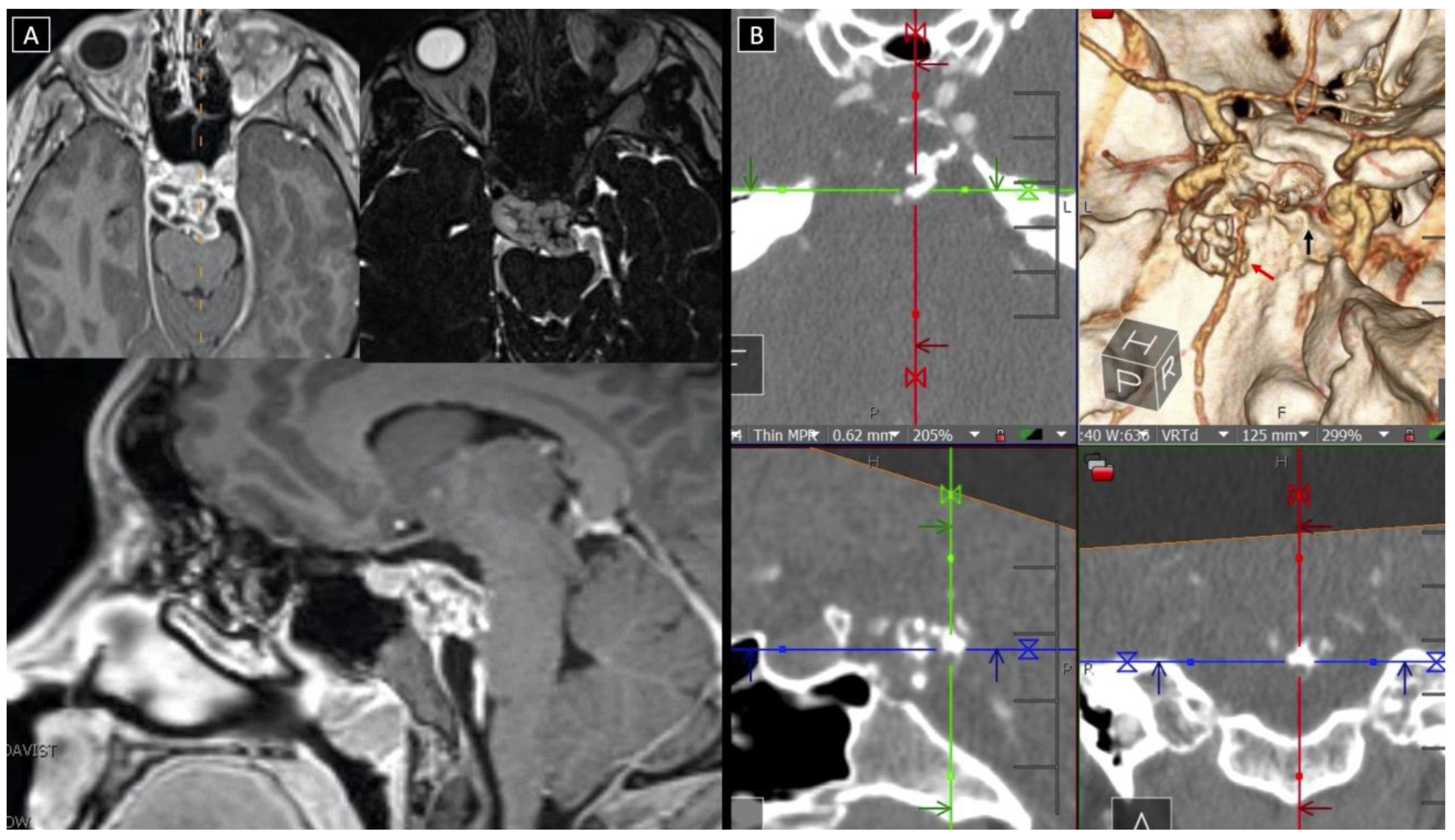
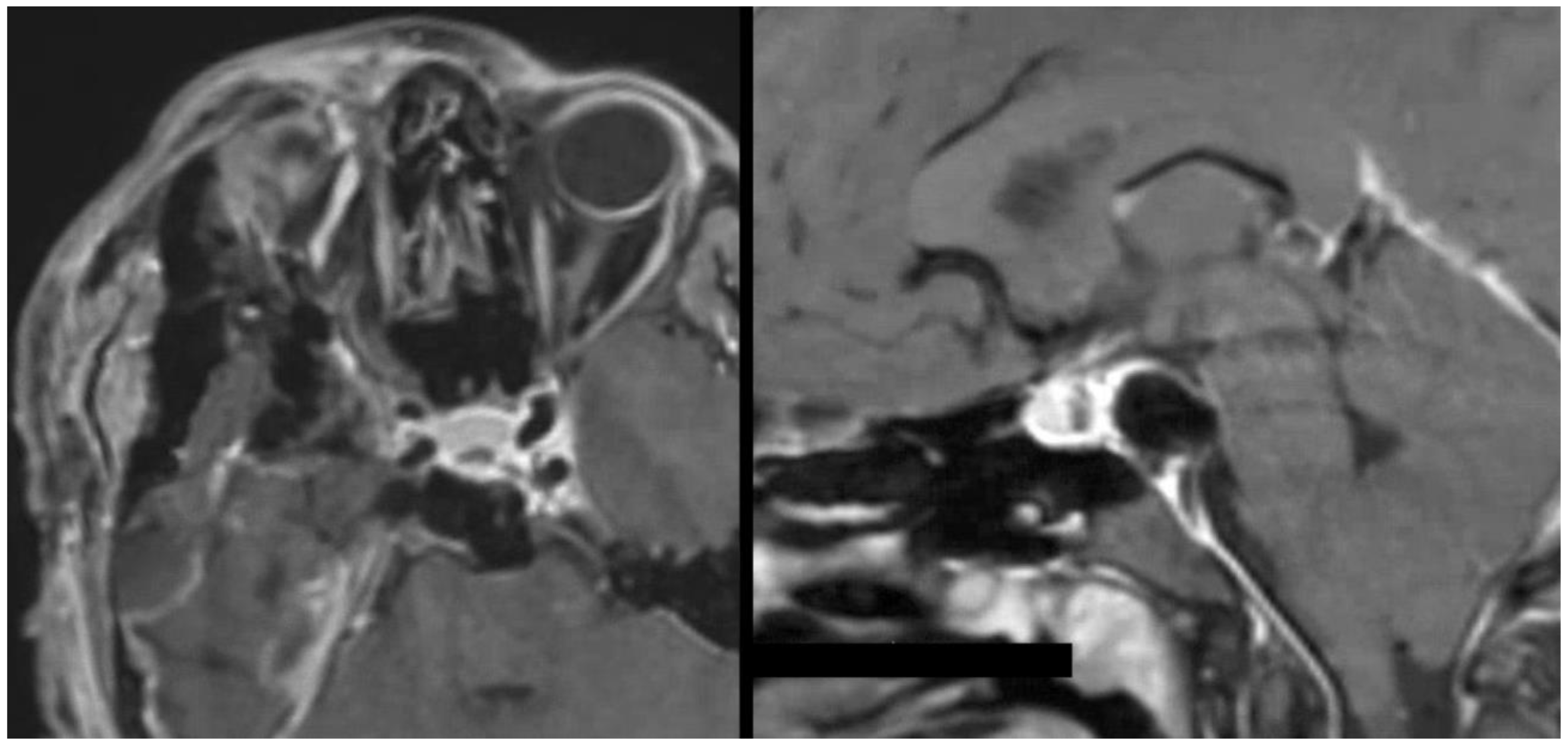
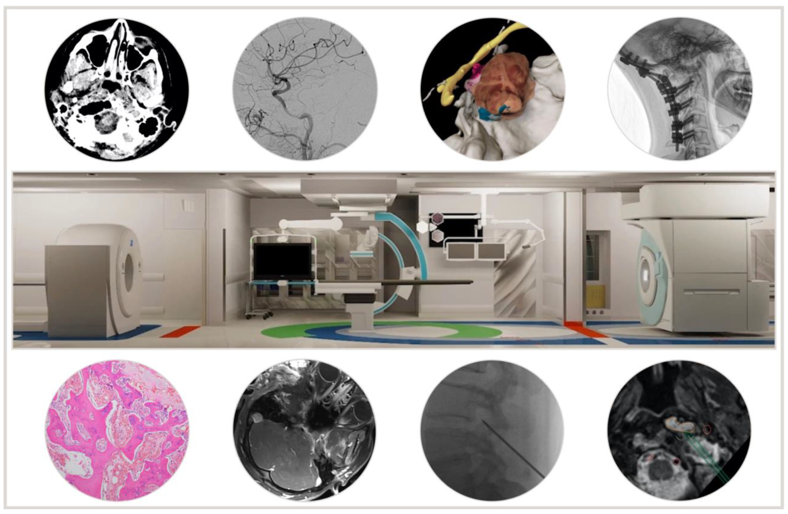
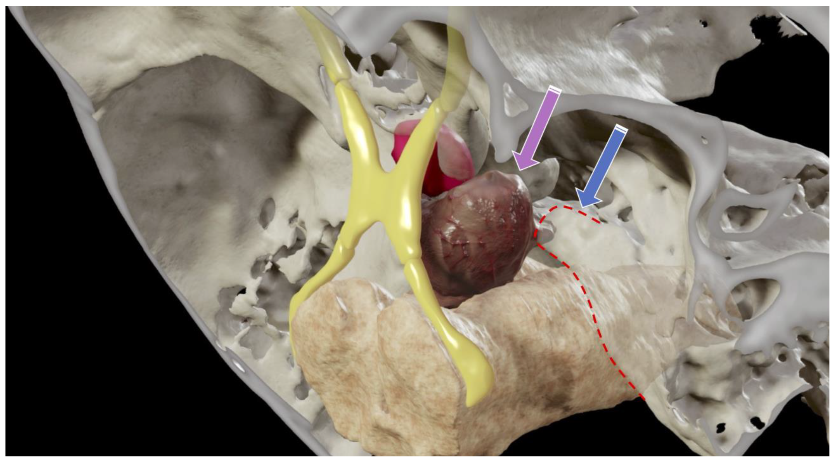
Multimodal Intraoperative Image-Driven Surgery for Skull Base Chordomas and Chondrosarcomas
 ,
,  , ,
, ,
Simple Summary
Abstract
1. Introduction
2. Methods
2.1. Operative and Perioperative Management in the AMIGO Suite
2.2. Volumetric Analysis
2.3. Clinical Outcomes and Follow-Up
3. Results
3.1. Patient Population, Tumor Characteristics, and Preoperative Imaging
3.2. Surgery and Intraoperative Imaging
3.3. Volumetric Analysis
3.4. Clinical Outcomes and Follow-Up
3.5. Case Examples
3.5.1. Case 1
3.5.2. Case 2
4. Discussion
5. Limitations
6. Conclusions
Supplementary Materials
Author Contributions
Funding
Institutional Review Board Statement
Informed Consent Statement
Data Availability Statement
Conflicts of Interest
Abbreviations
References
- Dolati, P.; Gokoglu, A.; Eichberg, D.; Zamani, A.; Golby, A.; Al-Mefty, O. Multimodal navigated skull base tumor resection using image-based vascular and cranial nerve segmentation: A prospective pilot study. Surg. Neurol. Int. 2015, 6, 172. [Google Scholar] [CrossRef] [Scilit] [PubMed]
- Salame, K.; Fliss, D.M.; Cohen, J.T.; Ouaknine, G.E. Technological advances in skull base surgery. Oper. Tech. Otolaryngol. Head Neck Surg. 2000, 11, 234–237. [Google Scholar] [CrossRef] [Scilit]
- Tempany, C.M.; Jayender, J.; Kapur, T.; Bueno, R.; Golby, A.; Agar, N.; Jolesz, F.A. Multimodal imaging for improved diagnosis and treatment of cancers. Cancer 2015, 121, 817–827. [Google Scholar] [CrossRef] [Scilit] [PubMed]
- Colli, B.O.; Al-Mefty, O. Chordomas of the skull base: Follow-up review and prognostic factors. Neurosurg. Focus 2001, 10, E1. [Google Scholar] [CrossRef] [Scilit] [PubMed]
- Almefty, K.; Pravdenkova, S.; Colli, B.O.; Al-Mefty, O.; Gokden, M. Chordoma and chondrosarcoma: Similar, but quite different, skull base tumors. Cancer 2007, 110, 2457–2467. [Google Scholar] [CrossRef] [Scilit] [PubMed]
- Fedorov, A.; Beichel, R.; Kalpathy-Cramer, J.; Finet, J.; Fillion-Robin, J.C.; Pujol, S.; Bauer, C.; Jennings, D.; Fennessy, F.; Sonka, M.; et al. 3D Slicer as an image computing platform for the Quantitative Imaging Network. Magn. Reson. Imaging 2012, 30, 1323–1341. [Google Scholar] [CrossRef] [Scilit] [PubMed]
- Essayed, W.I.; Mooney, M.A.; Al-Mefty, O. Transcavernous Resection of an Upper Clival Chondrosarcoma: “Cavernous Sinus as a Route”: 2-Dimensional Operative Video. Oper. Neurosurg. 2021, 20, E422–E423. [Google Scholar] [CrossRef] [Scilit]
- Jain, M.; Noseworthy, M.D. Current Status of Radiological Multimodality Imaging. Crit. Rev. Biomed. Eng. 2016, 44, 167–176. [Google Scholar] [CrossRef] [Scilit]
- Jones, D.W.; Stangenberg, L.; Swerdlow, N.J.; Alef, M.; Lo, R.; Shuja, F.; Schermerhorn, M.L. Image Fusion and 3-Dimensional Roadmapping in Endovascular Surgery. Ann. Vasc. Surg. 2018, 52, 302–311. [Google Scholar] [CrossRef] [Scilit]
- Santagata, S.; Eberlin, L.S.; Norton, I.; Calligaris, D.; Feldman, D.R.; Ide, J.L.; Liu, X.; Wiley, J.S.; Vestal, M.L.; Ramkissoon, S.H.; et al. Intraoperative mass spectrometry mapping of an onco-metabolite to guide brain tumor surgery. Proc. Natl. Acad. Sci. USA 2014, 111, 11121–11126. [Google Scholar] [CrossRef] [Scilit]
- Murphy, M.A.; O’Brien, T.J.; Morris, K.; Cook, M.J. Multimodality image-guided surgery for the treatment of medically refractory epilepsy. J. Neurosurg. 2004, 100, 452–462. [Google Scholar] [CrossRef] [Scilit] [PubMed]
- Mazzucchi, E.; La Rocca, G.; Ius, T.; Sabatino, G.; Della Pepa, G.M. Multimodality Imaging Techniques to Assist Surgery in Low-Grade Gliomas. World Neurosurg. 2020, 133, 423–425. [Google Scholar] [CrossRef] [Scilit] [PubMed]
- Nowell, M.; Rodionov, R.; Zombori, G.; Sparks, R.; Rizzi, M.; Ourselin, S.; Miserocchi, A.; McEvoy, A.; Duncan, J. A Pipeline for 3D Multimodality Image Integration and Computer-assisted Planning in Epilepsy Surgery. J. Vis. Exp. 2016, 111, e53450. [Google Scholar] [CrossRef] [Scilit]
- Ning, G.; Zhang, X.; Zhang, Q.; Wang, Z.; Liao, H. Real-time and multimodality image-guided intelligent HIFU therapy for uterine fibroid. Theranostics 2020, 10, 4676–4693. [Google Scholar] [CrossRef] [Scilit]
- Huang, B.; Yang, F.; Yin, M.; Mo, X.; Zhong, C. A Review of Multimodal Medical Image Fusion Techniques. Comput. Math. Methods Med. 2020, 2020, 8279342. [Google Scholar] [CrossRef] [Scilit] [PubMed]
- Hahn, B.S.; Park, J.Y. Incorporating New Technologies to Overcome the Limitations of Endoscopic Spine Surgery: Navigation, Robotics, and Visualization. World Neurosurg. 2021, 145, 712–721. [Google Scholar] [CrossRef] [Scilit]
- Zaidi, H.A.; De Los Reyes, K.; Barkhoudarian, G.; Litvack, Z.N.; Bi, W.L.; Rincon-Torroella, J.; Mukundan, S., Jr.; Dunn, I.F.; Laws, E.R., Jr. The utility of high-resolution intraoperative MRI in endoscopic transsphenoidal surgery for pituitary macroadenomas: Early experience in the Advanced Multimodality Image Guided Operating suite. Neurosurg. Focus 2016, 40, E18. [Google Scholar] [CrossRef] [Scilit]
- Olubiyi, O.I.; Ozdemir, A.; Incekara, F.; Tie, Y.; Dolati, P.; Hsu, L.; Santagata, S.; Chen, Z.; Rigolo, L.; Golby, A.J. Intraoperative Magnetic Resonance Imaging in Intracranial Glioma Resection: A Single-Center, Retrospective Blinded Volumetric Study. World Neurosurg. 2015, 84, 528–536. [Google Scholar] [CrossRef] [Scilit]
- Walcott, B.P.; Nahed, B.V.; Mohyeldin, A.; Coumans, J.V.; Kahle, K.T.; Ferreira, M.J. Chordoma: Current concepts, management, and future directions. Lancet Oncol. 2012, 13, e69–e76. [Google Scholar] [CrossRef] [Scilit]
- Koto, M.; Ikawa, H.; Kaneko, T.; Hagiwara, Y.; Hayashi, K.; Tsuji, H. Long-term outcomes of skull base chordoma treated with high-dose carbon-ion radiotherapy. Head Neck. 2020, 42, 2607–2613. [Google Scholar] [CrossRef] [Scilit]
- Kremenevski, N.; Schlaffer, S.M.; Coras, R.; Kinfe, T.M.; Graillon, T.; Buchfelder, M. Skull Base Chordomas and Chondrosarcomas. Neuroendocrinology 2020, 110, 836–847. [Google Scholar] [CrossRef] [Scilit] [PubMed]
- Erdem, E.; Angtuaco, E.C.; Van Hemert, R.; Park, J.S.; Al-Mefty, O. Comprehensive review of intracranial chordoma. Radiographics 2003, 23, 995–1009. [Google Scholar] [CrossRef] [Scilit]
- Bakker, S.H.; Jacobs, W.C.H.; Pondaag, W.; Gelderblom, H.; Nout, R.A.; Dijkstra, P.D.S.; Peul, W.C.; Vleggeert-Lankamp, C.L.A. Chordoma: A systematic review of the epidemiology and clinical prognostic factors predicting progression-free and overall survival. Eur. Spine J. 2018, 27, 3043–3058. [Google Scholar] [CrossRef] [Scilit] [PubMed]
- Kadri, P.A.S.; Al-Mefty, R.O.; Borba, L.A.B.; Ayoubi, S.; Al-Mefty, O. Recurrent Skull Base Chordomas. In Chordomas and Chondrosarcomas of the Skull Base and Spine; Elsevier: Amsterdam, The Netherlands, 2018; pp. 393–398. [Google Scholar]
- Zoli, M.; Milanese, L.; Bonfatti, R.; Faustini-Fustini, M.; Marucci, G.; Tallini, G.; Zenesini, C.; Sturiale, C.; Frank, G.; Pasquini, E.; et al. Clival chordomas: Considerations after 16 years of endoscopic endonasal surgery. J. Neurosurg. 2018, 128, 329–338. [Google Scholar] [CrossRef] [Scilit]
- Tzortzidis, F.; Elahi, F.; Wright, D.; Natarajan, S.K.; Sekhar, L.N. Patient outcome at long-term follow-up after aggressive microsurgical resection of cranial base chordomas. Neurosurgery 2006, 59, 230–237. [Google Scholar] [CrossRef] [Scilit] [PubMed]
- Sen, C.; Triana, A. Cranial chordomas: Results of radical excision. Neurosurg. Focus 2001, 10, E3. [Google Scholar] [CrossRef] [Scilit]
- Al-Mefty, O.; Borba, L.A. Skull base chordomas: A management challenge. J. Neurosurg. 1997, 86, 182–189. [Google Scholar] [CrossRef] [Scilit]
- Al-Mefty, O.; Almefty, R. 24 Chordomas: A Personal Perspective. Chordomas 2017, 179–186. [Google Scholar] [CrossRef] [Scilit]
- Sen, C.; Triana, A.I.; Berglind, N.; Godbold, J.; Shrivastava, R.K. Clival chordomas: Clinical management, results, and complications in 71 patients. J. Neurosurg. 2010, 113, 1059–1071. [Google Scholar] [CrossRef] [Scilit]
- Nimsky, C.; Ganslandt, O.; Kober, H.; Buchfelder, M.; Fahlbusch, R. Intraoperative magnetic resonance imaging combined with neuronavigation: A new concept. Neurosurgery 2001, 48, 1082–1089. [Google Scholar] [CrossRef] [Scilit]
- Metwali, H.; Samii, A.; Gerganov, V.; Giordano, M.; Fahlbusch, R.; Samii, M. The Significance of Intraoperative Magnetic Resonance Imaging in Resection of Skull Base Chordomas. World Neurosurg. 2019, 128, e185–e194. [Google Scholar] [CrossRef] [Scilit] [PubMed]



| Surg | Pts | Age | Sex | Symptoms | Tumor Location | Type of Tumor (Prior Surgeries) | Previous Radiation |
|---|---|---|---|---|---|---|---|
| 1 | 1 | 71 | F | Radiological progression | Rt CS | Recurrence (3) | Proton beam radiation |
| 2 | 2 | 58 | M | Increasing diplopia | Cl, PPS, Lt CS | Recurrence (2) | Proton beam radiation and Gamma knife |
| 3 | 3 | 39 | M | Increasing diplopia | Rt Cl, CS | Recurrence (1) | RT |
| 4 | 42 | M | Worsening vision in right eye | Rt Cl, MF, PPS, S, CS, NP | Recurrence (2) | RT | |
| 5 | 45 | M | Radiological progression | Rt Cl, MF, PPS, S, CS, NP | Recurrence (3) | RT | |
| 6 | 4 | 55 | M | Headaches, blurry vision | Cl, CS, S, PA | New | No |
| 7 | 5 | 45 | F | Right-sided jaw pain, neck pain | Cl, Bilateral condyles | New | No |
| 8 | 6 | 45 | F | Decreased hearing in the Rt | Rt PA, Cl | New | No |
| 9 | 7 | 51 | F | None | Rt PA, Cl | New | No |
| 10 | 8 | 64 | F | Worsening voice and swallowing | Cl, Bilateral condyles, retropharyngeal | Recurrence (1) | Proton beam radiation |
| 11 | 9 | 24 | F | Double vision | Upper Clivus | New | No |
| Complaints | Clinical Findings | Features | Total | % | |||||||
|---|---|---|---|---|---|---|---|---|---|---|---|
| Neuro-Ophthalmologic | % | CN Deficit | % | Clival | |||||||
| Abnormal eye movement | 5 | 45% | Optic | 2 | 18% | Upper | 9 | 82% | |||
| Double vision | 5 | 45% | Oculomotor | 4 | 36% | Middle | 9 | 82% | |||
| Eye Drop | 4 | 36% | Trochlear | 2 | 18% | Lower | 9 | 82% | |||
| Visual difficulties | 6 | 55% | Trigeminal | 1 | 9% | Extension | 0% | ||||
| Headaches | 2 | 18% | Abducens | 5 | 45% | Cavernous sinus | 8 | 73% | |||
| Neck pain | 2 | 18% | Facial | 0 | 0% | Sellar region | 7 | 64% | |||
| Voice hoarseness | 1 | 9% | Vestibulocochlear | 1 | 9% | Intradural | 5 | 45% | |||
| Swallowing difficulties | 1 | 9% | Glossopharyngeal | 1 | 9% | Petrous ridge | 8 | 73% | |||
| Vertigo | 1 | 9% | Vagus | 1 | 9% | Cerebellopontine angle/prepontine | 4 | 36% | |||
| Accessory | 0 | 0% | Jugular foramen | 2 | 18% | ||||||
| Hypoglossal | 2 | 18% | Retro pharynx | 4 | 36% | ||||||
| Long tracts | 1 | 9% | Infratemporal fossa | 2 | 18% | ||||||
| Cerebellar signs | 0 | 0% | Occipital condyles | 3 | 27% | ||||||
| Cervical spine | 1 | 9% | |||||||||
| Mean Tumor Volume (cm3) | 13.4 | SD 12 | |||||||||
| Surg | Modalities | Modalities Used | Residual iMRI | Complications | Major Events in the Long Term Follow-Up | |
|---|---|---|---|---|---|---|
| 1 | Right anterior preauricular | 1 | MRI | Anterior genu of the right carotid | None | No recurrence on the available one-year follow-up |
| 2 | Left preauricular and zygomatic | 2 | MRI, Endosc | Lt PPS, Lt CS, Post clinoid, Meckel’s cave | None | Multifocal disease including spinal cord, deceased |
| 3 | Right preauricular zygomatic middle fossa approach | 4 | MRI, CT, Endosc, Fluoro | Anterior CS | None | Local recurrence (S4) |
| 4 | Right preauricular zygomatic middle fossa approach | 3 | MRI, Endosc, Fluoro | Anterior CS | None | Local recurrence (S5) |
| 5 | Right preauricular zygomatic middle fossa approach | 3 | MRI, Endosc, Angio | Rt PPS, Anterior CS | Intracavernous carotid injury | Developed local recurrence and distal drop metastasis to the lumbar spine |
| 6 | Left preauricular middle fossa anterior petrosal approach (3 weeks after anterior transsphenoidal approach) | 4 | MRI, CT, Endosc, Fluoro | Petroclival/occipital clivus | Left V1-V2 hypoalgesia, L frontalis branch of facial | Radiation induced panhypopituitarism |
| 7 | Bilateral transcondylar approach with O-C5 fusion | 3 | MRI, Endosc, Fluoro | Base of the odontoid | DVT | Multifocal metastatic disease, including spine requiring radiation therapy, contralateral intradural infratentorial met requiring surgery in August 2020 |
| 8 | Right anterior preauricular | 3 | MRI, Endosc, Fluoro | None | No recurrent disease on the 5 year follow-up | |
| 9 | Right anterior preauricular | 3 | MRI, Endosc, Fluoro | Intracranial hypotension, Partial Rt VII | Initial progression of residual, stable after radiation therapy | |
| 10 | Rt far lateral | 2 | MRI, Endosc | Retropharyngeal | None | Aggressive recurrence on 3–4 month postoperative imaging |
| 11 | R orbitozygomatic transcavernous | 3 | MRI, Endosc, Fluoro | None | No recurrence on the available one-year follow-up |
| Extent of Resection | Patients | % | |
|---|---|---|---|
| GTR | 2 | 18.2% | |
| STR/partial resection | 9 | 81.8% | |
| Pathology | |||
| Chordoma | Classic | 8 | 72.7% |
| Chondroid | 0 | 0.0% | |
| Dedifferentiated | 0 | 0.0% | |
| Chondrosarcoma | Grade I | 3 | 27.3% |
| Myxoid | 2 | 18.1% | |
| Symptoms/Deficit | Active | 6 | 54.5% |
| Postoperative symptoms | Improvement | 2 | 18.2% |
| Stable | 7 | 63.6% | |
| Worsening | 1 | 9.1% | |
| Morbidity | Major | 1 | 9.1% |
| Minor | 4 | 36.4% | |
| Follow-up | Years | SD | |
| Progression-free survival (index surgery to clinical/rad progression) | 2.3 | 2.3 | |
| Postoperative follow-up (index surgery to last available follow-up) | 2.9 | 2.7 | |
| Overall follow-up (initial diagnosis-death/last follow-up in months) | 11.48* | 8.0 |
| Surgery | 1 | 2 | 3 | 4 | 5 | 6 | 7 | 8 | 9 | 10 | 11 |
|---|---|---|---|---|---|---|---|---|---|---|---|
| Preop Volume (cm3) | 3.78 | 16.82 | 7.15 | 10.28 | 25.84 | 22.23 | 40.74 | 2.56 | 2.56 | 10.03 | 5.50 |
| iMRI volume (cm3) | 0.13 | 0.13 | 0.60 | 0.28 | 2.74 | 0.11 | 2.88 | 0.00 | 0.44 | 1.02 | 0.00 |
| Intraop % of reduction | 96.6% | 99.2% | 91.6% | 97.3% | 89.4% | 99.5% | 92.9% | 100.0% | 82.8% | 89.8% | 100.0% |
| Postop MRI (cm3) | 0.13 | 0.13 | 0.07 | 0.09 | n/a | 0.00 | 0.76 | 0.00 | 0.2 | 1.39 | n/a |
| Postop % of reduction | 96.6% | 99.2% | 99.0% | 99.1% | 89.4% | 100.0% | 98.1% | 100.0% | 92.2% | n/a * | 100.0% |
Publisher’s Note: MDPI stays neutral with regard to jurisdictional claims in published maps and institutional affiliations. |
© 2022 by the authors. Licensee MDPI, Basel, Switzerland. This article is an open access article distributed under the terms and conditions of the Creative Commons Attribution (CC BY) license (https://creativecommons.org/licenses/by/4.0/).
Share and Cite
Essayed, W.I.; Juvekar, P.; Bernstock, J.D.; Rassi, M.S.; Almefty, K.; Zamani, A.A.; Golby, A.J.; Al-Mefty, O. Multimodal Intraoperative Image-Driven Surgery for Skull Base Chordomas and Chondrosarcomas. Cancers 2022, 14, 966. https://doi.org/10.3390/cancers14040966
Essayed WI, Juvekar P, Bernstock JD, Rassi MS, Almefty K, Zamani AA, Golby AJ, Al-Mefty O. Multimodal Intraoperative Image-Driven Surgery for Skull Base Chordomas and Chondrosarcomas. Cancers. 2022; 14(4):966. https://doi.org/10.3390/cancers14040966
Chicago/Turabian StyleEssayed, Walid I., Parikshit Juvekar, Joshua D. Bernstock, Marcio S. Rassi, Kaith Almefty, Amir Arsalan Zamani, Alexandra J. Golby, and Ossama Al-Mefty. 2022. "Multimodal Intraoperative Image-Driven Surgery for Skull Base Chordomas and Chondrosarcomas" Cancers 14, no. 4: 966. https://doi.org/10.3390/cancers14040966
APA StyleEssayed, W. I., Juvekar, P., Bernstock, J. D., Rassi, M. S., Almefty, K., Zamani, A. A., Golby, A. J., & Al-Mefty, O. (2022). Multimodal Intraoperative Image-Driven Surgery for Skull Base Chordomas and Chondrosarcomas. Cancers, 14(4), 966. https://doi.org/10.3390/cancers14040966