Article

Measurable Residual Disease Monitoring by Locked Nucleic Acid Quantitative Real-Time PCR Assay for IDH1/2 Mutation in Adult AML

1
Division of Hematology-Oncology, Department of Internal Medicine, Chang Gung Memorial Hospital at Linkou, Taoyuan 333423, Taiwan
2
College of Medicine, Chang Gung University, Taoyuan 333323, Taiwan
3
Division of Hematology and Oncology, Department of Internal Medicine, China Medical University Hospital, Taichung 404327, Taiwan
4
Division of Hematology and Oncology, Department of Internal Medicine, Chang Gung Memorial Hospital at Chiayi, Chiayi 613016, Taiwan
*
Author to whom correspondence should be addressed.
Cancers 2022, 14(24), 6205; https://doi.org/10.3390/cancers14246205
Submission received: 26 October 2022 / Revised: 5 December 2022 / Accepted: 13 December 2022 / Published: 15 December 2022
(This article belongs to the Special Issue 2nd Edition: Minimal Residual Disease of Cancers)

Abstract

:

Simple Summary

Measurable residual disease (MRD) monitoring is crucial in managing AML to predict the risk of relapse. A better understanding of which MRD technique and molecular target will have an effective clinical impact on AML is still required. Locked nucleic acid quantitative Real-Time PCR assay (LNA-qPCR) is sensitive and specific for quantifying oncogenetic single-nucleotide variation. We assessed the role of LNA-qPCR in the monitoring of IDH1/2 mutations MRD in eighty-eight AML patients from multiple centers. We found that IDH1/2 LNA-qPCR MRD correlates well with NPM1 qPCR MRD, predicts relapse-free survival and cumulative incidence of relapse, and is a potential MRD technique for IDH1/2-mutated AML patients with reduced IDH1/2 mutant levels after complete remission.

Abstract

Locked nucleic acid quantitative Real-Time PCR (LNA-qPCR) for IDH1/2 mutations in AML measurable residual disease (MRD) detection is rarely reported. LNA-qPCR was applied to quantify IDH1/2 mutants MRD kinetics in bone marrow from 88 IDH1/2-mutated AML patients, and correlated with NPM1-MRD, clinical characteristics, and outcomes. The median normalized copy number (NCN) of IDH1/2 mutants decreased significantly from 53,228 (range 87–980,686)/ALB × 106 at diagnosis to 773 (range 1.5–103,600)/ALB × 106 at first complete remission (CR). IDH1/2 LNA-qPCR MRD was concordant with remission status or NPM1-MRD in 79.5% (70/88) of patients. Younger patients and patients with FLT3 mutations had higher concordance. The Spearman correlation coefficient (rs) and concordance rate between the log reduction of IDH1/2 LNA-qPCR and NPM1-MRD were 0.68 and 81% (K = 0.63, 95% CI 0.50–0.74), respectively. IDH1/2-MRD > 2 log reduction at first CR predicted significantly better relapse-free survival (3-year RFS rates 52.9% vs. 31.9%, p = 0.007) and cumulative incidence of relapse (3-year CIR rates 44.5% vs. 64.5%, p = 0.012) compared to IDH1/2-MRD ≤ 2 log reduction. IDH1/2-MRD > 2 log reduction during consolidation is also associated with a significantly lower CIR rate than IDH1/2-MRD ≤ 2 log reduction (3-year CIR rates 42.3% vs. 68.8%, p = 0.019). LNA-qPCR for IDH1/2 mutation is a potential MRD technique to predict relapse in IDH1/2-mutated AML patients, especially for those with IDH1/2 MRD > 2 log reduction at first CR or a concurrent FLT3 mutation.

1. Introduction

Acute myeloid leukemia (AML) is a hematological malignancy characterized by clonal expansion of myeloid progenitors in the bone marrow (BM), peripheral blood, and other tissues. AML is a heterogeneous disease in terms of molecular genetics and phenotypic characteristics [1,2]. Molecular screening plays a significant role in prognostic categorization and decision of treatment strategies for AML [3].
Isocitrate dehydrogenases 1 and 2 (IDH1, IDH2) are metabolic enzymes that convert isocitrate to α-ketoglutarate, which are involved in diverse cellular processes, including adaptation to hypoxia, histone demethylation, and DNA modification [4,5]. Somatic mutations in IDH1 and IDH2 genes cause their protein dysfunction and an intracellular accumulation of aberrant 2-hydroxy-glutarate, a cancer-associated metabolite [6,7]. The incidence of IDH1/2 mutations in AML was 6%–14% for IDH1 [8,9,10,11], and 8%–19% for IDH2 [8,10,12]. Most of these mutations involve hot-spot codons R140 and R172 in exon 4 of IDH2 and codon R132 in exon 4 of IDH1. The prognostic impact of IDH1/2 mutations is still controversial. Mutated IDH1-R132 has been reported as an unfavorable [9,11] or non-significant factor for AML outcome [8,10]. Mutated IDH2-R140 has been described as favorable or with no impact on overall survival [10,13], whereas mutated IDH2-R172 was associated with a worse prognosis [9,13].
Minimal or measurable residual disease (MRD) has prognostic value in AML patients during or after treatment [14,15,16,17]. The use of IDH1/2 mutations as molecular markers for MRD monitoring is still under investigation. A few studies have reported the stability and suitability of IDH1/2 mutations to monitor MRD by digital PCR (ddPCR) [18,19], next-generation sequencing (NGS) [20,21,22], and locked nucleic acid (LNA) quantitative Real-Time PCR (LNA-qPCR) [23,24]. Persistent IDH1 or IDH2 mutations have been observed in AML patients at the time of morphological remission [22,25]. Two studies with large cohorts of patients have reported that persistent mutated IDH1/2 MRD detected by ddPCR or NGS in AML at the time of complete remission (CR) could predict relapse [19,20]. The impact of IDH1/2 MRD by LNA-qPCR on the outcome of AML patients has not been investigated in large cohorts previously.
NPM1 mutation is a stable MRD marker and NPM1 PCR assays are recommended in AML patients with NPM1 mutation [14,17,26]. In the present study, we aimed to evaluate the use of LNA-qPCR assay to monitor MRD after treatment in AML patients harboring IDH1-R132, IDH2-R140, or IDH2-R172 mutations, to correlate the MRD results of IDH1/2 mutations with those of NPM1 mutations, and to determine their impact on the outcome.

2. Materials and Methods

2.1. Patients and Samples

Newly diagnosed adult de novo AML patients who received induction therapy, including standard chemotherapy, azacitidine, or low-dose cytarabine, were enrolled. This study was conducted by the AML consortium of Taiwan, which aimed to detect genetic mutations and monitor MRD for AML patients in Taiwan. BM samples at the initial diagnosis were analyzed for cytogenetics and a panel of gene mutations. G-banding method was used for karyotypic analysis. AML classification and risk categorization followed the criteria of 2022 WHO classification and 2022 ELN recommendations [3,27]. The study was approved by the Institutional Review Board of Chang Gung Memorial Hospital (99-0112B, 102-3031B, 201700154B0, and 201701453A3) and the participating hospitals (CMUH-HO-AML-10701). Informed consent was obtained from enrolled patients.
BM samples were collected at diagnosis and at different time points after treatment, including the first CR documented after induction therapy, during consolidation (after one to two cycles of consolidation therapy), end of treatment, and relapse. Mononuclear cells were enriched by using Ficoll density gradient centrifugation and cryopreserved. Genomic DNA (gDNA) or RNA extraction and cDNA preparation were performed as previously described [28].

2.2. Detection of Gene Mutations

Diagnostic BM samples were examined for the presence of IDH1 and IDH2 mutations by using NGS (n = 64, MiSeq with SOPHiA DDM™ for Blood Cancers), pyro-sequencing (n = 35), and/or direct sequencing (n = 4). Mutations of NPM1, CEBPA, FLT3-ITD, FLT3-TKD, and myelodysplasia-related gene mutations were detected by NGS or methods described previously [28].

2.3. Monitoring of IDH1/2 MRD by LNA-qPCR Assay

Quantification of IDH1/2 mutation copy number was assessed by quantitative Real-Time PCR (qPCR) for gDNA using the TaqMan technology with LNA probes specific for the mutant allele to increase the specificity for single-nucleotide variation in IDH1-R132, IDH2-R140, and IDH2-R172. The primer sequences and DNA probes used to amplify IDH1-R132, IDH2-R140, and IDH2-R172 mutations are shown in Table 1. Probes, forward primers, and reverse primers for IDH2-R140Q, IDH2-R140W, IDH1-R132H, IDH1-R132S, IDH1-R132G, and ALB were the same as those previously reported [24,29]. The reverse primer of IDH2-R172K was modified by removing the R-LNA. Reverse primers of IDH1-R132C and IDH1-R132L were designed in our lab. A locked nucleic acid was fixed in the last 3′nucleotide of the reverse primers. Reaction mixtures of 25 µL contained the 100 ng gDNA, 2 × TaqMan Master Mix buffer (ThermoFisher), 0.08% BSA, 400 nM of each primer, and 200 nM probe. The PCR cycling protocol consisted of 2 min at 50 °C, 10 min of initial denaturation at 92 °C, and was followed by 50 cycles of 15 s at 95 °C, 1 min at 58 °C. Albumin (ALB) was used as a reference gene. IDH1/2 standard curves were constructed by serial dilution of IDH1-R132, IDH2-R140, and IDH2-R172 plasmids with wild-type IDH1/2 gDNA from normal peripheral blood (Figure 1a–d). Specificity was determined by negative control with wild-type IDH1/2 gDNA. The sensitivity of the assay was five copies for IDH1-R132C/S/G/L, ten copies for IDH1-R132H, five copies for IDH2-R140Q/W, and ten copies for IDH2-R172K with a quantitative range from 5–100 to 106 copies. The normalized copy number (NCN) of the IDH1/2 mutant of the sample was determined by the IDH1/2-mutant copies/ALB × 106 copies. The result of IDH1/2 MRD was expressed as log reduction by calculation of NCNfollow-up divided by NCNdiagnosis for the individual patients. The mean NCN (IDH1/2 copies/ALB × 106 copies) of 19 BM samples from healthy donors were 1119.4 for R132C, 0.2 for R132G, 462.2 for R132H, 11.3 for R132L, 77.2 for R132S, 337.3 for R140Q, 122.5 for R140W, and 81.1 for R172K (Table S1, Figure S1). The MRD negativity is defined as a NCN level that is less than the mean NCN of normal BM samples for each IDH1/2 mutation type.

2.4. Detection of NPM1 RT-qPCR MRD

NPM1 MRD was detected by quantitative Real-Time reverse transcriptase PCR (RT-qPCR) with the TaqMan assay using ABL1 as the internal control gene. Primers and probes used were according to the method described by Gorello et al. [30]. The MRD results were expressed as a log reduction of the NCN at follow-up compared to the NCN at diagnosis.

2.5. Statistical Analysis

Wilcoxon’s rank-sum test, Fisher’s exact test, and the χ2 analysis were used whenever appropriate to make comparisons between groups. The correlation between IDH1/2 LNA-qPCR, marrow blasts, and NPM1 RT-qPCR was assessed by Spearman’s rank correlation test. Cohen’s kappa (K) was used to evaluate the concordance between IDH1/2 LNA-qPCR and NPM1 RT-qPCR. The IDH1/2 MRD at first CR after induction therapy and during consolidation was subjected for outcome analysis. The overall survival (OS), relapse-free survival (RFS), and cumulative incidence of relapse (CIR) are based on the 2022 ELN AML recommendation [3]. Estimates of survival were calculated according to the Kaplan–Meier method. The log-rank test analyzed the differences in survival for significance. Differences in the CIR were assessed using the Gray test. In all analyses, the p-values were two-sided and considered statistically significant when values < 0.05. Statistical analysis was carried out by R version 4.1.3, R Foundation for Statistical Computing, Vienna, Austria.

3. Results

3.1. Patient Characteristics

Eighty-eight IDH1/2-mutated AML patients with post-treatment follow-up BM samples were enrolled for MRD evaluation. There were 25 IDH1 mutations (11 R132C, 9 R132H, 2 R132G, 2 R132S, 1 R132L) and 63 IDH2 mutations (49 R140Q, 2 R140W, 12 R172K). The median age was 54 (range 18–86) years. Cytogenetic normal, intermediate, and adverse risk groups based on the 2022 ELN AML recommendation account for 64%, 17%, and 9%, respectively (10% no metaphase). Eighty-four (95%) patients received standard “3 + 7” chemotherapy, two (2%) patients received azacitidine, and two (2%) patients received low-dose cytarabine as induction therapy. Thirteen patients received allogeneic hematopoietic stem cell transplantation (HSCT). Forty-two (48%) patients had concurrent NPM1 mutations. The detailed clinical and genetic characteristics of patients are summarized in Table 2.

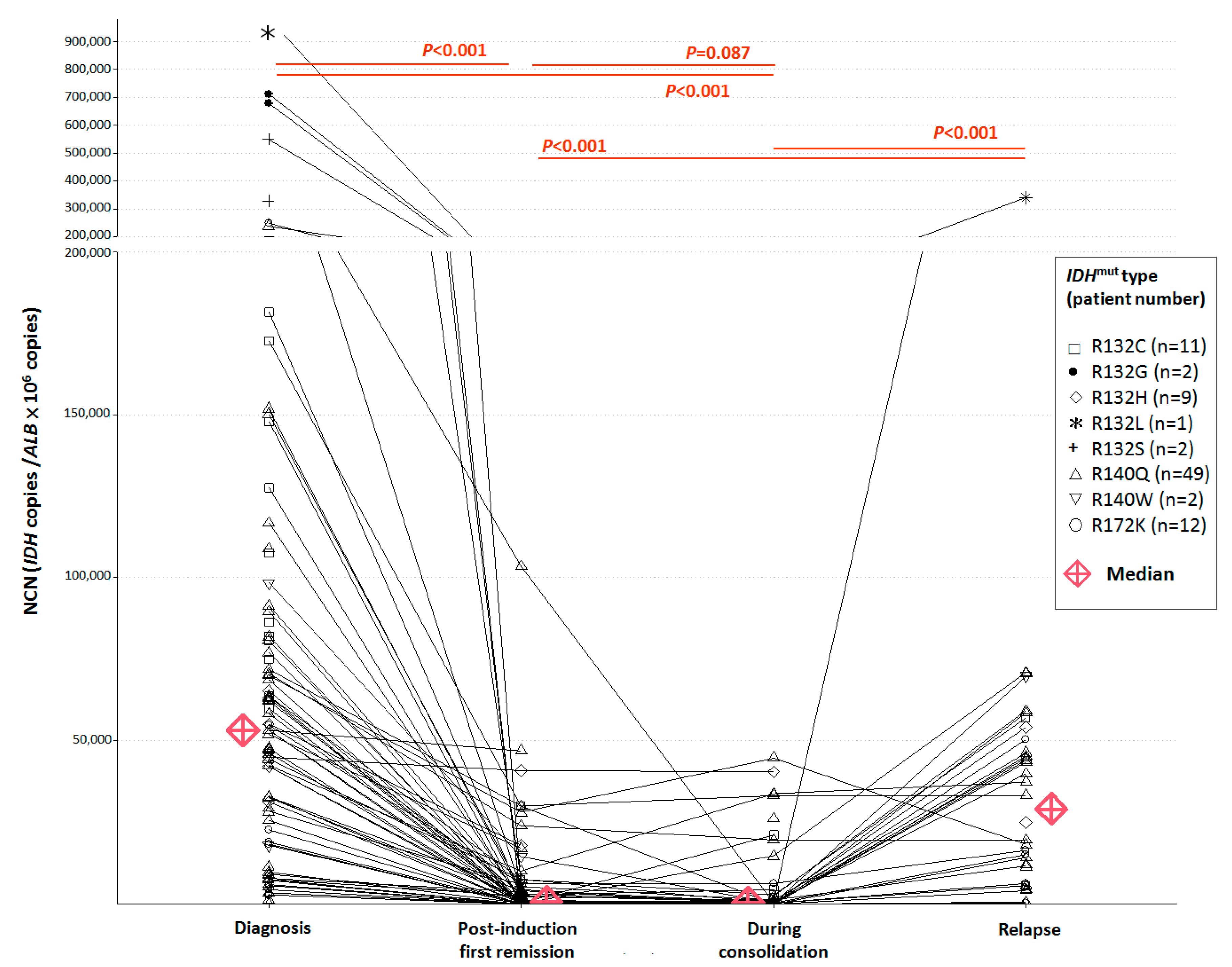
3.2. NCN of IDH1/2 Mutations in Pre-Treatment and Post-Treatment BM Samples

The median diagnostic NCN was 80,920 (31,343–980,686)/ALB × 106 for 25 IDH1-mutated patients and 46,156 (87–247,796)/ALB × 106 for 63 IDH2-mutated patients. IDH1/2-NCN at diagnosis (median 53,228, range 87–980,686/ALB × 106) was significantly higher than both IDH1/2-NCN at first CR (median IDH1/2 NCN at first CR 773, range 1.5–103,600/ALB × 106, p << 0.001) and IDH1/2-NCN during consolidation therapy (median 299, range 0–44,828/ALB × 106, p << 0.001). There was no significant difference between IDH1/2-NCN at first CR and IDH1/2-NCN during consolidation therapy (p = 0.087). IDH1/2-NCN at relapse from 34 patients (median 29,253, range 364–339,239/ALB × 106, p << 0.001) was significantly higher than IDH1/2-NCN at first CR and during consolidation therapy (Figure 2).

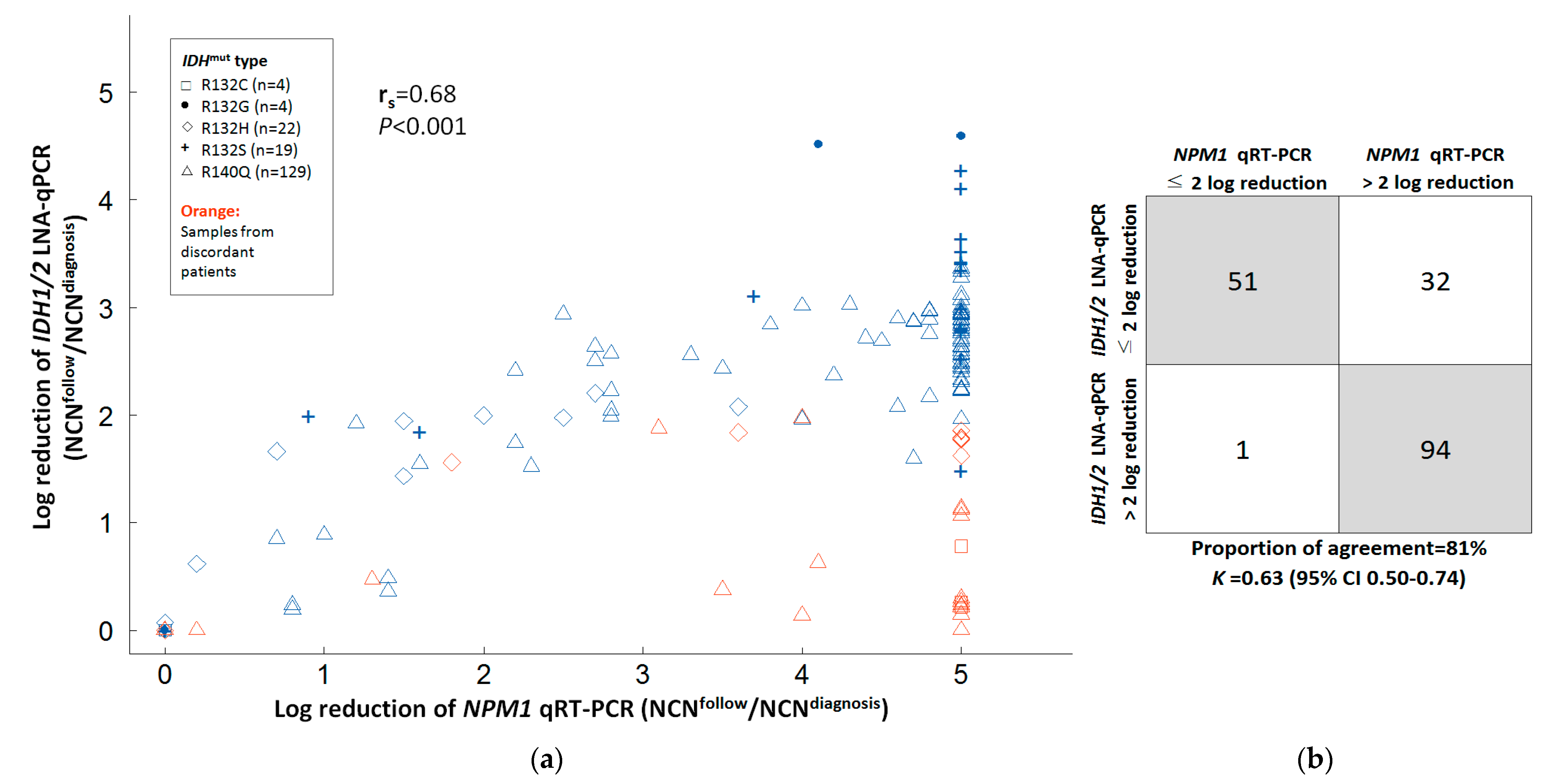
3.3. Correlation of IDH1/2 LNA-qPCR MRD with NPM1 RT-qPCR MRD and Hematologic Remission Status

Of the 25 IDH1-mutated AML patients, nine had NPM1 mutation RT-qPCR MRD for comparison. Of the nine coexisting NPM1 and IDH1 mutated patients, six had good correlations among IDH1 LNA-qPCR MRD, NPM1 RT-qPCR, and marrow blast percentage (Figure S2), while three had persistently high IDH1 LNA-qPCR MRD (≤2 log reduction) but with low NPM1 RT-qPCR MRD and marrow blasts <5% (Figure S3a). Of the 16 IDH1-mutated patients without NPM1 mutation, eight patients with marrow blasts <5% had IDH1 LNA-qPCR MRD > 2 log reduction (Figure S4), four patients with refractory AML and marrow blasts >5% (ranged 9.7% to 59.3%) had IDH1 LNA-qPCR MRD ≤ 2 log reduction (Figure S5), and four patients with marrow blasts <5% had persistent IDH1 qPCR MRD ≤ 2 log reduction (Figure S3b). The concordance rate of IDH1 LNA-qPCR MRD with remission status and/or NPM1 MRD was 72.0% (18/25).
Among the 63 IDH2-mutated AML patients, 22 had concurrent NPM1 mutation RT-qPCR MRD for comparison, 17 had good concordance between IDH2 LNA-qPCR MRD, NPM1 RT-qPCR (Figure S6), and marrow blasts, while five patients had persistently high IDH2 LNA-qPCR MRD but with low NPM1 RT-qPCR MRD and marrow blasts <5% (Figure S7a). Of the 41 IDH2-mutated patients without NPM1 mutation, 29 patients had good concordance between serial follow-up for IDH2 LNA-qPCR MRD and the percentage of marrow blasts (Figure S8). Of note, IDH2 LNA-qPCR MRD elevations preceded frank hematologic relapse (Figure S8. #24, #50, #59). Six patients with marrow blasts >5% had IDH2 qPCR MRD ≤ 2 log reduction (Figure S9), but another six patients with marrow blasts <5% had persistent IDH2 qPCR MRD ≤ 2 log reductions (Figure S7b). The concordance rate of IDH2 LNA-qPCR MRD with remission status and/or NPM1 MRD was 82.5% (52/63).
Of the 25 relapsed patients with good concordance between IDH1/2 LNA-qPCR MRD and diagnosis-remission status, all patients retained IDH1/2 mutation at relapse, and the stability of IDH1/2 LNA-qPCR at AML relapse was 100% (25/25). The IDH1/2 LNA-qPCR MRD kinetics are summarized in Figure S10 based on concordance and remission status. Overall, the concordance rate of IDH1/2 LNA-qPCR MRD with remission status or NPM1 MRD was 79.5% (70/88).

3.4. Correlation between IDH1/2 LNA-qPCR MRD, NPM1 RT-qPCR, and Marrow Blasts in Serial BM Samples

Of 554 serial BM samples from all patients (median sample number per patient 5.5, range 2–16), there was a positive correlation between the log reduction of IDH1/2 LNA-qPCR and BM blasts with Spearman correlation coefficient (rs) of 0.73 (p << 0.001, Figure 3a). The proportion of concordance was 70% (K = 0.44, 95% CI 0.37–0.51) between log reduction of IDH1/2 LNA-qPCR (≤2 log or >2 log) and BM blasts (≥5% or <5%) in all samples (Figure 3b).
In 178 serial BM samples from 25 patients with concurrent NPM1 mutation (median sample number per patient 4.5, range 2–12), there was a positive correlation between the log reduction of IDH1/2 LNA-qPCR and NPM1 RT-qPCR MRD with rs of 0.68 (p << 0.001, Figure 4a). The concordance rate was 81% (K = 0.63, 95% CI 0.50–0.74) between log reduction of IDH1/2 LNA-qPCR (≤2 log or >2 log) and NPM1 RT-qPCR (≤2 log or >2 log) MRD in all samples (Figure 4b).

3.5. Correlation of IDH1/2 LNA-qPCR MRD with Clinical and Genetic Characteristics of AML Patients

The difference in MRD results between NPM1 RT-qPCR MRD and IDH1/2 LNA-qPCR methods was analyzed for patients with good correlations and IDH1/2 LNA-qPCR MRD > 2 log reduction. The median log reduction of MRD was 5 log (range 2.7–5.0) by NPM1 RT-qPCR and 2.9 (range 2.0–4.6) by IDH1/2 LNA-qPCR. The level of MRD log reduction was significantly higher by NPM1 RT-qPCR than by IDH1/2 LNA-qPCR (p << 0.001), with a median difference of 2.0 log (range −0.4–2.8).
Patients with a good correlation between IDH1/2 LNA-qPCR MRD and AML remission status were significantly younger than patients without correlation (median age 53 vs. 61 years, p = 0.025). Patients with FLT3 mutations (either FLT3-ITD or FLT3-TKD) had significantly higher concordance rates between IDH1/2 LNA-qPCR MRD and AML remission status than patients without FLT3 mutations (96% vs. 71%, p = 0.012). The diagnostic IDH1/2 NCN, white blood cell counts, hemoglobin, platelet counts, percentages of circulating or marrow blasts, and the mutation status of NPM1 had no effect on the correlation between IDH1/2 LNA-qPCR MRD and AML remission status (Table 3).
To explore the clonal evolution of IDH1/2 mutated AML patients in the different stages, we detected co-mutations with allele frequencies in one patient with discordant IDH1-R132H MRD and another patient with concordant IDH2-R140Q MRD at the diagnosis, CR, and relapse samples. The patient with discordant IDH1-R132H MRD had stable clone sizes of DNMT3A mutation and IDH2-R140Q at diagnosis, CR, and relapse, but lost STAG2 mutation and acquired CEBPA mutation at relapse (Figure 5a). Another patient with concordant IDH2-R140Q had reduced allele frequency of IDH2-R140Q and other co-mutations at CR, but IDH2-R140Q and co-mutations at diagnosis re-expanded at relapse (Figure 5b).

3.6. Outcome Impact of IDH1/2 LNA-qPCR MRD at the End of Induction

For patients at first CR after induction therapy, those who achieved IDH1/2 LNA-qPCR MRD > 2 log reduction had significantly better RFS compared to patients with IDH1/2 LNA-qPCR MRD ≤ 2 log reduction (3-year RFS rates 52.9% [95% CI 35.0%–80.0%] vs. 31.9% [95% CI 18.7%–54.4%], p = 0.007, Figure 6a). IDH1/2 LNA-qPCR MRD > 2 log reduction did not predict a longer OS than patients with IDH1/2 LNA-PCR MRD ≤ 2 log reduction (3-year OS rates 71.8% [95% CI 55.7%–92.5%] vs. 41.4% [95% CI 26.3%–65.0%], p = 0.2; Figure 6b). A significantly higher CIR was observed for patients with IDH1/2 LNA-qPCR MRD ≤ 2 log reduction than patients with IDH1/2 qPCR MRD > 2 log reduction (3-year CIR 64.5% vs. 44.5%, p = 0.012; Figure 6c).
Based on IDH1/2 mutation subtypes, IDH2-R140 post-induction MRD ≤ 2 log reduction was associated with significantly inferior RFS (3-year RFS rates 24.2% [95% CI 11.0%–53.3%] vs. 48.0% [95% CI 24.6%–93.8%], p = 0.001) and higher CIR (3-year CIR rates 71.4% vs. 42.0%, p = 0.002) compared to post-induction IDH2-R140 MRD > 2 log reduction (n = 38), while IDH1-R132 mutation (n = 18) or IDH2-R172 mutation (n = 8) MRD had no impact on RFS and CIR.

3.7. Outcome Impact of IDH1/2 LNA-qPCR MRD during Consolidation

For CR patients after one or two cycles of consolidation chemotherapy, we did not observe significant differences in RFS (3-year RFS 56.1% [95% CI 40.1%–78.5%] vs. 31.1% [95% CI 16.8%–57.9%], p = 0.07, Figure 6d) and OS (3-year OS 59.5% [95% CI 44.0%–80.4%] vs. 54.8% [95% CI 37.3%–80.5%], p = 0.9, Figure 6e) between patients with IDH1/2 LNA-qPCR MRD > 2 log reduction and ≤ 2 log reduction. The CIR was significantly higher for patients with IDH1/2 LNA-qPCR MRD ≤ 2 log reduction than patients with IDH1/2 LNA- qPCR MRD > 2 log reduction (3-year CIR 68.8% vs. 42.3%, p = 0.019; Figure 6f)
Based on IDH1/2 mutation subtypes, IDH2-R140 MRD ≤ 2 log reduction during consolidation was associated with a trend of inferior RFS (3-year RFS rates 26.8% [95% CI 10.9%–66.0%] vs. 44.2% [95% CI 24.7%–79.1%], p = 0.06) and a significantly higher CIR rate (3-year CIR rates 73.2% vs. 44.8%, p = 0.013) compared to IDH2-R140 MRD > 2 log reduction during consolidation (n = 37), while IDH1-R132 mutation (n = 17) or IDH2-R172 mutation (n = 8) had no impact on RFS and CIR.

4. Discussion

Our cohort of 88 adult patients with IDH1/2-mutated AML showed that the concordance rate of IDH1/2 LNA-qPCR MRD with remission status was 79.5%. There was a positive correlation (rs = 0.68) and a substantial agreement (81%, K = 0.74) between IDH1/2 LNA-qPCR and NPM1 RT-qPCR MRD. The median difference in MRD results was 2 logs between IDH1/2 LNA-qPCR and NPM1 RT-qPCR. Patients with FLT3 mutation or younger age have a higher concordance rate between IDH1/2 LNA-qPCR MRD, remission status, or NPM1 RT-qPCR MRD. At the end of induction and during consolidation, IDH1/2 LNA-qPCR MRD had a significant impact on CIR or RFS.
Because of the single nucleotide substitution of IDH1/2 mutations, the sensitivity and specificity of IDH1/2-directed MRD are limited if there is a small proportion of IDH1/2 mutations in the wild-type background. IDH1/2-targeted MRD using ddPCR assays reached a sensitivity of 0.1% due to the problem of a relatively high background observed in negative controls, which consisted of false-positive droplets [19]. LNA primers could block the wild-type allele, avoid the false-positive signals resulting from polymerase errors during PCR amplification steps, and allow for higher sensitivity and specificity of IDH1/2 LNA-qPCR [24,31]. Previous studies have applied diverse methods to detect IDH1/2-targeted residual disease by using NGS, ddPCR, or LNA-qPCR. The sensitivity of IDH1/2 MRD was reported to be 0.1%–0.2% by ddPCR [18,19,32], < 0.001%–1% by NGS [20,21,22], and 10−4 by LNA-qPCR [24]. LNA-qPCR assay for IDH1/2 mutations theoretically has a sensitivity of 1 × 10⁻⁴, allowing for a deeper evaluation of molecular response. However, we observed a low copy number background of IDH1/2 mutations by LNA-qPCR in normal BM samples, especially for IDH1-R132C (c.394C > T) and IDH1-R132H (c.395G > A). There was also a higher mean variant allele fraction of IDH1-R132C and IDH1-R132H than other IDH1/2 mutant types detected by ddPCR assay on gDNA extracted from IDH1/2 wild-type pooled blood lymphocytes in the study by Ferret et al. [19]. The low copy number background of IDH1/2 LNA-qPCR in normal BM samples seems to be related to the mutant subtype rather than age (Figure S1). Clonal hematopoiesis is unlikely to be the cause of the low copy number background of IDH1/2 mutants. A possible explanation for this background noise is PCR-induced substitution error caused by cytosine deamination and increased C > T and G > A transition errors, which exhibit strong sequence context dependency [33,34].
We observed a persistently high level of IDH1/2 mutation at CR in 20% of IDH1/2-mutated patients. The discrepancy of persistent IDH1/2 LNA-qPCR MRD in CR might be attributed to clonal hematopoiesis in pre-leukemic hematopoietic stem cells, which provide a potential reservoir for leukemic progression [35,36]. This hypothesis is supported by the observation of persistent IDH1-R132H and DNMT3A mutation, loss of STAG2 mutation at CR, and acquisition of CEBPA mutation in one patient in this study. Previous studies reported that persistent IDH1/2 mutants in AML patients at CR occurred in 39–62% by NGS and 7% by ddPCR [19,20,21]. We found that NPM1 RT-qPCR MRD showed a median difference of two log reductions compared with IDH1/2 LNA-qPCR MRD in AML. Low copy number false-positive IDH1/2 LNA-qPCR background due to PCR-induced substitution errors of IDH1/2 mutant and detection of gDNA by IDH1/2 LNA-qPCR rather than cDNA by NPM1 RT-qPCR, where a single cell may contain multiple copies of RNAs, and low NCN of IDH1/2 mutants at initial diagnosis might all contribute to the limited sensitivity of IDH1/2 LNA-qPCR. Due to this, RT-qPCR MRD targeting NPM1 mutant rather than IDH1/2 mutant is recommended for AML patients with both NPM1 and IDH1/2 mutations.
The use of FLT3 mutations as a genetic marker for MRD is controversial. This is due to unstable FLT3 mutations that are acquired or lost during the disease course of AML [37,38]. It lacks standardization of the detection methods for FLT3 MRD regarding mutation types covered and limited sensitivity [39,40,41]. The ELN MRD working party recommends that the use of FLT3 mutations is appropriate in combination with other additional MRD markers [26]. NGS-MRD for multiple genetic markers is not widely available. Only one of 25 (4.0%) AML patients with concurrent IDH1/2 and FLT3 mutations had a discrepancy between IDH1/2 mutant MRD and remission-relapse status. Based on this observation, IDH1/2 mutation might be a helpful surrogate or additional genetic marker for MRD monitoring in patients with concurrent FLT3 mutation and wild-type NPM1.
The loss of IDH1/2 mutation at relapse of AML has been rarely reported. Brambat et al. reported the loss of IDH2 mutation at relapse in one of 14 post-transplantation AML patients [18]. All IDH1/2-mutated AML patients who had achieved CR retained IDH1/2 mutant at relapse. The small number of cases detected for IDH1/2-MRD at relapse in this cohort might preclude the finding of IDH1/2 disappearing at relapse. Whether IDH1/2 mutation could be a stable genetic MRD marker will need to be validated in a larger cohort. The stability of NPM1 mutation, a consensus recommended MRD marker for AML, was reported to be 99%–100% in large cohorts [14,42], but the loss of NPM1 mutation at relapse has been observed in 7% to 25% of AML patients in smaller series [17,43,44,45,46,47]. Because of the marked genetic heterogeneity of AML, no single genetic MRD marker can be applied to all patients. As the role of MRD in the management of AML patients is becoming more and more important, IDH1/2 mutation might be a surrogate MRD marker for a subset of AML patients without recurrent fusion genes or NPM1 mutations.
A large cohort study of the French ALFA group reported that IDH1/2 mutant allele burden < 0.2% by ddPCR after induction was a significant predictor of prolonged event-free survival [19]. The MD Anderson Cancer Center study showed that persistent IDH1/2 mutation in remission detected by NGS at a sensitivity of 1% predicted relapse in multivariate analyses [20]. The results of this study further support the impact of IDH1/2 MRD on relapse. In a recent study reported by Bill et al., IDH1-R132 and IDH2-R172 MRD positivity in remission at HSCT was associated with an increased risk of relapse, while IDH2-R140 mutations did not [48]. We observed that IDH1/2 LNA-qPCR MRD elevations preceded frank hematological relapse and confirmed that IDH1/2 LNA-qPCR MRD after induction therapy and during consolidation could predict relapse in AML patients but did not significantly predict OS. However, we found that IDH2-R140 MRD positivity post-induction and during consolidation could predict an increased risk of relapse, whereas IDH1-R132 or IDH2-R172 did not.
Studies showed that early pre-emptive interventions might improve the outcomes of AML patients in CR with persistent positive fusion genes or NPM1 mutation [49,50]. However, the feasibility, outcome prediction, and guidance of pre-emptive therapy based on IDH1/2-MRD in AML patients was rarely investigated and thus cannot be recommended, mainly due to the concern of IDH1/2 mutants as pre-leukemic clones as well as the possibility of loss or gain of IDH1/2 mutants at relapse [26]. For patients with IDH1/2 MRD > 2 log reduction at the end of induction, the IDH1/2 MRD correlated with NPM1 MRD and morphological remission status, and the loss of IDH1/2 mutation at relapse rarely occurred in the current and previous studies [18,19]. Patients with a deep IDH1/2 molecular response after achieving CR will benefit from serial IDH1/2 MRD monitoring to identify early relapse. On the other hand, patients with persistently high IDH1/2 MRD at first CR should not be followed by IDH1/2-targeted MRD alone. It is still unclear whether early pre-emptive therapy will benefit AML patients in CR who have increasing IDH1/2 mutant MRD during follow-up, which requires further evaluation in prospective clinical trials.

5. Conclusions

Our study showed that the correlation and stability of IDH1/2 MRD by LNA-qPCR was good in patients who had concordantly reduced IDH1/2 MRD in morphologic CR. IDH1/2 LNA-qPCR MRD > 2 log reduction at first CR and during consolidation is associated with improved CIR or RFS. IDH1/2 LNA-qPCR MRD has the potential to become a clinical tool to monitor deep treatment response and predict relapse for IDH1/2 mutated AML patients, especially for those who are younger, with reduced IDH1/2 MRD at first CR, or concurrent FLT3 mutation.

Supplementary Materials

The following supporting information can be downloaded at: https://www.mdpi.com/article/10.3390/cancers14246205/s1, Table S1. IDH1/2 LNA-qPCR of 19 normal bone marrow samples from healthy donors. Figure S1. IDH1/2 LNA-qPCR of 19 normal bone marrow samples from healthy donors according to age. Figure S2. MRD kinetics of individual patients with good correlation between IDH1 LNA-qPCR MRD, NPM1 RT-qPCR, or remission-relapse status. Figure S3. MRD kinetics of individual patients with discordant IDH1 LNA-qPCR MRD, NPM1 RT-qPCR MRD, or remission status. Figure S4. MRD kinetics of individual patients with good correlation between IDH1 LNA-qPCR MRD and hematological remission-relapse status. Figure S5. MRD kinetics of individual patients with persistent high IDH1 LNA-qPCR and relapse refractory status. Figure S6. MRD kinetics of individual patients with good correlation between IDH2 LNA-qPCR MRD, NPM1 RT-qPCR, or remission-relapse status. Figure S7. MRD kinetics of individual patients with discordant IDH2 LNA-qPCR MRD, NPM1 RT-qPCR MRD, and hematological remission status. Figure S8. MRD kinetics of individual patients with good correlation between IDH2 LNA-qPCR MRD and hematological remission-relapse status. Figure S9. MRD kinetics of individual patients with persistent high IDH2 LNA-qPCR and relapse refractory status. Figure S10. Summary of MRD kinetics according to the concordance between IDH1/2 LNA-qPCR MRD and the disease status.

Author Contributions

Conceptualization, L.-Y.S., H.-W.K. and M.-C.K.; Methodology, L.-Y.S., Y.-J.H. and C.-F.H.; Validation, L.-Y.S., H.-W.K. and Y.-J.H.; Formal Analysis, H.-W.K. and L.-Y.S.; Investigation, Y.-J.H., S.-F.H. and C.-F.H.; Resources, L.-Y.S., H.-W.K., M.-C.K., H.C., Y.-S.H., T.-L.L., C.-W.O., M.-Y.L., J.-H.W. and C.-C.C.; Data Curation, L.-Y.S., H.-W.K., M.-C.K., H.C., Y.-S.H., T.-L.L., C.-W.O., M.-Y.L., J.-H.W. and C.-C.C.; Writing—Original Draft Preparation, H.-W.K.; Writing—Review & Editing, L.-Y.S.; Visualization, H.-W.K.; Supervision, L.-Y.S.; Project Administration, L.-Y.S., H.-W.K. and M.-C.K.; Funding Acquisition, L.-Y.S., H.-W.K. and M.-C.K. All authors have read and agreed to the published version of the manuscript.

Funding

The project was funded by the Ministry of Health and Warfare, Taiwan (grant MOHW107-TDU-B-212-114011, MOHW108-TDU-B-212-124011, MOHW109-TDU-B-212-134011, MOHW110-TDU-B-212-144011 to LYS, MCK, HWK), Celgene (grant XPRPG3G0121~4 to LYS, HWK, MCK), and Chang Gung Memorial Hospital (grant CMRPG3D1521~4 to HWK).

Institutional Review Board Statement

The study was conducted at multiple medical centers (Chang Gung Memorial Hospital at Linkou, Kaohsiung, and Chiayi; China Medical University Hospital), approved by the Institutional Review Board of Chang Gung Memorial Hospital (99-0112B, 102-3031B, 201700154B0, and 201701453A3) and China Medical University Hospital (CMUH-HO-AML-10701), and conducted in accordance with the Declaration of Helsinki.

Informed Consent Statement

Informed consent was obtained from enrolled patients.

Data Availability Statement

The datasets used and/or analyzed in the current study are available from the corresponding author on reasonable request.

Acknowledgments

The authors are grateful to Po-Nan Wang, Shun-Ren Shih, Yi-Jun Su, Tran-Der Tan, Ming-Chung Wang, Jing-Yuan Kuo, Ci-En Huang, Qin-Yuan Wang, Yu-Ying Wu, and Sheng-Yan Xiao for contributing patients to this study.

Conflicts of Interest

LYS, HWK, and MCK received research funding from Celgene. LYS received research support from Novartis (Taiwan) Co., Ltd. The other authors declare no conflict of interest.

Abbreviations

BM: Bone marrow; IDH: Isocitrate dehydrogenase; MRD: Measurable residual disease; ddPCR: Digital PCR; NGS: Next-generation sequencing; LNA: Locked nucleic acid; qPCR: Quantitative Real-Time PCR; CR: Complete remission; ELN: European LeukemiaNet; gDNA: Genomic DNA; ALB: Albumin; NCN: Normalized copy number; RT-qPCR: Quantitative Real-Time reverse transcriptase PCR; OS: Overall survival; RFS: Relapse-free survival; CIR: Cumulative incidence of relapse; HSCT: Allogeneic hematopoietic stem cell transplantation; ITD: Internal tandem duplication; TKD: Tyrosine kinase domain.

References

  1. Dohner, H.; Weisdorf, D.J.; Bloomfield, C.D. Acute Myeloid Leukemia. N. Engl. J. Med. 2015, 373, 1136–1152. [Google Scholar] [CrossRef] [PubMed] [Green Version]
  2. Ley, T.J.; Miller, C.; Ding, L.; Raphael, B.J.; Mungall, A.J.; Robertson, A.; Hoadley, K.; Triche, T.J., Jr.; Laird, P.W.; Baty, J.D.; et al. Genomic and epigenomic landscapes of adult de novo acute myeloid leukemia. N. Engl. J. Med. 2013, 368, 2059–2074. [Google Scholar] [CrossRef] [PubMed] [Green Version]
  3. Döhner, H.; Wei, A.H.; Appelbaum, F.R.; Craddock, C.; DiNardo, C.D.; Dombret, H.; Ebert, B.L.; Fenaux, P.; Godley, L.A.; Hasserjian, R.P.; et al. Diagnosis and management of AML in adults: 2022 recommendations from an international expert panel on behalf of the ELN. Blood 2022, 140, 1345–1377. [Google Scholar] [CrossRef] [PubMed]
  4. Kats, L.M.; Reschke, M.; Taulli, R.; Pozdnyakova, O.; Burgess, K.; Bhargava, P.; Straley, K.; Karnik, R.; Meissner, A.; Small, D.; et al. Proto-oncogenic role of mutant IDH2 in leukemia initiation and maintenance. Cell Stem Cell 2014, 14, 329–341. [Google Scholar] [CrossRef] [PubMed] [Green Version]
  5. Lu, C.; Ward, P.S.; Kapoor, G.S.; Rohle, D.; Turcan, S.; Abdel-Wahab, O.; Edwards, C.R.; Khanin, R.; Figueroa, M.E.; Melnick, A.; et al. IDH mutation impairs histone demethylation and results in a block to cell differentiation. Nature 2012, 483, 474–478. [Google Scholar] [CrossRef] [Green Version]
  6. Losman, J.A.; Looper, R.E.; Koivunen, P.; Lee, S.; Schneider, R.K.; McMahon, C.; Cowley, G.S.; Root, D.E.; Ebert, B.L.; Kaelin, W.G., Jr. (R)-2-hydroxyglutarate is sufficient to promote leukemogenesis and its effects are reversible. Science 2013, 339, 1621–1625. [Google Scholar] [CrossRef]
  7. Gross, S.; Cairns, R.A.; Minden, M.D.; Driggers, E.M.; Bittinger, M.A.; Jang, H.G.; Sasaki, M.; Jin, S.; Schenkein, D.P.; Su, S.M.; et al. Cancer-associated metabolite 2-hydroxyglutarate accumulates in acute myelogenous leukemia with isocitrate dehydrogenase 1 and 2 mutations. J. Exp. Med. 2010, 207, 339–344. [Google Scholar] [CrossRef]
  8. Abbas, S.; Lugthart, S.; Kavelaars, F.G.; Schelen, A.; Koenders, J.E.; Zeilemaker, A.; van Putten, W.J.; Rijneveld, A.W.; Löwenberg, B.; Valk, P.J. Acquired mutations in the genes encoding IDH1 and IDH2 both are recurrent aberrations in acute myeloid leukemia: Prevalence and prognostic value. Blood 2010, 116, 2122–2126. [Google Scholar] [CrossRef]
  9. Boissel, N.; Nibourel, O.; Renneville, A.; Gardin, C.; Reman, O.; Contentin, N.; Bordessoule, D.; Pautas, C.; de Revel, T.; Quesnel, B.; et al. Prognostic Impact of Isocitrate Dehydrogenase Enzyme Isoforms 1 and 2 Mutations in Acute Myeloid Leukemia: A Study by the Acute Leukemia French Association Group. J. Clin. Oncol. 2010, 28, 3717–3723. [Google Scholar] [CrossRef]
  10. Marcucci, G.; Maharry, K.; Wu, Y.Z.; Radmacher, M.D.; Mrózek, K.; Margeson, D.; Holland, K.B.; Whitman, S.P.; Becker, H.; Schwind, S.; et al. IDH1 and IDH2 Gene Mutations Identify Novel Molecular Subsets Within De Novo Cytogenetically Normal Acute Myeloid Leukemia: A Cancer and Leukemia Group B Study. J. Clin. Oncol. 2010, 28, 2348–2355. [Google Scholar] [CrossRef]
  11. Schnittger, S.; Haferlach, C.; Ulke, M.; Alpermann, T.; Kern, W.; Haferlach, T. IDH1 mutations are detected in 6.6% of 1414 AML patients and are associated with intermediate risk karyotype and unfavorable prognosis in adults younger than 60 years and unmutated NPM1 status. Blood 2010, 116, 5486–5496. [Google Scholar] [CrossRef] [PubMed]
  12. Paschka, P.; Schlenk, R.F.; Gaidzik, V.I.; Habdank, M.; Krönke, J.; Bullinger, L.; Späth, D.; Kayser, S.; Zucknick, M.; Götze, K.; et al. IDH1 and IDH2 Mutations Are Frequent Genetic Alterations in Acute Myeloid Leukemia and Confer Adverse Prognosis in Cytogenetically Normal Acute Myeloid Leukemia With NPM1 Mutation without FLT3 Internal Tandem Duplication. J. Clin. Oncol. 2010, 28, 3636–3643. [Google Scholar] [CrossRef] [PubMed]
  13. Green, C.L.; Evans, C.M.; Zhao, L.; Hills, R.K.; Burnett, A.K.; Linch, D.C.; Gale, R.E. The prognostic significance of IDH2 mutations in AML depends on the location of the mutation. Blood 2011, 118, 409–412. [Google Scholar] [CrossRef] [Green Version]
  14. Ivey, A.; Hills, R.K.; Simpson, M.A.; Jovanovic, J.V.; Gilkes, A.; Grech, A.; Patel, Y.; Bhudia, N.; Farah, H.; Mason, J.; et al. Assessment of Minimal Residual Disease in Standard-Risk AML. N. Engl. J. Med. 2016, 374, 422–433. [Google Scholar] [CrossRef] [Green Version]
  15. Terwijn, M.; van Putten, W.L.; Kelder, A.; van der Velden, V.H.; Brooimans, R.A.; Pabst, T.; Maertens, J.; Boeckx, N.; de Greef, G.E.; Valk, P.J.; et al. High prognostic impact of flow cytometric minimal residual disease detection in acute myeloid leukemia: Data from the HOVON/SAKK AML 42A study. J. Clin. Oncol. 2013, 31, 3889–3897. [Google Scholar] [CrossRef] [PubMed]
  16. Yin, J.A.; O’Brien, M.A.; Hills, R.K.; Daly, S.B.; Wheatley, K.; Burnett, A.K. Minimal residual disease monitoring by quantitative RT-PCR in core binding factor AML allows risk stratification and predicts relapse: Results of the United Kingdom MRC AML-15 trial. Blood 2012, 120, 2826–2835. [Google Scholar] [CrossRef] [Green Version]
  17. Krönke, J.; Schlenk, R.F.; Jensen, K.O.; Tschürtz, F.; Corbacioglu, A.; Gaidzik, V.I.; Paschka, P.; Onken, S.; Eiwen, K.; Habdank, M.; et al. Monitoring of minimal residual disease in NPM1-mutated acute myeloid leukemia: A study from the German-Austrian acute myeloid leukemia study group. J. Clin. Oncol. 2011, 29, 2709–2716. [Google Scholar] [CrossRef]
  18. Brambati, C.; Galbiati, S.; Xue, E.; Toffalori, C.; Crucitti, L.; Greco, R.; Sala, E.; Crippa, A.; Chiesa, L.; Soriani, N.; et al. Droplet digital polymerase chain reaction for DNMT3A and IDH1/2 mutations to improve early detection of acute myeloid leukemia relapse after allogeneic hematopoietic stem cell transplantation. Haematologica 2016, 101, e157–e161. [Google Scholar] [CrossRef]
  19. Ferret, Y.; Boissel, N.; Helevaut, N.; Madic, J.; Nibourel, O.; Marceau-Renaut, A.; Bucci, M.; Geffroy, S.; Celli-Lebras, K.; Castaigne, S.; et al. Clinical relevance of IDH1/2 mutant allele burden during follow-up in acute myeloid leukemia. A study by the French ALFA group. Haematologica 2018, 103, 822–829. [Google Scholar] [CrossRef] [Green Version]
  20. Ok, C.Y.; Loghavi, S.; Sui, D.; Wei, P.; Kanagal-Shamanna, R.; Yin, C.C.; Zuo, Z.; Routbort, M.J.; Tang, G.; Tang, Z.; et al. Persistent IDH1/2 mutations in remission can predict relapse in patients with acute myeloid leukemia. Haematologica 2019, 104, 305–311. [Google Scholar] [CrossRef]
  21. Shumilov, E.; Flach, J.; Joncourt, R.; Porret, N.; Wiedemann, G.; Novak, U.; Gfeller, E.; Jeker, B.; Amstutz, U.; Pabst, T.; et al. Clinical value of molecular MRD monitoring by next-generation sequencing in patients with IDH2 mutated AML. Leuk. Lymphoma 2019, 60, 2588–2590. [Google Scholar] [CrossRef] [PubMed]
  22. Debarri, H.; Lebon, D.; Roumier, C.; Cheok, M.; Marceau-Renaut, A.; Nibourel, O.; Geffroy, S.; Helevaut, N.; Rousselot, P.; Gruson, B.; et al. IDH1/2 but not DNMT3A mutations are suitable targets for minimal residual disease monitoring in acute myeloid leukemia patients: A study by the Acute Leukemia French Association. Oncotarget 2015, 6, 42345–42353. [Google Scholar] [CrossRef] [PubMed] [Green Version]
  23. Jeziskova, I.; Razga, F.; Toskova, M.; Dvorakova, D.; Timilsina, S.; Mayer, J.; Racil, Z. Quantitative detection of IDH2 mutation for minimal residual disease monitoring in patients with acute myeloid leukemia and its comparison with mutations in NPM1 gene. Leuk. Lymphoma 2013, 54, 867–870. [Google Scholar] [CrossRef] [PubMed]
  24. Abdelhamid, E.; Besbes, S.; Renneville, A.; Nibourel, O.; Helevaut, N.; Preudhomme, C.; Soua, Z. Minimal Residual Disease assessment of IDH1/2 mutations in Acute Myeloid Leukemia by LNA-RQ-PCR. Tunis. Med. 2016, 94, 190–197. [Google Scholar]
  25. Chou, W.C.; Peng, K.Y.; Lei, W.C.; Ko, B.S.; Tsay, W.; Kuo, C.H.; Tien, H.F. Persistence of mutant isocitrate dehydrogenase in patients with acute myeloid leukemia in remission. Leukemia 2012, 26, 527–529. [Google Scholar] [CrossRef] [PubMed] [Green Version]
  26. Heuser, M.; Freeman, S.D.; Ossenkoppele, G.J.; Buccisano, F.; Hourigan, C.S.; Ngai, L.L.; Tettero, J.M.; Bachas, C.; Baer, C.; Béné, M.C. 2021 Update on MRD in acute myeloid leukemia: A consensus document from the European LeukemiaNet MRD Working Party. Blood 2021, 138, 2753–2767. [Google Scholar] [CrossRef]
  27. Khoury, J.D.; Solary, E.; Abla, O.; Akkari, Y.; Alaggio, R.; Apperley, J.F.; Bejar, R.; Berti, E.; Busque, L.; Chan, J.K.C.; et al. The 5th edition of the World Health Organization Classification of Haematolymphoid Tumours: Myeloid and Histiocytic/Dendritic Neoplasms. Leukemia 2022, 36, 1703–1719. [Google Scholar] [CrossRef]
  28. Kao, H.W.; Liang, D.C.; Wu, J.H.; Kuo, M.C.; Wang, P.N.; Yang, C.P.; Shih, Y.S.; Lin, T.H.; Huang, Y.H.; Shih, L.Y. Gene mutation patterns in patients with minimally differentiated acute myeloid leukemia. Neoplasia 2014, 16, 481–488. [Google Scholar] [CrossRef] [Green Version]
  29. Pongers-Willemse, M.J.; Verhagen, O.J.; Tibbe, G.J.; Wijkhuijs, A.J.; de Haas, V.; Roovers, E.; van der Schoot, C.E.; van Dongen, J.J. Real-time quantitative PCR for the detection of minimal residual disease in acute lymphoblastic leukemia using junctional region specific TaqMan probes. Leukemia 1998, 12, 2006–2014. [Google Scholar] [CrossRef] [Green Version]
  30. Gorello, P.; Cazzaniga, G.; Alberti, F.; Dell’Oro, M.G.; Gottardi, E.; Specchia, G.; Roti, G.; Rosati, R.; Martelli, M.F.; Diverio, D.; et al. Quantitative assessment of minimal residual disease in acute myeloid leukemia carrying nucleophosmin (NPM1) gene mutations. Leukemia 2006, 20, 1103–1108. [Google Scholar] [CrossRef] [Green Version]
  31. Di Giusto, D.A.; King, G.C. Strong positional preference in the interaction of LNA oligonucleotides with DNA polymerase and proofreading exonuclease activities: Implications for genotyping assays. Nucleic Acids Res. 2004, 32, e32. [Google Scholar] [CrossRef] [PubMed]
  32. Petrova, L.; Vrbacky, F.; Lanska, M.; Zavrelova, A.; Zak, P.; Hrochova, K. IDH1 and IDH2 mutations in patients with acute myeloid leukemia: Suitable targets for minimal residual disease monitoring? Clin. Biochem. 2018, 61, 34–39. [Google Scholar] [CrossRef] [PubMed]
  33. Ma, X.; Shao, Y.; Tian, L.; Flasch, D.A.; Mulder, H.L.; Edmonson, M.N.; Liu, Y.; Chen, X.; Newman, S.; Nakitandwe, J.; et al. Analysis of error profiles in deep next-generation sequencing data. Genome Biol. 2019, 20, 50. [Google Scholar] [CrossRef] [PubMed] [Green Version]
  34. Chen, G.; Mosier, S.; Gocke, C.D.; Lin, M.T.; Eshleman, J.R. Cytosine deamination is a major cause of baseline noise in next-generation sequencing. Mol. Diagn. Ther. 2014, 18, 587–593. [Google Scholar] [CrossRef] [PubMed] [Green Version]
  35. Shlush, L.I.; Zandi, S.; Mitchell, A.; Chen, W.C.; Brandwein, J.M.; Gupta, V.; Kennedy, J.A.; Schimmer, A.D.; Schuh, A.C.; Yee, K.W.; et al. Identification of pre-leukaemic haematopoietic stem cells in acute leukaemia. Nature 2014, 506, 328–333. [Google Scholar] [CrossRef] [Green Version]
  36. Cappelli, L.V.; Meggendorfer, M.; Baer, C.; Nadarajah, N.; Hutter, S.; Jeromin, S.; Dicker, F.; Kern, W.; Haferlach, T.; Haferlach, C.; et al. Indeterminate and oncogenic potential: CHIP vs. CHOP mutations in AML with NPM1 alteration. Leukemia 2022, 36, 394–402. [Google Scholar] [CrossRef]
  37. Cloos, J.; Goemans, B.F.; Hess, C.J.; van Oostveen, J.W.; Waisfisz, Q.; Corthals, S.; de Lange, D.; Boeckx, N.; Hählen, K.; Reinhardt, D.; et al. Stability and prognostic influence of FLT3 mutations in paired initial and relapsed AML samples. Leukemia 2006, 20, 1217–1220. [Google Scholar] [CrossRef]
  38. Murphy, K.M.; Levis, M.; Hafez, M.J.; Geiger, T.; Cooper, L.C.; Smith, B.D.; Small, D.; Berg, K.D. Detection of FLT3 internal tandem duplication and D835 mutations by a multiplex polymerase chain reaction and capillary electrophoresis assay. J. Mol. Diagn. 2003, 5, 96–102. [Google Scholar] [CrossRef] [Green Version]
  39. Carbonell, D.; Chicano, M.; Cardero, A.J.; Gómez-Centurión, I.; Bailén, R.; Oarbeascoa, G.; Martínez-Señarís, D.; Franco, C.; Muñiz, P.; Anguita, J.; et al. FLT3-ITD Expression as a Potential Biomarker for the Assessment of Treatment Response in Patients with Acute Myeloid Leukemia. Cancers 2022, 14, 4006. [Google Scholar] [CrossRef]
  40. Levis, M.J.; Perl, A.E.; Altman, J.K.; Gocke, C.D.; Bahceci, E.; Hill, J.; Liu, C.; Xie, Z.; Carson, A.R.; McClain, V.; et al. A next-generation sequencing-based assay for minimal residual disease assessment in AML patients with FLT3-ITD mutations. Blood Adv. 2018, 2, 825–831. [Google Scholar] [CrossRef]
  41. Scholl, S.; Krause, C.; Loncarevic, I.F.; Müller, R.; Kunert, C.; Wedding, U.; Sayer, H.G.; Clement, J.H.; Höffken, K. Specific detection of Flt3 point mutations by highly sensitive real-time polymerase chain reaction in acute myeloid leukemia. J. Lab. Clin. Med. 2005, 145, 295–304. [Google Scholar] [CrossRef]
  42. Schnittger, S.; Kern, W.; Tschulik, C.; Weiss, T.; Dicker, F.; Falini, B.; Haferlach, C.; Haferlach, T. Minimal residual disease levels assessed by NPM1 mutation-specific RQ-PCR provide important prognostic information in AML. Blood 2009, 114, 2220–2231. [Google Scholar] [CrossRef] [PubMed] [Green Version]
  43. Krönke, J.; Bullinger, L.; Teleanu, V.; Tschürtz, F.; Gaidzik, V.I.; Kühn, M.W.; Rücker, F.G.; Holzmann, K.; Paschka, P.; Kapp-Schwörer, S.; et al. Clonal evolution in relapsed NPM1-mutated acute myeloid leukemia. Blood 2013, 122, 100–108. [Google Scholar] [CrossRef] [PubMed] [Green Version]
  44. Papadaki, C.; Dufour, A.; Seibl, M.; Schneider, S.; Bohlander, S.K.; Zellmeier, E.; Mellert, G.; Hiddemann, W.; Spiekermann, K. Monitoring minimal residual disease in acute myeloid leukaemia with NPM1 mutations by quantitative PCR: Clonal evolution is a limiting factor. Br. J. Haematol. 2009, 144, 517–523. [Google Scholar] [CrossRef] [PubMed]
  45. Pettersson, L.; Levéen, P.; Axler, O.; Dvorakova, D.; Juliusson, G.; Ehinger, M. Improved minimal residual disease detection by targeted quantitative polymerase chain reaction in Nucleophosmin 1 type a mutated acute myeloid leukemia. Genes Chromosomes Cancer 2016, 55, 750–766. [Google Scholar] [CrossRef]
  46. Thol, F.; Kölking, B.; Damm, F.; Reinhardt, K.; Klusmann, J.H.; Reinhardt, D.; von Neuhoff, N.; Brugman, M.H.; Schlegelberger, B.; Suerbaum, S.; et al. Next-generation sequencing for minimal residual disease monitoring in acute myeloid leukemia patients with FLT3-ITD or NPM1 mutations. Genes Chromosomes Cancer 2012, 51, 689–695. [Google Scholar] [CrossRef] [PubMed]
  47. Hubmann, M.; Köhnke, T.; Hoster, E.; Schneider, S.; Dufour, A.; Zellmeier, E.; Fiegl, M.; Braess, J.; Bohlander, S.K.; Subklewe, M.; et al. Molecular response assessment by quantitative real-time polymerase chain reaction after induction therapy in NPM1-mutated patients identifies those at high risk of relapse. Haematologica 2014, 99, 1317–1325. [Google Scholar] [CrossRef] [PubMed]
  48. Bill, M.; Jentzsch, M.; Bischof, L.; Kohlschmidt, J.; Grimm, J.; Schmalbrock, L.K.; Backhaus, D.; Brauer, D.; Goldmann, K.; Franke, G.N.; et al. Impact of IDH1 and IDH2 mutation detection at diagnosis and in remission in patients with AML receiving allogeneic transplantation. Blood Adv. 2022. online ahead of print. [Google Scholar] [CrossRef]
  49. Bataller, A.; Oñate, G.; Diaz-Beyá, M.; Sampol, A.; Coll, R.; Vall-Llovera, F.; Oliver-Caldés, A. Acute myeloid leukemia with NPM1 mutation and favorable European LeukemiaNet category: Outcome after preemptive intervention based on measurable residual disease. Br. J. Haematol. 2020, 191, 52–61. [Google Scholar] [CrossRef]
  50. Platzbecker, U.; Middeke, J.M.; Sockel, K.; Herbst, R.; Wolf, D.; Baldus, C.D.; Oelschlägel, U.; Mütherig, A.; Fransecky, L.; Noppeney, R.; et al. Measurable residual disease-guided treatment with azacitidine to prevent haematological relapse in patients with myelodysplastic syndrome and acute myeloid leukaemia (RELAZA2): An open-label, multicentre, phase 2 trial. Lancet Oncol. 2018, 19, 1668–1679. [Google Scholar] [CrossRef]
Figure 1. Standard curves constructed by IDH1/2 mutant plasmids. (a) IDH1-R132H. (b) IDH2-R172K. (c) IDH2-R140Q. (d) IDH2-R140W.
Cancers 14 06205 g001
Figure 2. Changes in normalized copy numbers of IDH1/2 mutants at diagnosis, post-induction first complete remission, during consolidation therapy, and relapse.
Cancers 14 06205 g002
Figure 3. Correlation and agreement between IDH1/2 LNA-qPCR MRD and marrow blasts in serial bone marrow samples. (a) Correlation between IDH1/2 LNA-qPCR MRD and marrow blasts by log reduction. Gray dashed lines showed two-log reduction of IDH1/2 LNA-qPCR and 1.3-log (5%) of marrow blasts percentage. Orange points represent samples from patients with discordant reduction of IDH1/2 LNA-qPCR in complete remission status. Blue points represent samples from patients with regeneration marrow. (b) Agreement between IDH1/2 LNA-qPCR MRD and marrow blasts of all samples.
Cancers 14 06205 g003
Figure 4. Correlation and agreement between IDH1/2 LNA-qPCR MRD and NPM1 RT-qPCR MRD in serial bone marrow samples. (a) Correlation between IDH1/2 LNA-qPCR and NPM1 RT-qPCR MRD by log reduction. Gray dashed lines showed two-log reduction of IDH1/2 LNA-qPCR and NPM1 RT-qPCR MRD. Orange points represent samples from patients with discordant reduction of IDH1/2 LNA-qPCR in complete remission status. (b) Agreement between IDH1/2 LNA-qPCR and NPM1 RT-qPCR MRD for all samples.
Cancers 14 06205 g004
Figure 5. Changes in allele frequency of concurrent mutations (left) and hypothesized clonal evolution (right) at diagnosis, complete remission, and relapse of AML. (a) A patient with persistently high IDH1-R132H MRD, and (b) a patient with concordant IDH2-R140Q MRD.
Cancers 14 06205 g005
Figure 6. The relapse-free survival (a), overall survival (b), and cumulative incidence of relapse (c) of AML patients based on IDH1/2 LNA-qPCR MRD at post-induction of first complete remission. The relapse-free survival (d), overall survival (e), and cumulative incidence of relapse (f) of AML patients based on IDH1/2 LNA-qPCR MRD during consolidation therapy.
Cancers 14 06205 g006aCancers 14 06205 g006b
Table 1. Probes, forward primers, and reverse primers.
TargetReverse Primer (5′-3′)Forward Primer (5′-3′)Probe (5′-3′)
IDH1-R132CR-LNA-0:
GATCCCCATAAGCATGACA
CGGTCTTCAGAGAAGCCATTFAM-ATGGGTAAAACCTATCATC-MGB
IDH1-R132HR-LNA-0:
CTTGATCCCCATAAGCATGAT
IDH1-R132SR-LNA-0:
GATCCCCATAAGCATGACT
IDH1-R132GR-LNA-0:
GATCCCCATAAGCATGACC
IDH1-R132LR-LNA-0:
CTTGATCCCCATAAGCATGAA
IDH2-R140QR-LNA-0:
GTCCCCCCCAGGATGTTCT
AGTTCAAGCTGAAGAAGATGTGGFAM-AGTCCCAATGGAACTA-MGB
IDH2-R140WR-LNA-0:
GTCCCCCCCAGGATGTTCCA
IDH2-R172KGTCGCCATGGGCGTGCTTCCCACGCCTAGTCCCTGGCTGFAM-AGCCCATCACCAT-MGB
ALBCTCTCCTTCTCAGAAAGTGTGCATATTGAAACATACGTTCCCAAAGAGTTTFAM-TGCTGAAACATTCACCTTCCATGCAGA -TAMRA
R-LNA-0, a locked nucleic acid was fixed in the last 3′ nucleotide of the reverse primers.
Table 2. Clinical and genetic characteristics of IDH1/2-mutated AML patients.
VariablesAll (N = 88)IDH1 (N = 25)IDH2 (N = 63)p
Median age, range (years) *54 (18–86)54 (36–68)55 (18–86)0.582
Gender 0.587
Female, n (%)†47 (53)15 (60)32 (51)
Male, n (%)†41 (47)10 (40)31 (49)
Hemoglobin, range (g/dL) * 7.9 (3.7–14.3)8.1 (3.7–12.6)7.8 (3.8–14.3)0.627
White blood cell count, range (× 109/L) * 12.8 (0.7–354.6)11.2 (0.9–162.0)13.2 (0.7–355.0)0.701
Platelet count, range (× 109/L) *66 (0–382)47 (7–194)70 (0–382)0.134
Circulating blasts, range (%) *47 (0–99)80 (6–99)37 (0–99)0.005
Marrow blasts, range (%) *67 (19–96)86 (20–96)61 (19–94)0.627
2022 WHO AML classification †
AML with NPM1 mutation42 (48)11 (44)31 (49)
AML with CEBPA mutation2 (2)0 (0)2 (3)
AML with TP53 mutation1 (1)1 (4)0 (0)
AML, myelodysplasia-related18 (20)5 (20)13 (21)
AML with minimal differentiation1 (1)0 (0)1 (2)
AML without maturation6 (7)4 (16)2 (3)
AML with maturation16 (18)3 (12)13 (21)
Acute myelomonocytic leukemia2 (2)1 (4)1 (2)
Cytogenetic risk † 0.755
Normal, n (%)56 (64)14 (56)42 (67)
Intermediate risk, n (%)15 (17))4 (16)11 (17))
Adverse risk, n (%)8 (9)3 (12)5 (8)
Unknown, n (%)9 (10)4 (16)5 (8)
2022 ELN risk group † 0.384
Favorable, n (%)31 (35)6 (24)25 (40)
Intermediate, n (%)32 (36)9 (36)23 (37)
Unfavorable, n (%)22 (25)8 (32)14 (22)
Unknown 3 (4)2 (8)1 (2)
Treatment † 0.534
Standard chemotherapy, n (%)84 (95)24 (96)60 (95)
Azacitidine, n (%)2 (2)1 (4)1 (2)
Low-dose cytarabine, n (%)2 (2)0 (0)2 (3)
IDH1/2 mutation †
IDH1-R132C/H/G/S/L, n (%)25 (28)11/9/2/2/1 -
IDH2-R140Q/W, n (%)51 (58)-49/2 (81)
IDH1-R172K, n (%)12 (14)-12 (19)
Median IDH1/2 NCN at diagnosis, range (/ALB × 106)53,228 (87–980,686)80,920 (31,343–980,686)46,156 (87–247,796)<< 0.001
Concurrent gene mutations †
FLT3-ITD, n (%)20 (23)8 (32)12 (19)
FLT3-TKD, n (%)6 (7)2 (8) #4 (6)
EZH2, n (%)1 (1)0 (0)1 (2)
RUNX1, n (%)7 (8)4 (16)3 (5)
ASXL1, n (%)4 (5)1 (4)3 (5)
STAG2, n (%)4 (5)1 (4)3 (5)
SF3B1, n (%)1 (1)0 (0)1 (2)
BCOR, n (%)3 (4)1 (4)2 (3)
SRSF2, n (%)1 (1)0 (0)1 (2)
* Data are expressed as median (minimum-maximum). † Data are expressed as number of patients (percentage). # one patient with both FLT3-ITD and FLT3-TKD. NCN, normalized copy number; ELN, European LeukemiaNet; ITD, internal tandem duplication; TKD, tyrosine kinase domain.
Table 3. Clinical characteristics and the correlation between IDH1/2 qPCR MRD and hematologic remission status.
Factors With Correlation (n = 69) Without Correlation (n = 19) p
Age, range (years) * 53 (18–86) 61 (23–74) 0.025
NCN at diagnosis (/ALB × 106) *53,281 (1252–980,686) 44,929 (87–181,539) 0.903
Marrow blasts, range (%) *71 (19–96) 60 (23–94) 0.231
WBC, range (× 109/L) *20.4 (0.7–355.0) 10.3 (1.4–162.0) 0.271
Hemoglobin, range (g/dL) * 7.8 (3.7–13.6) 8.1 (4.3–14.3) 0.520
Platelet, range (× 109/L) * 65 (7–382) 69 (0–201) 0.594
Circulating blasts, range (%) *52 (0–98) 44 (0–99) 0.294
FLT3-ITD/TKD 0.012
Positive241
Negative4518
NPM1 mutation 0.458
Positive3111
Negative388
* Data are expressed as median (minimum-maximum). † Data are expressed as number of patients. NCN, normalized copy number; qPCR, quantitative RT-PCR; MRD, measurable residual disease; CR, complete remission; ITD, internal tandem duplication; TKD, tyrosine kinase domain.
Publisher’s Note: MDPI stays neutral with regard to jurisdictional claims in published maps and institutional affiliations.

