Review

Role of CAR T Cell Metabolism for Therapeutic Efficacy

by
Judit Rial Saborido
1,
Simon Völkl
1,
Michael Aigner
1,
Andreas Mackensen
1,2 and
Dimitrios Mougiakakos
1,2,3,*
1
Department of Internal Medicine 5, Hematology and Oncology, Friedrich-Alexander-Universität and University Hospital Erlangen, 91054 Erlangen, Germany
2
Deutsches Zentrum für Immuntherapie (DZI), Friedrich-Alexander-Universität and University Hospital Erlangen, 91054 Erlangen, Germany
3
Medical Center, Department of Hematology and Oncology, Otto-von-Guericke University, 39120 Magdeburg, Germany
*
Author to whom correspondence should be addressed.
Cancers 2022, 14(21), 5442; https://doi.org/10.3390/cancers14215442
Submission received: 6 October 2022 / Revised: 30 October 2022 / Accepted: 2 November 2022 / Published: 4 November 2022
(This article belongs to the Special Issue CAR-T Cells: Past, Present, and Future)

Abstract

:

Simple Summary

Chimeric antigen receptor (CAR) T cell therapy has heralded a new era in cancer treatment, in particular for hematological malignancies. Despite the current progress in CAR T cell research and development, frequent occurrence of exhausted and/or terminally differentiated CAR T cells can lead to poor tumor infiltration, limited persistence, lack of effector functions, and finally tumor immune escape. In fact, key functions and even the differentiation of T cells are tightly interconnected with the cells’ bioenergetics. Tumor cells and their microenvironment (TME) in turn can impact T cell metabolism in a variety of ways, including depletion of critical nutrients (e.g., glucose or tryptophan), accumulation of bioactive metabolites (e.g., lactic acid or reactive oxygen species) or via immunological checkpoints. Given this strong link between T cell metabolism and functional features that represent prerequisites for an efficient CAR T cell therapy, it is of great interest to explore metabolic modulation as a mean to improve clinical efficacy and even tolerability.

Abstract

Chimeric antigen receptor (CAR) T cells hold enormous potential. However, a substantial proportion of patients receiving CAR T cells will not reach long-term full remission. One of the causes lies in their premature exhaustion, which also includes a metabolic anergy of adoptively transferred CAR T cells. T cell phenotypes that have been shown to be particularly well suited for CAR T cell therapy display certain metabolic characteristics; whereas T-stem cell memory (TSCM) cells, characterized by self-renewal and persistence, preferentially meet their energetic demands through oxidative phosphorylation (OXPHOS), effector T cells (TEFF) rely on glycolysis to support their cytotoxic function. Various parameters of CAR T cell design and manufacture co-determine the metabolic profile of the final cell product. A co-stimulatory 4-1BB domain promotes OXPHOS and formation of central memory T cells (TCM), while T cells expressing CARs with CD28 domains predominantly utilize aerobic glycolysis and differentiate into effector memory T cells (TEM). Therefore, modification of CAR co-stimulation represents one of the many strategies currently being investigated for improving CAR T cells’ metabolic fitness and survivability within a hostile tumor microenvironment (TME). In this review, we will focus on the role of CAR T cell metabolism in therapeutic efficacy together with potential targets of intervention.

1. Introduction

Since its first clinical application in 2011, CAR-T cells have revolutionized the field of cancer therapy. As outcome, several approvals by the Food and Drug Administration (FDA) have followed [1]. CARs are synthetic constructs that bind to a specific target antigen in a major histocompatibility complex (MHC)-independent fashion. MHC binding triggers a vigorous T cell activation cascade leading to the target cells’ elimination. The currently approved CAR T cell constructs consist of four components: an extracellular target antigen-binding domain, a hinge region, a transmembrane domain, and one or more intracellular signaling domains [2,3,4]. In fact, co-stimulation not only acts as an enhancer of T cell signaling and promotion of a memory-like phenotype, it also controls the CAR T cells’ metabolic phenotype, which is pivotal for function, differentiation, and longevity [5]. T cells that carry CARs with a 4-1BB co-stimulatory domain have higher OXPHOS rates and an increased respiratory reserve, while cells with CD28 are glycolytic, which could both be beneficial in the respective scenarios of a glucose-depleted and/or hypoxic TME. The third generation CARs even incorporate a second co-stimulatory signaling domain to foster T cell viability, proliferation, and effector functions [6]. Moreover, numerous additional approaches are being pursued to optimize CAR T cell treatment. These include the combination with other therapeutic modalities (e.g., immune checkpoint blockade [7,8,9] or immunomodulatory drugs (IMIDs) [10,11,12]), the use of “off-the-shelf” allogeneic CAR T cells [13,14] as well as the co-targeting of multiple antigens (e.g., CD19xCD20 or CD19xCD22) [15,16].

2. Current Limitations of CAR T Cell Therapy

Despite the aforementioned progress, CAR T cell therapy still faces several obstacles, including treatment-related toxicity, limited efficacy against solid tumors, loss of target antigen, poor tumor infiltration, and limited persistence, driven, in part, by the immunosuppressive TME (as summarized in Figure 1).

2.1. CAR T Cell-Associated Toxicities

Several CAR-related (i.e., target antigen, co-stimulatory domain) and disease-related (i.e., type of tumor, tumor mass, pro-inflammatory activity) factors determine the incidence and extent of therapy-associated toxicities [3]. The most relevant include cytokine-release syndrome (CRS) [4], immune effector cell-associated neurotoxicity syndrome (ICANS) [2,4], and long-lasting hematotoxicity [17]. CRS results from extensive T cell activation and massive cytokine release, mostly IL-6, by myeloid cells. Therefore, IL-6 receptor blockade by tocilizumab represents the first line treatment [18]. The pathophysiology of ICANS is not fully elucidated yet. Unlike CRS, IL-6 does not play a prominent role and first line treatment consists of steroids. The situation is similar for hematotoxicity, where therapy is symptomatic with, e.g., transfusions, autologous stem cell boost, and use of thrombopoietin receptor agonists [19,20,21]. For instance, it has been shown that CD28 CAR T cells induce more severe adverse effects than 4-1BB CAR T cells, including higher frequency of grade III-IV CRS, grade I-II neurotoxicity and episodes of severe ICANS [22].

2.2. Poor Trafficking and Infiltration in Solid Tumors

Treatment of solid malignancies is decisively dependent on the CAR T cells’ ability to infiltrate the tumor site. Additionally, clinical efficacy has not been nearly as good as for hematological malignancies. Strategies to overcome this obstacle include local CAR T cell administration [23,24], armament of CAR T cells with chemokine receptors responsive to tumor-derived chemokines [25], and expression of enzymes (e.g., heparinase) that degrade the extracellular matrix [26]. Interestingly, T cell metabolism can influence cell motility and consequently tumor tissue infiltration. T cell motility mainly relies on amoeboid-like motion [27]. Naturally, both glycolysis and OXPHOS fuel this energy-consuming process [28,29]. In addition, depletion of certain nutrients such as tryptophan and arginine [30,31] or the accumulation of lactic acid [29] within the TME can inhibit T cell motility.

2.3. Loss of Target Antigen

One of the best-described immune escape mechanisms is the loss of the target antigen because of the immunological pressure. Countermeasures include the development of multi-specific CARs (e.g., dual or tandem CARs) that target more than one tumor antigen. Signals from clinical trials are encouraging [20,21].

2.4. Immunosuppressive TME

The TME is a hostile milieu for (CAR) T cells, also from an immunometabolic perspective. Nutrients required for proper T cell function, such as glucose or arginine, are depleted, whereas detrimental metabolites such as kynurenine, reactive oxygen species (ROS), and lactic acid accumulate at the same time. The expression of immune checkpoint molecules such as programmed cell death ligand 1 (PD-L1) interfere with T cell function and bioenergetics [32]. Moreover, the presence of tolerance-promoting cell subsets such as myeloid-derived suppressor cells (MDSCs), tumor-associated macrophages (TAMs) or regulatory T cells (TRegs) antagonizes tumor-directed immune responses [33]. Interestingly, metabolic crosstalk can be part of this process. For instance, MDSCs paralyze T cells by transferring the metabolite methylglyoxal, which is a byproduct of MDSCs metabolism that was found to be enriched in patient-derived MDSCs. After co-culture of T cells and MDSCs, it was found that T cells displayed enhanced concentrations of this metabolite, resulting in impaired glycolysis, cell proliferation and T cell function, as evidenced by reduced cytokine production [34]. On the other hand, TRegs inhibit T cell function in the TME via several mechanisms. First, TRegs deprive effector T cells from IL-2 given their high expression of IL-2R. In addition, they secrete inhibitory cytokines, including TGF-β, IL-10 and IL-35, and are involved in immune checkpoint-related pathways, such as the interaction between PD-1/PD-L1 or the impairment of antigen presentation by downregulation of CD80/86 expression caused by CTLA-4 expressed on TRegs [35]. In relation to metabolism, TRegs modulate the REDOX balance, convert ATP into adenosine and induce the expression of indoleamine 2,3-dioxygenase (IDO) in dendritic cells, which leads to T cell exhaustion via essential amino acid depletion [35,36]. All of the aforementioned factors can attenuate T cell-based immune responses and lead to (premature) exhaustion.

3. Link between T Cell Differentiation and Metabolism

As mentioned above, therapeutic efficacy and persistence of CAR T cells significantly correlate with their differentiation state (Figure 2). The different stages of T cell differentiation include the following: naïve T cells (TN), stem central memory T cells (TSCM), central memory T cells (TCM), effector memory T cells (TEM), effector T cells (TEFF), and terminally differentiated T cells (TEMRA). CAR T cell products with a high content of TN, TCM, and TSCM cells display a superior antitumor response and in vivo persistence [37,38]. This observation is most likely because less differentiated T cell subsets (such as TN, TCM, and TSCM) are characterized by an enhanced capacity for self-renewal [39]. The differentiation status of T cells and metabolic phenotypes are tightly interconnected and since adoptive immune cell therapies appear to benefit from products rich in less differentiated CAR T cells, it is important to understand how metabolism shapes differentiation and vice versa.
As TN leave the thymus, they mostly rely on OXPHOS fueled by glucose-derived pyruvate or by fatty acid oxidation (FAO) [40,41,42]. Once in the periphery, TN are likely to encounter antigens, to undergo T cell receptor (TCR) activation, and differentiation towards TEFF cells. This process is paralleled by metabolic skewing towards aerobic glycolysis despite the presence of sufficient oxygen for performing OXPHOS [43,44]. This metabolic reprogramming allows production of metabolic intermediates required for rapid cell growth and proliferation (upon stimulation), and the maintenance of the cellular redox balance during stress conditions. This process is tightly regulated by the key metabolic regulator, mammalian target of rapamycin (mTOR) [45,46,47]. In addition to glycolysis, it has been proven that mitochondrial activity is also important to support T cell effector function. Additionally, this is via the generation of reactive oxygen species (ROS) in the electron transport chain (ETC) since ROS have been shown to be essential to maintain the T cell activation-induced metabolic shift and support T cell proliferation and IL-2 production [48,49,50].
Antigen encounter and its successful clearance are followed by a contraction of the TEFF cell population. However, a small subset persists as long-lived memory T cells, characterized by rapid activation, proliferation, and effector function upon antigen re-exposure [51]. In contrast to TEFF cells, mTOR inhibition via rapamycin enhances memory T cell formation in vivo [52]. In this case, 5′ adenosine monophosphate-activated protein kinase (AMPK) regulates memory T cell metabolism [53]. Memory T cells fuel their energetic demands through FAO (and OXPHOS). This leads to increased spare respiratory capacity (SRC), which allows T cells to rapidly meet the energetic demands in situations of stress or nutrient restriction, such as those encountered during an infection or within the TME [53,54]. Within the memory subset, there are slight differences between TCM and TEM. Whereas TCM strongly rely on FAO and OXPHOS to maintain their robust proliferative response and cytokine production upon re-stimulation, TEM tend to be less metabolically dependent on OXPHOS [55]. Similar to TCM and TEM, TSCM also display reduced glucose metabolism with a preference for lipid metabolism, characterized by high SRC. However, TSCM are characterized by a lower mitochondrial membrane potential [56].
Different T cell subsets rely on distinct metabolic programs that determine the function, longevity, persistence, and consequently success of T cell-based immunotherapies. Several clinical T cell trials report a positive correlation between the infusion of less differentiated T cells and better clinical outcome. For instance, the percentage of TN or TCM in the infused products has been positively correlated with the prolonged presence of transferred T cells in the peripheral blood and with an overall response in different malignancies, including melanoma [57,58,59], neuroblastoma [60], chronic lymphocytic leukemia [61], multiple myeloma [62], pancreatic carcinoma [63] and B cell lymphoma [64]. Strategies to promote stemness include, amongst others, application of cytokines (e.g., IL-7 or IL-15), activation of certain signaling pathways (e.g., Wnt and Notch) or epigenetic modulation (e.g., bromodomain inhibitors). In line with these observations, CD19 CAR T cells derived from TSCM cells yield better long-term responses as compared to their TEFF counterparts [37].

4. T Cell Metabolism in the TME

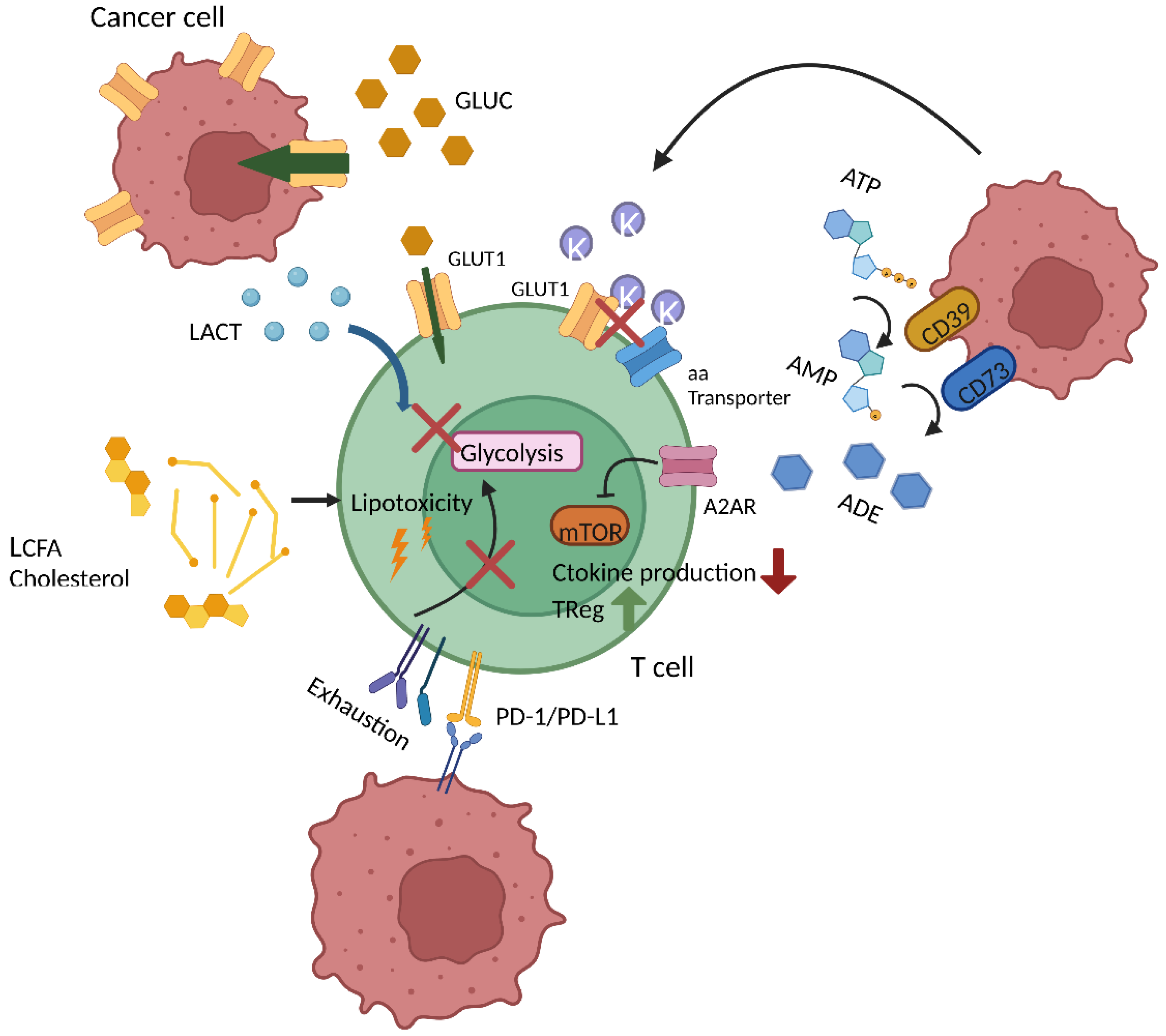
T cell metabolism is not only impacted by the state of T cell differentiation and activation, etc., but also by the environmental conditions, which can be rather burdensome in the case of the TME (Figure 3). One of the reasons is that activated T cells and malignant cells have a similar metabolic profile and thus metabolic requirements, which leads to competition over nutrients such as glucose [65]. This competition can impede T cell responses as one exemplary study showed an inverse correlation between expression of the glucose transporter 1 (Glut1) in renal cell carcinoma with T cell function and infiltration [66]. Another indirect mechanism whereby glycolytic tumors suppress T cell function is via excessive lactic acid release. Lactic acid has inhibitory effects on T cell metabolism, in particular glycolysis, which results from an inauspicious lactic acid gradient between the extracellular milieu and the cytoplasm [67,68]. In fact, numerous tumor-derived metabolic by-products hold a suppressive potential. The accumulation of potassium [K+] within the TME blocks amino acid and glucose transporters of T cells [69]. The ectonucleotidases CD39 and CD73, which can be found highly expressed on tumors, convert extracellular ATP into adenosine. The latter interferes with NfκB signaling, limiting the T cells’ anti-tumor activity and promoting the induction of TRegs [70,71]. Adenosine also directly impacts T cell metabolism and function, which correlates with the abundance of the adenosine receptor (A2AR). Indeed, adenosine blunts mTOR target, S6, phosphorylation, which leads to a reduction in glycolytic metabolism. Adenosine also seems to reduce OXPHOS, but the effects are not as strong. In addition to impairing metabolism, adenosine reduces cytokine production and T cell degranulation activity [72].
Moreover, increased concentrations of lipids (e.g., cholesterol) within the TME can trigger so-called lipotoxicity leading to cell death and exhaustion of T cells [73]. In a different study it was also shown that accumulation of long-chain fatty acids in the TME induced T cell dysfunction in pancreatic cancer [74]. T cell metabolic dysfunction in the TME has been described in other studies where, for example, T cells from CLL patients show reduced glucose uptake and increased SRC and membrane potential, accompanied by increased ROS [75]. Similarly, in a model of melanoma, intratumoral T cells were characterized by depolarized mitochondria and functional exhaustion [76].
In addition to the aforementioned molecules, different cytokines in the TME also have an impact on T cell metabolism and differentiation. On the one hand, IL-2 sustains the glycolytic shift via sustained mTOR activation, and this has been shown to favor TRegs generation and suppressive activity given the high expression of the IL-2 receptor in this subset [77,78]. On the contrary, cytokines such as IL-7, IL-15 and IL-21 are known to drive mitochondrial and fatty acid metabolism via the increase in glycerol uptake, SRC, CPT1α expression and even the expression of anti-oxidant molecules (glutathione reductase, thioredoxin reductase 1, peroxiredoxin and superoxide dismutase) that compensate for the increase in ROS following OXPHOS [79,80,81,82]. The prevalence of mitochondrial metabolism is linked to a less differentiated pool of T cells. In the TME, inflammatory cytokines, such as IFN-γ, IL-1β, IL-23 and TNF-α, are abundant. All of these cytokines contribute to the enrichment of differentiated effector T cells mainly via mTOR activation [83,84,85,86]. Conversely, anti-inflammatory cytokines, such as TGF-β, have the potential to inhibit mTOR activity, limiting glycolysis and subsequently shifting the balance towards a mitochondrial-based less differentiated phenotype [87]. To date, immunological checkpoints such as PD-L1 are well-established drivers of tumor immune escape. Recent data suggest that they exert part of their effects by interfering with T cell metabolism. The binding of PD-L1 to its cognate receptor PD-1, expressed on T cells, impairs their effector functions by reducing glycolytic activity. At the same time FAO is enhanced but PD-1+ T cells display an overall reduced SRC [32]. Moreover, PD1+ T cells also show a reduced peroxisome proliferator-activated receptor gamma coactivator 1-alpha (PGC1α) expression, which controls mitochondrial biogenesis [32]. A recent study highlighted the importance of mitochondrial biogenesis for manufacturing effective CAR T cells in chronic lymphocytic leukemia (CLL) [75]. Interestingly, PD-L1 expressed on tumor cells boosts their glucose uptake, further limiting glucose availability for infiltrating T cells [65]. The second very well-described inhibitory receptor, CTLA-4, downregulates glutamine transporters (SNAT1 and SNAT2) and Glut1 [32]. These results suggest that the immune checkpoint blockade as currently employed could restore immunometabolic fitness, which needs to be further investigated. In this direction, several clinical trials (NTC00586391, NTC01822652, NCT02650999, NCT02706402, NCT02926833, NCT03310619, NCT03726515, NCT02862028, NCT03081715, NCT02867332, NCT02867345, NCT02793856, NCT03044743) focus on the combination of CAR T cells with an immune checkpoint blockade (CPB). These studies mostly focus on the inhibition of the PD-1/PD-L1 axis via different strategies, which include the administration of anti-PD1 and PD-L1 antibodies, the design of anti-PD-1-blocker secreting CAR T cells and the genetic modification of CAR T cells to knock out the PD-1 receptor. Most of these studies show a benefit of the combination treatment in terms of T cell function and proliferation. However, they are limited by the short half-life of blocking antibodies and associated toxicities [7,8,9,88]. Therefore, although promising, these strategies need to be optimized.

5. Impact of CAR Design on T Cell Metabolism

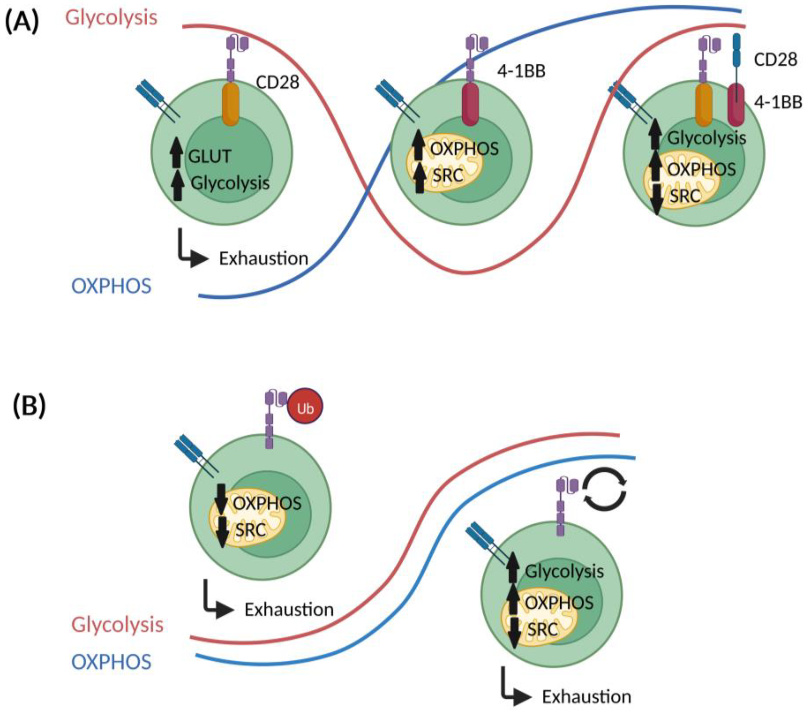
A less differentiated T cell phenotype appears advantageous for the clinical efficacy of CAR T cells. The design of certain CAR components could co-determine the metabolic phenotype together with stemness (Figure 4).

5.1. Co-Stimulatory Domains

Various co-stimulatory domains are used in CARs. Currently, most constructs belong to one of the following superfamilies: the tumor necrosis factor receptor superfamily (4-1BB/CD137, OX40) or the immunoglobulin superfamily (CD28, ICOS/CD278) [89]. Most of the studies that compare 4-1BB vs. CD28 involved CARs directed against CD19 or mesothelin. Kawalekar and colleagues describe a higher basal oxygen consumption rate (OCR), which is a surrogate for OXPHOS, and SRC in 4-1BB- as compared to CD28-carrying CARs [90]. This difference was strongest for the TCM and TN populations. On the contrary, metabolic flux analyses revealed enhanced extracellular acidification rates (ECAR), a surrogate for aerobic glycolysis, in CD28 CAR T cells. The reliance of 4-1BB CAR T cells on FAO, which represents a feature of memory-like T cells, was confirmed using heavy-carbon-labelled palmitic acid. In accordance with the superior ECAR, CD28 CAR T cells display an increased expression of key genes involved in glycolysis, including GLUT1, phosphoglycerate kinase (PGK), and glucose-6-phosphate dehydrogenase (G6PD). Enhanced FAO, SRC, and OXPHOS in 4-1BB CAR T cells is supported by an enhanced mitochondrial biogenesis as well as fusion, which is also seen in memory T cells. These differences might arise from the molecular pathways that are triggered by each co-stimulatory domain. For instance, CD28 interacts with PI3K leading to Akt activation, which in turn induces the expression of glucose transporters and other enzymes involved in glucose metabolism [46,91]. Alternatively, 4-1BB activates the p38-MAPK signaling axis, which results in PGC1α overexpression and subsequent mitochondrial fusion and biogenesis [92,93].
Those metabolic features might explain the preferential skewing towards a TCM phenotype, slower effector responses, and longer in vivo persistence of 4-1BB CAR T cells. In a similar study, Liu and colleagues reported that OCR is similar for CAR T cells with both types of co-stimulatory domains but 4-1BB CARs display an enhanced SRC [94]. The introduction of 4-1BB into the CAR construct led to a downregulation of genes encoding for glycolytic molecules and an upregulation of genes (such as the fatty acid-binding protein 5) involved in FAO and mitochondrial metabolism.
Overall, the data to date indicates that 4-1BB co-stimulation skews CAR T cell metabolism towards OXPHOS and FAO and promotes a less differentiated phenotype (with a higher replicative potential). Interestingly, third generation CAR T cells, which bare both CD28 and 4-1BB co-stimulatory domains, seem to preserve the high mitochondrial metabolism of 4-1BB CAR T cells but are accompanied by increased glycolytic metabolism. This overall enhanced metabolic activity prompted a sustained tonic TCR signaling, proliferation and metabolic fitness in dual CAR T cells over time [95].

5.2. Other CAR Construct-Related Factors

In addition to the co-stimulatory domains, other aspects of CAR design can influence the metabolism of CAR T cells. Li and colleagues showed that ubiquitination-deficient CD19 CAR T cells displayed higher OCR and SRC [96]. The inhibition of ubiquitination led to a recycling of CAR constructs to the surface and enhanced lysosomal signaling, which promotes OXPHOS and, subsequently, memory T cell formation. A different study focused on the dynamics of CAR T cell stimulation, in particular in TRegs with tonic CAR receptors, which uncouple antigen recognition from CAR activation signaling. Here, they reported that tonic CAR signaling led to an exhausted T cell phenotype with higher ECAR and OCR but low SRC. Moreover, T cells carrying a tonic CAR construct relied more on glycolysis in terms of energy [97].

6. Strategies to Target CAR T Cell Metabolism

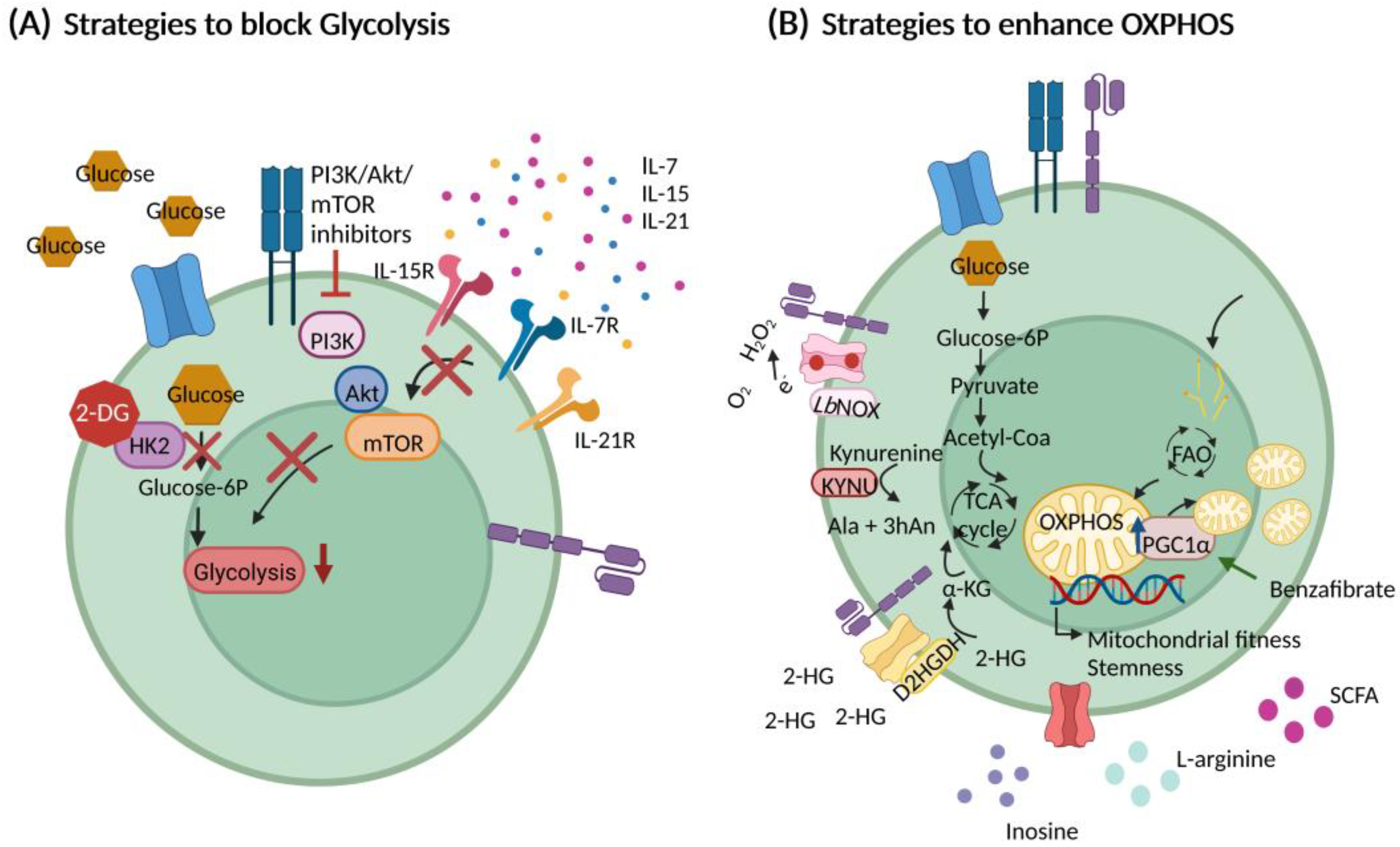
With increasing data regarding the importance of energy metabolism for the function and persistence of CAR T cells, many strategies of intervention are currently being exploited, which we summarize in the follow (Figure 5).

6.1. Optimizing Cell Culturing Conditions

Culture conditions during CAR T cell manufacturing can determine the differentiation state and metabolic phenotype. One important factor is serum supplementation. Serum, either of animal or human origin, is an important component of media to support cell viability and growth. The traditional formulations include fetal bovine serum (FBS) and human serum (HS). Recent studies explored the usage of human platelet lysates (HPL) that are used for the expansion of mesenchymal stem cells (MSCs) [98]. CAR T cells expanded in presence of HPL (as opposed to cells cultured with FBS- or HS-containing media) displayed an enrichment of TCM cells, a superior in vivo expansion, and an enhanced anti-tumor activity [99,100]. The metabolic effects of HPL supplementation remain to be investigated. As an alternative, serum can be replaced by PhysiologixTM (Phx), which is an extract of whole blood-derived growth factors. Phx enhanced the functionality and in vivo expansion of CAR T cells. At the same time, metabolism was skewed away from glycolysis towards OXPHOS, which is triggered by an enrichment of the dipeptide carnosine by neutralization of the extracellular H+ [101].
The duration of the CAR T cell production protocol can affect differentiation and function. Typically, the CAR T cell expansion lasts between 9 and 14 days. However, shorter intervals allow for the generation of CAR T cells with higher proliferative capacity, less differentiation, and greater cytotoxic activity. These optimized protocols range from three to five days [102].
The most commonly used cytokine for CAR T cell production is IL-2. However, IL-2 promotes glycolysis for rapid proliferation but leads to the formation of short-lived, highly differentiated, and exhausted CAR T cells [103,104]. Efforts to substitute IL-2 have focused on alternative γ-chain cytokines, including IL-7, IL-15, and IL-21. Evidence supports that IL-15 preserves stemness and induces higher anti-tumor activity and proliferation via mTOR inhibition. Interfering with mTOR signaling leads to reduced glycolysis and a shift towards OXPHOS [105]. Furthermore, combining IL-15 and IL-7 further boosts generations of TSCM cells [106]. In addition, IL-21 represents an interesting candidate since it enhances FAO, thus supporting TCM cell generation and in vivo persistence. Furthermore, IL-21 might yield a superior anti-tumor activity as compared to IL-15 [107]. Interestingly, the administration of a PD-1/IL-21 fusion protein improved the delivery of IL-21 to PD-1 expressing T cells, which was superior compared to systemic delivery [108,109]. Other strategies include the combination of IL-21 with IL-4 and IL-7, which maintained stemness and reduced the expression of inhibitory receptors, including PD-1 and TIM3 in CAR T cells [110].

6.2. Interfering with Glycolysis

The TME is often characterized by a shortage of nutrients. Malignant cells and T cells compete over bioenergetic substrates. Generally, one should assume that lack of nutrients is detrimental for (T) cells. However, recent studies suggest that metabolic stress, particularly glucose deprivation, may promote T cell persistence. One of the most studied glycolysis inhibitors is 2-deoxy-D-glucose (2-DG). 2-DG is a glucose derivative that enters cells via glucose transporters and is then phosphorylated by hexokinase 2 (HK2), the pacemaker enzyme of glycolysis. The resulting 2-DG-6-phosphate cannot be further metabolized and HK2 activity decreases significantly due to end product inhibition [111]. As anticipated, the treatment of T cells during in vitro expansion with 2-DG favored formation of memory T cells [112]. More recently, it was shown that transient glucose restriction improves the T cell anti-tumor function via increased pentose phosphate pathway (PPP) activity [113]. The PPP is a key pathway that generates the reduced form of Nicotinamide adenine dinucleotide phosphate (NADPH), pentoses and ribose-5-phosphate, which serve as antioxidants and nucleotide precursors, respectively. In the latter study, by transiently restricting glycolysis, they could show that T cells had a more reduced state and more PPP-generated intermediates to sustain their functionality.
One of the main pathways activated upon TCR engagement (and further boosted by CD28 stimulation) is the PI3K/Akt/mTOR signaling axis, which promotes glycolysis to support rapid T cell proliferation and differentiation. Therefore, interfering with this signaling pathway could lead to less-differentiated CAR T cells. First, Perkins and colleagues tested the expansion of B cell maturation antigen (BCMA)-directed CAR T cells in the presence of a PI3K inhibitor. The infusion of these CAR T cells to Burkitt lymphoma- and multiple myeloma-bearing mice resulted in long-term tumor regression. One of the key findings was the enhanced frequency of CD8+ CD62L+ memory T cells [114]. Similarly, Petersen and colleagues targeted the delta subunit of PI3K (δPI3K), expressed specifically in lymphocytes. In this case, both murine and human cytotoxic lymphocytes (CTLs) displayed a less differentiated phenotype as compared to their untreated counterparts [115]. The effects of tonic CAR signaling that can promote glycolysis and exhaustion can also be antagonized by pharmacological PI3K inhibition as shown for CD33-specific CAR T cells [116].
In equivalence to PI3K inhibition, Akt blockade skews metabolism towards FAO and OXPHOS, which is accompanied by enhanced stemness. However, these effects were independent of a reduced glycolytic flux. Accordingly, inhibition of Akt in EpCAM-specific CAR T cells prevented terminal differentiation without having an impact on viability and proliferation [115]. Furthermore, treated CAR T cells display a superior expansion and antitumor activity in preclinical colon cancer models. Inhibition of mTOR causes similar phenomena. Treatment of IL-2-expanded CAR T cells led to reduced glycolysis and a less-differentiated phenotype [105]. It is worth noting that intervention in the PI3k/Akt/mTOR axis not only promotes the induction of memory T cells via the blockade of glycolysis, but also via increased autophagy. This process of clearing debris and damaged organelles is very important for cellular homeostasis [117]. Additional targets of the PI3K/Akt/mTOR axis with potential (direct) impact on stemness and memory T cell formation include, amongst others, FOXO, Wnt/β-catenin and STAT3 [118,119]. Therefore, further research is required to better understand the contribution of glycolysis in the context of PI3K/Akt/mTOR blockade.
At the intersection between glycolysis and OXPHOS is pyruvate, which enters the mitochondria via the mitochondrial pyruvate carrier (MPC). A recent study showed that genetic MPC deletion favored a memory phenotype (through a preferential fueling of the TCA with fatty acids and glutamine) without affecting CD8+ T cell effector functions. Interestingly, MPC deletion did not improve CAR T cell effector functions in a nutrient-deprived TME. However, this approach remains interesting in view of the in vitro CAR T cell expansion. In fact, pre-treatment of CAR T cells with a small molecule inhibitor of MPC led to higher proportions of memory cells and a superior in vivo anti-tumor activity [120].

6.3. Promoting Mitochondrial Metabolism and Fitness

Given the fact that mitochondrial metabolism is linked to stemness and in vivo persistence of CAR T cells, several strategies (beyond the choice of co-stimulation) are currently being investigated to skew the CAR T cells’ bioenergetics accordingly. A key regulator of mitochondrial biogenesis is PGC1α. PGC1α is triggered by enhanced energetic demands, such as those imposed by T cell activation in the TME. It is activated by phosphorylation following AMPK activation and functions via the binding and activation of DNA-binding transcription factors, including nuclear respiratory factors, NRF1 and NRF2, which are involved in controlling the excess of ROS generated upon increased mitochondrial activity [121,122]. Several studies have addressed the impact PGC1α overexpression. Bengsch and colleagues reported that during chronic viral infection, T cells undergo exhaustion and are unable to meet their bioenergetic demands as both glycolysis and OXPHOS are impaired [55]. This “metabolic paralysis” was caused by mitochondrial depolarization, which did not allow the build-up of a proton gradient across the mitochondrial membrane. Membrane potential and function were restored by PGC1α overexpression. In another study, the same approach also led to increased mitochondrial biogenesis, and subsequently increased the frequency of memory CD8+ T cells [86]. Several studies have demonstrated the benefits of combining CAR T cells with an immune checkpoint blockade [7,8,9]. In fact, interfering with PD-L1/PD-1 interaction leads to a metabolic switch towards FAO and OXPHOS in T cells, thereby promoting cell survival and self-renewal [54]. Combining anti-PD-1 treatment with the PGC1α agonist benzafibrate in a murine melanoma model led to increased OXPHOS and reduced apoptosis in T cells [32]. Interestingly, in a different study, it was shown that the mitochondrial function of T cells represents a marker for responsiveness towards immune checkpoint blockade treatment [87]. As discussed above, 4-1BB co-stimulation shifts metabolism towards OXPHOS while promoting mitochondrial biogenesis and fusion [92].
Modulating mitochondrial dynamics (i.e., fusion or fission) represents an alternative strategy for bolstering mitochondrial fitness. This is achieved by pharmacological means, such as Mdivi-1, a mitochondrial division inhibitor, or ablation of genes such as dynamin-related GTPase (DRP1) or mitochondrial division dynamin I (DNM1) [89]. Both approaches led to fused mitochondria, increased mitochondrial fitness, and anti-tumor activity of the tumor-infiltrating lymphocytes.
Recent studies have exploited the potential of (over-)expressing metabolic enzymes to render CAR T cells more resilient towards the TME. Two candidate molecules are Lactobacillus brevis NADH oxidase (LbNOX) and D-2-hydroxyglutarate dehydrogenase (D2HGDH) [123], which catalyze the transfer of electrons from O2 to H2O2 and oxidation of D-2-hydroxyglutarate (D-2-HG) to alpha-ketoglutarate (αKG), respectively. Co-expressing LbNOX and CD28ζ CAR induced higher levels of baseline OCR as compared to the control group. In addition, these CAR T cells were not as affected by OXPHOS inhibitors such as antimycin A and rotenone. Overall, survivability was increased but not in vivo efficacy. D2HGDH-expressing CAR T cells metabolize D-2-HG, which is abundant within the TME. However, basal and maximal respiration is reduced, while the frequency of TME-infiltrating TEM cells is increased. The latter could also explain the improved anti-tumor activity and emphasizes that the relationship between metabolism and function is not linear, but more complex. Kynurenine accumulation is also frequently found in the TME and results from an overexpression of indoleamine 2,3-dioxygenase 1 (IDO1) or tryptophan 2,3-dioxygenase 2 (TDO) in tumor and/or tumor-associated cells (e.g., MDSCs or cancer-associated fibroblasts). It has a strong (CAR) T cell-inhibitory effect (compared to others’ metabolic interference [124]) and can limit the efficacy of immune-based therapies [125,126]. CAR T cells which were modified to express the kynurenine-degrading kynureninase showed a better proliferative and tumor-eradicating capacity but glucose uptake was actually also increased. As described above, adenosine also negatively impacts T cell metabolic fitness and function. Therefore, an interesting approach is the expression of adenosine deaminase (ADA), which is a characteristic of CD26-expressing T cells [127]. It seems that ADA metabolizes adenosine, which is suppressive for T cells, into inosine, which can be used as an alternative carbon source under nutrient-deprived conditions. Its ribose subunit provides ATP and biosynthetic precursors from both glycolysis and the PPP. Besides supporting carbon metabolism, inosine has been proven to have boosting anti-tumor effects on effector T cells, even in the absence of glucose. For instance, inosine enhanced cytokine production (granzyme B, IFN-γ and TNF-α) and the tumor killing capacity of T cells, even in glucose-deprived conditions [128].

6.4. Nutritional Support

Given the competition over nutrients within the TME, several efforts are currently focusing on the potential supplementation of amino acids and nucleotides. For example, L-arginine can improve T cell anti-tumor activity both in vitro and in vivo, by enhancing OXPHOS and memory cell formation [129]. Regarding nucleotides, inosine seems to be an attractive supplement given its potential to enter glycolysis and the pentose phosphate pathway. For instance, in mouse models, it enhanced tumor clearance even under nutrient-deprived conditions. Similar to inosine, methionine can enter the carbon cycle and support T cell metabolic fitness and effector functions, mostly due to its role as methyl donor in nucleotide methylations and the epigenetic reprogramming needed for T cell differentiation. Furthermore, methionine is the amino acid required to start protein synthesis [124].
Other nutrients to investigate are fatty acids. As specified above, the accumulation of long-chain fatty acids is detrimental to T cells. However, short chain fatty acids (SCFAs), such as butyrate, propionate and acetate, seem to have the opposite effect. SCFAs are generated by bacteria present in the gut and are incorporated into T cells either via passive diffusion or binding to different transporters. How SCFAs affect T function depends on the cytokine milieu and T cell activation conditions. For instance, whereas CD4+ T cell activation in the presence of TGF-β1, IL-2 and 0,1 mM butyrate induces TReg generation [130], higher concentrations of butyrate, propionate and acetate support Th1 and Th17 differentiation [131]. In addition, CD8+ T cells cultured in the presence of 1 mM propionate and butyrate were shown to enhance IFN-γ and Granzyme B (GrzB) production. Interestingly, SCFAs were shown to promote long-lasting memory phenotypes through the upregulation of FoxO1, a transcription factor required for memory formation. This memory transition was supported by favoring the use of fatty acids and glutaminolysis to fuel the TCA rather than glycolysis [132,133]. In a different study, SCFAs could enhance CAR T cell anti-tumor activity via mTOR activation and the production of effector molecules, including TNF-α [134]. Therefore, depending on the concentration and culture conditions, SCFA can either favor a memory-like phenotype or induce the differentiation towards a specific effector T cell subset, both of which have their advantages, whereas memory-like T cells render the advantage of long-term responses and are required to establish an adaptive immune response [135], more differentiated effector T cells are essential to rapidly eliminate tumor cells [136,137]. Therefore, a balance between both types of T cells might provide the optimal result.
As mentioned above, the TME is characterized by a high concentration of ROS. Therefore, the supplementation of antioxidants might have a beneficial result in maintaining T cell fitness and function. The use of N-acetylcysteine (NAC) was proposed in a study to limit ROS metabolism during T cell activation [138]. It was shown that CD19 CAR T cells supplemented with NAC remained less differentiated (TSCM) and displayed overall reduced glycolytic metabolism, as evidenced by the reduced mTOR activity, lower expression of key glycolytic genes, such as Glyceraldehyde 3-phosphate dehydrogenase (GAPDH), Enolase 2 (ENO2), Pyruvate kinase (PKM) and Lactate dehydrogenase (LDHA), and reduced glucose uptake. In addition, NAC-treated cells showed increased expression of CPT1α, supporting FAO.
Self-evidently, metabolic re-modeling of the TME by interfering with the metabolic pathways and signaling in tumor and/or tumor-associated cells also represents an interesting alternative strategy for improving the environmental conditions for immune cells and is discussed in detail elsewhere [139,140].

7. Conclusions

Despite having revolutionized the field of cancer treatment, CAR T cell therapy still has several obstacles to overcome, including toxicity and efficacy. The latter is in most cases the result of successful tumor immune escape phenomena that impact the CAR T cells’ functionality and range from inadequate motility to reduced secretion of effector cytokines. In an attempt to overcome these limitations, the current focus is set on optimizing CAR T cell design with strategies such as including on/off switches to modulate CAR T cell activity and toxicity, CAR T cells redirected for universal cytokine-mediated killing, known as TRUCKs [141], universal CARs [142] or armored CAR T cells [143]. Currently ongoing clinical trials mostly focus on hematological malignancies. In fact, the combination regimens of CAR T cells and CPB are close to finding a cure to such diseases. On the other hand, intensive efforts are still ongoing in targeting several antigens expressed on solid tumors.
In addition to the currently developed CAR generations, significant effort is focused on optimizing CAR T cell culture, manufacturing and transfer with the aim of altering certain aspects of T cell fitness and metabolism. Emerging evidence emphasizes the interconnection between T cell metabolism and function, differentiation, and persistence. For this reason, it seems inevitable to evaluate metabolic modulation as a strategy to improve CAR T cell therapies. Easier-to-incorporate approaches deal with the optimization of culture conditions that can extend from the supplementation of cytokines and amino acids to the pharmacological control of metabolic pathways. The design of the CAR construct, especially the costimulatory domain, can strongly influence the metabolism, phenotype, persistence and possibly also toxicity. In the future, we expect a much stronger focus on the metabolic impact of the utilized signaling domains, which will at least co-determine CAR design. In addition, further genetic interventions are also conceivable which will allow certain key metabolic molecules to be more or less strongly expressed. Overall, these approaches focus on the generation of preferentially stem-like CAR T cells with high anti-tumor activity, superior survivability within the TME, and long-lasting persistence. In spite of all the possibilities and opportunities of such concepts, it should not be forgotten that we are talking about a combined cell-, immune-, and gene-therapy with corresponding regulatory requirements. Even small changes in manufacturing protocols may take a long time to reach clinical implementation (if they do at all).

Author Contributions

Writing—investigation and original draft preparation J.R.S.; review and editing, S.V., M.A., A.M. and D.M., supervision, D.M. All authors have read and agreed to the published version of the manuscript.

Funding

This research received no external funding.

Institutional Review Board Statement

Not applicable.

Informed Consent Statement

Not applicable.

Data Availability Statement

Not applicable.

Acknowledgments

Figures were created with BioRender.com, accessed on 25 August 2022.

Conflicts of Interest

The authors declare no conflict of interest.

References

  1. Roex, G.; Timmers, M.; Wouters, K.; Campillo-Davo, D.; Flumens, D.; Schroyens, W.; Chu, Y.; Berneman, Z.N.; Lion, E.; Luo, F.; et al. Safety and clinical efficacy of BCMA CAR-T-cell therapy in multiple myeloma. J. Hematol. Oncol. 2020, 13, 164. [Google Scholar] [CrossRef] [PubMed]
  2. Kennedy, V.E.; Wong, C.; Huang, C.-Y.; Wolf, J.L.; Martin, T.; Shah, N.; Wong, S.W. Macrophage Activation Syndrome-like Manifestations (MAS-L) Following BCMA-Directed CAR T-Cells in Multiple Myeloma. Blood 2020, 136, 7–8. [Google Scholar] [CrossRef]
  3. Shimabukuro-Vornhagen, A.; Gödel, P.; Subklewe, M.; Stemmler, H.J.; Schlößer, H.A.; Schlaak, M.; Kochanek, M.; Böll, B.; von Bergwelt-Baildon, M.S. Cytokine release syndrome. J. Immunother. Cancer 2018, 6, 56. [Google Scholar] [CrossRef] [PubMed] [Green Version]
  4. Siegler, E.L.; Kenderian, S.S. Neurotoxicity and Cytokine Release Syndrome After Chimeric Antigen Receptor T Cell Therapy: Insights Into Mechanisms and Novel Therapies. Front. Immunol. 2020, 11, 1973. [Google Scholar] [CrossRef]
  5. Neelapu, S.S.; Locke, F.L.; Bartlett, N.L.; Lekakis, L.J.; Miklos, D.B.; Jacobson, C.A.; Braunschweig, I.; Oluwole, O.O.; Siddiqi, T.; Lin, Y.; et al. Axicabtagene Ciloleucel CAR T-Cell Therapy in Refractory Large B-Cell Lymphoma. N. Engl. J. Med. 2017, 377, 2531–2544. [Google Scholar] [CrossRef]
  6. Maude, S.L.; Laetsch, T.W.; Buechner, J.; Rives, S.; Boyer, M.; Bittencourt, H.; Bader, P.; Verneris, M.R.; Stefanski, H.E.; Myers, G.D.; et al. Tisagenlecleucel in Children and Young Adults with B-Cell Lymphoblastic Leukemia. N. Engl. J. Med. 2018, 378, 439–448. [Google Scholar] [CrossRef]
  7. Yin, Y.; Boesteanu, A.C.; Binder, Z.A.; Xu, C.; Reid, R.A.; Rodriguez, J.L.; Cook, D.R.; Thokala, R.; Blouch, K.; McGettigan-Croce, B.; et al. Checkpoint Blockade Reverses Anergy in IL-13Rα2 Humanized scFv-Based CAR T Cells to Treat Murine and Canine Gliomas. Mol. Ther. Oncolytics 2018, 11, 20–38. [Google Scholar] [CrossRef] [Green Version]
  8. Li, A.M.; Hucks, G.E.; Dinofia, A.M.; Seif, A.E.; Teachey, D.T.; Baniewicz, D.; Callahan, C.; Fasano, C.; McBride, B.; Gonzalez, V.; et al. Checkpoint Inhibitors Augment CD19-Directed Chimeric Antigen Receptor (CAR) T Cell Therapy in Relapsed B-Cell Acute Lymphoblastic Leukemia. Blood 2018, 132, 556. [Google Scholar] [CrossRef]
  9. Chong, E.A.; Melenhorst, J.J.; Lacey, S.F.; Ambrose, D.E.; Gonzalez, V.; Levine, B.L.; June, C.H.; Schuster, S.J. PD-1 blockade modulates chimeric antigen receptor (CAR)-modified T cells: Refueling the CAR. Blood 2017, 129, 1039–1041. [Google Scholar] [CrossRef] [Green Version]
  10. Neumeister, P.; Schulz, E.; Pansy, K.; Szmyra, M.; Deutsch, A.J. Targeting the Microenvironment for Treating Multiple Myeloma. Int. J. Mol. Sci. 2022, 23, 7627. [Google Scholar] [CrossRef]
  11. Zhang, L.; Jin, G.; Chen, Z.; Yu, C.; Li, Y.; Li, Y.; Chen, J.; Yu, L. Lenalidomide improves the antitumor activity of CAR-T cells directed toward the intracellular Wilms Tumor 1 antigen. Hematology 2021, 26, 818–826. [Google Scholar] [CrossRef] [PubMed]
  12. Wang, Z.; Zhou, G.; Risu, N.; Fu, J.; Zou, Y.; Tang, J.; Li, L.; Liu, H.; Liu, Q.; Zhu, X. Lenalidomide Enhances CAR-T Cell Activity Against Solid Tumor Cells. Cell Transpl. 2020, 29, 963689720920825. [Google Scholar] [CrossRef] [PubMed]
  13. Depil, S.; Duchateau, P.; Grupp, S.A.; Mufti, G.; Poirot, L. ‘Off-the-shelf’ allogeneic CAR T cells: Development and challenges. Nat. Rev. Drug Discov. 2020, 19, 185–199. [Google Scholar] [CrossRef] [PubMed]
  14. Martínez Bedoya, D.; Dutoit, V.; Migliorini, D. Allogeneic CAR T Cells: An Alternative to Overcome Challenges of CAR T Cell Therapy in Glioblastoma. Front. Immunol. 2021, 12, 640082. [Google Scholar] [CrossRef] [PubMed]
  15. Han, X.; Wang, Y.; Wei, J.; Han, W. Multi-antigen-targeted chimeric antigen receptor T cells for cancer therapy. J. Hematol. Oncol. 2019, 12, 128. [Google Scholar] [CrossRef]
  16. Spiegel, J.Y.; Patel, S.; Muffly, L.; Hossain, N.M.; Oak, J.; Baird, J.H.; Frank, M.J.; Shiraz, P.; Sahaf, B.; Craig, J.; et al. CAR T cells with dual targeting of CD19 and CD22 in adult patients with recurrent or refractory B cell malignancies: A phase 1 trial. Nat. Med. 2021, 27, 1419–1431. [Google Scholar] [CrossRef]
  17. Rejeski, K.; Perez, A.; Sesques, P.; Hoster, E.; Berger, C.; Jentzsch, L.; Mougiakakos, D.; Frölich, L.; Ackermann, J.; Bücklein, V.; et al. CAR-HEMATOTOX: A model for CAR T-cell–related hematologic toxicity in relapsed/refractory large B-cell lymphoma. Blood 2021, 138, 2499–2513. [Google Scholar] [CrossRef]
  18. Lee, D.W.; Gardner, R.; Porter, D.L.; Louis, C.U.; Ahmed, N.; Jensen, M.; Grupp, S.A.; Mackall, C.L. Current concepts in the diagnosis and management of cytokine release syndrome. Blood 2014, 124, 188–195. [Google Scholar] [CrossRef] [Green Version]
  19. Schubert, M.L.; Schmitt, M.; Wang, L.; Ramos, C.A.; Jordan, K.; Müller-Tidow, C.; Dreger, P. Side-effect management of chimeric antigen receptor (CAR) T-cell therapy. Ann. Oncol. 2021, 32, 34–48. [Google Scholar] [CrossRef]
  20. Yáñez, L.; Alarcón, A.; Sánchez-Escamilla, M.; Perales, M.A. How I treat adverse effects of CAR-T cell therapy. ESMO Open 2020, 4, e000746. [Google Scholar] [CrossRef]
  21. Luo, W.; Li, C.; Zhang, Y.; Du, M.; Kou, H.; Lu, C.; Mei, H.; Hu, Y. Adverse effects in hematologic malignancies treated with chimeric antigen receptor (CAR) T cell therapy: A systematic review and Meta-analysis. BMC Cancer 2022, 22, 98. [Google Scholar] [CrossRef] [PubMed]
  22. Zhao, X.; Yang, J.; Zhang, X.; Lu, X.-A.; Xiong, M.; Zhang, J.; Zhou, X.; Qi, F.; He, T.; Ding, Y.; et al. Efficacy and Safety of CD28- or 4-1BB-Based CD19 CAR-T Cells in B Cell Acute Lymphoblastic Leukemia. Mol. Ther.-Oncolytics 2020, 18, 272–281. [Google Scholar] [CrossRef] [PubMed]
  23. Priceman, S.J.; Tilakawardane, D.; Jeang, B.; Aguilar, B.; Murad, J.P.; Park, A.K.; Chang, W.C.; Ostberg, J.R.; Neman, J.; Jandial, R.; et al. Regional Delivery of Chimeric Antigen Receptor-Engineered T Cells Effectively Targets HER2+ Breast Cancer Metastasis to the Brain. Clin. Cancer Res. 2018, 24, 95–105. [Google Scholar] [CrossRef] [PubMed] [Green Version]
  24. Brown, C.E.; Aguilar, B.; Starr, R.; Yang, X.; Chang, W.C.; Weng, L.; Chang, B.; Sarkissian, A.; Brito, A.; Sanchez, J.F.; et al. Optimization of IL13Rα2-Targeted Chimeric Antigen Receptor T Cells for Improved Anti-tumor Efficacy against Glioblastoma. Mol. Ther. 2018, 26, 31–44. [Google Scholar] [CrossRef] [Green Version]
  25. Whilding, L.M.; Halim, L.; Draper, B.; Parente-Pereira, A.C.; Zabinski, T.; Davies, D.M.; Maher, J. CAR T-Cells Targeting the Integrin αvβ6 and Co-Expressing the Chemokine Receptor CXCR2 Demonstrate Enhanced Homing and Efficacy against Several Solid Malignancies. Cancers 2019, 11, 674. [Google Scholar] [CrossRef] [PubMed] [Green Version]
  26. Caruana, I.; Savoldo, B.; Hoyos, V.; Weber, G.; Liu, H.; Kim, E.S.; Ittmann, M.M.; Marchetti, D.; Dotti, G. Heparanase promotes tumor infiltration and antitumor activity of CAR-redirected T lymphocytes. Nat. Med. 2015, 21, 524–529. [Google Scholar] [CrossRef] [PubMed] [Green Version]
  27. Yamada, K.M.; Sixt, M. Mechanisms of 3D cell migration. Nat. Rev. Mol. Cell Biol. 2019, 20, 738–752. [Google Scholar] [CrossRef]
  28. Campello, S.; Lacalle, R.A.; Bettella, M.; Mañes, S.; Scorrano, L.; Viola, A. Orchestration of lymphocyte chemotaxis by mitochondrial dynamics. J. Exp. Med. 2006, 203, 2879–2886. [Google Scholar] [CrossRef]
  29. Haas, R.; Smith, J.; Rocher-Ros, V.; Nadkarni, S.; Montero-Melendez, T.; D’Acquisto, F.; Bland, E.J.; Bombardieri, M.; Pitzalis, C.; Perretti, M.; et al. Lactate Regulates Metabolic and Pro-inflammatory Circuits in Control of T Cell Migration and Effector Functions. PLoS Biol. 2015, 13, e1002202. [Google Scholar] [CrossRef]
  30. Rhoads, J.M.; Chen, W.; Gookin, J.; Wu, G.Y.; Fu, Q.; Blikslager, A.T.; Rippe, R.A.; Argenzio, R.A.; Cance, W.G.; Weaver, E.M.; et al. Arginine stimulates intestinal cell migration through a focal adhesion kinase dependent mechanism. Gut 2004, 53, 514–522. [Google Scholar] [CrossRef]
  31. Gu, K.; Liu, G.; Wu, C.; Jia, G.; Zhao, H.; Chen, X.; Tian, G.; Cai, J.; Zhang, R.; Wang, J. Tryptophan improves porcine intestinal epithelial cell restitution through the CaSR/Rac1/PLC-γ1 signaling pathway. Food Funct. 2021, 12, 8787–8799. [Google Scholar] [CrossRef] [PubMed]
  32. Patsoukis, N.; Bardhan, K.; Chatterjee, P.; Sari, D.; Liu, B.; Bell, L.N.; Karoly, E.D.; Freeman, G.J.; Petkova, V.; Seth, P.; et al. PD-1 alters T-cell metabolic reprogramming by inhibiting glycolysis and promoting lipolysis and fatty acid oxidation. Nat. Commun. 2015, 6, 6692. [Google Scholar] [CrossRef] [PubMed] [Green Version]
  33. Quail, D.F.; Joyce, J.A. Microenvironmental regulation of tumor progression and metastasis. Nat. Med. 2013, 19, 1423–1437. [Google Scholar] [CrossRef] [PubMed]
  34. Baumann, T.; Dunkel, A.; Schmid, C.; Schmitt, S.; Hiltensperger, M.; Lohr, K.; Laketa, V.; Donakonda, S.; Ahting, U.; Lorenz-Depiereux, B.; et al. Regulatory myeloid cells paralyze T cells through cell-cell transfer of the metabolite methylglyoxal. Nat. Immunol. 2020, 21, 555–566. [Google Scholar] [CrossRef] [PubMed]
  35. Yan, Z.; Garg, S.K.; Kipnis, J.; Banerjee, R. Extracellular redox modulation by regulatory T cells. Nat. Chem. Biol. 2009, 5, 721–723. [Google Scholar] [CrossRef] [PubMed] [Green Version]
  36. Kim, J.H.; Kim, B.S.; Lee, S.K. Regulatory T Cells in Tumor Microenvironment and Approach for Anticancer Immunotherapy. Immune Netw. 2020, 20, e4. [Google Scholar] [CrossRef] [PubMed]
  37. Sabatino, M.; Hu, J.; Sommariva, M.; Gautam, S.; Fellowes, V.; Hocker, J.D.; Dougherty, S.; Qin, H.; Klebanoff, C.A.; Fry, T.J.; et al. Generation of clinical-grade CD19-specific CAR-modified CD8+ memory stem cells for the treatment of human B-cell malignancies. Blood 2016, 128, 519–528. [Google Scholar] [CrossRef]
  38. Xu, Y.; Zhang, M.; Ramos, C.A.; Durett, A.; Liu, E.; Dakhova, O.; Liu, H.; Creighton, C.J.; Gee, A.P.; Heslop, H.E.; et al. Closely related T-memory stem cells correlate with in vivo expansion of CAR.CD19-T cells and are preserved by IL-7 and IL-15. Blood 2014, 123, 3750–3759. [Google Scholar] [CrossRef] [Green Version]
  39. Klebanoff, C.A.; Gattinoni, L.; Restifo, N.P. CD8+ T-cell memory in tumor immunology and immunotherapy. Immunol. Rev. 2006, 211, 214–224. [Google Scholar] [CrossRef] [Green Version]
  40. Pearce, E.L.; Pearce, E.J. Metabolic pathways in immune cell activation and quiescence. Immunity 2013, 38, 633–643. [Google Scholar] [CrossRef]
  41. Wang, R.; Dillon, C.P.; Shi, L.Z.; Milasta, S.; Carter, R.; Finkelstein, D.; McCormick, L.L.; Fitzgerald, P.; Chi, H.; Munger, J.; et al. The transcription factor Myc controls metabolic reprogramming upon T lymphocyte activation. Immunity 2011, 35, 871–882. [Google Scholar] [CrossRef] [PubMed] [Green Version]
  42. Fox, C.J.; Hammerman, P.S.; Thompson, C.B. Fuel feeds function: Energy metabolism and the T-cell response. Nat. Rev. Immunol. 2005, 5, 844–852. [Google Scholar] [CrossRef] [PubMed]
  43. MacIver, N.J.; Michalek, R.D.; Rathmell, J.C. Metabolic regulation of T lymphocytes. Annu. Rev. Immunol. 2013, 31, 259–283. [Google Scholar] [CrossRef] [PubMed] [Green Version]
  44. Vander Heiden, M.G.; Cantley, L.C.; Thompson, C.B. Understanding the Warburg effect: The metabolic requirements of cell proliferation. Science 2009, 324, 1029–1033. [Google Scholar] [CrossRef] [PubMed] [Green Version]
  45. Jones, R.G.; Thompson, C.B. Revving the engine: Signal transduction fuels T cell activation. Immunity 2007, 27, 173–178. [Google Scholar] [CrossRef] [PubMed] [Green Version]
  46. Frauwirth, K.A.; Riley, J.L.; Harris, M.H.; Parry, R.V.; Rathmell, J.C.; Plas, D.R.; Elstrom, R.L.; June, C.H.; Thompson, C.B. The CD28 signaling pathway regulates glucose metabolism. Immunity 2002, 16, 769–777. [Google Scholar] [CrossRef] [Green Version]
  47. Wieman, H.L.; Wofford, J.A.; Rathmell, J.C. Cytokine stimulation promotes glucose uptake via phosphatidylinositol-3 kinase/Akt regulation of Glut1 activity and trafficking. Mol. Biol. Cell 2007, 18, 1437–1446. [Google Scholar] [CrossRef] [Green Version]
  48. Devadas, S.; Zaritskaya, L.; Rhee, S.G.; Oberley, L.; Williams, M.S. Discrete generation of superoxide and hydrogen peroxide by T cell receptor stimulation: Selective regulation of mitogen-activated protein kinase activation and fas ligand expression. J. Exp. Med. 2002, 195, 59–70. [Google Scholar] [CrossRef] [Green Version]
  49. Chaudhri, G.; Hunt, N.H.; Clark, I.A.; Ceredig, R. Antioxidants inhibit proliferation and cell surface expression of receptors for interleukin-2 and transferrin in T lymphocytes stimulated with phorbol myristate acetate and ionomycin. Cell Immunol. 1988, 115, 204–213. [Google Scholar] [CrossRef]
  50. Previte, D.M.; O’Connor, E.C.; Novak, E.A.; Martins, C.P.; Mollen, K.P.; Piganelli, J.D. Reactive oxygen species are required for driving efficient and sustained aerobic glycolysis during CD4+ T cell activation. PLoS ONE 2017, 12, e0175549. [Google Scholar] [CrossRef]
  51. Williams, M.A.; Bevan, M.J. Effector and memory CTL differentiation. Annu. Rev. Immunol. 2007, 25, 171–192. [Google Scholar] [CrossRef] [PubMed]
  52. Araki, K.; Turner, A.P.; Shaffer, V.O.; Gangappa, S.; Keller, S.A.; Bachmann, M.F.; Larsen, C.P.; Ahmed, R. mTOR regulates memory CD8 T-cell differentiation. Nature 2009, 460, 108–112. [Google Scholar] [CrossRef] [PubMed] [Green Version]
  53. Pearce, E.L.; Walsh, M.C.; Cejas, P.J.; Harms, G.M.; Shen, H.; Wang, L.-S.; Jones, R.G.; Choi, Y. Enhancing CD8 T-cell memory by modulating fatty acid metabolism. Nature 2009, 460, 103–107. [Google Scholar] [CrossRef] [PubMed] [Green Version]
  54. van der Windt, G.J.; Everts, B.; Chang, C.H.; Curtis, J.D.; Freitas, T.C.; Amiel, E.; Pearce, E.J.; Pearce, E.L. Mitochondrial respiratory capacity is a critical regulator of CD8+ T cell memory development. Immunity 2012, 36, 68–78. [Google Scholar] [CrossRef] [Green Version]
  55. O’Sullivan, D. The metabolic spectrum of memory T cells. Immunol. Cell Biol. 2019, 97, 636–646. [Google Scholar] [CrossRef]
  56. Sukumar, M.; Liu, J.; Mehta, G.U.; Patel, S.J.; Roychoudhuri, R.; Crompton, J.G.; Klebanoff, C.A.; Ji, Y.; Li, P.; Yu, Z.; et al. Mitochondrial Membrane Potential Identifies Cells with Enhanced Stemness for Cellular Therapy. Cell Metab. 2016, 23, 63–76. [Google Scholar] [CrossRef] [Green Version]
  57. Johnson, L.A.; Morgan, R.A.; Dudley, M.E.; Cassard, L.; Yang, J.C.; Hughes, M.S.; Kammula, U.S.; Royal, R.E.; Sherry, R.M.; Wunderlich, J.R.; et al. Gene therapy with human and mouse T-cell receptors mediates cancer regression and targets normal tissues expressing cognate antigen. Blood 2009, 114, 535–546. [Google Scholar] [CrossRef] [Green Version]
  58. Rosenberg, S.A.; Yang, J.C.; Sherry, R.M.; Kammula, U.S.; Hughes, M.S.; Phan, G.Q.; Citrin, D.E.; Restifo, N.P.; Robbins, P.F.; Wunderlich, J.R.; et al. Durable Complete Responses in Heavily Pretreated Patients with Metastatic Melanoma Using T-Cell Transfer Immunotherapy. Clin. Cancer Res. 2011, 17, 4550–4557. [Google Scholar] [CrossRef] [Green Version]
  59. Krishna, S.; Lowery, F.J.; Copeland, A.R.; Bahadiroglu, E.; Mukherjee, R.; Jia, L.; Anibal, J.T.; Sachs, A.; Adebola, S.O.; Gurusamy, D.; et al. Stem-like CD8 T cells mediate response of adoptive cell immunotherapy against human cancer. Science 2020, 370, 1328–1334. [Google Scholar] [CrossRef]
  60. Louis, C.U.; Savoldo, B.; Dotti, G.; Pule, M.; Yvon, E.; Myers, G.D.; Rossig, C.; Russell, H.V.; Diouf, O.; Liu, E.; et al. Antitumor activity and long-term fate of chimeric antigen receptor-positive T cells in patients with neuroblastoma. Blood 2011, 118, 6050–6056. [Google Scholar] [CrossRef]
  61. Fraietta, J.A.; Lacey, S.F.; Orlando, E.J.; Pruteanu-Malinici, I.; Gohil, M.; Lundh, S.; Boesteanu, A.C.; Wang, Y.; O’Connor, R.S.; Hwang, W.T.; et al. Author Correction: Determinants of response and resistance to CD19 chimeric antigen receptor (CAR) T cell therapy of chronic lymphocytic leukemia. Nat. Med. 2021, 27, 561. [Google Scholar] [CrossRef] [PubMed]
  62. Cohen, A.D.; Garfall, A.L.; Stadtmauer, E.A.; Melenhorst, J.J.; Lacey, S.F.; Lancaster, E.; Vogl, D.T.; Weiss, B.M.; Dengel, K.; Nelson, A.; et al. B cell maturation antigen-specific CAR T cells are clinically active in multiple myeloma. J. Clin. Investig. 2019, 129, 2210–2221. [Google Scholar] [CrossRef] [Green Version]
  63. Liu, Y.; Guo, Y.; Wu, Z.; Feng, K.; Tong, C.; Wang, Y.; Dai, H.; Shi, F.; Yang, Q.; Han, W. Anti-EGFR chimeric antigen receptor-modified T cells in metastatic pancreatic carcinoma: A phase I clinical trial. Cytotherapy 2020, 22, 573–580. [Google Scholar] [CrossRef] [PubMed]
  64. Deng, Q.; Han, G.; Puebla-Osorio, N.; Ma, M.C.J.; Strati, P.; Chasen, B.; Dai, E.; Dang, M.; Jain, N.; Yang, H.; et al. Characteristics of anti-CD19 CAR T cell infusion products associated with efficacy and toxicity in patients with large B cell lymphomas. Nat. Med. 2020, 26, 1878–1887. [Google Scholar] [CrossRef] [PubMed]
  65. Chang, C.H.; Qiu, J.; O’Sullivan, D.; Buck, M.D.; Noguchi, T.; Curtis, J.D.; Chen, Q.; Gindin, M.; Gubin, M.M.; van der Windt, G.J.; et al. Metabolic Competition in the Tumor Microenvironment Is a Driver of Cancer Progression. Cell 2015, 162, 1229–1241. [Google Scholar] [CrossRef] [Green Version]
  66. Singer, K.; Kastenberger, M.; Gottfried, E.; Hammerschmied, C.G.; Büttner, M.; Aigner, M.; Seliger, B.; Walter, B.; Schlösser, H.; Hartmann, A.; et al. Warburg phenotype in renal cell carcinoma: High expression of glucose-transporter 1 (GLUT-1) correlates with low CD8+ T-cell infiltration in the tumor. Int. J. Cancer 2011, 128, 2085–2095. [Google Scholar] [CrossRef]
  67. Fischer, K.; Hoffmann, P.; Voelkl, S.; Meidenbauer, N.; Ammer, J.; Edinger, M.; Gottfried, E.; Schwarz, S.; Rothe, G.; Hoves, S.; et al. Inhibitory effect of tumor cell-derived lactic acid on human T cells. Blood 2007, 109, 3812–3819. [Google Scholar] [CrossRef] [Green Version]
  68. Bröer, S. Lactate transportation is required for lymphocyte activation. Nat. Chem. Biol. 2005, 1, 356–357. [Google Scholar] [CrossRef]
  69. De Vita, V.T., Jr.; Chu, E. A history of cancer chemotherapy. Cancer Res. 2008, 68, 8643–8653. [Google Scholar] [CrossRef] [Green Version]
  70. Ohta, A.; Sitkovsky, M. Extracellular adenosine-mediated modulation of regulatory T cells. Front. Immunol. 2014, 5, 304. [Google Scholar] [CrossRef]
  71. Ohta, A.; Madasu, M.; Subramanian, M.; Kini, R.; Jones, G.; Choukèr, A.; Ohta, A.; Sitkovsky, M. Hypoxia-induced and A2A adenosine receptor-independent T-cell suppression is short lived and easily reversible. Int. Immunol. 2014, 26, 83–91. [Google Scholar] [CrossRef] [PubMed] [Green Version]
  72. Mastelic-Gavillet, B.; Navarro Rodrigo, B.; Décombaz, L.; Wang, H.; Ercolano, G.; Ahmed, R.; Lozano, L.E.; Ianaro, A.; Derré, L.; Valerio, M.; et al. Adenosine mediates functional and metabolic suppression of peripheral and tumor-infiltrating CD8+ T cells. J. Immunother. Cancer 2019, 7, 257. [Google Scholar] [CrossRef] [PubMed]
  73. Ma, X.; Bi, E.; Lu, Y.; Su, P.; Huang, C.; Liu, L.; Wang, Q.; Yang, M.; Kalady, M.F.; Qian, J.; et al. Cholesterol Induces CD8+ T Cell Exhaustion in the Tumor Microenvironment. Cell Metab. 2019, 30, 143–156.e5. [Google Scholar] [CrossRef] [PubMed]
  74. Manzo, T.; Prentice, B.M.; Anderson, K.G.; Raman, A.; Schalck, A.; Codreanu, G.S.; Nava Lauson, C.B.; Tiberti, S.; Raimondi, A.; Jones, M.A.; et al. Accumulation of long-chain fatty acids in the tumor microenvironment drives dysfunction in intrapancreatic CD8+ T cells. J. Exp. Med. 2020, 217, e20191920. [Google Scholar] [CrossRef] [PubMed]
  75. van Bruggen, J.A.C.; Martens., A.W.J.; Fraietta, J.A.; Hofland, T.; Tonino, S.H.; Eldering, E.; Levin, M.-D.; Siska, P.J.; Endstra, S.; Rathmell, J.C.; et al. Chronic lymphocytic leukemia cells impair mitochondrial fitness in CD8(+) T cells and impede CAR T-cell efficacy. Blood 2019, 134, 44–58. [Google Scholar] [CrossRef] [PubMed]
  76. Yu, Y.-R.; Imrichova, H.; Wang, H.; Chao, T.; Xiao, Z.; Gao, M.; Rincon-Restrepo, M.; Franco, F.; Genolet, R.; Cheng, W.-C.; et al. Disturbed mitochondrial dynamics in CD8+ TILs reinforce T cell exhaustion. Nat. Immunol. 2020, 21, 1540–1551. [Google Scholar] [CrossRef]
  77. Ray, J.P.; Staron, M.M.; Shyer, J.A.; Ho, P.C.; Marshall, H.D.; Gray, S.M.; Laidlaw, B.J.; Araki, K.; Ahmed, R.; Kaech, S.M.; et al. The Interleukin-2-mTORc1 Kinase Axis Defines the Signaling, Differentiation, and Metabolism of T Helper 1 and Follicular B Helper T Cells. Immunity 2015, 43, 690–702. [Google Scholar] [CrossRef] [Green Version]
  78. Ye, C.; Brand, D.; Zheng, S.G. Targeting IL-2: An unexpected effect in treating immunological diseases. Signal Transduct. Target. Ther. 2018, 3, 2. [Google Scholar] [CrossRef] [Green Version]
  79. Kesarwani, P.; Al-Khami, A.A.; Scurti, G.; Thyagarajan, K.; Kaur, N.; Husain, S.; Fang, Q.; Naga, O.S.; Simms, P.; Beeson, G.; et al. Promoting thiol expression increases the durability of antitumor T-cell functions. Cancer Res. 2014, 74, 6036–6047. [Google Scholar] [CrossRef] [Green Version]
  80. Kaur, N.; Naga, O.S.; Norell, H.; Al-Khami, A.A.; Scheffel, M.J.; Chakraborty, N.G.; Voelkel-Johnson, C.; Mukherji, B.; Mehrotra, S. T cells expanded in presence of IL-15 exhibit increased antioxidant capacity and innate effector molecules. Cytokine 2011, 55, 307–317. [Google Scholar] [CrossRef]
  81. Vignali, D.; Cantarelli, E.; Bordignon, C.; Canu, A.; Citro, A.; Annoni, A.; Piemonti, L.; Monti, P. Detection and Characterization of CD8+ Autoreactive Memory Stem T Cells in Patients With Type 1 Diabetes. Diabetes 2018, 67, 936–945. [Google Scholar] [CrossRef] [PubMed] [Green Version]
  82. Buck, M.D.; O’Sullivan, D.; Klein Geltink, R.I.; Curtis, J.D.; Chang, C.H.; Sanin, D.E.; Qiu, J.; Kretz, O.; Braas, D.; van der Windt, G.J.; et al. Mitochondrial Dynamics Controls T Cell Fate through Metabolic Programming. Cell 2016, 166, 63–76. [Google Scholar] [CrossRef] [PubMed] [Green Version]
  83. Stoycheva, D.; Deiser, K.; Stärck, L.; Nishanth, G.; Schlüter, D.; Uckert, W.; Schüler, T. IFN-γ regulates CD8+ memory T cell differentiation and survival in response to weak, but not strong, TCR signals. J. Immunol. 2015, 194, 553–559. [Google Scholar] [CrossRef] [PubMed] [Green Version]
  84. Chung, Y.; Chang, S.H.; Martinez, G.J.; Yang, X.O.; Nurieva, R.; Kang, H.S.; Ma, L.; Watowich, S.S.; Jetten, A.M.; Tian, Q.; et al. Critical regulation of early Th17 cell differentiation by interleukin-1 signaling. Immunity 2009, 30, 576–587. [Google Scholar] [CrossRef] [PubMed] [Green Version]
  85. McGeachy, M.J.; Chen, Y.; Tato, C.M.; Laurence, A.; Joyce-Shaikh, B.; Blumenschein, W.M.; McClanahan, T.K.; O’Shea, J.J.; Cua, D.J. The interleukin 23 receptor is essential for the terminal differentiation of interleukin 17-producing effector T helper cells in vivo. Nat. Immunol. 2009, 10, 314–324. [Google Scholar] [CrossRef] [PubMed] [Green Version]
  86. Davignon, J.-L.; Rauwel, B.; Degboé, Y.; Constantin, A.; Boyer, J.-F.; Kruglov, A.; Cantagrel, A. Modulation of T-cell responses by anti-tumor necrosis factor treatments in rheumatoid arthritis: A review. Arthritis Res. Ther. 2018, 20, 229. [Google Scholar] [CrossRef] [Green Version]
  87. Delisle, J.S.; Giroux, M.; Boucher, G.; Landry, J.R.; Hardy, M.P.; Lemieux, S.; Jones, R.G.; Wilhelm, B.T.; Perreault, C. The TGF-β-Smad3 pathway inhibits CD28-dependent cell growth and proliferation of CD4 T cells. Genes Immun. 2013, 14, 115–126. [Google Scholar] [CrossRef]
  88. Grosser, R.; Cherkassky, L.; Chintala, N.; Adusumilli, P.S. Combination Immunotherapy with CAR T Cells and Checkpoint Blockade for the Treatment of Solid Tumors. Cancer Cell 2019, 36, 471–482. [Google Scholar] [CrossRef]
  89. Weinkove, R.; George, P.; Dasyam, N.; McLellan, A.D. Selecting costimulatory domains for chimeric antigen receptors: Functional and clinical considerations. Clin. Transl. Immunol. 2019, 8, e1049. [Google Scholar] [CrossRef] [Green Version]
  90. Kawalekar, O.U.; O’Connor, R.S.; Fraietta, J.A.; Guo, L.; McGettigan, S.E.; Posey, A.D., Jr.; Patel, P.R.; Guedan, S.; Scholler, J.; Keith, B.; et al. Distinct Signaling of Coreceptors Regulates Specific Metabolism Pathways and Impacts Memory Development in CAR T Cells. Immunity 2016, 44, 380–390. [Google Scholar] [CrossRef]
  91. Garçon, F.; Patton, D.T.; Emery, J.L.; Hirsch, E.; Rottapel, R.; Sasaki, T.; Okkenhaug, K. CD28 provides T-cell costimulation and enhances PI3K activity at the immune synapse independently of its capacity to interact with the p85/p110 heterodimer. Blood 2008, 111, 1464–1471. [Google Scholar] [CrossRef] [PubMed]
  92. Menk, A.V.; Scharping, N.E.; Rivadeneira, D.B.; Calderon, M.J.; Watson, M.J.; Dunstane, D.; Watkins, S.C.; Delgoffe, G.M. 4-1BB costimulation induces T cell mitochondrial function and biogenesis enabling cancer immunotherapeutic responses. J. Exp. Med. 2018, 215, 1091–1100. [Google Scholar] [CrossRef] [PubMed] [Green Version]
  93. Cannons, J.L.; Choi, Y.; Watts, T.H. Role of TNF receptor-associated factor 2 and p38 mitogen-activated protein kinase activation during 4-1BB-dependent immune response. J. Immunol. 2000, 165, 6193–6204. [Google Scholar] [CrossRef] [PubMed] [Green Version]
  94. Liu, F.; Liu, W.; Zhou, S.; Yang, C.; Tian, M.; Jia, G.; Wang, H.; Zhu, B.; Feng, M.; Lu, Y.; et al. Identification of FABP5 as an immunometabolic marker in human hepatocellular carcinoma. J. Immunother. Cancer 2020, 8, e000501. [Google Scholar] [CrossRef]
  95. Hirabayashi, K.; Du, H.; Xu, Y.; Shou, P.; Zhou, X.; Fucá, G.; Landoni, E.; Sun, C.; Chen, Y.; Savoldo, B.; et al. Dual Targeting CAR-T Cells with Optimal Costimulation and Metabolic Fitness enhance Antitumor Activity and Prevent Escape in Solid Tumors. Nat. Cancer 2021, 2, 904–918. [Google Scholar] [CrossRef]
  96. Li, W.; Qiu, S.; Chen, J.; Jiang, S.; Chen, W.; Jiang, J.; Wang, F.; Si, W.; Shu, Y.; Wei, P.; et al. Chimeric Antigen Receptor Designed to Prevent Ubiquitination and Downregulation Showed Durable Antitumor Efficacy. Immunity 2020, 53, 456–470.e6. [Google Scholar] [CrossRef]
  97. Lamarche, C.; Novakovsky, G.E.; Qi, C.N.; Weber, E.W.; Mackall, C.L.; Levings, M.K. Repeated stimulation or tonic-signaling chimeric antigen receptors drive regulatory T cell exhaustion. bioRxiv 2020. [Google Scholar] [CrossRef]
  98. Torres Chavez, A.; McKenna, M.K.; Canestrari, E.; Dann, C.T.; Ramos, C.A.; Lulla, P.; Leen, A.M.; Vera, J.F.; Watanabe, N. Expanding CAR T cells in human platelet lysate renders T cells with in vivo longevity. J. Immunother. Cancer 2019, 7, 330. [Google Scholar] [CrossRef]
  99. Barro, L.; Burnouf, P.A.; Chou, M.L.; Nebie, O.; Wu, Y.W.; Chen, M.S.; Radosevic, M.; Knutson, F.; Burnouf, T. Human platelet lysates for human cell propagation. Platelets 2021, 32, 152–162. [Google Scholar] [CrossRef]
  100. Canestrari, E.; Steidinger, H.R.; McSwain, B.; Charlebois, S.J.; Dann, C.T. Human Platelet Lysate Media Supplement Supports Lentiviral Transduction and Expansion of Human T Lymphocytes While Maintaining Memory Phenotype. J. Immunol. Res. 2019, 2019, 3616120. [Google Scholar] [CrossRef]
  101. Ghassemi, S.; Martinez-Becerra, F.J.; Master, A.M.; Richman, S.A.; Heo, D.; Leferovich, J.; Tu, Y.; García-Cañaveras, J.C.; Ayari, A.; Lu, Y.; et al. Enhancing Chimeric Antigen Receptor T Cell Anti-tumor Function through Advanced Media Design. Mol. Ther.-Methods Clin. Dev. 2020, 18, 595–606. [Google Scholar] [CrossRef] [PubMed]
  102. Ghassemi, S.; Nunez-Cruz, S.; O’Connor, R.S.; Fraietta, J.A.; Patel, P.R.; Scholler, J.; Barrett, D.M.; Lundh, S.M.; Davis, M.M.; Bedoya, F.; et al. Reducing Ex Vivo Culture Improves the Antileukemic Activity of Chimeric Antigen Receptor (CAR) T Cells. Cancer Immunol. Res. 2018, 6, 1100–1109. [Google Scholar] [CrossRef] [Green Version]
  103. Gershovich, P.M.; Karabelskii, A.V.; Ulitin, A.B.; Ivanov, R.A. The Role of Checkpoint Inhibitors and Cytokines in Adoptive Cell-Based Cancer Immunotherapy with Genetically Modified T Cells. Biochemistry 2019, 84, 695–710. [Google Scholar] [CrossRef]
  104. Chan, J.D.; Lai, J.; Slaney, C.Y.; Kallies, A.; Beavis, P.A.; Darcy, P.K. Cellular networks controlling T cell persistence in adoptive cell therapy. Nat. Rev. Immunol. 2021, 21, 769–784. [Google Scholar] [CrossRef] [PubMed]
  105. Alizadeh, D.; Wong, R.A.; Yang, X.; Wang, D.; Pecoraro, J.R.; Kuo, C.F.; Aguilar, B.; Qi, Y.; Ann, D.K.; Starr, R.; et al. IL15 Enhances CAR-T Cell Antitumor Activity by Reducing mTORC1 Activity and Preserving Their Stem Cell Memory Phenotype. Cancer Immunol. Res. 2019, 7, 759–772. [Google Scholar] [CrossRef] [PubMed]
  106. Quintarelli, C.; Orlando, D.; Boffa, I.; Guercio, M.; Polito, V.A.; Petretto, A.; Lavarello, C.; Sinibaldi, M.; Weber, G.; Del Bufalo, F.; et al. Choice of costimulatory domains and of cytokines determines CAR T-cell activity in neuroblastoma. Oncoimmunology 2018, 7, e1433518. [Google Scholar] [CrossRef] [Green Version]
  107. Loschinski, R.; Böttcher, M.; Stoll, A.; Bruns, H.; Mackensen, A.; Mougiakakos, D. IL-21 modulates memory and exhaustion phenotype of T-cells in a fatty acid oxidation-dependent manner. Oncotarget 2018, 9, 13125–13138. [Google Scholar] [CrossRef] [Green Version]
  108. Shen, S.; Sckisel, G.; Sahoo, A.; Lalani, A.; Otter, D.D.; Pearson, J.; DeVoss, J.; Cheng, J.; Casey, S.C.; Case, R.; et al. Engineered IL-21 Cytokine Muteins Fused to Anti-PD-1 Antibodies Can Improve CD8+ T Cell Function and Anti-tumor Immunity. Front. Immunol. 2020, 11, 832. [Google Scholar] [CrossRef]
  109. Li, Y.; Cong, Y.; Jia, M.; He, Q.; Zhong, H.; Zhao, Y.; Li, H.; Yan, M.; You, J.; Liu, J.; et al. Targeting IL-21 to tumor-reactive T cells enhances memory T cell responses and anti-PD-1 antibody therapy. Nat. Commun. 2021, 12, 951. [Google Scholar] [CrossRef]
  110. Ptáčková, P.; Musil, J.; Štach, M.; Lesný, P.; Němečková, Š.; Král, V.; Fábry, M.; Otáhal, P. A new approach to CAR T-cell gene engineering and cultivation using piggyBac transposon in the presence of IL-4, IL-7 and IL-21. Cytotherapy 2018, 20, 507–520. [Google Scholar] [CrossRef]
  111. Marchesi, F.; Vignali, D.; Manini, B.; Rigamonti, A.; Monti, P. Manipulation of Glucose Availability to Boost Cancer Immunotherapies. Cancers 2020, 12, 2940. [Google Scholar] [CrossRef] [PubMed]
  112. Sukumar, M.; Liu, J.; Ji, Y.; Subramanian, M.; Crompton, J.G.; Yu, Z.; Roychoudhuri, R.; Palmer, D.C.; Muranski, P.; Karoly, E.D.; et al. Inhibiting glycolytic metabolism enhances CD8+ T cell memory and antitumor function. J. Clin. Investig. 2013, 123, 4479–4488. [Google Scholar] [CrossRef] [PubMed]
  113. Klein Geltink, R.I.; Edwards-Hicks, J.; Apostolova, P.; O’Sullivan, D.; Sanin, D.E.; Patterson, A.E.; Puleston, D.J.; Ligthart, N.A.M.; Buescher, J.M.; Grzes, K.M.; et al. Metabolic conditioning of CD8+ effector T cells for adoptive cell therapy. Nat. Metab. 2020, 2, 703–716. [Google Scholar] [CrossRef] [PubMed]
  114. Perkins, M.R.; Grande, S.; Hamel, A.; Horton, H.M.; Garrett, T.E.; Miller, S.M.; Latimer, H.J.; Horvath, C.J.; Kuczewski, M.; Friedman, K.M.; et al. Manufacturing an Enhanced CAR T Cell Product By Inhibition of the PI3K/Akt Pathway During T Cell Expansion Results in Improved In Vivo Efficacy of Anti-BCMA CAR T Cells. Blood 2015, 126, 1893. [Google Scholar] [CrossRef]
  115. Zhang, Q.; Ding, J.; Sun, S.; Liu, H.; Lu, M.; Wei, X.; Gao, X.; Zhang, X.; Fu, Q.; Zheng, J. Akt inhibition at the initial stage of CAR-T preparation enhances the CAR-positive expression rate, memory phenotype and in vivo efficacy. Am. J. Cancer Res. 2019, 9, 2379–2396. [Google Scholar]
  116. Zheng, W.; O’Hear, C.E.; Alli, R.; Basham, J.H.; Abdelsamed, H.A.; Palmer, L.E.; Jones, L.L.; Youngblood, B.; Geiger, T.L. PI3K orchestration of the in vivo persistence of chimeric antigen receptor-modified T cells. Leukemia 2018, 32, 1157–1167. [Google Scholar] [CrossRef]
  117. Xu, X.; Araki, K.; Li, S.; Han, J.-H.; Ye, L.; Tan, W.G.; Konieczny, B.T.; Bruinsma, M.W.; Martinez, J.; Pearce, E.L.; et al. Autophagy is essential for effector CD8+ T cell survival and memory formation. Nat. Immunol. 2014, 15, 1152–1161. [Google Scholar] [CrossRef] [Green Version]
  118. Cui, W.; Liu, Y.; Weinstein, J.S.; Craft, J.; Kaech, S.M. An interleukin-21-interleukin-10-STAT3 pathway is critical for functional maturation of memory CD8+ T cells. Immunity 2011, 35, 792–805. [Google Scholar] [CrossRef] [Green Version]
  119. van der Waart, A.B.; van de Weem, N.M.P.; Maas, F.; Kramer, C.S.M.; Kester, M.G.D.; Falkenburg, J.H.F.; Schaap, N.; Jansen, J.H.; van der Voort, R.; Gattinoni, L.; et al. Inhibition of Akt signaling promotes the generation of superior tumor-reactive T cells for adoptive immunotherapy. Blood 2014, 124, 3490–3500. [Google Scholar] [CrossRef] [Green Version]
  120. Wenes, M.; Jaccard, A.; Wyss, T.; Maldonado-Pérez, N.; Teoh, S.T.; Lepez, A.; Renaud, F.; Franco, F.; Waridel, P.; Yacoub Maroun, C.; et al. The mitochondrial pyruvate carrier regulates memory T cell differentiation and antitumor function. Cell Metab. 2022, 34, 731–746.e9. [Google Scholar] [CrossRef]
  121. Austin, S.; St-Pierre, J. PGC1α and mitochondrial metabolism—Emerging concepts and relevance in ageing and neurodegenerative disorders. J. Cell Sci. 2012, 125, 4963–4971. [Google Scholar] [CrossRef] [PubMed]
  122. Ventura-Clapier, R.; Garnier, A.; Veksler, V. Transcriptional control of mitochondrial biogenesis: The central role of PGC-1alpha. Cardiovasc. Res. 2008, 79, 208–217. [Google Scholar] [CrossRef] [PubMed] [Green Version]
  123. Yang, Q.; Hao, J.; Chi, M.; Wang, Y.; Li, J.; Huang, J.; Zhang, J.; Zhang, M.; Lu, J.; Zhou, S.; et al. D2HGDH-mediated D2HG catabolism enhances the anti-tumor activities of CAR-T cells in an immunosuppressive microenvironment. Mol. Ther. 2022, 30, 1188–1200. [Google Scholar] [CrossRef]
  124. Sinclair, L.V.; Howden, A.J.; Brenes, A.; Spinelli, L.; Hukelmann, J.L.; Macintyre, A.N.; Liu, X.; Thomson, S.; Taylor, P.M.; Rathmell, J.C.; et al. Antigen receptor control of methionine metabolism in T cells. Elife 2019, 8, e44210. [Google Scholar] [CrossRef]
  125. Rad Pour, S.; Morikawa, H.; Kiani, N.A.; Yang, M.; Azimi, A.; Shafi, G.; Shang, M.; Baumgartner, R.; Ketelhuth, D.F.J.; Kamleh, M.A.; et al. Exhaustion of CD4+ T-cells mediated by the Kynurenine Pathway in Melanoma. Sci. Rep. 2019, 9, 12150. [Google Scholar] [CrossRef] [PubMed] [Green Version]
  126. Fallarino, F.; Grohmann, U.; Vacca, C.; Orabona, C.; Spreca, A.; Fioretti, M.C.; Puccetti, P. T cell apoptosis by kynurenines. Adv Exp. Med. Biol. 2003, 527, 183–190. [Google Scholar] [CrossRef]
  127. Bailey, S.R.; Nelson, M.H.; Majchrzak, K.; Bowers, J.S.; Wyatt, M.M.; Smith, A.S.; Neal, L.R.; Shirai, K.; Carpenito, C.; June, C.H.; et al. Human CD26(high) T cells elicit tumor immunity against multiple malignancies via enhanced migration and persistence. Nat. Commun. 2017, 8, 1961. [Google Scholar] [CrossRef] [Green Version]
  128. Wang, T.; Gnanaprakasam, J.N.R.; Chen, X.; Kang, S.; Xu, X.; Sun, H.; Liu, L.; Rodgers, H.; Miller, E.; Cassel, T.A.; et al. Inosine is an alternative carbon source for CD8+-T-cell function under glucose restriction. Nat. Metab. 2020, 2, 635–647. [Google Scholar] [CrossRef]
  129. Geiger, R.; Rieckmann, J.C.; Wolf, T.; Basso, C.; Feng, Y.; Fuhrer, T.; Kogadeeva, M.; Picotti, P.; Meissner, F.; Mann, M.; et al. L-Arginine Modulates T Cell Metabolism and Enhances Survival and Anti-tumor Activity. Cell 2016, 167, 829–842.e13. [Google Scholar] [CrossRef] [Green Version]
  130. Furusawa, Y.; Obata, Y.; Fukuda, S.; Endo, T.A.; Nakato, G.; Takahashi, D.; Nakanishi, Y.; Uetake, C.; Kato, K.; Kato, T.; et al. Commensal microbe-derived butyrate induces the differentiation of colonic regulatory T cells. Nature 2013, 504, 446–450. [Google Scholar] [CrossRef]
  131. Kespohl, M.; Vachharajani, N.; Luu, M.; Harb, H.; Pautz, S.; Wolff, S.; Sillner, N.; Walker, A.; Schmitt-Kopplin, P.; Boettger, T.; et al. The Microbial Metabolite Butyrate Induces Expression of Th1-Associated Factors in CD4+ T Cells. Front. Immunol. 2017, 8, 1036. [Google Scholar] [CrossRef] [PubMed]
  132. Balmer, M.L.; Ma, E.H.; Bantug, G.R.; Grählert, J.; Pfister, S.; Glatter, T.; Jauch, A.; Dimeloe, S.; Slack, E.; Dehio, P.; et al. Memory CD8+ T Cells Require Increased Concentrations of Acetate Induced by Stress for Optimal Function. Immunity 2016, 44, 1312–1324. [Google Scholar] [CrossRef] [PubMed] [Green Version]
  133. Bachem, A.; Makhlouf, C.; Binger, K.J.; de Souza, D.P.; Tull, D.; Hochheiser, K.; Whitney, P.G.; Fernandez-Ruiz, D.; Dähling, S.; Kastenmüller, W.; et al. Microbiota-Derived Short-Chain Fatty Acids Promote the Memory Potential of Antigen-Activated CD8+ T Cells. Immunity 2019, 51, 285–297.e5. [Google Scholar] [CrossRef] [PubMed]
  134. Luu, M.; Riester, Z.; Baldrich, A.; Reichardt, N.; Yuille, S.; Busetti, A.; Klein, M.; Wempe, A.; Leister, H.; Raifer, H.; et al. Microbial short-chain fatty acids modulate CD8+ T cell responses and improve adoptive immunotherapy for cancer. Nat. Commun. 2021, 12, 4077. [Google Scholar] [CrossRef]
  135. Busch, D.H.; Fräßle, S.P.; Sommermeyer, D.; Buchholz, V.R.; Riddell, S.R. Role of memory T cell subsets for adoptive immunotherapy. Semin. Immunol. 2016, 28, 28–34. [Google Scholar] [CrossRef] [Green Version]
  136. Oh, D.Y.; Fong, L. Cytotoxic CD4+ T cells in cancer: Expanding the immune effector toolbox. Immunity 2021, 54, 2701–2711. [Google Scholar] [CrossRef]
  137. Raskov, H.; Orhan, A.; Christensen, J.P.; Gögenur, I. Cytotoxic CD8+ T cells in cancer and cancer immunotherapy. Br. J. Cancer 2021, 124, 359–367. [Google Scholar] [CrossRef]
  138. Pilipow, K.; Scamardella, E.; Puccio, S.; Gautam, S.; De Paoli, F.; Mazza, E.M.; De Simone, G.; Polletti, S.; Buccilli, M.; Zanon, V.; et al. Antioxidant metabolism regulates CD8+ T memory stem cell formation and antitumor immunity. JCI Insight 2018, 3, e122299. [Google Scholar] [CrossRef] [PubMed] [Green Version]
  139. Li, J.; Eu, J.Q.; Kong, L.R.; Wang, L.; Lim, Y.C.; Goh, B.C.; Wong, A.L.A. Targeting Metabolism in Cancer Cells and the Tumour Microenvironment for Cancer Therapy. Molecules 2020, 25, 4831. [Google Scholar] [CrossRef]
  140. Bader, J.E.; Voss, K.; Rathmell, J.C. Targeting Metabolism to Improve the Tumor Microenvironment for Cancer Immunotherapy. Molecular Cell 2020, 78, 1019–1033. [Google Scholar] [CrossRef]
  141. Chmielewski, M.; Abken, H. TRUCKs: The fourth generation of CARs. Expert Opin. Biol. Ther. 2015, 15, 1145–1154. [Google Scholar] [CrossRef] [PubMed]
  142. Bachmann, M. The UniCAR system: A modular CAR T cell approach to improve the safety of CAR T cells. Immunol. Lett. 2019, 211, 13–22. [Google Scholar] [CrossRef] [PubMed]
  143. Hawkins, E.R.; D’Souza, R.R.; Klampatsa, A. Armored CAR T-Cells: The Next Chapter in T-Cell Cancer Immunotherapy. Biologics 2021, 15, 95–105. [Google Scholar] [CrossRef] [PubMed]
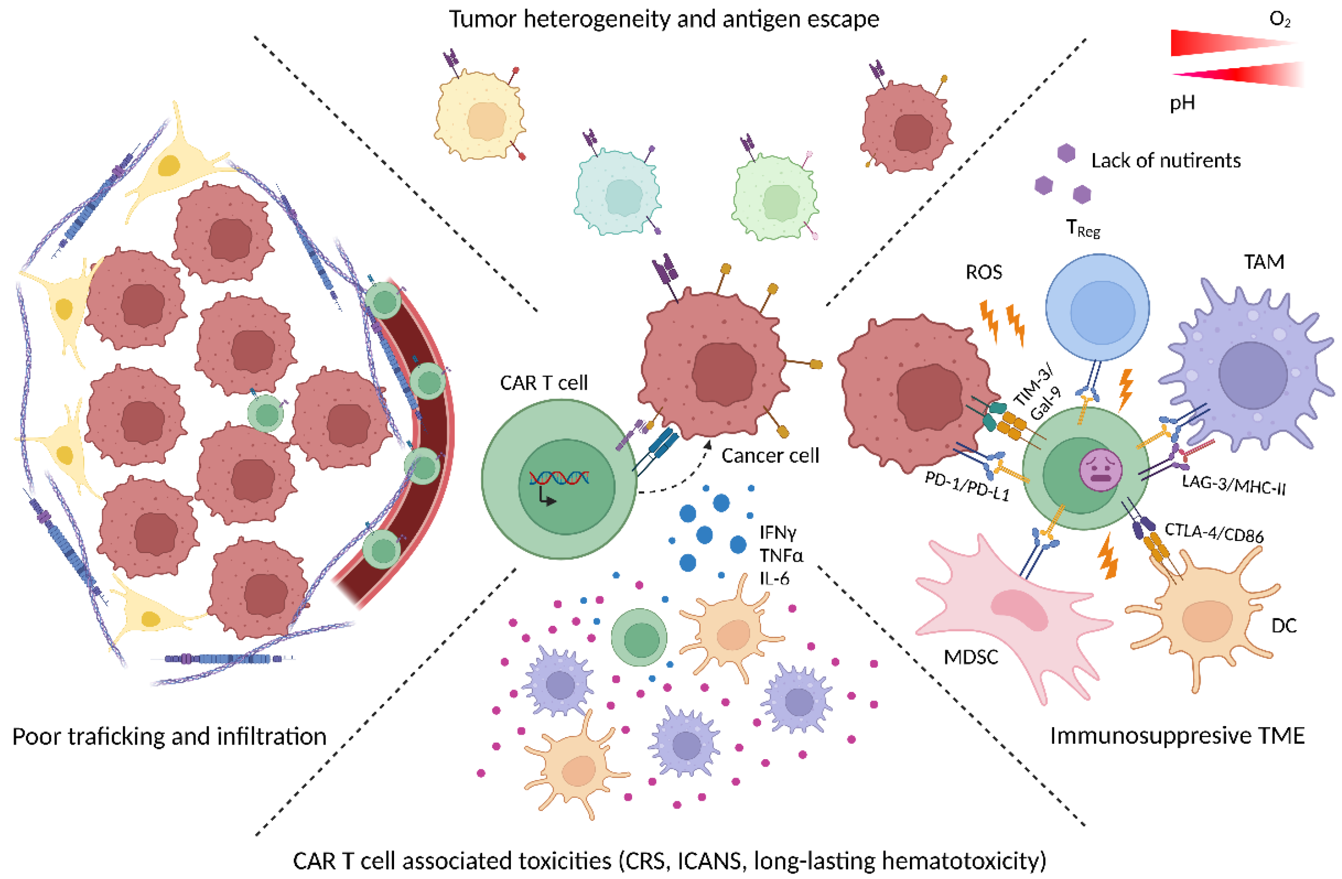
Figure 1. Current barriers in CAR T cell therapy. Clinical efficacy of CAR T cell therapy can be limited by various factors. The heterogeneity of tumors can give rise to variants that do not carry the target antigen on their surface. Immune checkpoint molecules (e.g., PD-L1 on tumor and/or stroma cells that binds to PD-1 on CAR T cells) can slow down anti-tumor activity. Overall, CAR T cells enter an immunosuppressive TME with tolerance-promoting cells such as myeloid-derived suppressor cells (MDSCs), tumor associated macrophages (TAM) or TRegs and inhospitable metabolic conditions (i.e., hypoxia, acidosis, oxidative stress, depletion of critical nutrients, etc.). In fact, the TME and the tumor cells within can represent an impermeable obstacle for CAR T cells due to their poor trafficking resulting in an insufficient infiltration of tumor tissue. In addition, CAR T cells’ (over)activation can lead to severe and potentially life-threatening toxicities with cytokine release (CRS); immune effector cell-associated neurotoxicity syndrome (ICANS) and long-lasting hematotoxicity as the most common.
Cancers 14 05442 g001
Figure 2. T cell differentiation is interconnected to metabolism. As naïve T cells (TN) leave the thymus and undergo antigen presentation, they experience a metabolic switch from oxidative phosphorylation (OXPHOS) to glycolysis as they become effector T cells (TEFF). After antigen clearance, a small subset of T cells become stem central (TSCM) and central memory (TCM) T cells, which mostly rely on OXPHOS. In case of TCR re-engagement, effector memory T cells (TEM) rapidly shift to a high glycolytic activity that supports their strong effector function. Lastly, terminally differentiated T cells (TEMRA) appeared rather senescent. T cell subsets that rely more on OXPHOS (TN, TSCM, TCM) are characterized by stemness, self-renewal and proliferation capacity. On the contrary, glycolysis-fueled cells (TEM, TEFF) display high cytotoxic function that ultimately leads them to senescence (TEMRA).
Cancers 14 05442 g002
Figure 3. Metabolic barriers for T cells in the TME. The metabolic fitness of T cells can be compromised in a variety of ways within the TME. Critical nutrients such as glucose (GLUC) are depleted. Bioenergetically active tumors secrete bioactive molecules such as lactic acid (LACT), which impedes glycolysis. Dying cancer cells release vast amounts of potassium (K) that limits glucose and amino acid uptake. The two ectonucleotidases CD39/CD73 degrade ATP into adenosine (ADE), which also impairs metabolic T cell fitness. Increased levels of lipids together with an increased fatty acid uptake of TME-infiltrating T cells promotes exhaustion and lipotoxicity.
Cancers 14 05442 g003
Figure 4. Impact of CAR design on CAR T cell metabolism. (A) Co-stimulatory signaling domains (CD28, 4-1BB, CD28+4-1BB) of CARs can impact several metabolic parameters such expression of glucose transporter (GLUT), extracellular acidification rate (ECAR), oxygen consumption rate (OCR) or spared respirator capacity (SRC). (B) Ubiquitination (Ub) and tonicity of CARs with impact on bioenergetics (and exhaustion).
Cancers 14 05442 g004
Figure 5. Strategies to modulate CAR T cell metabolism. (A) Strategies to block glycolysis. Cell culturing conditions can be improved by optimizing the use serum-containing and/or serum-free media. Treatment with cytokines such as IL-7, IL-15 or IL-21 led to the generation of the preferred T cell phenotype. Metabolic skewing (towards FAO and OXPHOS) can also be achieved by interfering with glycolysis by interfering with glycolytic enzymes such as hexokinase 2 (HK2) or glycolytic signaling such as the PI3K/Akt/mTOR axis. (B) Strategies to enhances oxidative phosphorylation (OXPHOS). Genetic engineering can be utilized for driving mitochondrial biogenesis and fitness (by, e.g., PGC1α) or metabolizing/redirect substrates such as 2-HG (by, e.g., D2HGDH), kynurenine (by, e.g., kynureninase) or O2 (by, e.g., LbNOX). Supplementation of nutrients such as inosine, L-arginine or short chain fatty acids (SCFAs) could also have a beneficial effect for CAR T cell metabolism and stemness. Abbreviations: Phosphoinositide 3-kinase (PI3K), protein kinase B (Akt), mammalian target of rapamycin (mTOR), interleukin (IL), interleukin receptor (IL-R), fatty acid oxidation (FAO), oxidative phosphorylation (OXPHOS), Peroxisome proliferator-activated receptor-gamma coactivator alpha (PGC1α), short chain fatty acids (SCFA), 2-Deoxy-D-glucose (2-DG), hexokinase 2 (HK2), glucose-6-phosphate (glucose-6P), tricarboxylic acid (TCA), alpha-ketoglutarate (α-KG), 2-hydroxyglutarate (2-HG), D-2-hydroxyglutarate dehydrogenase (D2HGDH), Kynureninase (KYNU), 3-hydroxyanthranilic acid (3hAn), alanine (Ala), Lactobacillus brevis NADH oxidase (LbNOX).
Cancers 14 05442 g005
Publisher’s Note: MDPI stays neutral with regard to jurisdictional claims in published maps and institutional affiliations.