Simple Summary
Percutaneous thermal ablation (TA) could be a safe and efficient nephron-sparing treatment for treating renal cell carcinoma (RCCs) associated with Birt–Hogg–Dubé (BHD) syndrome, a rare hereditary condition at greater risk of repeated treatments, even in the case of advanced chronic kidney disease, and should be systematically discussed as a treatment option. Indications for nephron-sparing management of these tumors in BHD patients depends on many factors, including size, number, and location of the tumor(s) in each kidney, patient surgical history, and renal function. The role of the tumor board including radiologists, interventional radiologists, urologists, and oncologists is essential.
Abstract
BHD syndrome is characterized by an increased risk of bilateral and multifocal renal cell carcinoma (RCCs), but is rarely metastatic. Our report aims to analyze the outcome of patients with BHD syndrome who underwent percutaneous thermal ablation (TA). The present report included six BHD syndrome patients (five men) with a mean age of 66 ± 11 (SD) years who had a proven germline FLCN gene mutation and underwent TA for a renal tumor. Nineteen renal tumors (median two tumors per patient; range: 1–3), including seven chromophobe RCCs, five clear-cell RCCs, four papillary RCCs, two clear-cell papillary RCC, and one hybrid oncocytic/chromophobe tumor were treated in 14 ablation sessions. The mean size of the tumors was 21 ± 11 (SD) mm (median: 20 mm; interquartile range (IQR): 14–25 mm) for a mean volume of 7 ± 11 (SD) mL (median: 3; IQR: 1–5 mL). Technical success was achieved in all ablation sessions (primary success rate, 100%). The procedure was well tolerated under conscious sedation with no significant Clavien–Dindo complication (grade 2, 3, 4). All patients were alive with no distant metastasis during a median follow-up period of 74 months (range: 33–83 months). No local tumor progression was observed. The mean decrease in estimated glomerular filtration rate was 8 mL/min/1.73 m2. No patients required dialysis or renal transplantation. In this case series, percutaneous TA appeared as a safe and efficient nephron-sparing treatment for treating RCCs associated with BHD syndrome, even in the case of advanced chronic kidney disease.
1. Introduction
Birt–Hogg–Dubé (BHD) syndrome, initially described in 1977, is a rare autosomal dominant condition due to a germline mutation in the FLCN gene located on chromosome 17p11.2 identified in 2002 [1,2]. Today, several hundred families with BHD syndrome with FLCN mutations have been reported worldwide, but BHD syndrome is probably underdiagnosed because of the wide variability in its clinical expression [3]. BHD syndrome is characterized by skin fibrofolliculomas, multiple lung cysts, and spontaneous pneumothorax [2]. However, its most severe complication is a seven-fold increased risk of renal cell carcinoma (RCC), which tend to be bilateral and multifocal in more than half of patients, but is rarely metastatic [2,3,4,5]. Renal cancers occur in up to 30% of the patients during follow-up, at a mean age of 50 years (range 25–75 years) [3]. Renal tumors are more frequently indolent, such as chromophobe RCC (chRCC) and hybrid oncocytic/chromophobe renal tumors [3]. Other histological subtypes, such as clear-cell RCC (ccRCC) and papillary RCC (pRCC), including several mixed patterns have been also reported [3]. To date, no international guidelines have been established for the management of renal tumors in BHD syndrome patients [6]. It should take into account the indolent course of small renal tumors, the renal function preservation and the risk of multiple synchronous or metachronous tumors [7]. Percutaneous thermal ablation (PTA) is an effective treatment for RCC with minimal invasiveness and similar oncologic efficacy when the procedure can be repeated. Moreover, it allows repeated tumor ablation with minimal deterioration of the renal function [8,9]. It should, thus, be considered as an alternative option for patients with hereditary RCC who require repeated treatment for multiple tumors [10,11,12]. Previous reports have suggested the usefulness of radiofrequency (RFA) for RCC in patients with von Hippel–Lindau disease [10,11,12], but the efficacy of percutaneous thermal ablation (PTA) for RCC associated with BHD syndrome has been poorly studied [3,13]. The purpose of this report was to analyze the outcome of patients with BHD syndrome who underwent PTA (radiofrequency ablation (RFA), microwave ablation (MWA), and cryotherapy) of renal tumors.
2. Materials and Methods
Our study is a single-institution retrospective report based on our prospectively maintained database and was approved by the institutional review board (IRB number: CRM-2206-287).
2.1. Patients
All patients presenting with BHD syndrome and referred for PTA were included from January 2007 to May 2021 and are followed by the Reference Center of the National Network for Rare Cancers in Adult PREDIR (inherited predispositions to RCC) labeled by the French NCI (INCa). The PTA indication was approved by the institutional multidisciplinary tumor board. A renal biopsy was performed for six lesions of significant size above 1 cm. The remaining lesions exhibiting similar pattern at contrast-enhanced Computed Tomography (CE-CT) or contrast-enhanced Magnetic Resonance Imaging (CE-MRI) did not undergo renal biopsy and was considered as having similar pathology. If during follow-up one of the renal masses was growing faster, a targeted biopsy was performed. Patients lacking post-operative CT or MRI control were excluded as well. The patient was informed of the benefits and potential complications during a dedicated consultation and clinical findings were collected, including, the Eastern Cooperative Oncology Group (ECOG) status, the tumor history, the comorbidity factors, the renal status, the bleeding risk, and the results of the pre-procedural imaging examination. Written informed consent for PTA was obtained from all patients before initiating any procedure.
2.2. Procedure
The PTA technique (RFA, MWA, or cryoablation) was discussed during the interventional radiology meeting between three interventional radiologists with 5 to 20 years of experience in renal PTA. Pre-ablation blood tests included at least complete blood count, coagulation tests, and serum creatinine level.
2.3. Follow-Up
The follow-up imaging protocol consisted of both unenhanced and triphasic CE-CT and CE-MRI performed the following morning and at 2, 6, and 12 months after the procedure and then annually. Two senior radiologists with 5 to 25 years of experience in renal imaging reviewed each imaging examination for complication and potential local persisting disease. The appearance of a focal enhancing area within or adjacent to the ablation zone indicated local tumor progression [14,15]. Technical success was defined completing of the planned ablation protocol and complete coverage of the tumor by the ablation zone [14]. After each PTA procedure, the other renal masses were carefully followed.
Outcomes of thermal ablation were assessed, including primary success rate, complications, change in renal function, local tumor progression, development of metastases, survival rate after ablation, and global progression-free survival. Clinical success (primary success rate) was defined as no recurrence or metastasis. The median follow-up period was of 74 months (range: 33–83 months). Adverse events were graded according to the Clavien-Dingo classification [14]. Serum creatinine levels (sCr) (μmol/L) and estimated Glomerular Function Rate (eGFR) were recorded before each ablation and at last follow-up [16].
All information related to the procedure were prospectively collected, including the ablation technique, the number of needles/probes, and the duration of the procedure.
2.4. Descriptive Statistics
Categorical variables were reported as counts and percentages, and continuous variables as means (standard deviation) or median (range).
3. Results
3.1. Patients
Six patients (five men and one woman) with BHD syndrome and proven germline FLCN gene mutation were included. The mean age at referral for TA was 66 ± 11 (SD) years (median: 68 years; range: 52–84). Four patients (67%) had a history of partial nephrectomy (PN) for RCC on the kidney ipsilateral to the tumor, and one of them (17%) also had a total nephrectomy (TN) of the contralateral kidney. Two patients (33%) were treated with a curative dose of oral anticoagulants for ischemic heart disease with a coronary stent stopped 5 days before the PFA. The median ASA score was 3 (range: 2–4). Patient characteristics are summarized in Table 1.

Table 1.
Clinical, genetic, and histological characteristics of the six patients with renal cell carcinoma and Birt–Hogg–Dubé syndrome.
3.2. Renal Tumors
A total of 19 renal tumors (median: 2 tumors per patient; range: 1–3) were treated using PTA on 14 ablation sessions. An 18-gauge needle biopsy was performed for 6 tumors (32%). For 13 masses, the CE-CT and/or CE-MRI was consistent with previous pathologically proven RCC for 13 tumors. Finally, seven chRCC, five ccRCCs, four pRCC, two clear-cell papillary renal tumors (ex clear-cell papillary RCC) (ccPRT), and 1 hybrid oncocytic/chromophobe tumor (HOCT) were included. The median size of the tumors was 20 ± 11 (SD) mm (mean: 21 mm; interquartile range (IQR): 14–25 mm) for a mean volume of 7 ± 11 (SD) mL (median: 3; IQR: 1–5 mL). The mean distance between the skin surface and the center of the lesion was 102 ± 15 (SD) mm (median: 98 mm; IQR: 91–115 mm). Fifteen renal tumors (79%) were located in the right kidney and four in the left, while six were more than 50% exophytic, one was less than 50% exophytic, and eleven entirely endophytic according to the RENAL Score [17]. Nine tumors (47%) were nearness to the collecting system or the sinus (< 4 mm), four (21%) were closed to the colon, three (16%) were in contact with the liver, and one (5%) was closed to the spleen and pancreas. All tumors were solid.
Renal tumor characteristics are summarized in Table 2.

Table 2.
Characteristics of the renal cell carcinomas and thermal ablation procedures.
3.3. Thermal Ablation Procedures
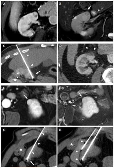
Seventeen RF ablations, 1 MW ablation, and 1 cryoablation were performed percutaneously under monitored anesthesia care (MAC) using US and CT guidance. Pain was controlled with local anesthesia and conscious sedation. Hydrodissection using 60 mL of 30% dextrose administered through a 20-Gauge needle inserted between the target tumor and the colon was used in 3 (16%) procedures to prevent bowel injury (Figure 1). Nine tumors were close to the collecting system or the sinus. For three of them, pyeloperfusion was performed using a 6 Fr ureteral stent to avoid thermal damage [18]. For the remaining lesions, a safety margin of more than 3 mm was found and considered to be sufficient according to our experience to avoid specific maneuvers.

Figure 1.
RFA of a 68-year-old man with Birt–Hogg–Dubé syndrome who had a history of partial nephrectomy and presents two ccRCCs treated in two separate sessions. Dynamic axial T1w sequences with fat suppression after gadolinium injection (A) and axial T2w image with fat saturation (B) show the first ccRCC (arrow) treated. Computed tomography (CT) images in the axial plane obtained during the RFA show the transhepatic probe (arrowheads) into the lesion (arrow) (C). CT images in the axial plane on the follow-up 6 months later show a RFA scar (arrow) (D). Dynamic axial T1w sequences with fat suppression after gadolinium injection (E) and axial T2w image with fant saturation (F) show the second ccRCC (arrow) developed in the left kidney 26 months after the initial RFA session. CT images in the axial plane obtained during the 2nd session of RFA show the RFA probe (arrowheads) into two of the lesions (G), and hydrodissection with 60 cc of serum glucose 30% was performed to protect the colon (star) (H). The patient was alive with no local progression or distant metastasis 33 months after the initial thermal ablation.
RFA was performed using a single 17-gauge internally cooled electrode (Cool-tip™, Medtronic, UK) and a generator (E-series, Medtronic, UK). Depending on the tumor size, an electrode with an active tip length of 20 or 30 mm was used. After insertion of the electrode, a 6 to 12 min of ablation.
Cryoablation was conducted using an argon-based cryoablation system (Visual Ice™ Boston-Galil Medical Inc., St Paul, MN, USA) and 17-Gauge cryoprobes (IceRod™1.5 I-Thaw™, Boston-Galil Medical Inc., St Paul, MN, USA). The standard ablation protocol included two 10 min freezing cycles separated by 9 min of passive thawing and 1 min of active thawing. CT was performed to assess the size and position of the iceball at the end of each freezing cycle. The ablation procedure aimed to cover the target lesion with an ice ball margin larger than 5 mm, according to Georgiades et al. [19].
MWA was performed using a single 17-Gauge antenna (NeuWave™, Ethicon, Johnson & Johnson, New Brunswick, NJ, USA). The duration of the treatment was 8 min at 65 Watts.
Thermal ablation procedure characteristics are summarized in Table 2.
3.4. Efficiency and Adverse Effect
All tumors were successfully ablated after a single session with no imaging findings of persisting local tumors for all tumors at the end of the follow-up period (19/19, 100%). A single adverse event consisted of a subcapsular renal hematoma (52 × 48 × 21 mm) found incidentally at the post-procedural systematic CT. It did not result in a hemoglobin drop and did not require a blood transfusion (Clavien Dindo grade 0). No additional complications were reported.
Treatment outcome is presented in Table 3. Multiple synchronous and/or asynchronous tumors were treated in each patient (Figure 1). No local tumor progression was observed during a median follow-up period of 74 months (range: 33–83 months). All patients were alive with no distant metastasis during the same follow-up period.

Table 3.
Patient outcomes.
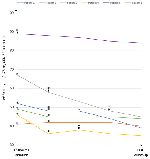
Before thermal ablation, two patients had chronic kidney disease (CKD) stage 2 and three patients had CKD stage 3 (Figure 1 and Table 4). The median increase in sCr during follow-up was 13 μmol/L [2,3,4,5,6,7,8,9,10,11,12,13,14,15,16,17,18,19,20,21,22,23,24,25,26,27,28] corresponding to a median decrease in eGFR of 8 mL/min/1.73 m2 [4,5,6,7,8,9,10,11,12,13,14,15] (Figure 2 and Table 4). No patients developed end stage kidney disease.

Table 4.
Evolution of the renal function of the six patients after each thermal ablation procedure.

Figure 2.
The renal function during the follow-up period for each of the six patients. * (one ablation) and ** (two ablations): Se Creatinine level at the moment of TA for each ablation procedure.
4. Discussion
In this case series we reported six BHD syndrome patients who underwent TA for a renal tumor. Technical success was achieved in all ablation sessions and the procedure was well tolerated under conscious sedation with no significant Clavien–Dindo complication. All patients were alive with no distant metastasis during a median follow-up period of 74 months (range: 33–83 months), with no local tumor progression. The mean decrease in estimated glomerular filtration rate was 8 mL/min/1.73 m2 and no patients required dialysis or renal transplantation.
RCC occurs in up to 30% of patients with BHD syndrome [20]. Histological subtypes of BHD-associated renal tumors include mostly indolent tumors (HOCT, chromophobe RCC, oncocytoma), but also clear cell RCC and papillary RCC [21]. A close lifelong check-up is indicated for them and their relatives over 20 years old, as far as possible, to detect renal tumors [3,22,23]. The French National Cancer Institute Network PREDIR recommends MRI surveillance every 3 years with annual ultrasound surveillance in between [24]. To date, no international guidelines have been established so far for the clinical management of these patients [6].
Our report shows that PTA in patients with BHD syndrome is safe and provides good oncological control. The slow decrease in the renal function can be attributed to CKD, eventually accelerated by previous renal surgery or percutaneous ablation. The alteration of the renal function remains acceptable even in patients with multiple ablation procedures. It is important to note that all patients were alive with no distant metastasis at last follow-up and no dialysis was required.
Stamatakis L et al. [5] recommended abdominal imaging every 36 months in individuals without renal lesions at initial screening, and then, once a tumor is detected, follow-up imaging at regular intervals until the largest tumor reaches 3 cm in maximum diameter at which time nephron-sparing surgery should ideally be performed. Although the histology of renal tumors may vary in BHD, most tumors have a relatively indolent natural history and do not require adjuvant therapy if resected when localized in the kidney. This approach aims to achieve a curative oncologic outcome and to limit the impact on renal.
Percutaneous thermal ablation can be performed using several techniques, including radiofrequency ablation, microwave ablation, or cryoablation. The choice between these methods depends on the experience of the operator, the availability of the equipment as well as the size and position of the tumor and the cost of the probes. PTA can be conducted under conscious sedation, improving the tolerance of the procedure, particularly in the elderly population. As in any other hereditary renal tumors with multifocal, bilateral, asynchronous renal tumors such as von Hippel–Lindau disease, the key advantage of PTA is the reduced impact on renal function, even when compared to partial nephrectomy [25,26]. Moreover, the percutaneous TA can be repeated to treat other tumors without the main surgical limitation, i.e., the perirenal fibrosis that makes the dissection of the kidney extremely difficult [13].
Furthermore, Matsui et al. [13] and Gaillard et al. [26] showed that several lesions can be treated simultaneously. However, the need for multiple sessions versus a single intervention is debatable. Thanks to training and imaging performance, post-ablation imaging can detect persistence of viable tumor or local recurrence [27,28,29]. In addition, it appears that the only patients progressing to metastatic disease are those with ccCCR [5], which tends to be more aggressive than the more common oncocytic and chromophobe hybrid masses, which goes against aggressive management of the renal masses. Finally, it is notable that general anesthesia can be challenging in BHD patients with pulmonary cysts, particularly in the elderly BHD population. Indeed, excessive positive pressure ventilation is at risk of rupture of a pulmonary cyst which could lead to an associated tension pneumothorax [5]. Since PTA can be performed under conscious sedation, it has the advantage over surgery (performed under general anesthesia) in avoiding this risk.
The present report has some limitations. This monocentric retrospective report includes a limited number of tumors and patients, even though it is the largest cohort of BHD-associated renal tumors with Matsui et al. [13]. The ablation technique combined RFA, MWA, and cryoablation depending on the size and position of the tumors. However, most of the procedures were performed using RFA. Finally, the report was not designed to randomly compare partial nephrectomy and PTA efficacy and tolerance, and no information can be gathered for confirming the theoretical advantages of thermal ablation over surgery.
Indications for nephron-sparing management of these tumors in BHD patients depends on many factors, including size, number, and location of the tumor(s) in each kidney, patient surgical history, and renal function [3]. The role of the tumor board including radiologists, interventional radiologists, urologists, and oncologists is essential. Despite the lack of scientific data for discussing the appropriate timing of intervention [3,15,30], it is necessary to take into account that partial surgery can become very difficult after PTA in patients with new tumors or local tumor progression [5].
5. Conclusions
The present report confirms promising long-term oncologic and renal functional outcomes of PTA for BHD-associated multifocal renal tumors. Because of its minimal invasiveness and repeatability, PTA could be an effective and safe mini-invasive nephron-sparing treatment option for this rare hereditary condition at greater risk of repeated treatments and CKD.
Author Contributions
Conceptualization, S.B. and J.-M.C.; methodology, S.B. and J.-M.C.; software, S.B.; validation, S.B., D.J., M.-O.T., A.M., O.H. and S.R.; formal analysis, S.B., S.G. and I.B.; investigation, S.B. and I.B.; resources, C.D. (Charles Dariane), C.D. (Christophe Delavaud), V.V.; data curation, S.B.; writing—original draft preparation, S.B. and I.B.; writing—review and editing, S.B., I.B., C.D. (Charles Dariane), D.J., S.R., J.-M.C. and O.H.; visualization, S.B.; supervision, O.H. and J.-M.C.; project administration, O.H.; All authors have read and agreed to the published version of the manuscript.
Funding
This research received no external funding.
Institutional Review Board Statement
The report was conducted in accordance with the Declaration of Helsinki, and approved by the Institutional Review Board of Comité d’Ethique pour la Recherche en Imagerie Médicale (IRB number: CRM-2206-287, 26 July 2022).
Informed Consent Statement
Patients were informed that their deidentified data would be used for research in conformity with current French legislation.
Data Availability Statement
The data presented in this study are available in this article.
Acknowledgments
BHD Foundation/Myrovlitis Trust (SR).
Conflicts of Interest
The authors declare no conflict of interest.
References
- Nickerson, M.L.; Warren, M.B.; Toro, J.R.; Matrosova, V.; Glenn, G.; Turner, M.L.; Duray, P.; Merino, M.; Choyke, P.; Pavlovich, C.P.; et al. Mutations in a Novel Gene Lead to Kidney Tumors, Lung Wall Defects, and Benign Tumors of the Hair Follicle in Patients with the Birt-Hogg-Dubé Syndrome. Cancer Cell 2002, 2, 157–164. [Google Scholar] [CrossRef]
- Menko, F.H.; van Steensel, M.A.; Giraud, S.; Friis-Hansen, L.; Richard, S.; Ungari, S.; Nordenskjöld, M.; Hansen, T.V.; Solly, J.; Maher, E.R. Birt-Hogg-Dubé Syndrome: Diagnosis and Management. Lancet Oncol. 2009, 10, 1199–1206. [Google Scholar] [CrossRef]
- Cornelis, F.H.; Bernhard, J.-C. Diagnostic and Interventional Radiology Is a Milestone in the Management of Renal Tumors in Birt-Hugg-Dubé Syndrome. Diagn. Interv. Imaging 2019, 100, 657–658. [Google Scholar] [CrossRef]
- Schmidt, L.S.; Linehan, W.M. Clinical Features, Genetics and Potential Therapeutic Approaches for Birt–Hogg–Dubé Syndrome. Expert Opin. Orphan Drugs 2015, 3, 15–29. [Google Scholar] [CrossRef]
- Stamatakis, L.; Metwalli, A.R.; Middelton, L.A.; Marston Linehan, W. Diagnosis and Management of BHD-Associated Kidney Cancer. Fam. Cancer 2013, 12, 397–402. [Google Scholar] [CrossRef]
- Finelli, A.; Ismaila, N.; Bro, B.; Durack, J.; Eggener, S.; Evans, A.; Gill, I.; Graham, D.; Huang, W.; Jewett, M.A.S.; et al. Management of Small Renal Masses: American Society of Clinical Oncology Clinical Practice Guideline. J. Clin. Oncol. 2017, 35, 668–680. [Google Scholar] [CrossRef]
- Hasumi, H.; Baba, M.; Hasumi, Y.; Furuya, M.; Yao, M. Birt–Hogg–Dubé Syndrome: Clinical and Molecular Aspects of Recently Identified Kidney Cancer Syndrome. Int. J. Urol. 2016, 23, 204–210. [Google Scholar] [CrossRef]
- Vroomen, L.G.P.H.; Petre, E.N.; Cornelis, F.H.; Solomon, S.B.; Srimathveeravalli, G. Irreversible Electroporation and Thermal Ablation of Tumors in the Liver, Lung, Kidney and Bone: What Are the Differences? Diagn. Interv. Imaging 2017, 98, 609–617. [Google Scholar] [CrossRef]
- Cornelis, F.H. The Future of Percutaneous Renal Ablation. Diagn. Interv. Imaging 2017, 98, 285–286. [Google Scholar] [CrossRef]
- Park, B.K.; Kim, C.K.; Park, S.Y.; Shen, S.-H. Percutaneous Radiofrequency Ablation of Renal Cell Carcinomas in Patients with von Hippel Lindau Disease: Indications, Techniques, Complications, and Outcomes. Acta Radiol. 2013, 54, 418–427. [Google Scholar] [CrossRef]
- Iwamoto, Y.; Kanda, H.; Yamakado, K.; Soga, N.; Arima, K.; Takeda, K.; Sugimura, Y. Management of Renal Tumors in Von Hippel-Lindau Disease by Percutaneous CT Fluoroscopic Guided Radiofrequency Ablation: Preliminary Results. Fam. Cancer 2011, 10, 529–534. [Google Scholar] [CrossRef]
- Gobara, H.; Hiraki, T.; Iguchi, T.; Fujiwara, H.; Nasu, Y.; Kanazawa, S. Percutaneous CT-Guided Radiofrequency Ablation for Renal Cell Carcinoma in von Hippel-Lindau Disease: Midterm Results. Interv. Radiol. 2016, 1, 1–6. [Google Scholar] [CrossRef][Green Version]
- Matsui, Y.; Hiraki, T.; Gobara, H.; Iguchi, T.; Tomita, K.; Uka, M.; Araki, M.; Nasu, Y.; Furuya, M.; Kanazawa, S. Percutaneous Thermal Ablation for Renal Cell Carcinoma in Patients with Birt-Hogg-Dubé Syndrome. Diagn. Interv. Imaging 2019, 100, 671–677. [Google Scholar] [CrossRef]
- Ahmed, M.; Solbiati, L.; Brace, C.L.; Breen, D.J.; Callstrom, M.R.; Charboneau, J.W.; Chen, M.-H.; Choi, B.I.; de Baère, T.; Dodd, G.D.; et al. Image-Guided Tumor Ablation: Standardization of Terminology and Reporting Criteria--a 10-Year Update. Radiology 2014, 273, 241–260. [Google Scholar] [CrossRef]
- Tsivian, M.; Kim, C.Y.; Caso, J.R.; Rosenberg, M.D.; Nelson, R.C.; Polascik, T.J. Contrast Enhancement on Computed Tomography After Renal Cryoablation: An Evidence of Treatment Failure? J. Endourol. 2012, 26, 330–335. [Google Scholar] [CrossRef]
- Levey, A.S.; Stevens, L.A.; Schmid, C.H.; Zhang, Y.L.; Castro, A.F.; Feldman, H.I.; Kusek, J.W.; Eggers, P.; Van Lente, F.; Greene, T.; et al. A New Equation to Estimate Glomerular Filtration Rate. Ann. Intern. Med. 2009, 150, 604–612. [Google Scholar] [CrossRef]
- Kutikov, A.; Uzzo, R.G. The R.E.N.A.L. Nephrometry Score: A Comprehensive Standardized System for Quantitating Renal Tumor Size, Location and Depth. J. Urol. 2009, 182, 844–853. [Google Scholar] [CrossRef]
- Mauri, G.; Nicosia, L.; Varano, G.M.; Bonomo, G.; Della Vigna, P.; Monfardini, L.; Orsi, F. Tips and Tricks for a Safe and Effective Image-Guided Percutaneous Renal Tumour Ablation. Insights Imaging 2017, 8, 357–363. [Google Scholar] [CrossRef]
- Georgiades, C.; Rodriguez, R.; Azene, E.; Weiss, C.; Chaux, A.; Gonzalez-Roibon, N.; Netto, G. Determination of the Nonlethal Margin Inside the Visible “Ice-Ball” During Percutaneous Cryoablation of Renal Tissue. Cardiovasc. Interv. Radiol. 2013, 36, 783–790. [Google Scholar] [CrossRef]
- Benusiglio, P.R.; Giraud, S.; Deveaux, S.; Méjean, A.; Correas, J.-M.; Joly, D.; Timsit, M.-O.; Ferlicot, S.; Verkarre, V.; Abadie, C.; et al. Renal Cell Tumour Characteristics in Patients with the Birt-Hogg-Dubé Cancer Susceptibility Syndrome: A Retrospective, Multicentre Study. Orphanet J. Rare Dis. 2014, 9, 163. [Google Scholar] [CrossRef]
- Pavlovich, C.P.; Walther, M.M.; Eyler, R.A.; Hewitt, S.M.; Zbar, B.; Linehan, W.M.; Merino, M.J. Renal Tumors in the Birt-Hogg-Dubé Syndrome. Am. J. Surg. Pathol. 2002, 26, 1542–1552. [Google Scholar] [CrossRef]
- Lee, E.; Sayyouh, M.; Haggerty, J.E.; Kazerooni, E.; Agarwal, P.P. Role of Radiologists in the Diagnosis of Unsuspected Birt-Hogg-Dubé Syndrome in a Tertiary Clinical Practice. Am. J. Roentgenol. 2019, 213, 792–797. [Google Scholar] [CrossRef]
- Gupta, S.; Kang, H.C.; Ganeshan, D.; Morani, A.; Gautam, R.; Choyke, P.L.; Kundra, V. The ABCs of BHD: An In-Depth Review of Birt-Hogg-Dubé Syndrome. AJR Am. J. Roentgenol. 2017, 209, 1291–1296. [Google Scholar] [CrossRef]
- Réseau National de Référence Pour Cancers Rares de l’Adulte PREDIR (PREDIspositions Aux Tumeurs Du Rein). Available online: https://predir.org/View/index.aspx (accessed on 5 July 2022).
- Marcelin, C.; Ambrosetti, D.; Bernhard, J.C.; Roy, C.; Grenier, N.; Cornelis, F.H. Percutaneous Image-Guided Biopsies of Small Renal Tumors: Current Practice and Perspectives. Diagn. Interv. Imaging 2017, 98, 589–599. [Google Scholar] [CrossRef]
- Gaillard, V.; Tricard, T.; Garnon, J.; Cazzato, R.L.; Dalili, D.; Gangi, A.; Lang, H. Repeat Ablative Therapy in Hereditary or Multifocal Renal Cancer: Functional and Oncological Outcomes. Urol. Oncol. Semin. Orig. Investig. 2020, 38, 797.e15–797.e20. [Google Scholar] [CrossRef]
- Wile, G.E.; Leyendecker, J.R.; Krehbiel, K.A.; Dyer, R.B.; Zagoria, R.J. CT and MR Imaging after Imaging-Guided Thermal Ablation of Renal Neoplasms. Radiographics 2007, 27, 325–339. [Google Scholar] [CrossRef]
- Cornelis, F.; Tricaud, E.; Lasserre, A.S.; Petitpierre, F.; Bernhard, J.C.; Le Bras, Y.; Yacoub, M.; Bouzgarrou, M.; Ravaud, A.; Grenier, N. Routinely Performed Multiparametric Magnetic Resonance Imaging Helps to Differentiate Common Subtypes of Renal Tumours. Eur. Radiol. 2014, 24, 1068–1080. [Google Scholar] [CrossRef]
- Ganguli, S.; Brennan, D.D.; Faintuch, S.; Rayan, M.E.; Goldberg, S.N. Immediate Renal Tumor Involution after Radiofrequency Thermal Ablation. J. Vasc. Interv. Radiol. 2008, 19, 412–418. [Google Scholar] [CrossRef]
- Walther, M.M.; Choyke, P.L.; Glenn, G.; Lyne, J.C.; Rayford, W.; Venzon, D.; Linehan, W.M. Renal Cancer in Families with Hereditary Renal Cancer: Prospective Analysis Of A Tumor Size Threshold for Renal Parenchymal Sparing Surgery. J. Urol. 1999, 161, 1475–1479. [Google Scholar] [CrossRef]
Publisher’s Note: MDPI stays neutral with regard to jurisdictional claims in published maps and institutional affiliations. |
© 2022 by the authors. Licensee MDPI, Basel, Switzerland. This article is an open access article distributed under the terms and conditions of the Creative Commons Attribution (CC BY) license (https://creativecommons.org/licenses/by/4.0/).