Imaging of Uveal Melanoma—Current Standard and Methods in Development
by
 , , , , , ,
Małgorzata Solnik
1,2,† ,
,
Natalia Paduszyńska
1,2,†,
Anna M. Czarnecka
2,3 ,
,
Kamil J. Synoradzki
3,4,*,
Yacoub A. Yousef
5,
Tomasz Chorągiewicz
6,
Robert Rejdak
6,
Mario Damiano Toro
6,7 ,
,
Sandrine Zweifel
8,
Katarzyna Dyndor
9 and
Michał Fiedorowicz
4 1
Faculty of Medicine, Medical University of Warsaw, 02-091 Warsaw, Poland
2
Department of Soft Tissue/Bone Sarcoma and Melanoma, Maria Sklodowska-Curie National Research Institute of Oncology, 5 Roentgen Str., 02-781 Warsaw, Poland
3
Department of Experimental Pharmacology, Mossakowski Medical Research Institute, Polish Academy of Sciences, 5 Pawinskiego Str., 02-106 Warsaw, Poland
4
Small Animal Magnetic Resonance Imaging Laboratory, Mossakowski Medical Research Institute, Polish Academy of Sciences, 5 Pawinskiego Str., 02-106 Warsaw, Poland
5
Department of Surgery (Ophthalmology), King Hussein Cancer Centre, Amman 11941, Jordan
6
Department of General and Pediatric Ophthalmology, Medical University of Lublin, Chmielna 1, 20-079 Lublin, Poland
7
Eye Clinic, Public Health Department, Federico II University, via Pansini 5, 80131 Naples, Italy
8
Department of Ophthalmology, University of Zurich, 8091 Zurich, Switzerland
9
Department of Radiography, Medical University of Lublin, 8 Jaczewskiego Str., 20-090 Lublin, Poland
*
Author to whom correspondence should be addressed.
†
These authors contributed equally to this work.
Cancers 2022, 14(13), 3147; https://doi.org/10.3390/cancers14133147
Submission received: 29 April 2022
/
Revised: 6 June 2022
/
Accepted: 13 June 2022
/
Published: 27 June 2022
(This article belongs to the Special Issue Uveal Melanoma: From Diagnosis to Therapy)
Simple Summary
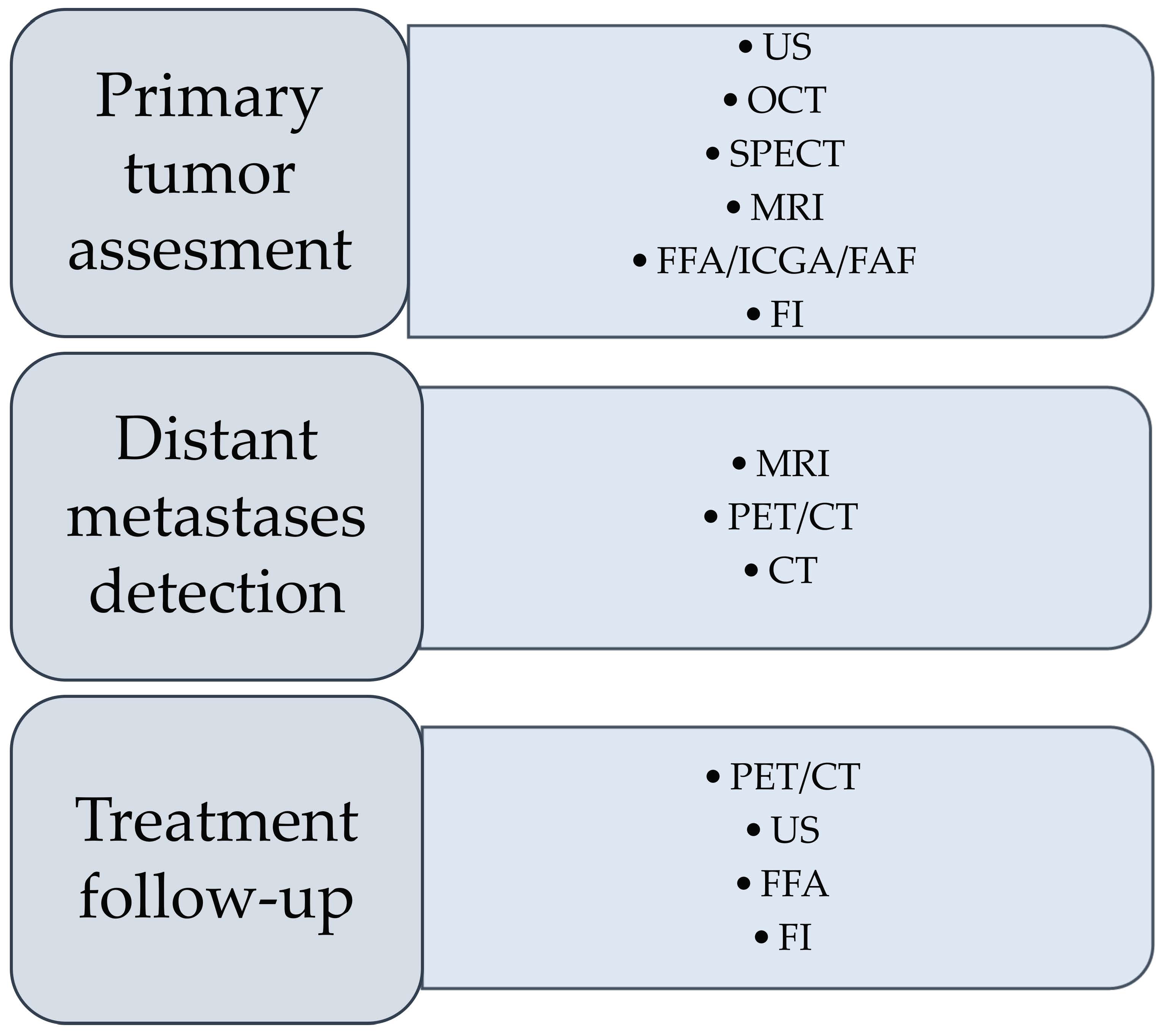
Uveal melanoma is the most prevalent intraocular tumor in adults, derived from melanocytes; the liver is the most common site of its metastases. Due to troublesome tumor localization, different imaging techniques are utilized in diagnostics, i.e., fundus imaging (FI), ultrasonography (US), optical coherence tomography (OCT), single-photon emission computed tomography (SPECT), positron emission tomography/computed tomography (PET/CT), magnetic resonance imaging (MRI), fundus fluorescein angiography (FFA), indocyanine green angiography (ICGA), or fundus autofluorescence (FAF). Specialists eagerly use these techniques, but sometimes the precision and quality of the obtained images are imperfect, raising diagnostic doubts and prompting the search for new ones. In addition to analyzing the currently utilized methods, this review also introduces experimental techniques that may be adapted to clinical practice in the future. Moreover, we raise the topic and present a perspective for personalized medicine in uveal melanoma treatment.
Abstract
Uveal melanoma is the most common primary intraocular malignancy in adults, characterized by an insidious onset and poor prognosis strongly associated with tumor size and the presence of distant metastases, most commonly in the liver. Contrary to most tumor identification, a biopsy followed by a pathological exam is used only in certain cases. Therefore, an early and noninvasive diagnosis is essential to enhance patients’ chances for early treatment. We reviewed imaging modalities currently used in the diagnostics of uveal melanoma, including fundus imaging, ultrasonography (US), optical coherence tomography (OCT), single-photon emission computed tomography (SPECT), fundus fluorescein angiography (FFA), indocyanine green angiography (ICGA), fundus autofluorescence (FAF), as well as positron emission tomography/computed tomography (PET/CT) or magnetic resonance imaging (MRI). The principle of imaging techniques is briefly explained, along with their role in the diagnostic process and a summary of their advantages and limitations. Further, the experimental data and the advancements in imaging modalities are explained. We describe UM imaging innovations, show their current usage and development, and explain the possibilities of utilizing such modalities to diagnose uveal melanoma in the future.
Keywords:
uveal melanoma; diagnosis; imaging; MRI; PET; CT; SPECT; OCT; ultrasonography