Three-Dimensional Radiological Assessment of Ablative Margins in Hepatocellular Carcinoma: Pilot Study of Overlay Fused CT/MRI Imaging with Automatic Registration
by
 , , , , and
Yasunori Minami
1,* ,
,
Tomohiro Minami
1, 
Kazuomi Ueshima
1,
Yukinobu Yagyu
2,
Masakatsu Tsurusaki
2 ,
,
Takuya Okada
3,
Masatoshi Hori
3,
Masatoshi Kudo
1 and
and
Takamichi Murakami
3 1
Department of Gastroenterology and Hepatology, Faculty of Medicine, Kindai University, 377-2 Ohno-Higashi Osaka-Sayama, Osaka 589-8511, Japan
2
Department of Radiology, Faculty of Medicine, Kindai University, 377-2 Ohno-Higashi Osaka-Sayama, Osaka 589-8511, Japan
3
Department of Radiology, Kobe University Graduate School of Medicine, 7-5-2 Kusunoki-cho, Chuo-ku, Kobe, Hyogo 650-0017, Japan
*
Author to whom correspondence should be addressed.
Cancers 2021, 13(6), 1460; https://doi.org/10.3390/cancers13061460
Submission received: 23 January 2021
/
Revised: 6 March 2021
/
Accepted: 16 March 2021
/
Published: 23 March 2021
(This article belongs to the Collection Advances in the Management of Hepatocellular Carcinoma)
Simple Summary
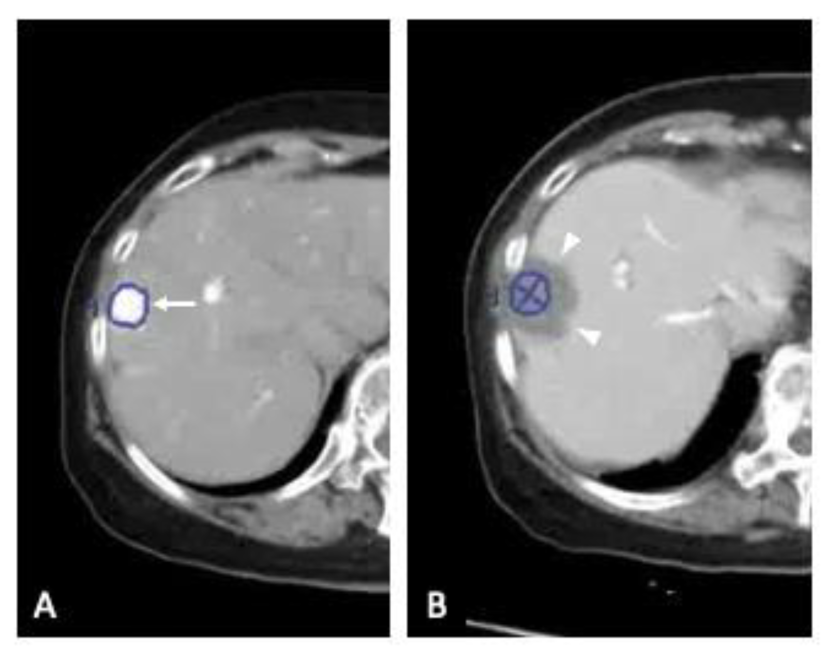
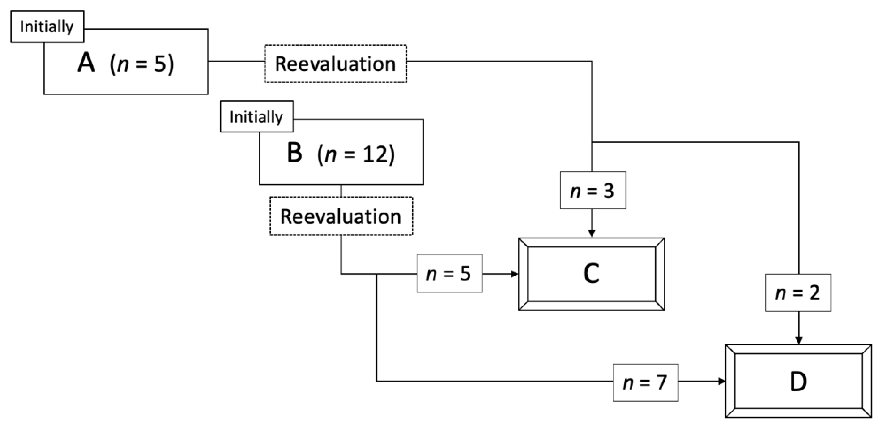
Recent advances in fusion imaging technology have made it easier to visualize and estimate ablative margins. This study was conducted to assess the clinical feasibility of a computed tomography (CT)/magnetic resonance imaging (MRI) fusion application for evaluation of the ablative margin in radiofrequency ablation (RFA) for hepatocellular carcinoma (HCC). Seventeen patients developed local tumor progressions (LTPs) due to wrong initial evaluations of technical success through a side-by-side comparison, and we reevaluated the ablative margins using the CT/MRI overlay fusion application. Eight patients were categorized into grade C (margin-zero ablation) and nine patients into grade D (existence of residual HCC). LTP occurred in re-graded C patients within 4 to 30.3 months (median, 14.3 months), and in re-graded D patients within 2.4 to 6.7 months (median, 4.2 months) (p = 0.006). Overlay fused CT/MRI imaging can allow us to evaluate HCC ablative margin three-dimensionally with high accuracy.
Abstract
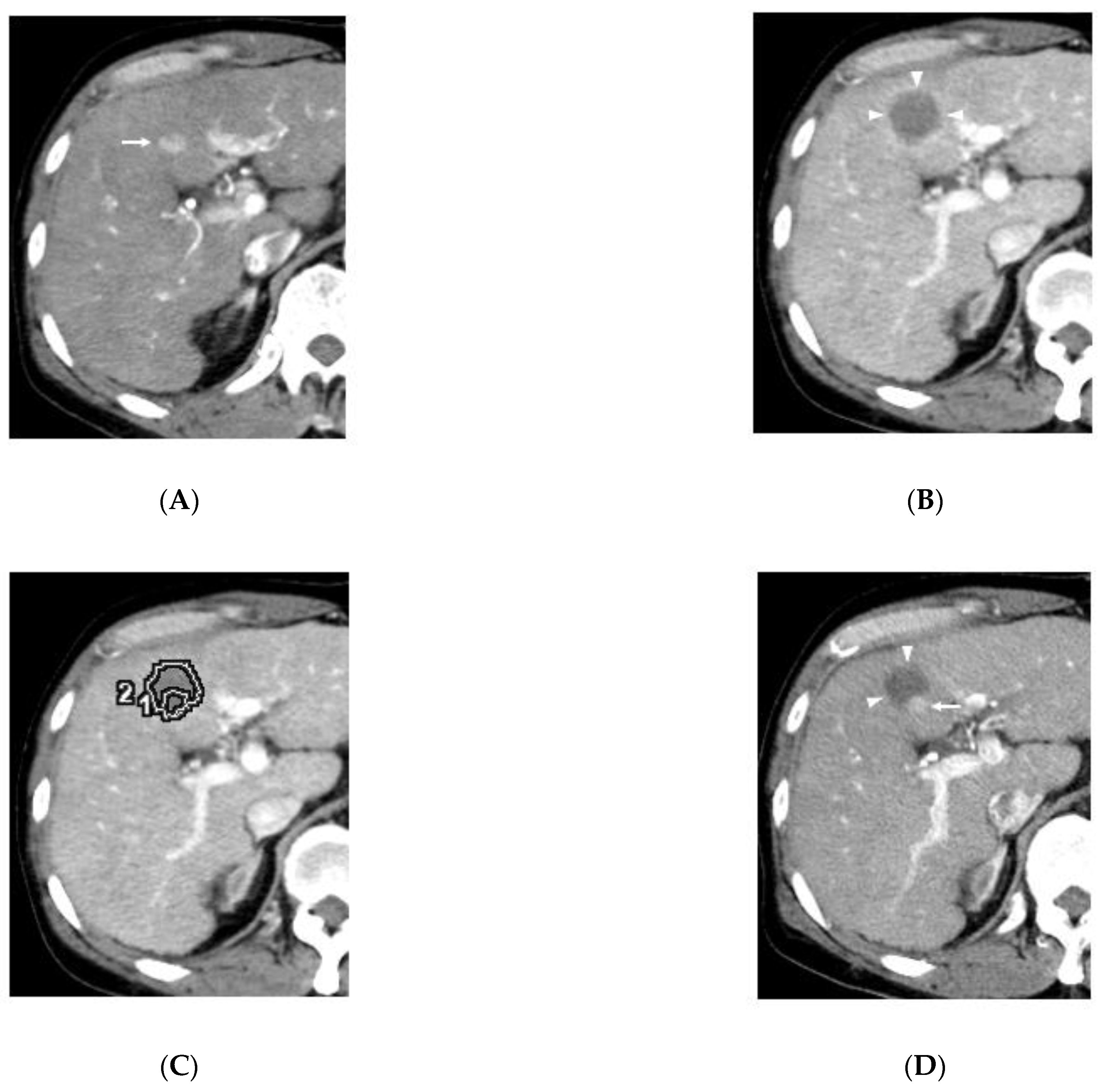
Background: We investigate the feasibility of image fusion application for ablative margin assessment in radiofrequency ablation (RFA) for hepatocellular carcinoma (HCC) and possible causes for a wrong initial evaluation of technical success through a side-by-side comparison. Methods: A total of 467 patients with 1100 HCCs who underwent RFA were reviewed retrospectively. Seventeen patients developed local tumor progressions (LTPs) (median size, 1.0 cm) despite initial judgments of successful ablation referring to contrast-enhanced images obtained in the 24 h after ablation. The ablative margins were reevaluated radiologically by overlaying fused images pre- and post-ablation. Results: The initial categorizations of the 17 LTPs had been grade A (absolutely curative) (n = 5) and grade B (relatively curative) (n = 12); however, the reevaluation altered the response categories to eight grade C (margin-zero ablation) and nine grade D (existence of residual HCC). LTP occurred in eight patients re-graded as C within 4 to 30.3 months (median, 14.3) and in nine patients re-graded as D within 2.4 to 6.7 months (median, 4.2) (p = 0.006). Periablational hyperemia enhancements concealed all nine HCCs reevaluated as grade D. Conclusion: Side-by-side comparisons carry a risk of misleading diagnoses for LTP of HCC. Overlay fused imaging technology can be used to evaluate HCC ablative margin with high accuracy.