Article

Diagnostic, Clinical and Post-SARS-CoV-2 Scenarios in Cancer Patients with SARS-CoV-2: Retrospective Analysis in Three German Cancer Centers

1
Department of Hematology and Medical Oncology, University Medicine Göttingen (UMG), 37075 Göttingen, Germany
2
Department of Thorax Oncology, Franziskus-Hospital Harderberg, Niels-Stensen-Kliniken, 49124 Georgsmarienhütte, Germany
3
Department of Medical Oncology and Hematology, Franziskus-Hospital Harderberg, Niels-Stensen-Kliniken, 41924 Georgsmarienhütte, Germany
4
Medical Clinic A, Haematology, Haemostasiology, Oncology and Pulmonology, University Hospital Münster, 48149 Münster, Germany
5
Department of Hematology and Central Hematology Laboratory, Inselspital, Bern University Hospital, University of Bern, 3010 Bern, Switzerland
6
Institute of Infection Control and Infectious Diseases, University Medicine Göttingen (UMG), 37075 Göttingen, Germany
*
Authors to whom correspondence should be addressed.
Cancers 2021, 13(12), 2917; https://doi.org/10.3390/cancers13122917
Submission received: 23 April 2021 / Revised: 1 June 2021 / Accepted: 8 June 2021 / Published: 11 June 2021
(This article belongs to the Collection The Impact of COVID-19 Infection in Cancer)

Abstract

:

Simple Summary

The study investigated diagnostic, clinical and post-SARS-CoV-2 scenarios in cancer patients with SARS-CoV-2 aiming to improve management of SARS-CoV-2 infections and cancer afterwards. Around half of patients were initially asymptomatic and were diagnosed with SARS-CoV-2 during routine or contact tracing screening. Of them, 33% developed COVID-19 lately. Eventually, predominant part of patients had asymptomatic SARS-CoV-2 or mild COVID-19 course. Lymphocytopenia preceding SARS-CoV-2 was associated with a significantly increased risk for severe or critical COVID-19 course. Commonly patients experienced a treatment delay post-SARS-CoV-2; one fifth developed progressive disease (PD) within that time and/or had to undergo therapy modifications following deterioration of the performance status or PD post-COVID-19. This study provides knowledge of real-life clinical courses of SARS-CoV-2 in oncology and contributes to improving therapeutic strategies for cancer patients in the COVID-19 pandemic.

Abstract

Oncologists face challenges in the management of SARS-CoV-2 infections and post-SARS-CoV-2 cancer treatment. We analyzed diagnostic, clinical and post-SARS-CoV-2 scenarios in patients from three German cancer centers with RT-PCR confirmed SARS-CoV-2 infection. Sixty-three patients with SARS-CoV-2 and hematologic or solid neoplasms were included. Thirty patients were initially asymptomatic, 10 of whom developed COVID-19 symptoms subsequently. Altogether 20 (32%) patients were asymptomatic, 18 (29%) had mild, 12 (19%) severe and 13 (20%) critical courses. Lymphocytopenia increased risk of severe/critical COVID-19 three-fold (p = 0.015). Asymptomatic course was not associated with age, remission status, therapies or co-morbidities. Secondary bacterial infection accompanied more than one third of critical COVID-19 cases. Treatment was delayed post-SARS-CoV-2 in 46 patients, 9 of whom developed progressive disease (PD). Cancer therapy was modified in 8 SARS-CoV-2 survivors because of deteriorating performance or PD. At the last follow-up, 17 patients had died from COVID-19 (n = 8) or PD (n = 9) giving an estimated 73% four-month overall survival rate. SARS-CoV-2 infection has a heterogenous course in cancer patients. Lymphocytopenia carries a significant risk of severe/critical COVID-19. SARS-CoV-2 disruption of therapy is as serious as SARS-CoV-2 infection itself. Careful surveillance will allow early restart of the anti-cancer treatment.

1. Introduction

Declared a pandemic by the WHO in March 2020 corona virus disease 2019 (COVID-19) caused by the novel coronavirus 2019 (SARS-CoV-2) continues to pose many new challenges to the medical community worldwide. In view of the high contagiosity of the disease, as well as of its increased propensity for causing severe symptoms, patients with a compromised immune system belong to the most relevant risk group [1]. Due to the immunosuppressive effects of systemic anticancer therapy, cancer patients are one of the most vulnerable groups for COVID-19. With around 17 million new cancer cases diagnosed worldwide in 2018 [2] alone, the magnitude of possible SARS-CoV-2 consequences in the field of oncology can be very intimidating. Along these lines, several studies have documented a significantly higher risk for severe COVID-19 events among cancer patients compared with non-cancer individuals [3,4,5,6,7,8,9,10]. The UK Coronavirus Cancer Monitoring Project and the collaboration of 19 European cancer centers recently reported death rates of 28% and 33.6% in 800 and 890 oncological patients with COVID-19, respectively. This is dramatically higher than in the general population. Interestingly, age, gender and comorbidities were significantly associated with the lethality rate in both studies, whereas ongoing chemotherapy, targeted or immunotherapy, and the time point of its application did not worsen mortality [11,12]. Hematologists and oncologists must therefore carefully balance the tremendous risk of severe SARS-CoV-2 complications against the need to continue the cancer treatment, as the latter is automatically put on hold until recovery from COVID-19. Thus, questions such as the appropriate time to restart the cancer treatment, the duration of contagiousness, as well as about persistence of immune memory after COVID-19 and the risk of SARS-CoV-2 re-infection under systemic anticancer therapy will arise in virtually every case. Therefore, the knowledge of real-life clinical courses of SARS-CoV-2 infections, as well as of its diagnostic and therapeutic pitfalls in cancer patients can provide a basis for optimal decision-making strategies. Even though many efforts have been made to highlight an increased risk for cancer patients in the COVID-19 era, practice-oriented clinical analyses of SARS-CoV-2 infections and post-SARS-CoV-2 follow-up are underrepresented. Aiming to fill this gap, we analyzed the diagnostic and therapeutic strategies with regard to SARS-CoV-2 infections, as well as impact of the infection on the cancer course and treatment algorithms of 63 hematological and oncological patients from three German cancer centers.

2. Materials and Methods

2.1. Patients

Between March 2020 and February 2021, all patients with active or non-active cancer and a SARS-CoV-2 infection confirmed by real-time reverse transcriptase polymerase chain reaction (RT-PCR) presenting in the University Hospital of Göttingen, the University Hospital of Münster or the Departments of Thoracic Oncology and Medical Oncology and Hematology of the Franziskus-Hospital Harderberg, Georgsmarienhütte, Germany, were enrolled in the study. Both, hospitalized as well as outpatients with a SARS-CoV-2 infection with any cancer entity were analyzed. The retrospective data analysis was approved by decisions of the local ethics committees (Ethics Committee of the University Medical Center Göttingen No 31/10/20; Ethics Committee of the Lower Saxony Medical Association No 31/10/20; Ethics Committee of the Westphalia-Lippe Medical Association No 2020-955-b-S).

2.2. Methods

The diagnosis of SARS-CoV-2 was made by RT-PCR on nasopharyngeal and oropharyngeal swab material. Following commercial SARS-CoV-2 RT-PCR test systems were used: SARS-CoV-2 Cobas® 6800/8800 (Roche, Basel, Switzerland), Alinity m SARS-CoV-2 assay (Abbott, Chicago, IL, USA), Xpert® Xpress SARS-CoV-2 (Cepheid, Sunnyvale, CA, USA) and Genesic SARS-CoV-2 (G Healthcare, Chicago, IL, USA). Antibody response to SARS-CoV-2 was evaluated by enzyme-linked immunosorbent assays (ELISA) with Euroimmun (Lübeck, Germany) (1. Generation, IgA/IgG) or Abbott (Chicago, IL, USA) (IgG) kits.

2.3. Definitions

Patients with active cancer were defined as those with recurrent, regionally advanced or metastatic cancer for which treatment had been administered within six months in any setting (curative, palliative, radical, adjuvant or neoadjuvant). Patients with non-active cancer were defined as cancer survivors who did meet the above-mentioned criteria and were undergoing follow-up surveillance at the time of the SARS-CoV-2 infection. The COVID-19 severity categories were determined according to WHO guidelines: asymptomatic, mild (i.e., general symptoms, outpatient care), severe (i.e., hospital admission with need for oxygen supplementation) and critical (i.e., need for life support therapy) [13]. Seroconversion was defined as a transition of the test results for immunoglobulin G (IgG) or immunoglobulin A (IgA) against SARS-CoV-2 from negative to positive in sequential samples. Lymphocytopenia was documented when a lymphocyte count was below 1.0 × 10⁹/L. Routine SARS-CoV-2 screening was referred to in- and outpatients subjected randomly to PCR-testing without exhibiting any signs or symptoms of COVID-19. Contract tracing was defined as PCR-testing performed for patients with the history of close contacts to individuals with laboratory-confirmed or probable COVID-19. The post-SARS-CoV-2 follow-up implied monitoring of patients from the first positive SARS-CoV-2 RT-PCR result.

2.4. Analyzed Data

The patients were analyzed with regard to (A) demographics and cancer data including the last cancer therapy and remission status, as well as comorbidities at the time of first RT-PCR SARS-CoV-2 positive result, (B) the course of the SARS-CoV-2 infection including symptoms, severity and duration as well as laboratory and radiological findings, and finally (C) the cancer course in the post-SARS-CoV-2 follow-up. To this end, the impact of SARS-CoV-2 on overall survival, cancer therapy and remission status was evaluated. Patients were assessed for the presence of common comorbidities such as cardiovascular diseases (arterial hypertension, coronary artery disease, chronic heart failure, atrial fibrillation), metabolic diseases (diabetes mellitus, obesity), chronic respiratory and liver diseases, renal insufficiency, cerebrovascular diseases and autoimmune disorders.

2.5. Statistics

Categorical variables were summarized as frequencies and percentages, and continuous variables were summarized as medians and ranges. Probability of overall survival (OS) was calculated using the Kaplan-Meier method. Cumulative incidence curves were used for COVID-19 and cancer mortality in a competing risk. The Chi-square test of independence was used to determine significance of relationship between lymphocytopenia, histologic type of cancer (hematologic vs. solid), age and severity of COVID-19 course. The association between clinical characteristics of cancer patients and the occurrence of asymptomatic SARS-CoV-2 infection was assessed by Spearman’s correlation. A p-value less than 0.05 was considered statistically significant. Statistical analyses were performed with SPSS, version 26.0 (SPSS, Chicago, IL, USA) and R, version 3.6.2 (RStudio, Boston, MA, USA) (https://www.R-project.org/ (accessed on 25 February 2020)).

3. Results

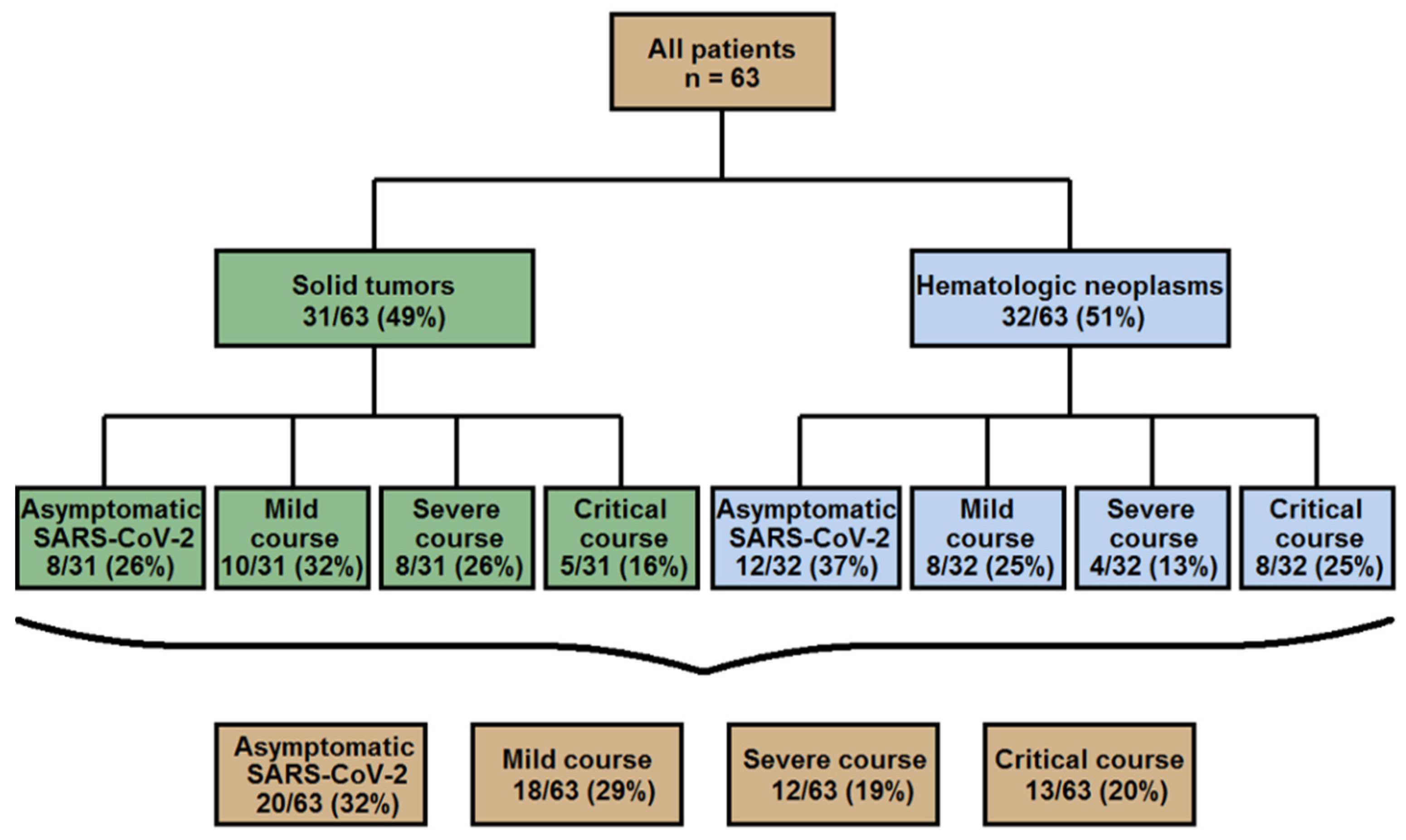
3.1. Characteristics of Cancer Patients Positive for SARS-CoV-2

Sixty-three patients tested positive for SARS-CoV-2 were included in this study. Patient and disease characteristics at the time of the confirmed SARS-CoV-2 test are presented in Table 1. The median age was 62 (range, 19–85) with a slight predominance of males (35 males: 28 females). Hematologic and solid neoplasms were equally represented (32 vs. 31) with lymphomas the more frequent hematologic, and lung, breast or gastrointestinal tumors the most frequent solid neoplasms (Table 1 for details). In this case, 56 patients (89%) were categorized as active cancer cases, while the remaining seven were under follow-up surveillance after systemic cancer treatment. For details of the therapeutic modalities see Table 1. Particularly, 17 patients (30%) underwent immunotherapy-based treatment (either as monotherapy or combined with cytostatic agents or radiotherapy) presented predominantly by anti-CD20 antibodies (n = 9/17) and followed by programmed death-1/programmed death-ligand 1 (PD-1/PD-L1) (n = 6/17) and anti-CD38 antibodies (n = 1/17), as well as chimeric antigen receptor (CAR) T-cell therapy (n = 1/17). Before testing positive for SARS-CoV-2, thirty patients had received one therapy line (48%) and 2 (3%) received no therapy yet, whereas the remaining had undergone up to four or more therapy lines (Table 1). At the time of study enrollment 12 patients were in complete remission (CR), 12 in partial remission (PR), 11 had a stable disease (SD) and 20 had relapsed/progressive disease. Remission status had not yet been assessed in eight patients because treatment begin, or cancer diagnosis were so recent. Eighteen patients had no relevant comorbidity, while the others had up to four or more pre-existing conditions (Table 1).

3.2. Clinical, Laboratory and Imaging Findings in 63 Cancer Patients with SARS-CoV-2

The laboratory, imaging and clinical findings of the patients are given in Table 2. The time from the last cancer treatment varied markedly from zero to nearly three years with a median of 13 days (Table 2). Approximately half of the patients exhibited COVID-19 symptoms primarily, while remaining patients were asymptomatic and tested within routine or contact tracing screening. A third of the initially asymptomatic patients subsequently developed typical COVID-19 symptoms giving a total of 43 patients with COVID-19 (Table 2, Figure 1). The median values for white blood cell count were in the normal range, while those for procalcitonin, C-reactive protein and lactate dehydrogenase (LDH) were elevated but there was a large spread in all parameters. Forty-six percent of the patients with an available differential blood count (n = 17/37) had lymphocytopenia (see Table 2). Even though immunophenotyping data was not available at the time point of SARS-CoV-2 diagnosis, B-cell depletion could be suggested in at least 35% (n = 6/17) of these patients who underwent either anti-CD20 immunotherapy (n = 5/6) or induction therapy within the GMALL (German Multicenter Study Group for Adult Acute Lymphoblastic Leukemia) protocol (n = 1/6) before. Chest imaging was documented as having been performed in 39 patients, and evidence of pneumonia was detected in 23 (59%) of them. Seroconversion with detectable IgA and/or IgG was seen in ten (63%) of the 16 patients who were tested for this with a median of 13 days. In six out of 43 COVID-19 cases (14%) secondary bacterial infection was confirmed subsequently. In four of them at least two microorganisms have been detected simultaneously, thereof two had the same and two different species, respectively. Bacteria could be cultivated from the following specimen: trachealbronchial fluid (n = 4/6), blood culture (n = 3/6) and urine (n = 2/6). The causative microorganisms were presented predominantly (n = 5/6) by Gram-negative bacteria: Klebsiella pneumoniae (n = 4/6), in one case together with Klebsiella oxytoca, Escherichia coli (n = 2/6) and Pseudomonas aeruginosa (n = 1/6). The remaining patient had evidence of Staphylococcus aureus in trachealbronchial fluid. Of note, five of six patients with secondary bacterial infection had critical COVID-19 compromising 38% (n = 5/13) of all critical cases. The remaining patient had severe COVID-19 (8%, n = 1/12).

3.3. Course of the SARS-CoV-2 Infection

Data on the course of the SARS-CoV-2 infection are presented in Table 3 and Figure 2. As mentioned above, 43 (68%) of the patients had COVID-19 symptoms, whereas 20 (32%) had an asymptomatic course.

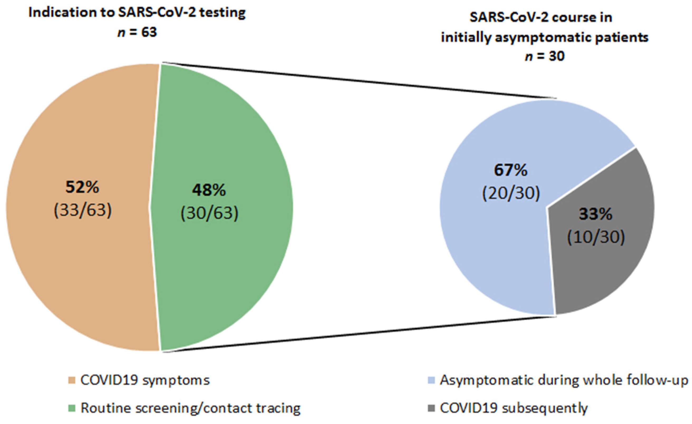
3.3.1. Asymptomatic SARS-CoV-2 Patients

Details of asymptomatic SARS-CoV-2 patients are listed in Table S1. Asymptomatic cases were seen among patients with hematologic 60% (n = 12/20) or solid 40% (n = 8/20) malignancies (p = 0.379). Analyzing the whole cohort of these patients, the occurrence of an asymptomatic course did not correlate with age (range, 21–81) (p = 0.328), the number of comorbidities (p = 0.816), therapy lines (p = 0.783) or level of remission (p = 0.718). Lymphoid neoplasms (83%, 10/12) were predominant among the hematologic malignancies: lymphomas (7/10), acute lymphatic leukemias (2/10) and multiple myeloma (1/10). Gastrointestinal cancer (4/8) was the most common malignancy among solid tumors followed by sarcomas (2/8), and breast and lung cancer (each 1/8). Of 20 patients, only three (15%) were under follow-up surveillance at the time point of SARS-CoV-2 diagnosis. The remaining patients were undergoing active cancer treatment with a broad spectrum of intensity and action principles. Particularly, cases with high intensity treatment (Table S1), such as advanced Hodgkin lymphoma in early post-transplant aplasia phase following high-dose chemotherapy/autologous stem cell transplantation (HDCT/ASCT) (pt #5), acute lymphatic leukemia under induction therapy with GMALL protocol (pt #10), pancreatic cancer under FOLFIRINOX (leucovorin, fluorouracil, irinotecan, oxaliplatin) (pt #15) or sarcoma (pt #20) under doxorubicin/ifosfamide regimen were documented. Regarding all patients with hematologic malignancies (n = 32) and comparing asymptomatic cases (n = 12) with those having COVID-19 (n = 20), asymptomatic patients tended to be younger (median age 59 vs. 65), had less co-morbidities (median number 1 vs. 2), were less heavily pretreated (median number therapy lines 1 vs. 2 prior SARS-CoV-2) and received less frequent immunotherapy (25% vs. 45%) at the time point of SARS-CoV-2 diagnosis (Table S2).

3.3.2. SARS-CoV-2 Infection with COVID-19 Symptoms

Of the patients with COVID-19, 29% (n = 18/63) had a mild, 19% (n = 12/63) severe and 20% (n = 13/63) critical course. Patients with hematologic malignancies tended to have a critical course more frequently (25%, n = 8/32) than those presenting with solid tumors (16%, n = 5/31). In contrast, patients with solid tumors more frequently had a severe course of the disease (26%; n = 8/31 vs. 13%; n = 4/32) (Figure 2). Of note, the risk of a severe or critical COVID-19 course was significantly higher for patients with lymphocytopenia diagnosed within two weeks preceding the SARS-CoV-2 infection or at its presentation (59%) than for those with normal lymphocyte counts (59% vs. 20%, p = 0.015). The patients with a severe/critical COVID-19 course had a trend towards worse remission (64% with stable or progressive disease) and general health status (56% with ≥2 co-morbidities). Yet, an advanced age (<60 vs. ≥60) at diagnosis of SARS-CoV-2 correlated significantly with a severe/critical COVID-19 course (p = 0.039). The majority (63%) of the patients was treated as inpatients, mostly because of COVID-19 symptoms, but also because they were already in the hospital when SARS-CoV-2 was detected, or were admitted for other indications such as pregnancy or severe lymphodepletion (see Table 3). Of all patients, 28 (44%) remained on the ward, while 12 (19%) had to be admitted to intermediate or intensive care unit (IMC/ICU). Of the latter, nine patients (75%) received respiratory support beyond simple oxygen administration presented either by high-flow oxygen (17%, n = 2/12) and non-invasive ventilation (25%, n = 3/12), or invasive (mechanical) ventilation (33%, n = 4/12). The median length of hospital stay of the patients admitted because of COVID-19 was 14 days (range 1–43). They were treated with a variety of modalities including antivirals, convalescent plasma, dexamethasone, antibodies against IL-6 or IL-6 receptor or serine protease inhibitor (see Table 3).

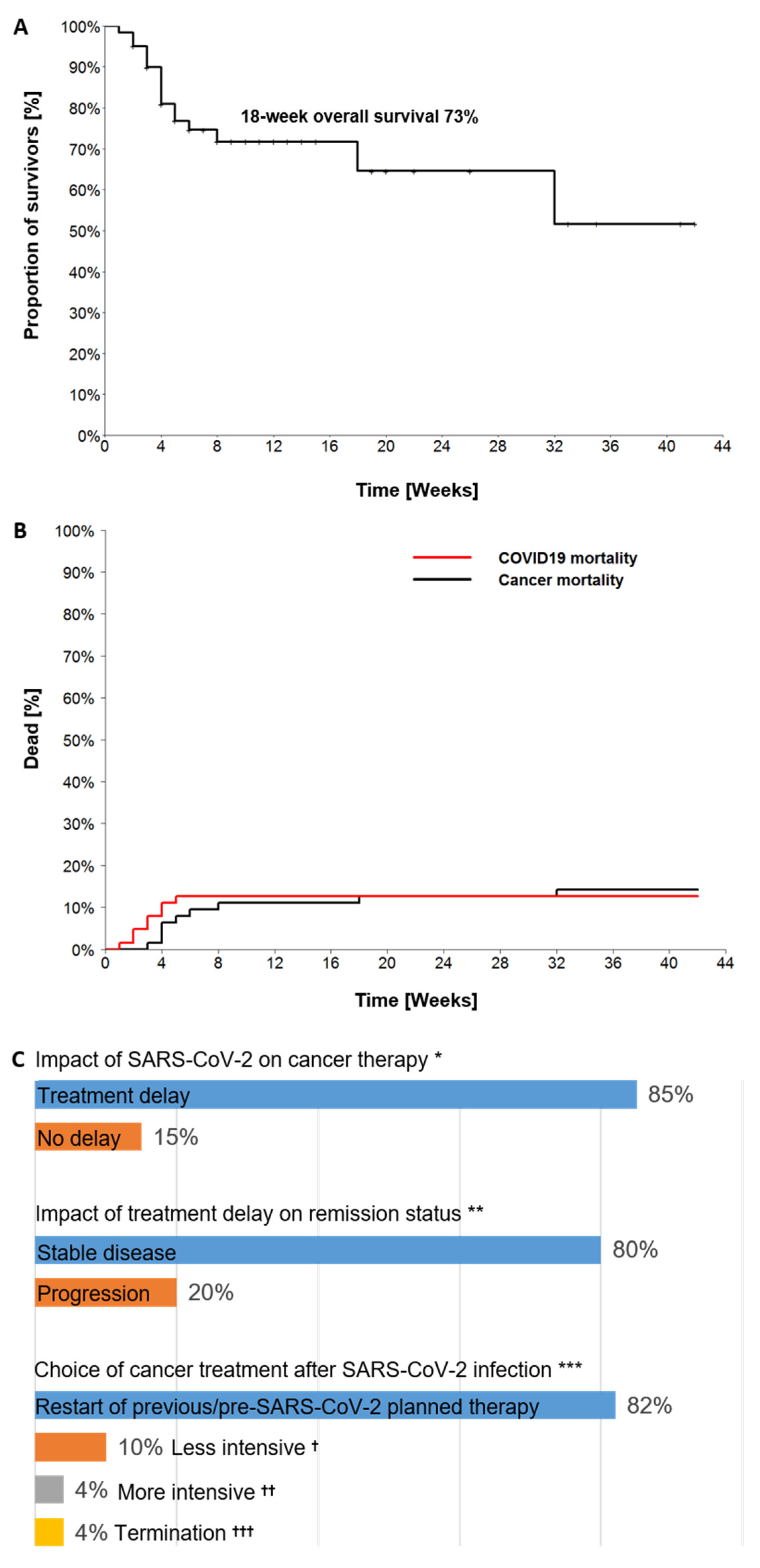
3.3.3. Outcomes of SARS-CoV-2 Infection with Impact on Cancer Course

Outcomes and impact of the SARS-CoV-2 infection on the subsequent cancer course are presented in Table 3 and Figure 3A–C. Median time from SARS-CoV-2 positivity to the last follow-up was 6 weeks (range 0–42). At the time of the last follow-up, 17 patients (27%) had died. Of them, COVID-19 was the cause of death in eight patients (47%) and the malignancy in nine (53%). The calculated four-month overall survival rate (Kaplan-Meier) post-SARS-CoV-2 was 73% (Figure 3A). Of the eight patients that succumbed to COVID-19, four had lung cancer and four a hematologic malignancy: either B-non-Hodgkin lymphoma (B-NHL) (3/8) or acute myeloid leukemia (1/8). Seven of the deceased were men, and the median age was 66 years (range 50–82). Notably, COVID-19 associated deaths occurred within the first month post-SARS-CoV-2, whereas cancer-associated deaths occurred later in the following time (Figure 3B).
Cancer treatment was delayed in 46 (85%) of the 54 patients with ongoing or scheduled cancer therapy prior to their testing positive for SARS-CoV-2 (Figure 3C). The median treatment delay was 25 days (range 4–66). Nine of these patients (20%) developed progressive disease (PD) during the SARS-CoV-2 infection, which may have been associated with the treatment delay. Four of these nine patients later died of the relapsed/progressive disease, and only one of those four had been able to resume cancer therapy. The median treatment delay in these nine patients was 31 days from the positive SARS-CoV-2 test (range 21–54).
Therapy was restarted in 37 of the 45 SARS-CoV-2 survivors with active cancer (82%) and modified for the other eight patients after the SARS-CoV-2 infection. In six patients the therapy was either reduced in intensity (4/6) or terminated (2/6) due to a deterioration of the performance status post-COVID-19. In two other patients more intensive regimens were necessary due to PD developed during the delay in therapy (Figure 3C). At the time of the last follow-up, 20% of the patients (n = 13) had CR, 16% (n = 10) had PR, 32% (n = 20) had SD and 32% (n = 20) had relapsed/progressive disease.

4. Discussion

Here, we comprehensively analyzed different diagnostic and therapeutic strategies in a cohort of 63 SARS-CoV-2-positive tested cancer patients with the aim of providing real-life data on the management and prognosis of these vulnerable patients.
Our patient cohort encompassed the whole spectrum of solid and hematological neoplasms as well as cancer treatment modalities in outpatient and inpatient care settings. The patients’ characteristics reflected everyday clinical practice as far as more than half of the patients were ≥60 years old, had received ≥1 therapy lines, had predominantly SD or relapsed/progressive disease and had at least on comorbidity. Of note, around half of cancer patients were initially asymptomatic and were diagnosed with SARS-CoV-2 during routine or contact tracing screening. Thirty-three percent of these patients developed COVID-19 after a median interval of approximately two weeks after the first positive SARS-CoV-2 test result. Thus, clinical vigilance together with regular or on-demand testing should be considered to prevent the spread of SARS-CoV-2 among these highly vulnerable patients. Only around two third of the patients tested for SARS-CoV-2 antibodies in our study met the criteria for seroconversion. Several other studies have recently reported a lower level of antibody development in cancer patients [14,15]. Therefore, the risk of SARS-CoV-2 re-infection may be increased in cancer patients, and all established preventive measure should be continued in the post-SARS-CoV-2 follow-up period.
Overall, around 60% of our patients were either asymptomatic or had a mild COVID-19 course. The asymptomatic cases (33%) were not associated with a younger age, a lack of co-morbidities, better remission status, number of ongoing therapies or histologic type of cancer (hematologic vs. solid neoplasms). Indeed, a significant number of these patients were more than 60 years old and presented with relapsed/progressive or stable disease, two or more comorbidities and two or more therapy lines. However, our data is consistent with those of the previous studies, which reported a high incidence of asymptomatic SARS-CoV-2 infections among cancer patients. The incidence of severe and critical courses was 39%, which was comparable with other large multicenter studies in cancer patients with COVID-19 [11,16]. Particularly, patients with hematologic neoplasms were more prone to develop a critical COVID-19 course (25%) which could be explained by a stronger immune dysregulation due to underlying disease and/or applied cancer treatment modalities. Accordingly, a large meta-analysis of 3377 cases with hematologic malignancy and COVID-19 found a 34% risk of death for this patient collective [17]. The most frequently recorded COVID-19 symptoms among our patients were in agreement with those documented for unselected patients [18] and encompassed fever, cough and dyspnea (56%–67%), whereas gastrointestinal complains and chest pain were found in fewer than 15% of the COVID-19 cases.
Particularly, lymphocytopenia preceding SARS-CoV-2 was associated with an almost three-fold increased risk for the development of a severe or critical course compared with cancer patients without lymphocytopenia in our study. This was corresponding to the risk of severe COVID-19 courses in the case of lymphocytopenia in a meta-analysis including 2282 non-cancer SARS-CoV-2 cases [19]. Considering the high frequency of post-treatment lymphodepletion in cancer patients (46% in our study), especially those patients with peri-SARS-CoV-2 lymphocytopenia should be monitored particularly closely. It should be noted that nineteen percent of the patients in our study required admission to IMC/ICU due to COVID-19. For ICU admission, recent studies reported a wide range between 7% and 36% among cancer patients [11,16,20,21]. The ICU admission rate in our cohort was comparable with unselected COVID-19 patients in Germany [22,23]. 75% of all IMC/ICU admitted patients in our study required respiratory therapy beyond standard oxygen administration. Particularly, invasive (mechanical) ventilation was applied for a third of the patients admitted to intensive care whereas second third received non-invasive respiratory support. Notably, the percentage of invasive ventilation was considerably lower than the approximately 54% rate in unselected COVID-19 patients in ICU [23]. Such a difference could be partly explained by the refusal of mechanical ventilation by a medical team due to a poor overall prognosis and/or a patient wish at the time point of COVID-19. In addition, secondary bacterial infection, presented mostly by gram-negative bacteria, accompanied more than third of critical COVID-19 cases in our study and should therefore be seriously considered in diagnostic and treatment workup for cancer patients admitted to ICU. Particularly, Klebsiella pneumoniae was the most common pathogen and documented also commonly in non-cancer patients with critical COVID-19 [24,25].
In line with previous studies [11,12], the overall-case mortality averaged 27% in our cohort with an estimated four month-OS of 73% post-SARS-CoV-2. However, only half of all deaths’ cases were due to COVID-19, with the remaining patients succumbing to progressive malignancy in the course of the SARS-CoV-2 infection. Notably, the deaths from COVID-19 were equally distributed between lung cancer and hematologic malignancy. Eighty-seven percent of the deceased were men. These results were in correspondence with the literature demonstrating the increased risk profile for a severe COVID-19 course of the respective patients [16,26,27,28,29].
Previous studies mainly reported short post-SARS-CoV-2 observation periods of two to three weeks following SARS-CoV-2 detection [11,12,16,20]. The strength of our study was a longer median follow-up (six weeks) post-SARS-CoV-2 with more than 30% patients monitored for 2.5 months or more after the SARS-CoV-2 infection. This enabled us to assess the long-term impact of the SARS-CoV-2 infection on the subsequent cancer management. Indeed, treatment was delayed in 85% of the patients post-SARS-CoV-2 with a median delay time of three and a half weeks. This may have contributed to 20% of the patients developing progressive disease during the delay. Notably, only around four-fifths of the patients were able to resume the previous or pre-SARS-CoV-2 planned therapy, whereas the therapy of the remaining patients was modified, predominantly with a switch to a less intensive regimen or to best-supportive care. Consequently, oncologists face two challenges in the SARS-CoV-2 pandemic: managing SARS-CoV-2 infection with the least possible harm without delaying subsequent cancer management. Careful clinical surveillance together with intensive testing for SARS-CoV-2 should be conducted for the earliest possible re-start of cancer therapy. The role of testing for antibodies to SARS-CoV-2 has not been conclusively answered and requires further investigation. Given the morbidity of COVID-19 and subsequent treatment delay, patients with cancer should be considered high-priority for the COVID-19 vaccination. Considering the efficacy of SARS-CoV-2 vaccine among per se immunocompromised cancer patients, many questions may arise especially for those developing B- and/or T-cell depletion following cancer treatment. As a result, the appropriate time point for vaccination of this patient collective remains undefined. Recently, one study reported that the first dose of a COVID-19 vaccine (either Pfizer-BioNTech or Oxford-AstraZeneca) was able to induce an immune response in around 70% of patients with multiple myeloma. Yet, patients with worse remission status (SD/PD) and suppression of polyclonal (physiologic) immunoglobulins were more likely to lack a seropositive response following a vaccination [30]. Even though the initial clinical studies of the COVID-19 vaccines did not include cancer patients, the European Society for Medical Oncology (ESMO), the Society for Immunotherapy of Cancer (SITC) and the National Comprehensive Cancer Network (NCCN) COVID-19 Vaccination Advisory Committee have recently released their preliminary recommendations supporting vaccination of all patients with cancer, including those receiving active therapy [31,32,33].
Our study has three main limitations. First, our pilot study includes only a relatively small number of patients. Second, the patient group is heterogenic and included different entities and treatment modalities at the time point of enrollment in the study. Third, the data were retrospectively collected from the registries of three cancer centers and patients treated in oncological outpatient clinics are underrepresented.

5. Conclusions

Based on our data and the current literature we summarize the following key-points:
  • At diagnosis of SARS-CoV-2 infection in cancer patients, the likelihood of an asymptomatic or mild course of COVID-19 seems to be greater than that of a severe or critical one.
  • Cancer patients diagnosed in an asymptomatic phase of a SARS-CoV-2 infection have around a 30% probability for the subsequent development of COVID-19.
  • Diagnostic and clinical pitfalls such as a lack of seroconversion post-SARS-CoV-2 in some of the cancer patients should be considered when planning the management of SARS-CoV-2 infection and subsequent cancer therapy.
  • Cancer patients with peri-SARS-CoV-2 lymphocytopenia are especially at risk for the development of a severe or critical COVID-19 course and should be monitored carefully.
  • Secondary bacterial infection, presented predominantly by gram-negative bacteria, is common in cancer patients with critical COVID-19 and should be included in a diagnostic and treatment workup for such cases.
  • Even though the COVID-19 mortality is higher in cancer patients as compared to non-cancer individuals, the frequency of ICU admission does not necessarily differ significantly compared with non-cancer COVID-19 patients.
  • A delay in cancer treatment is extremely likely following SARS-CoV-2 infection and requires thorough clinical and diagnostic surveillance to enable for the earliest possible restart of treatment.

Supplementary Materials

The following is available online at https://www.mdpi.com/article/10.3390/cancers13122917/s1, Table S1: clinical characteristics and outcomes in asymptomatic patients with SARS-CoV-2. Table S2: Comparison of asymptomatic and symptomatic SARS-CoV-2 patients with hematologic malignancies.

Author Contributions

Conceptualization, E.S. and A.B.; methodology, E.S., A.B., P.H. and A.K.; software, E.S. and N.S.; validation, E.S., P.H., R.P., S.V., N.S., A.K. and A.B.; investigation, E.S., P.H., R.K., R.P., S.V., M.P. and A.K.; data curation, U.B., S.S., L.T. and G.L.; writing—original draft preparation, E.S. and A.B.; writing—review and editing, P.H., R.K., U.B., M.P., S.S., L.T. and G.L.; supervision, A.B., R.K., U.B., S.S., L.T. and G.L. All authors have read and agreed to the published version of the manuscript.

Funding

This research received no external funding. E.S. is funded by the Deutsche Forschungsgemeinschaft (DFG, German Research Foundation)—413501650.

Institutional Review Board Statement

The study was conducted according to the guidelines of the Declaration of Helsinki, and approved by the following local Ethics Committees: Ethics Committee of the University Medical Center Göttingen No 31/10/20; Ethics Committee of the Lower Saxony Medical Association No 31/10/20; Ethics Committee of the Westphalia-Lippe Medical Association No 2020-955-b-S.

Informed Consent Statement

Informed consent was not sought as a retrospective study design was used.

Data Availability Statement

The data presented in this study are available on request from the corresponding author.

Acknowledgments

The authors wish to thank all staff members of the coronavirus units involved in the care of the patients reported in this study.

Conflicts of Interest

The authors declare no conflict of interest.

References

  1. Shields, A.M.; Burns, S.O.; Savis, S.; Richter, A.G. COVID-19 in patients with primary and secondary immunodeficiency: The United Kingdom experience. J. Allergy Clin. Immunol. 2021, 147, 870–875.e1. [Google Scholar] [CrossRef] [PubMed]
  2. Cancer Research UK. Worldwide Cancer Incidence Statistics. 2018. Available online: https://www.cancerresearchuk.org/health-professional/cancer-statistics/worldwide-cancer/incidence#heading-Zero (accessed on 14 November 2018).
  3. Liang, W.; Guan, W.; Chen, R.; Wang, W.; Li, J.; Xu, K.; Li, C.; Ai, Q.; Lu, W.; Liang, H.; et al. Cancer patients in SARS-CoV-2 infection: A nationwide analysis in China. Lancet Oncol. 2020, 21, 335–337. [Google Scholar] [CrossRef]
  4. Zhang, L.; Zhu, F.; Xie, L.; Wang, C.; Wang, J.; Chen, R.; Jia, P.; Guan, H.Q.; Peng, L.; Chen, Y.; et al. Clinical characteristics of COVID-19-infected cancer patients: A retrospective case study in three hospitals within Wuhan, China. Ann. Oncol. 2020, 31, 894–901. [Google Scholar] [CrossRef]
  5. Desai, A.; Sachdeva, S.; Parekh, T.; Desai, R. COVID-19 and Cancer: Lessons from a Pooled Meta-Analysis. JCO Glob. Oncol. 2020, 6, 557–559. [Google Scholar] [CrossRef]
  6. Saini, K.S.; Tagliamento, M.; Lambertini, M.; McNally, R.; Romano, M.; Leone, M.; Curigliano, G.; De Azambuja, E. Mortality in patients with cancer and coronavirus disease 2019: A systematic review and pooled analysis of 52 studies. Eur. J. Cancer 2020, 139, 43–50. [Google Scholar] [CrossRef] [PubMed]
  7. Erdal, G.S.; Polat, O.; Erdem, G.U.; Korkusuz, R.; Hindilerden, F.; Yilmaz, M.; Yasar, K.K.; Isiksacan, N.; Tural, D. The mortality rate of COVID-19 was high in cancer patients: A retrospective single-center study. Int. J. Clin. Oncol. 2021, 26, 826–834. [Google Scholar] [CrossRef] [PubMed]
  8. Dai, M.; Liu, D.; Liu, M.; Zhou, F.; Li, G.; Chen, Z.; Zhang, Z.; You, H.; Wu, M.; Zheng, Q.; et al. Patients with Cancer Appear More Vulnerable to SARS-CoV-2: A Multicenter Study during the COVID-19 Outbreak. Cancer Discov. 2020, 10, 783–791. [Google Scholar]
  9. Mehta, V.; Goel, S.; Kabarriti, R.; Cole, D.; Goldfinger, M.; Acuna-Villaorduna, A.; Pradhan, K.; Thota, R.; Reissman, S.; Sparano, J.A.; et al. Case Fatality Rate of Cancer Patients with COVID-19 in a New York Hospital System. Cancer Discov. 2020, 10, 935–941. [Google Scholar] [CrossRef]
  10. Bernard, A.; Cottenet, J.; Bonniaud, P.; Piroth, L.; Arveux, P.; Tubert-Bitter, P.; Quantin, C. Comparison of cancer patients to non-cancer patients among COVID-19 inpatients at a national level. Cancers 2021, 13, 1436. [Google Scholar] [CrossRef]
  11. Lee, L.Y.W.; Cazier, J.B.; Angelis, V.; Arnold, R.; Bisht, V.; Campton, N.A.; Chackathayil, J.; Cheng, V.W.; Curley, H.M.; Fittall, M.W.; et al. COVID-19 mortality in patients with cancer on chemotherapy or other anticancer treatments: A prospective cohort study. Lancet 2020, 395, 1919–1926. [Google Scholar] [CrossRef]
  12. Pinato, D.J.; Zambelli, A.; Aguilar-Company, J.; Bower, M.; Sng, C.C.T.; Salazar, R.; Bertuzzi, A.; Brunet, J.; Mesia, R.; Segui, E.; et al. Clinical Portrait of the SARS-CoV-2 Epidemic in European Patients with Cancer. Cancer Discov. 2020, 10, 1465–1474. [Google Scholar] [CrossRef]
  13. Wu, Z.; McGoogan, J.M. Characteristics of and Important Lessons from the Coronavirus Disease 2019 (COVID-19) Outbreak in China: Summary of a Report of 72/314 Cases from the Chinese Center for Disease Control and Prevention. JAMA 2020, 323, 1239–1242. [Google Scholar] [CrossRef]
  14. Bi, J.; Lin, Y.; Zhong, R.; Jiang, G.; Verma, V.; Shi, H.; Li, J.; Tong, X.; Li, Y.; Hu, D.; et al. Prevalence and clinical characterization of cancer patients with asymptomatic SARS-CoV-2 infection history. J. Infect. 2020, 81, e22–e24. [Google Scholar] [CrossRef]
  15. Hempel, L.; Piehler, A.; Pfaffl, M.W.; Molnar, J.; Kirchner, B.; Robert, S.; Veloso, J.; Gandorfer, B.; Trepotec, Z.; Mederle, S.; et al. SARS-CoV-2 infections in cancer outpatients—Most infected patients are asymptomatic carriers without impact on chemotherapy. Cancer Med. 2020, 9, 8020–8028. [Google Scholar] [CrossRef]
  16. Rüthrich, M.M.; Giessen-Jung, C.; Borgmann, S.; Classen, A.Y.; Dolff, S.; Grüner, B.; Hanses, F.; Isberner, N.; Köhler, P.; Lanznaster, J.; et al. COVID-19 in cancer patients: Clinical characteristics and outcome-an analysis of the LEOSS registry. Ann. Hematol. 2021, 100, 383–393. [Google Scholar] [CrossRef] [PubMed]
  17. Vijenthira, A.; Gong, I.Y.; Fox, T.A.; Booth, S.; Cook, G.; Fattizzo, B.; Martin-Moro, F.; Razanamahery, J.; Riches, J.C.; Zwicker, J.; et al. Outcomes of patients with hematologic malignancies and COVID-19: A systematic review and meta-analysis of 3377 patients. Blood 2020, 136, 2881–2892. [Google Scholar] [CrossRef] [PubMed]
  18. Grant, M.C.; Geoghegan, L.; Arbyn, M.; Mohammed, Z.; McGuiness, L.; Clarke, E.L.; Wade, R.G. The prevalence of symptoms in 24,410 adults infected by the novel coronavirus (SARS-CoV-2; COVID-19): A systematic review and meta-analysis of 148 studies from 9 countries. PLoS ONE 2020, 15, e0234765. [Google Scholar] [CrossRef] [PubMed]
  19. Zhao, Q.; Meng, M.; Kumar, R.; Wu, Y.; Huang, J.; Deng, Y.; Weng, Z.; Yang, L. Lymphopenia is associated with severe coronavirus disease 2019 (COVID-19) infections: A systemic review and meta-analysis. Int. J. Infect. Dis. IJID Off. Publ. Int. Soc. Infect. Dis. 2020, 96, 131–135. [Google Scholar] [CrossRef] [PubMed]
  20. Kuderer, N.M.; Choueiri, T.K.; Shah, D.P.; Shyr, Y.; Rubinstein, S.M.; Rivera, D.R.; Shete, S.; Hsu, C.Y.; Desai, A.; De Lima Lopes, G.; et al. Clinical impact of COVID-19 on patients with cancer (CCC19): A cohort study. Lancet 2020, 395, 1907–1918. [Google Scholar] [CrossRef]
  21. Shoumariyeh, K.; Biavasco, F.; Ihorst, G.; Rieg, S.; Nieters, A.; Kern, W.V.; Miething, C.; Duyster, J.; Engelhardt, M.; Bertz, H. Covid-19 in patients with hematological and solid cancers at a Comprehensive Cancer Center in Germany. Cancer Med. 2020, 9, 8412–8422. [Google Scholar] [CrossRef]
  22. Karagiannidis, C.; Mostert, C.; Hentschker, C.; Voshaar, T.; Malzahn, J.; Schillinger, G.; Klauber, J.; Janssens, U.; Marx, G.; Weber-Carstens, S.; et al. Case characteristics, resource use, and outcomes of 10 021 patients with COVID-19 admitted to 920 German hospitals: An observational study. Lancet Respir. Med. 2020, 8, 853–862. [Google Scholar] [CrossRef]
  23. Karagiannidis, C.; Windisch, W.; McAuley, D.F.; Welte, T.; Busse, R. Major differences in ICU admissions during the first and second COVID-19 wave in Germany. Lancet Respir. Med. 2021, 9, e47–e48. [Google Scholar] [CrossRef]
  24. Arcari, G.; Raponi, G.; Sacco, F.; Bibbolini, G.; Di Lella, F.M.; Alessandri, F.; Coletti, M.; Trancassini, M.; Deales, A.; Pugliese, F.; et al. Klebsiella pneumoniae infections in COVID-19 patients: A 2-month retrospective analysis in an Italian hospital. Int. J. Antimicrob. Agents 2021, 57, 106245. [Google Scholar] [CrossRef] [PubMed]
  25. Belvisi, V.; Del Borge, C.; Vita, S.; Redaelli, P.; Dolce, P.; Pacella, D.; Kertusha, B.; Carraro, A.; Marocco, R.; De Masi, M.; et al. Impact of SARS CoV-2 pandemic on carbapenemase-producing Klebsiella pneumoniae prevention and control programme: Convergent or divergent action? J. Hosp. Infect. 2021, 109, 29–31. [Google Scholar] [CrossRef]
  26. Luo, J.; Rizvi, H.; Preeshagul, I.R.; Egger, J.V.; Hoyos, D.; Bandlamudi, C.; McCarthy, C.G.; Falcon, C.J.; Schoenfeld, A.J.; Arbour, K.C.; et al. COVID-19 in patients with lung cancer. Ann Oncol. 2020, 31, 1386–1396. [Google Scholar] [CrossRef]
  27. Nie, L.; Dai, K.; Wu, J.; Zhou, X.; Hu, J.; Zhang, C.; Than, Y.; Song, Y.; Fan, W.; Hu, Z.; et al. Clinical characteristics and risk factors for in-hospital mortality of lung cancer patients with COVID-19: A multicenter, retrospective, cohort study. Thoracic. Cancer 2021, 12, 57–65. [Google Scholar] [CrossRef]
  28. Wood, W.A.; Neuberg, D.S.; Thompson, J.C.; Tallman, M.S.; Sekeres, M.A.; Sehn, L.H.; Anderson, K.C.; Goldberg, A.D.; Pennell, N.A.; Niemeyer, C.M.; et al. Outcomes of patients with hematologic malignancies and COVID-19: A report from the ASH Research Collaborative Data Hub. Blood Adv. 2020, 4, 5966–5975. [Google Scholar] [CrossRef] [PubMed]
  29. Rogado, J.; Pangua, C.; Serrano-Montero, G.; Obispo, B.; Marino, A.M.; Pérez-Pérez, M.; López-Alfonso, A.; Gullón, P.; Lara, M.A. Covid-19 and lung cancer: A greater fatality rate? Lung Cancer 2020, 146, 19–22. [Google Scholar] [CrossRef]
  30. Bird, S.; Panopoulou, A.; Shea, R.L.; Tsui, M.; Saso, R.; Sud, A.; West, S.; Smith, K.; Barwood, J.; Kaczmarek, E.; et al. Response to first vaccination against SARS-CoV-2 in patients with multiple myeloma. Lancet Haematol. 2021, 8, e389–e392. [Google Scholar] [CrossRef]
  31. SITC Statement on SARS-CoV-2 Vaccination and Cancer Immunotherapy. SIT Cancer. 2020. Available online: https://www.sitcancer.org/aboutsitc/press-releases/2020/sitc-statement-sars-cov-2-vaccination-cancer-immunotherapy (accessed on 23 December 2020).
  32. National Comprehensive Cancer Network. Preliminary Recommendations of the NCCN COVID-19 Vaccination Advisory Committee Version 1.0 1/22/2021. NCCN, 2021. Available online: https://www.nccn.org/covid-19/pdf/COVID-19_Vaccination_Guidance_V1.0.pdf (accessed on 22 January 2021).
  33. Garassino, M.C.; Vyas, M.; De Vries, E.G.E.; Kanesvaran, R.; Giuliani, R.; Peters, S. The ESMO Call to Action on COVID-19 vaccinations and patients with cancer: Vaccinate. Monitor. Educate. Ann. Oncol. 2021, 32, 579–581. [Google Scholar] [CrossRef]
Figure 1. Indication for SARS-CoV-2 testing.
Cancers 13 02917 g001
Figure 2. Distribution of SARS-CoV-2 infection severity.
Cancers 13 02917 g002
Figure 3. (A) Overall survival in the 63 cancer patients with SARS-CoV-2 infection from this study. (B) Cumulative percentage of deaths due to COVID-19 and cancer. (C) Impact of SARS-CoV-2 infection on the subsequent cancer course. * Results of 54 out of 63 patients with ongoing or planned cancer therapy before testing positive for SARS-CoV-2; 85% of those were observed with delay of cancer therapy. ** Results in 46 patients with treatment delay due to SARS-CoV-2 infection; 80% had stable and 20% progressive disease; *** Illustration of subsequent cancer treatment in 45 SARS-CoV-2 survivors with active malignancy requiring further treatment. 10% of these patients received less intensive therapy due to deterioration of general condition following COVID-19; †† 4% received more intensive therapy due to progressive disease following therapy delay after SARS-CoV-2 infection; ††† 4% experienced termination of therapy due to poor general condition following COVID-19.
Cancers 13 02917 g003
Table 1. Clinical characteristics of 63 cancer patients at the time of SARS-CoV-2 diagnosis. M, male; F, female; n, number; B-NHL, B-cell non-hodgkin lymphoma; T-NHL, T-cell NHL; ALL, acute lymphoblastic leukemia; AML, acute myeloid leukemia; PNH, paroxysmal nocturnal hemoglobinuria.
ParameterAll Patients, n = 63
Gender (M/F), n (ratio)35/281.2
Median age, years (range)6219–85
Cancer entities, n (%)n = 63 (100%)
Solid tumors, n (%)31/63(49%)
Lung cancer812%
Breast cancer711%
Gastrointestinal cancer711%
Sarcoma23%
Gynecologic cancer23%
Endocrine cancer23%
Glioma12%
Melanoma12%
Urogenital cancer12%
Hematologic neoplasms, n (%)32/63(51%)
Lymphoma2235%
- B-NHL2031%
- T-NHL12%
- Hodgkin lymphoma12%
Multiple myeloma23%
Acute leukemia711%
- ALL46%
- AML35%
PNH12%
Cancer treatment preceding SARS-CoV-2 positivity, n (%)
Conventional chemotherapy3759%
- cytostatic agents only2032%
- combined with immunotherapy1016%
- combined with targeted therapy69%
- combined with radiotherapy12%
Immunotherapy711%
- immunotherapy monoregimen58%
- combined with radiotherapy23%
Targeted therapy46%
Surgery46%
Hormonal therapy12%
Radiotherapy12%
No therapy yet due to first diagnosis or “watch and wait” strategy23%
Aftercare following systemic cancer treatment711%
Number of therapy lines at SARS-CoV-2 positivity, n (%)
No therapy yet23%
One therapy line3048%
Two therapy lines2032%
Three therapy lines35%
≥Four therapy lines812%
Remission status at SARS-CoV-2 positivity, n (%)
Complete remission1219%
Partial remission1219%
Stable disease1117%
Relapsed/progressive disease2032%
Not yet assessed813%
Comorbidities, n (%)
Cardiovascular disease3759%
- arterial hypertension3352%
- coronary artery disease914%
- chronic heart failure58%
- atrial fibrillation35%
Diabetes mellitus914%
Metachronous cancer in the history914%
Chronic respiratory disease711%
Chronic kidney failure711%
Obesity58%
Cerebrovascular disease46%
Autoimmune disorders46%
Chronic liver disease23%
No comorbidities1829%
Coincidence of comorbidities, n (%)
One comorbidity1625%
Two comorbidities1524%
Three comorbidities914%
≥Four comorbidities58%
Table 2. Laboratory, imaging and clinical findings in 63 cancer patients with SARS-CoV-2. RT-PCR, real-time reverse transcriptase polymerase chain reaction; n, number; CT, computed tomography; WBCs, white blood cells; CRP, c-reactive protein; PCT, procalcitonin; LDH, lactate dehydrogenase; pts, patients.
ParameterAll Patients, n = 63
Median time from last cancer treatment to first positive SARS-CoV-2 RT-PCR test, days (range)130–904
Indication for SARS-CoV-2 testing, n (%)
Patients with COVID-19 symptoms3352%
Routine or contact tracing screening of asymptomatic patients3048%
- subsequent onset of COVID-19 symptoms10/3333%
- asymptomatic SARS-CoV-2 during entire follow-up20/3367%
- median time from positive SARS-CoV-2 test to onset of COVID-19 symptoms days (range)13(6–24)
COVID-19 symptoms (multiple answers possible), n (%)43/6368%
Fever29/4367%
Cough25/4358%
Dyspnea24/4356%
Gastrointestinal symptoms6/4314%
Chest pain6/4314%
Laboratory results at SARS-CoV-2 detection
WBCs × 109/L, median (range) *4.6(0.2–12.5)
CRP mg/dl, median (range) **18.9(1.0–207)
PCT ng/mL, median (range) ***0.19(0.03–2.2)
LDH U/L, median (range) ****259(86–1301)
Lymphocytopenia (<1.0 × 10⁹/L) (data available for 37 patients)17/3746%
Chest imaging for diagnosing COVID-19, n (%) (data available for 39 patients)
X-ray24/3961%
CT24/3961%
Ultrasound3/398%
Imaging results, n (%)
Signs of pneumonia23/3959%
No indication of pneumonia16/3941%
SARS-CoV-2 seroconversion , n (%) (data available for 16 patients)
- seroconversion achieved10/1663%
- median time from SARS-CoV-2 diagnosis by RT-PCR to seroconversion or last negative antibody test, days, range13(3 ††–133)
Confirmed secondary bacterial infections in COVID-19 cases, n (%)6/4314%
Detected microorganisms (multiple answers possible), n (%)
Gram-negative bacteria5/683%
- Klebsiella pneumonia467%
- Klebsiella oxytoca117%
- Escherichia coli234%
- Pseudomonas aeriginosa117%
Gram-positive bacteria1/617%
- Staphylococcus aureus117%
Positive specimen/compartment (multiple answers possible), n (%)
- trachealbronchial fluid4/667%
- blood culture3/650%
- urine (including catheter urine)2/633%
* available for 38/63 patients; ** available for 32/63 patients; *** available for 16/63 patients; **** available for 33/63 patients; defined as transition of IgG and/or IgA against SARS-CoV-2 from negative to positive result; †† one asymptomatic patient was found to have SARS-CoV-2 antibodies at day 3 following RT-PCR based diagnosis of SARS-CoV-2.
Table 3. Outcomes of SARS-CoV-2 infection in 63 cancer patients. N, number; IMC, intermediate care; ICU, intensive care unit; PD, progressive disease.
ParameterAll Patients, n = 63
Severity of SARS-CoV-2 infection, n (%)
Asymptomatic SARS-CoV-2 course2032%
COVID-194368%
- mild course1829%
- severe course1219%
- critical course1320%
Patient care, n (%)
Outpatients2337%
Inpatients4063%
- admitted because of COVID-19 symptoms2641%
- routine admission prior to positive SARS-CoV-2 test, of them:1219%
a. subsequent COVID-19813%
b. asymptomatic SARS-CoV-246%
- admission of asymptomatic SARS-CoV-2 cases for observation23%
Requiring wards during hospitalization, n (%)
General ward2844%
IMC/ICU1219%
Non- and invasive respiratory support on IMC/ICU, n (%)9/1275%
Non-invasive respiratory support5/1242%
- high-flow oxygen2/1217%
- non-invasive ventilation3/1225%
Invasive (mechanical) ventilation4/1233%
Median length of hospital stay of patients admitted because of COVID-19, days, range141–43
Treatment modalities related to SARS-CoV-2 infection, n (%)
No therapy1930%
Symptomatic therapy only1829%
Specific COVID-19 therapy1117%
- remdesevir914%
- convalescent plasma711%
- dexamethasone23%
- anti-IL-6 receptor/anti-IL-6 antibody23%
- serine protease inhibitor12%
Antibiotic therapy1930%
Treatment delay due to SARS-CoV-2 among 54/63 patients with ongoing or planned cancer therapy before, n (%)46/5485%
Median time of treatment delay, days, range254–66
Median time from SARS-CoV-2 detection to last follow-up, weeks, range60–42
Remission status at last follow-up, n (%)
Complete remission1320%
Partial remission1016%
Stable disease2032%
Relapse/progressive disease2032%
Survival status at last follow-up, n (%)
Alive4673%
Dead1727%
Causes of death (n = 17)
COVID-19847%
Relapsed/refractory malignancy953%
Publisher’s Note: MDPI stays neutral with regard to jurisdictional claims in published maps and institutional affiliations.

