Article

Exploring the Role of Mutations in Fanconi Anemia Genes in Hereditary Cancer Patients

by
Jesús del Valle
1,2,3,
Paula Rofes
1,2,3,
José Marcos Moreno-Cabrera
1,2,3,
Adriana López-Dóriga
4,5,
Sami Belhadj
1,2,3,
Gardenia Vargas-Parra
1,2,3,
Àlex Teulé
1,2,6,
Raquel Cuesta
1,2,3,
Xavier Muñoz
1,2,3,
Olga Campos
1,2,3,
Mónica Salinas
1,2,
Rafael de Cid
7,
Joan Brunet
1,2,3,
Sara González
1,2,3,
Gabriel Capellá
1,2,3,
Marta Pineda
1,2,3,
Lídia Feliubadaló
1,2,3 and
Conxi Lázaro
1,2,3,*
1
Hereditary Cancer Program, Catalan Institute of Oncology, IDIBELL-IGTP-IDIBGI, 08908 Hospitalet de Llobregat, Spain
2
Program in Molecular Mechanisms and Experimental Therapy in Oncology (Oncobell), IDIBELL, 08908 Hospitalet de Llobregat, Spain
3
Centro de Investigación Biomédica en Red de Cáncer (CIBERONC), 28029 Madrid, Spain
4
Oncology Data Analytics Program (ODAP), Catalan Institute of Oncology, 08908 Hospitalet de Llobregat, Spain
5
Consortium for Biomedical Research in Epidemiology and Public Health (CIBERESP), 28029 Madrid, Spain
6
Medical Oncology Department, Catalan Institute of Oncology, IDIBELL, 08908 Hospitalet de Llobregat, Spain
7
Genomes for Life—GCAT lab Group, Institut Germans Trias i Pujol (IGTP), 08916 Badalona, Spain
*
Author to whom correspondence should be addressed.
Cancers 2020, 12(4), 829; https://doi.org/10.3390/cancers12040829
Submission received: 7 February 2020 / Revised: 23 March 2020 / Accepted: 25 March 2020 / Published: 30 March 2020
(This article belongs to the Special Issue Molecular Genetics of Breast and Ovary Cancer)

Abstract

:
Fanconi anemia (FA) is caused by biallelic mutations in FA genes. Monoallelic mutations in five of these genes (BRCA1, BRCA2, PALB2, BRIP1 and RAD51C) increase the susceptibility to breast/ovarian cancer and are used in clinical diagnostics as bona-fide hereditary cancer genes. Increasing evidence suggests that monoallelic mutations in other FA genes could predispose to tumor development, especially breast cancer. The objective of this study is to assess the mutational spectrum of 14 additional FA genes (FANCA, FANCB, FANCC, FANCD2, FANCE, FANCF, FANCG, FANCI, FANCL, FANCM, FANCP, FANCQ, FANCR and FANCU) in a cohort of hereditary cancer patients, to compare with local cancer-free controls as well as GnomAD. A total of 1021 hereditary cancer patients and 194 controls were analyzed using our next generation custom sequencing panel. We identified 35 pathogenic variants in eight genes. A significant association with the risk of breast cancer/breast and ovarian cancer was found for carriers of FANCA mutations (odds ratio (OR) = 3.14 95% confidence interval (CI) 1.4–6.17, p = 0.003). Two patients with early-onset cancer showed a pathogenic FA variant in addition to another germline mutation, suggesting a modifier role for FA variants. Our results encourage a comprehensive analysis of FA genes in larger studies to better assess their role in cancer risk.

1. Introduction

Fanconi anemia (FA) is a rare genetic condition originated from a DNA repair deficiency that causes a broad spectrum of clinical features of variable penetrance, mainly, progressive bone marrow failure (depending on the affected gene), congenital defects and cancer predisposition [1]. FA is usually inherited as an autosomal recessive genetic disease, although X-linked inheritance and dominant inheritance have also been described.
Hitherto, 22 genes have been described as FA genes: FANCA, FANCB, FANCC, FANCD1/BRCA2, FANCD2, FANCE, FANCF, FANCG/XRCC9, FANCI, FANCJ/BRIP1, FANCL/PHF9, FANCM, FANCN/PALB2, FANCO/RAD51C, FANCP/SLX4, FANCQ/ERCC4, FANCR/RAD51, FANCS/BRCA1, FANCT/UBE2T, FANCU/XRCC2, FANCV/REV7 and FANCW/RFWD3 [2]. The proteins encoded by these genes participate in the FA pathway involving DNA repair and genome maintenance processes when cell DNA damage occurs. These proteins are essential for inter-strand crosslink repair, and they also participate in homologous recombination and non-homologous end joining [3]. The FANC-A, -B, -C, -E, -F, -G, -L and -M genes encode the proteins that form the core complex, which monoubiquitinates the FANCI/FANCD complex formed by the dimer of FANCD2 and FANCI. The remaining proteins are downstream effectors in the FA pathway and their deficiency does not abolish the monoubiquitination of the I/D complex [4]. However, a recent publication described that biallelic FANCM mutations do not cause classical FA and therefore should not be considered a canonical FA gene [5], although these biallelic carriers showed risk for breast cancer, chemotherapy toxicity and may display chromosome fragility.
Apart from conditions caused by biallelic mutations in FA genes, it is well known that monoallelic mutations in certain FA genes (BRCA1, BRCA2, BRIP1, PALB2 and RAD51C) are clearly related with hereditary breast and/or ovarian cancer predisposition [6], and these genes are bona-fide hereditary breast and ovarian cancer (HBOC) predisposition genes. Hence, cancer risks have been estimated for heterozygous mutations in these genes, and clinical management is also well established and accepted. However, the role of monoallelic mutations in the remaining FA genes regarding cancer predisposition is a matter of discussion. Over the last few years, several case-controls studies have indicated that monoallelic FANCM [7,8,9,10,11,12,13,14,15] truncating mutations are breast cancer risk factors; in addition, there are inconsistent results regarding FANCA [16,17,18,19], FANCC [20,21,22,23,24], SLX4 [25,26,27] and XRCC2 [28,29,30].
In the midst of these conflicting results, the use of comprehensive next generation sequencing (NGS) gene panels could shed some light on the role of FA genes in the context of hereditary cancer in general. For this reason, we analyzed these FA genes in our entire cohort of hereditary cancer patients, not just breast and ovarian cancer. Our I2HCP panel [31] contains, besides the five bona-fide HBOC genes, the following 14 FA genes: FANCA, FANCB, FANCC, FANCD2, FANCE, FANCF, FANCG/XRCC9, FANCI, FANCL/PHF9, FANCM, FANCP/SLX4, FANCQ/ERCC4, FANCR/RAD51 and FANCU/XRCC2. Here, we present the mutation profile of these 14 genes in our cohort of 1021 hereditary cancer patients and compare it with the mutational spectrum found in a control population consisting of 194 cancer-free individuals from our region as well as the GnomAD (genome aggregation database) non-cancer, European non-Finnish cohort.

2. Results

A prospective cohort of 1021 unrelated cancer cases with clinical suspicion of hereditary cancer was screened for mutations in the following 14 FA genes: FANCA, FANCB, FANCC, FANCD2, FANCE, FANCF, FANCG/XRCC9, FANCI, FANCL/PHF9, FANCM, FANCP/SLX4, FANCQ/ERCC4, FANCR/RAD51 and FANCU/XRCC2. The sequence of all coding regions and exon–intron boundaries (±20) was obtained by NGS and was also used to determine putative copy number variations (CNVs), which were validated by MLPA analysis. Other pathogenic variants identified in the clinical testing workflow, according to the clinical cascades presented in Feliubadaló et al. [32], are depicted in Table S1.
Our study identified 35 heterozygous carriers of 22 pathogenic/likely pathogenic variants in the patient cohort. The most frequently mutated genes were FANCA, FANCL and FANCM, whereas no mutation was identified in FANCB, FANCD2, FANCF, FANCG, SLX4, ERCC4 and XRCC2 (Table 1). Six mutations were identified in our set of 194 healthy controls. Overall, a monoallelic mutation in a FA gene was identified in 3.4% of patients in our hereditary cancer cohort, a percentage very similar to that identified in the control cohort studied here (3.1%). However, distribution of mutations by clinical phenotype evidenced that pathogenic variants were mainly present in patients with a history of breast cancer, or breast and ovarian cancer. The percentage of pathogenic mutations increased to 4.6% (counting only women) in cases with breast cancer, being higher (5.5% counting only women) in those with breast and ovarian cancer (Figure 1).
Details of all identified mutations and the clinical characteristics of the carriers are depicted in Table S2. Intriguingly, in three cases, an additional mutation in a hereditary cancer gene was also identified. Two of them were carriers of a deleterious variant in FANCA, one corresponds to a female with breast cancer at age 35 (patient ID 19136 in Table S2), carrier of a pathogenic variant in ATM and the other was diagnosed with ovarian cancer at age 49 and also harbors a mutation in SDHB (patient ID 6988 in Table S2). The third case, with a deleterious mutation in FANCL, is a Lynch syndrome patient with a mutation in MLH1 who suffered colorectal cancer at age 29 (patient ID 19012 in Table S2). Interestingly, six of the mutations were identified in more than one individual, p.(Thr372Asnfs*13) in FANCL was identified in 10 individuals and p.(Arg1931*) in FANCM in 3 individuals, the remaining were identified in two cases each (Table S2).
DECoN (Detection of Exon Copy Number) analysis of NGS data identified 14 putative CNVs in the patient cohort that were validated by MLPA. Two turned out to be true positives consisting of a deletion of exons 6–13 in FANCL and a deletion of exons 11–37 in FANCA. Furthermore, 1605 variants of unknown significance (VUS) were identified in both cohorts, 589 unique (Table S3). Some of these VUS were predicted, by multiple in-silico tools, to alter correct splicing. Among them we were able to obtain lymphocytes for RNA analysis in five patients harboring the following mutations: FANCA: c.523-25_523-20delTTGTTT, c.576C > T, c.2217G > A and c.2602-9_2602-8delCT and FANCM: c.4222 + 5G > A. RNA analysis of these five variants did not identify any aberrant transcript (data not shown), so they remained classified as VUS.
Lastly, we compared the mutational profile of our cohort of patients with data from the European (non-Finnish, non-cancer) GnomAD 2.1 population. After the first analysis, a possible association was only found with breast and ovarian cancer, we stratified the different populations by gender, counting only women (analysis without this stratification is shown in Table S4). By this means, only FANCA mutations showed a statistically significant association with an increased cancer risk (Table 1) in the combined group of hereditary breast cancer (HBC) and HBOC (odds ratio (OR) = 3.14 (95% confidence interval (CI) 1.4–6.17) p = 0.003). However, this association must be taken with caution since 3% of our in-house control cohort (from GCAT, Genomes for Life Cohort) carried deleterious FANCA mutations compared with 0.6% of the European non-Finnish cohort, being 0.98% in our complete cohort of hereditary cancer patients.

3. Discussion

In this study, we have evaluated the presence of deleterious mutations in 14 FA genes in a cohort of 1021 patients in the context of hereditary cancer. In total, 3.4% of the patients have a pathogenic variant in one of these genes. This percentage is higher in the group of women patients with breast cancer (4.4%) and increases in the group of women patients with a history of breast and ovarian cancer (5.4%). We analyzed these genes in two European populations, a general adult population cohort from Spain (GCAT) and in the European non-Finnish GnomAD cohort, identifying pathogenic variants in 3.1% and 3.0% of control individuals, respectively. If only women are considered, the percentages increase to 5% in GCAT and 3.2% in GnomAD. The NGS analysis performed allowed us not only to detect single nucleotide variants but also to screen for CNVs. By this means, we identified two large intragenic deletions in FANCL and FANCA, highlighting the importance of searching for this type of variant when analyzing FA genes in patients with Fanconi anemia.
In general, the genes most frequently mutated in our cohort of patients were FANCA (n = 10), FANCL (n = 10) and FANCM (n = 7). Few cases were identified with mutations in FANCI, FANCE, FANCC (n = 2, in each gene) and FANCF and RAD51 (n = 1, in each gene). No mutations were identified in FANCB, FANCD2, FANCF, FANCG, ERCC4 and XRCC2, and only one pathogenic variant was identified in SLX4, but in a sample corresponding to a healthy control. Hence, it seems that most of these 14 FA genes do not play a major role in hereditary cancer, although our data cannot discard their relation with rare cancer syndromes or their role as modifier genes. To assess these possibilities, larger cohorts of patients with different tumor types and the use of polygenic risk score methodologies should be applied.
It is worth mentioning that one of the most frequently mutated genes in our series, as well as in the European non-Finnish GnomAD cohort, is FANCL. This fact is due to the high number of patients carrying the c.1111_1114dup mutation. This alteration, located in the last exon of the gene, produces a frameshift that lengthens the protein by three amino acids more than wild-type. This mutation has been described in a patient with FA, a compound heterozygote with another FANCL mutation [33]. Functional analysis of this mutation identified a partial correction of G2/M cell cycle arrest that results in an intermediate phenotype compatible with a hypomorphic mutation. So, the contribution to cancer risk of this variant in monoallelic carriers could be very limited but deserves further study. In our series, we also detected an enrichment of the c.5791C > T variant in FANCM. This alteration is the most common pathogenic FANCM variant in Southern Europe [34] and was associated with estrogen receptorER-negative breast cancer risk (OR = 1.96; p = 0.006) in a large case-control study with more than 50,000 cases and controls [15]. However, in the present study, we could not find a significant association with breast cancer risk (odds ratio = 1.46 (95% confidence interval 0.3–4.8) p = 0.467).

4. Materials and Methods

4.1. Patients and Controls

A total of 1021 hereditary cancer-suspected index cases, referred through our genetic counselling units, that underwent NGS panel testing based on clinical suspicion [32], were included in this study (Table 2). Genetic counselors followed international guidelines to request germline genetic tests under the suspicion of a hereditary cancer syndrome. Informed written consent for both diagnostics and research purposes was obtained from all patients included in the study and the study protocol was approved by the Ethics Committee of IDIBELL (Bellvitge Biomedical Research Institute, PR278/19).
GCAT (Cohort Study of the Genomes of Catalonia Study) is a biomedical research project designed for the study of genetic, epigenetic and environmental factors that lead to the appearance of different complex inheritance diseases in the general population [35]. Briefly, the subjects of the present study are part of the GCAT project, a prospective study that includes a cohort of a total of 19,267 participants recruited from the general population of Catalonia, a western Mediterranean region in the Northeast of Spain. All are cancer-free general population volunteers between 40 and 65 years of age. All eligible participants signed an informed consent agreement form. The GCAT study was approved by the local ethics committee (IRB00002131) (Germans Trias University Hospital) in 2013.

4.2. DNA Isolation

Genomic DNA was extracted from peripheral blood lymphocytes using the FlexiGene DNA Kit (Qiagen GmbH, Hilden, Germany) in the patient cohort and the ReliaPrep DNA Kit (Promega, Wisconsin, USA) in the GCAT cohort.

4.3. NGS Panel Testing

All patients and controls were analyzed by our validated custom NGS panel I2HCP, which comprises 122–135 hereditary cancer-associated genes, depending on the version used [31]. This panel includes the FANCA, FANCB, FANCC, FANCD2, FANCE, FANCF, FANCG/XRCC9, FANCI, FANCL/PHF9, FANCM, FANCP/SLX4, FANCQ/ERCC4, FANCR/RAD51 and FANCU/XRCC2 genes. Library preparation methods and bioinformatics pipeline were described previously [31]. The regions of interest analyzed include all coding regions and ±20 nucleotides intron/exon boundaries. For this study we considered as a pathogenic or likely pathogenic variant (pathogenic variant hereinafter) mutations that originate a premature stop codon, missense variants described in the literature as clearly pathogenic in FA patients and mutations affecting canonical splice site positions (+1, +2, −1,−2). All pathogenic variants were confirmed by Sanger sequencing.
Copy number analysis was performed from NGS data using the DECoN [36] tool with parameter optimization for our panel (Moreno et al., submitted manuscript). However, the FANCB gene was not included in this analysis as it is located on the X chromosome, which greatly complicates the identification of CNVs with our pipeline. Likewise, FANCD2 was also excluded from this analysis due to the presence of pseudogenes, which generate false positives in both directions (deletions and duplications). For the rest of the genes, we used the Bayesian-factor value, which is a good predictor of the reliability of the DECoN’s result to select the most likely true positive copy number alterations to be confirmed. All samples with a suspicion of alteration were subsequently analyzed by MLPA using custom probes according to the instructions provided by MRC-Holland in order to validate or discard the presence of CNVs (https://support.mlpa.com/downloads/files/designing-synthetic-mlpa-probes).

4.4. RNA Analysis

Lymphocytes were isolated by centrifugation of peripheral blood samples from carriers and controls. Cells were cultured in PB-Max medium for 5 to 7 days and treated with puromycin 4 to 6 h before RNA extraction in order to prevent the potential degradation of unstable transcripts by nonsense-mediated decay (NMD). Total RNA was isolated using TRIzol reagent according to the manufacturer’s instructions. One microgram of total RNA was reverse transcribed using the iScript cDNA Synthesis kit (Bio-Rad Laboratories, Hercules, CA, USA). cDNA amplification was performed with specific primers that encompassed the region of interest. Transcriptional profiles from carriers were compared to those derived from control lymphocytes cultures, both by agarose gel analysis and Sanger sequencing. Primer sequences and PCR conditions are available upon request.

4.5. GnomAD Analysis

The GnomAD non-Finnish European, non-cancer subpopulation (Genome Aggregation Database, v2.1.1, http://gnomad.broadinstitute.org/) was used as a control population. Variants were exported and filtered to identify predicted loss of function variants in FA genes.

4.6. Statistical Analysis

Differences in allele frequency between cases and controls were determined by the Fisher exact test. Odds ratios (OR) and the corresponding 95% confidence intervals (CI) were determined for two by two comparisons. Statistical tests were carried out using R v.3.5.1. (R Foundation for Statistical Computing, Vienna, Austria).

5. Conclusions

Our study identified an increased number of pathogenic mutations in FANCA in the HBC/HBOC group (p = 0.003). In addition, we observed a higher number of mutations in the remaining genes (5.4% versus 3.2%) in the group of patients with a history of breast and ovarian cancer. Two out of the three cases with additional mutations in other moderate/high-penetrance genes, had been diagnosed with cancer at a very young age, suggesting a modifier role for FA mutations. Altogether, our results encourage further studies in larger cohorts to assess the role and risks of deleterious variants in these genes to determine their potential future use in clinical settings.

Supplementary Materials

The following are available online at https://www.mdpi.com/2072-6694/12/4/829/s1: Table S1: List of pathogenic variants in other hereditary cancer genes, Table S2: List of (likely) pathogenic variants with clinical detail of the patients’ tumors and family cancer history, Table S3: List of VUS variants identified, Table S4: Summary of (Likely) Pathogenic variants in 14 FA genes in the different groups without gender stratification.

Author Contributions

J.d.V., L.F. and C.L. conceived, designed, and planned the study; J.d.V., P.R., J.M.M.-C., A.L.-D., S.B., G.V.-P., R.C., X.M., O.C. and M.P. contributed to the acquisition, analysis, and/or interpretation of the molecular data; À.T., M.S., R.d.C., V.M., J.B., S.G. and G.C. provided samples and clinical data; J.V., P.R. and C.L. drafted the manuscript. All authors critically reviewed or revised the manuscript for intellectual content and approved the final version. All authors have read and agreed to the published version of the manuscript.

Funding

Contract grant sponsor: Supported by the Carlos III National Health Institute funded by FEDER funds—a way to build Europe—(PI19/00553; PI16/00563; PI16/01898 and CIBERONC); the Government of Catalonia (Pla estratègic de recerca i innovació en salut (PERIS_MedPerCan and URDCat projects), 2017SGR1282 and 2017SGR496) and GCAT Cession Research Project 2017_01HC_GCAT.

Acknowledgments

We thank CERCA Program/Generalitat de Catalunya for their institutional support. We wish also to thank all the members of the ICO Hereditary Cancer Program. This study makes use of data generated by the GCAT, Genomes for Life Project. Cohort study of the Genomes of Catalonia”, IGTP. IGTP is part of the CERCA Program/Generalitat de Catalunya. A full list of the investigators who contributed to the generation of the data is available from www.genomesforlife.com.

Conflicts of Interest

The authors declare no conflict of interest.

References

  1. Mamrak, N.E.; Shimamura, A.; Howlett, N.G. Recent discoveries in the molecular pathogenesis of the inherited bone marrow failure syndrome Fanconi anemia. Blood Rev. 2017, 31, 93–99. [Google Scholar] [CrossRef] [PubMed] [Green Version]
  2. Asur, R.S.; Kimble, D.C.; Lach, F.P.; Jung, M.; Donovan, F.X.; Kamat, A.; Noonan, R.J.; Thomas, J.W.; Park, M.; Chines, P.; et al. Somatic mosaicism of an intragenic FANCB duplication in both fibroblast and peripheral blood cells observed in a Fanconi anemia patient leads to milder phenotype. Mol. Genet. Genom. Med. 2018, 6, 77–91. [Google Scholar] [CrossRef] [PubMed] [Green Version]
  3. Kottemann, M.C.; Smogorzewska, A. Fanconi anaemia and the repair of Watson and Crick DNA crosslinks. Nature 2013, 493, 356–363. [Google Scholar] [CrossRef] [PubMed] [Green Version]
  4. Bogliolo, M.; Surrallés, J. Fanconi anemia: A model disease for studies on human genetics and advanced therapeutics. Curr. Opin. Genet. Dev. 2015, 33, 32–40. [Google Scholar] [CrossRef] [PubMed] [Green Version]
  5. Catucci, I.; Osorio, A.; Arver, B.; Neidhardt, G.; Bogliolo, M.; Zanardi, F.; Riboni, M.; Minardi, S.; Pujol, R.; Azzollini, J.; et al. Individuals with FANCM biallelic mutations do not develop Fanconi anemia, but show risk for breast cancer, chemotherapy toxicity and may display chromosome fragility. Genet. Med. 2018, 20, 452–457. [Google Scholar] [CrossRef] [Green Version]
  6. Daly, M.B.; Pilarski, R.; Berry, M.; Buys, S.S.; Farmer, M.; Friedman, S.; Garber, J.E.; Kauff, N.D.; Khan, S.; Klein, C.; et al. NCCN Guidelines Insights: Genetic/Familial High-Risk Assessment: Breast and Ovarian, Version 2. J. Natl. Compr. Cancer Netw. 2017, 15, 9–20. [Google Scholar] [CrossRef]
  7. Gracia-Aznarez, F.J.; Fernandez, V.; Pita, G.; Peterlongo, P.; Dominguez, O.; de la Hoya, M.; Duran, M.; Osorio, A.; Moreno, L.; Gonzalez-Neira, A.; et al. Whole exome sequencing suggests much of non-BRCA1/BRCA2 familial breast cancer is due to moderate and low penetrance susceptibility alleles. PLoS ONE 2013, 8, e55681. [Google Scholar] [CrossRef] [Green Version]
  8. Kiiski, J.I.; Pelttari, L.M.; Khan, S.; Freysteinsdottir, E.S.; Reynisdottir, I.; Hart, S.N.; Shimelis, H.; Vilske, S.; Kallioniemi, A.; Schleutker, J.; et al. Exome sequencing identifies FANCM as a susceptibility gene for triple-negative breast cancer. Proc. Natl. Acad. Sci. USA 2014, 111, 15172–15177. [Google Scholar] [CrossRef] [Green Version]
  9. Peterlongo, P.; Catucci, I.; Colombo, M.; Caleca, L.; Mucaki, E.; Bogliolo, M.; Marin, M.; Damiola, F.; Bernard, L.; Pensotti, V.; et al. FANCM c.5791C > T nonsense mutation (rs144567652) induces exon skipping, affects DNA repair activity and is a familial breast cancer risk factor. Hum. Mol. Genet. 2015, 24, 5345–5355. [Google Scholar] [CrossRef] [Green Version]
  10. Neidhardt, G.; Hauke, J.; Ramser, J.; Groß, E.; Gehrig, A.; Müller, C.R.; Kahlert, A.K.; Hackmann, K.; Honisch, E.; Niederacher, D.; et al. Association Between Loss-of-Function Mutations Within the FANCM Gene and Early-Onset Familial Breast Cancer. JAMA Oncol. 2017, 3, 1245–1248. [Google Scholar] [CrossRef]
  11. Dicks, E.; Song, H.; Ramus, S.J.; Oudenhove, E.V.; Tyrer, J.P.; Intermaggio, M.P.; Kar, S.; Harrington, P.; Bowtell, D.D.; Group, A.S.; et al. Germline whole exome sequencing and large-scale replication identifies. Oncotarget 2017, 8, 50930–50940. [Google Scholar] [CrossRef] [PubMed]
  12. Nguyen-Dumont, T.; Myszka, A.; Karpinski, P.; Sasiadek, M.M.; Akopyan, H.; Hammet, F.; Tsimiklis, H.; Park, D.J.; Pope, B.J.; Slezak, R.; et al. FANCM and RECQL genetic variants and breast cancer susceptibility: Relevance to South Poland and West Ukraine. BMC Med. Genet. 2018, 19, 12. [Google Scholar] [CrossRef] [PubMed] [Green Version]
  13. Schubert, S.; van Luttikhuizen, J.L.; Auber, B.; Schmidt, G.; Hofmann, W.; Penkert, J.; Davenport, C.F.; Hille-Betz, U.; Wendeburg, L.; Bublitz, J.; et al. The identification of pathogenic variants in BRCA1/2 negative, high risk, hereditary breast and/or ovarian cancer patients: High frequency of FANCM pathogenic variants. Int. J. Cancer 2019, 144, 2683–2694. [Google Scholar] [CrossRef] [PubMed]
  14. Nurmi, A.; Muranen, T.A.; Pelttari, L.M.; Kiiski, J.I.; Heikkinen, T.; Lehto, S.; Kallioniemi, A.; Schleutker, J.; Bützow, R.; Blomqvist, C.; et al. Recurrent moderate-risk mutations in Finnish breast and ovarian cancer patients. Int. J. Cancer 2019, 145, 2692–2700. [Google Scholar] [CrossRef] [PubMed]
  15. Figlioli, G.; Bogliolo, M.; Catucci, I.; Caleca, L.; Lasheras, S.V.; Pujol, R.; Kiiski, J.I.; Muranen, T.A.; Barnes, D.R.; Dennis, J.; et al. The FANCM:p.Arg658* truncating variant is associated with risk of triple-negative breast cancer. NPJ Breast Cancer 2019, 5, 38. [Google Scholar] [CrossRef] [PubMed]
  16. Seal, S.; Barfoot, R.; Jayatilake, H.; Smith, P.; Renwick, A.; Bascombe, L.; McGuffog, L.; Evans, D.G.; Eccles, D.; Easton, D.F.; et al. Evaluation of Fanconi Anemia genes in familial breast cancer predisposition. Cancer Res. 2003, 63, 8596–8599. [Google Scholar]
  17. Haiman, C.A.; Hsu, C.; de Bakker, P.I.; Frasco, M.; Sheng, X.; Van Den Berg, D.; Casagrande, J.T.; Kolonel, L.N.; Le Marchand, L.; Hankinson, S.E.; et al. Comprehensive association testing of common genetic variation in DNA repair pathway genes in relationship with breast cancer risk in multiple populations. Hum. Mol. Genet. 2008, 17, 825–834. [Google Scholar] [CrossRef]
  18. Solyom, S.; Winqvist, R.; Nikkilä, J.; Rapakko, K.; Hirvikoski, P.; Kokkonen, H.; Pylkäs, K. Screening for large genomic rearrangements in the FANCA gene reveals extensive deletion in a Finnish breast cancer family. Cancer Lett. 2011, 302, 113–118. [Google Scholar] [CrossRef]
  19. Abbasi, S.; Rasouli, M. A rare FANCA gene variation as a breast cancer susceptibility allele in an Iranian population. Mol. Med. Rep. 2017, 15, 3983–3988. [Google Scholar] [CrossRef] [Green Version]
  20. van der Heijden, M.S.; Yeo, C.J.; Hruban, R.H.; Kern, S.E. Fanconi anemia gene mutations in young-onset pancreatic cancer. Cancer Res. 2003, 63, 2585–2588. [Google Scholar]
  21. Couch, F.J.; Johnson, M.R.; Rabe, K.; Boardman, L.; McWilliams, R.; de Andrade, M.; Petersen, G. Germ line Fanconi anemia complementation group C mutations and pancreatic cancer. Cancer Res. 2005, 65, 383–386. [Google Scholar] [PubMed]
  22. Berwick, M.; Satagopan, J.M.; Ben-Porat, L.; Carlson, A.; Mah, K.; Henry, R.; Diotti, R.; Milton, K.; Pujara, K.; Landers, T.; et al. Genetic heterogeneity among Fanconi anemia heterozygotes and risk of cancer. Cancer Res. 2007, 67, 9591–9596. [Google Scholar] [CrossRef] [PubMed] [Green Version]
  23. Thompson, E.R.; Doyle, M.A.; Ryland, G.L.; Rowley, S.M.; Choong, D.Y.; Tothill, R.W.; Thorne, H.; Barnes, D.R.; Li, J.; Ellul, J.; et al. Exome sequencing identifies rare deleterious mutations in DNA repair genes FANCC and BLM as potential breast cancer susceptibility alleles. PLoS Genet. 2012, 8, e1002894. [Google Scholar] [CrossRef] [PubMed] [Green Version]
  24. Dörk, T.; Peterlongo, P.; Mannermaa, A.; Bolla, M.K.; Wang, Q.; Dennis, J.; Ahearn, T.; Andrulis, I.L.; Anton-Culver, H.; Arndt, V.; et al. Two truncating variants in FANCC and breast cancer risk. Sci. Rep. 2019, 9, 12524. [Google Scholar] [CrossRef] [PubMed]
  25. Bakker, J.L.; van Mil, S.E.; Crossan, G.; Sabbaghian, N.; De Leeneer, K.; Poppe, B.; Adank, M.; Gille, H.; Verheul, H.; Meijers-Heijboer, H.; et al. Analysis of the novel fanconi anemia gene SLX4/FANCP in familial breast cancer cases. Hum. Mutat. 2013, 34, 70–73. [Google Scholar] [CrossRef]
  26. de Garibay, G.R.; Díaz, A.; Gaviña, B.; Romero, A.; Garre, P.; Vega, A.; Blanco, A.; Tosar, A.; Díez, O.; Pérez-Segura, P.; et al. Low prevalence of SLX4 loss-of-function mutations in non-BRCA1/2 breast and/or ovarian cancer families. Eur. J. Hum. Genet. 2013, 21, 883–886. [Google Scholar] [CrossRef] [Green Version]
  27. Shah, S.; Kim, Y.; Ostrovnaya, I.; Murali, R.; Schrader, K.A.; Lach, F.P.; Sarrel, K.; Rau-Murthy, R.; Hansen, N.; Zhang, L.; et al. Assessment of SLX4 Mutations in Hereditary Breast Cancers. PLoS ONE 2013, 8, e66961. [Google Scholar] [CrossRef]
  28. Park, D.J.; Lesueur, F.; Nguyen-Dumont, T.; Pertesi, M.; Odefrey, F.; Hammet, F.; Neuhausen, S.L.; John, E.M.; Andrulis, I.L.; Terry, M.B.; et al. Rare mutations in XRCC2 increase the risk of breast cancer. Am. J. Hum. Genet. 2012, 90, 734–739. [Google Scholar] [CrossRef] [Green Version]
  29. Hilbers, F.S.; Wijnen, J.T.; Hoogerbrugge, N.; Oosterwijk, J.C.; Collee, M.J.; Peterlongo, P.; Radice, P.; Manoukian, S.; Feroce, I.; Capra, F.; et al. Rare variants in XRCC2 as breast cancer susceptibility alleles. J. Med. Genet. 2012, 49, 618–620. [Google Scholar] [CrossRef]
  30. Kluźniak, W.; Wokołorczyk, D.; Rusak, B.; Huzarski, T.; Gronwald, J.; Stempa, K.; Rudnicka, H.; Kashyap, A.; Dębniak, T.; Jakubowska, A.; et al. Inherited variants in XRCC2 and the risk of breast cancer. Breast Cancer Res. Treat. 2019, 178, 657–663. [Google Scholar] [CrossRef] [Green Version]
  31. Castellanos, E.; Gel, B.; Rosas, I.; Tornero, E.; Santín, S.; Pluvinet, R.; Velasco, J.; Sumoy, L.; Del Valle, J.; Perucho, M.; et al. A comprehensive custom panel design for routine hereditary cancer testing: Preserving control, improving diagnostics and revealing a complex variation landscape. Sci. Rep. 2017, 7, 39348. [Google Scholar] [CrossRef] [PubMed]
  32. Feliubadalo, L.; Lopez-Fernandez, A.; Pineda, M.; Diez, O.; Del Valle, J.; Gutierrez-Enriquez, S.; Teule, A.; Gonzalez, S.; Stjepanovic, N.; Salinas, M.; et al. Opportunistic testing of BRCA1, BRCA2 and mismatch repair genes improves the yield of phenotype driven hereditary cancer gene panels. Int. J. Cancer 2019, 145, 2682–2691. [Google Scholar] [CrossRef] [PubMed]
  33. Ali, A.M.; Kirby, M.; Jansen, M.; Lach, F.P.; Schulte, J.; Singh, T.R.; Batish, S.D.; Auerbach, A.D.; Williams, D.A.; Meetei, A.R. Identification and characterization of mutations in FANCL gene: A second case of Fanconi anemia belonging to FA-L complementation group. Hum. Mutat. 2009, 30, E761–E770. [Google Scholar] [CrossRef] [PubMed] [Green Version]
  34. Figlioli, G.; Kvist, A.; Tham, E.; Soukupova, J.; Kleiblova, P.; Muranen, T.A.; Andrieu, N.; Azzollini, J.; Balmaña, J.; Barroso, A.; et al. The Spectrum of FANCM Protein Truncating Variants in European Breast Cancer Cases. Cancers (Basel) 2020, 12, 292. [Google Scholar] [CrossRef] [PubMed] [Green Version]
  35. Obón-Santacana, M.; Vilardell, M.; Carreras, A.; Duran, X.; Velasco, J.; Galván-Femenía, I.; Alonso, T.; Puig, L.; Sumoy, L.; Duell, E.J.; et al. GCAT|Genomes for life: A prospective cohort study of the genomes of Catalonia. BMJ Open 2018, 8, e018324. [Google Scholar] [CrossRef] [Green Version]
  36. Fowler, A.; Mahamdallie, S.; Ruark, E.; Seal, S.; Ramsay, E.; Clarke, M.; Uddin, I.; Wylie, H.; Strydom, A.; Lunter, G.; et al. Accurate clinical detection of exon copy number variants in a targeted NGS panel using DECoN. Wellcome Open Res. 2016, 1, 20. [Google Scholar] [CrossRef] [Green Version]
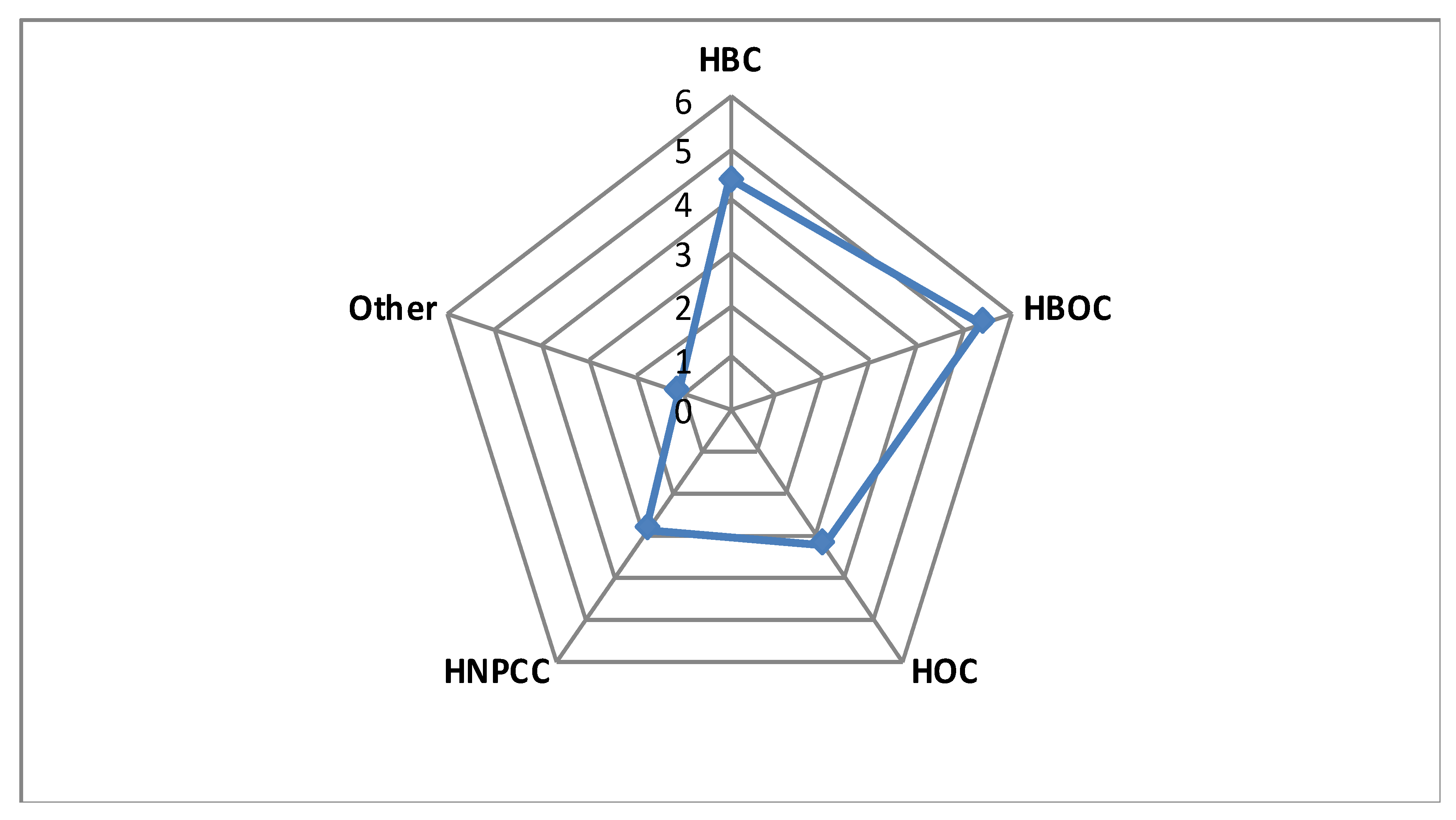
Figure 1. The diagram represents the percentage of pathogenic variants in the 14 Fanconi anemia (FA) genes analyzed per clinical suspicion group. HBC: Hereditary Breast Cancer Patients; HOC: Hereditary Ovarian Cancer Patients; HBOC: Hereditary Breast and Ovarian Cancer Patients; HNPCC: Hereditary non-polyposis colorectal cancer.
Cancers 12 00829 g001
Table 1. Summary of (Likely) Pathogenic Variants in 14 FA genes in the different clinical groups (only women are counted).
Clinical SuspicionGCAT Women Cohort (n = 100)GnomAD European >23,000 women βStudy Cohort Versus NFE γ, Non-Cancer GnomAD (OR/95%CI/p-Value)
GenesPathogenic VariantsBreast (HBC)Ovary (HOC)Breast + Ovary (HBOC)HNPCC αOtherAll PatientsHBC + HOC + HBOCHBC + HBOC
FANCA107021031471.94/0.91–3.7/0.0472.34/1.04–4.59/0.023.14/1.4–6.17/0.003*
FANCL83131011871.22/0.52–2.46/0.5491.42/0.56–3/0.3561.63/0.59–3.64/0.283
FANCM62301001591.07/0.38–2.39/0.8281.19/0.38–2.85/0.6180.63/0.08–2.34/0.774
FANCI1100000251.14/0.03–7/0.5931.12/0,04–9.29/0.4922.02/0.05–12.4/0.399
FANCE2100100171.14/0.03–6.97/0.5931.52/0.04–9.3/0.4922.03/0.05–12.4/0.399
FANCC2110000441.29/0.15–4.97/0.671.72/0.2–6.63/0.3321.15/0.03–6.78/0.586
FANCF1100000261.09/0.02–6.6/0.6081.45/0.04–8.88/0.5061.94/0.05–11.87/0.412
RAD51110000047.12/0.14–72/0.1599.49/0.19–96/0.12212.7/ 0.26–128/0.093
SLX4000000136NANANA
ERCC4000000022NANANA
FANCB00000000NANANA
FANCD2000000021NANANA
FANCG000000043NANANA
XRCC2000000022NANANA
TOTAL311755405753
α Hereditary non-polyposis colorectal cancer; β The number of GnomAD non-Finnish, non-cancer women is slightly variable per gene but in all cases was greater than 23,000 γ NFE: non-Finnish European.
Table 2. Summary of the hereditary cancer cohort by clinical suspicion.
Clinical SuspicionNumber of Patients (Women)
Hereditary breast cancer, HBC385 (370)
Hereditary non-polyposis colon cancer, HNPCC210 (130)
Hereditary ovarian cancer, HOC154 (154)
Other hereditary cancer conditions102 (55)
Hereditary breast and ovarian cancer, HBOC93 (90)
Familial (attenuated) adenomatous polyposis, FAP/AFAP77 (19)
Total1021 (818)
A set of 194 cancer-free controls (100 women) from GCAT, Genomes for Life Cohort, was also analyzed.