Review

The Cooperative Relationship between STAT5 and Reactive Oxygen Species in Leukemia: Mechanism and Therapeutic Potential

1
Department of Pediatrics, Division of Hem/Onc/BMT and Aflac Cancer and Blood Disorders Center, Emory University School of Medicine, 1760 Haygood Dr. NE, HSRB E308, Atlanta, GA 30322, USA
2
Emory Winship Cancer Institute, Atlanta, GA 30322, USA
*
Author to whom correspondence should be addressed.
Cancers 2018, 10(10), 359; https://doi.org/10.3390/cancers10100359
Submission received: 30 August 2018 / Revised: 21 September 2018 / Accepted: 24 September 2018 / Published: 27 September 2018
(This article belongs to the Special Issue Tyrosine Kinase Signaling Pathways in Cancer)

Abstract

:
Reactive oxygen species (ROS) are now recognized as important second messengers with roles in many aspects of signaling during leukemogenesis. They serve as critical cell signaling molecules that regulate the activity of various enzymes including tyrosine phosphatases. ROS can induce inactivation of tyrosine phosphatases, which counteract the effects of tyrosine kinases. ROS increase phosphorylation of many proteins including signal transducer and activator of transcription-5 (STAT5) via Janus kinases (JAKs). STAT5 is aberrantly activated through phosphorylation in many types of cancer and this constitutive activation is associated with cell survival, proliferation, and self-renewal. Such leukemic activation of STAT5 is rarely caused by mutation of the STAT5 gene itself but instead by overactive mutant receptors with tyrosine kinase activity as well as JAK, SRC family protein tyrosine kinases (SFKs), and Abelson murine leukemia viral oncogene homolog (ABL) kinases. Interestingly, STAT5 suppresses transcription of several genes encoding antioxidant enzymes while simultaneously enhancing transcription of NADPH oxidase. By doing so, STAT5 activation promotes an overall elevation of ROS level, which acts as a feed-forward loop, especially in high risk Fms-related tyrosine kinase 3 (FLT3) mutant leukemia. Therefore, efforts have been made recently to target ROS in cancer cells. Drugs that are able to either quench ROS production or inversely augment ROS-related signaling pathways both have potential as cancer therapies and may afford some selectivity by activating feedback inhibition of the ROS-STAT5 kinome. This review summarizes the cooperative relationship between ROS and STAT5 and explores the pros and cons of emerging ROS-targeting therapies that are selective for leukemia characterized by persistent STAT5 phosphorylation.

1. Introduction

Reactive oxygen species (ROS) are cellular molecules that are generated primarily as a by-product of mitochondrial oxidative metabolism or NADPH oxidase enzymes [1]. ROS consist of radical and non-radical oxygen species including superoxide anion (O2−), hydrogen peroxide (H2O2), and hydroxyl radical (·OH). They have been recognized as crucial cell signaling molecules and regulate a wide variety of important cellular processes including cell survival, proliferation, differentiation, apoptosis, and DNA damage response [2]. It has been widely accepted that ROS are associated with cancer due to the fact that they may be able to induce transformation by causing DNA damage and that transformed cells have higher levels of ROS production than normal cells [3]. An increased production of ROS has also been associated with genomic instability and enhanced DNA damage including double strand breaks and also performs a signaling function to promote cell proliferation and migration, thus, contributing to leukemic cell transformation [4]. For example, in more than 60% of acute myeloid leukemia (AML) patients, ROS production is heavily increased due to NADPH oxidase (NOX) activation and strongly promotes AML proliferation [5]. Interestingly, protein tyrosine phosphatases (PTPs) are also among the various ROS targets. ROS inactivate PTPs transiently by oxidizing a reactive cysteine on the catalytic domain [6]. By doing so, ROS induce tyrosine phosphorylation and activation of many important signaling molecules including mitogen-activated protein kinase (MAPK), c-JUN N-terminal kinase (JNK), and p38 MAPK, therefore establishing themselves as important factors in growth factor signaling pathways [2,7].
ROS also contribute to tyrosine phosphorylation of signal transducer and activator of transcription (STAT) family members [8]. STATs carry out two functions: Transducing cellular signals and activating transcription of a diverse set of genes including those that are involved in cancer development. STATs are constitutively active in many solid tumors and hematologic malignancies due to enforced upstream kinase signals [9]. The Janus kinase-signal transducer and activator of transcription (JAK-STAT) pathway controls survival, proliferation, and differentiation in several cell types. STATs are also persistently active in a number of cancers, especially myeloproliferative neoplasms and leukemias [10]. Among the STAT proteins, STAT5 is encoded by two closely related genes STAT5a and STAT5b and plays a major role in regulating vital cellular functions such as proliferation, differentiation, and apoptosis of hematopoietic and immune cells [11,12]. STAT5 is activated by phosphorylation of a single tyrosine residue (Y694 in STAT5A and Y699 in STAT5B) and negatively regulated by dephosphorylation. A wide variety of growth factors and cytokines can activate STAT5 through the JAK-STAT pathway. The activation of STAT5 is transient and tightly regulated in normal cells but can become dysregulated in hematologic malignancies (Figure 1).

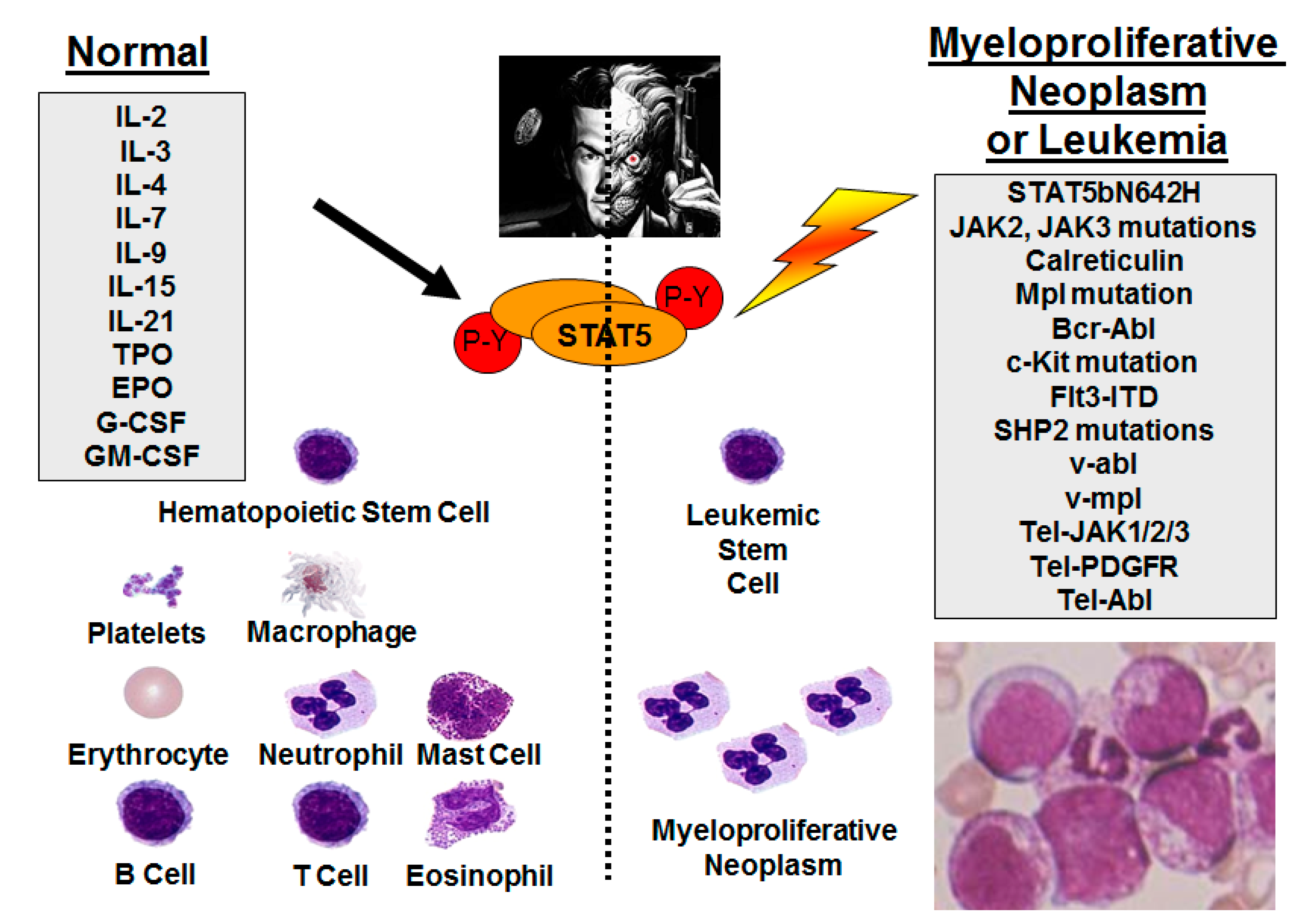
2. Persistent STAT5 Activation through Upstream Mutant Tyrosine Kinases

Persistent activation of STAT5 is mostly caused by activating mutations in upstream tyrosine kinases in a number of cancers. For example, in chronic myelogenous leukemia (CML), the Bcr-Abl translocation leads to expression of the Bcr-Abl fusion protein and constitutive activation of its tyrosine kinase activity. That causes aberrant activation of STAT5 [13]. By using a Bcr-Abl inhibitor, constitutive activation of STAT5 can be blocked [14]. In acute myeloid leukemia (AML), overexpression and constitutive activation of the FLT3 receptor tyrosine kinase induces a strong activation of STAT5, which can be inhibited by an FLT3 protein tyrosine kinase inhibitor [15]. Moreover, in mast cell (MC) neoplasms, the D816V-mutated variant of Kit promotes growth of hematopoietic progenitor cells and proliferation of neoplastic MCs through constitutive activation of the STAT5-PI3K signaling pathway [16]. Additionally, in chronic myelomonocytic leukemia, the TEL/PDGFβR tyrosine kinase fusion protein activates STAT5 [17].
JAK2 and JAK3 mutations can also persistently activate STAT5 by encoding constitutively active or hyperactive JAK proteins in many hematopoietic malignancies [10]. For instance, a gain-of-function JAK2 V617F somatic mutation constitutively activates the JAK-STAT pathway and is identified in the majority of patients with myeloproliferative neoplasm [18].
Apart from that, STAT5 can also be activated by members of the Src family kinases (SFKs) and in particular, c-Src through growth factor receptor signaling, which is different from the cytokine receptor signaling in the JAK pathway [19]. It has been shown that SFKs are frequently constitutively activated in leukemia cell lines and inhibitors of Src tyrosine kinases block constitutive activation of STAT5 in these cells [20].

3. STAT5b Gene Mutations in Leukemia/Lymphoma

Although there are relatively few cases of STAT5 mutation reported in human cancers and most of the aberrant activation of STAT5 is caused by mutations in upstream kinases, there are still some recent discoveries and identification of STAT5 mutations in patients leading to increased phosphorylation and activation of STAT5. For example, in patients with large granular lymphocytic (LGL) leukemia, two mutations: Y665F and N642H were identified, both located on exon 16 of STAT5b in the Src-like homology 2 (SH2) domain. The frequency of STAT5b mutation in all LGL leukemia patients is around 2%. However, no STAT5a mutations have been detected in LGL leukemia patients. From an in vitro analysis, both Y665F and N642H mutants increased tyrosine phosphorylation of STAT5b and also its transcriptional activity. Additionally, N642H mutant cells demonstrated a more prominent increase in STAT5b phosphorylation and transcriptional activity compared with Y665F. Accordingly, patients with the N642H mutation suffered from a more aggressive and fatal disease whereas typical LGL leukemia patients had a relatively favorable outcome [21,22]. Such activating mutations on STAT5b have also been observed in lymphomas derived from γδ-T or NK cells with a high frequency, especially in γδ-T-cell-derived lymphomas and enteropathy-associated T-cell lymphoma type II, where STAT5b mutations were observed in more than 30% of tumor samples [23]. The N642H mutation on STAT5b has also been identified as a driver mutation in both adult and pediatric acute lymphoblastic leukemia (T-ALL) prolymphocytic leukemia, NK/T cell lymphoma, and enteropathy-associated T cell lymphoma [24,25,26,27], whereas STAT5b T628S, Y665F, and V712E mutations have been discovered in multiple cases [24,27]. A few additional rare mutations in STAT5b such as E438K, G492C, P702A, I704L, and Q706L have been reported although the effects on STAT5b function are still unknown. In a transgenic mouse model, STAT5bN642H-expressing mice rapidly developed T cell and hepatosplenic T cell lymphoma but lacked a hematopoietic phenotype [28].

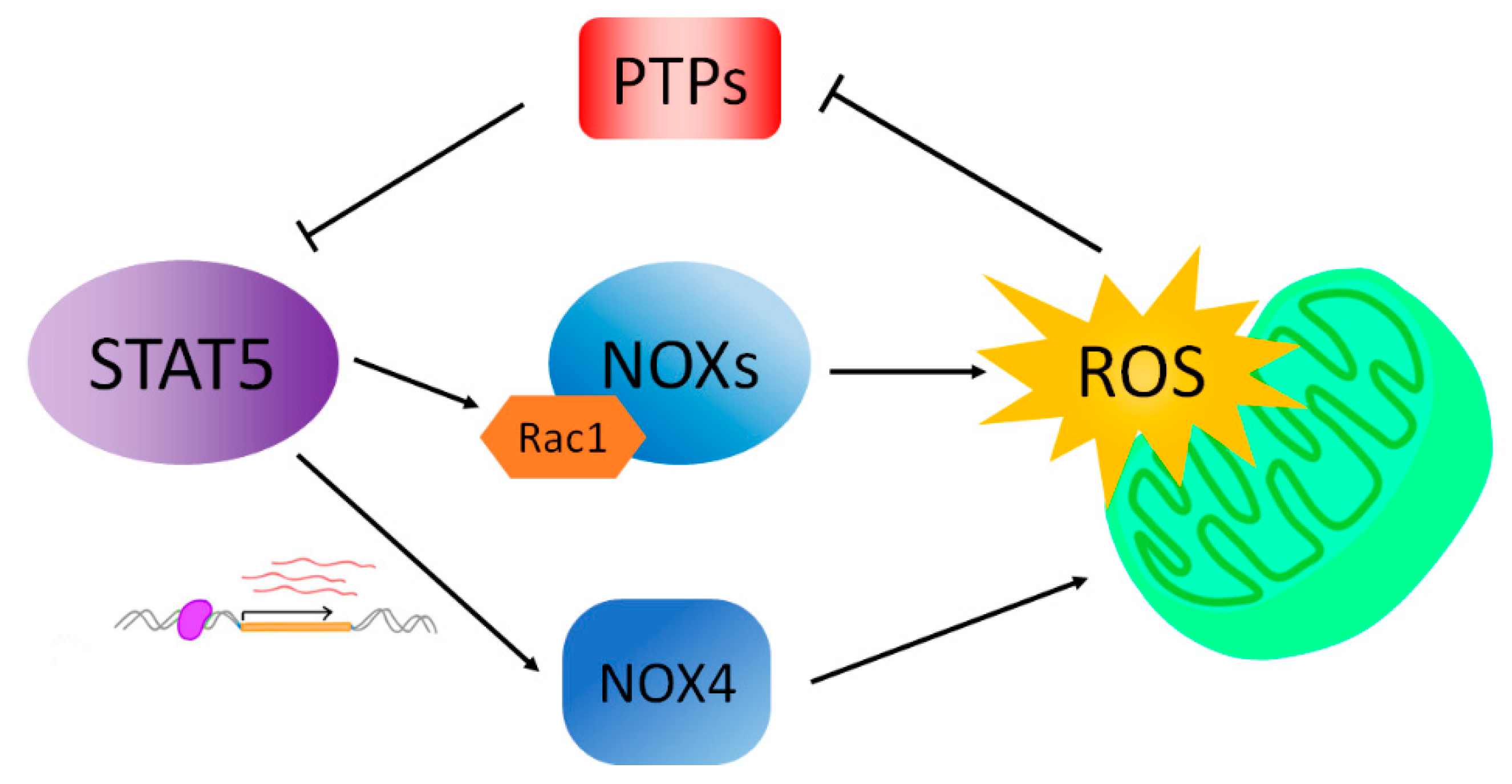
4. STAT5 Drives ROS Formation

Constitutive activation of STAT5 in cancer is associated with increased production of ROS through multiple mechanisms [29]. This has been proven by increased levels of cellular ROS upon enforced expression of STAT5 in BCR-ABL1 transformed cell lines by retrovirus transduction and further confirmed by decreased ROS production after the introduction of shRNA specific for STAT5 in K562 cells [30]. Such STAT5-mediated increase of ROS production has also been validated in vivo, where mice injected with transformed cells ectopically expressing STAT5 had a higher ROS level in the spleen compared with mice injected with transformed cells only expressing GFP [30].
STAT5 mediates ROS production through a mechanism independent of JAK2 or mitochondrial respiration [30]. In FLT3/ITD-positive AML cells, increased ROS levels appear to be produced through the direct interaction of tyrosine phosphorylated STAT5 with RAC1. RAC1 is a small GTPase protein and serves as an essential component of the NOX holoenzyme [31]. NOX is a family of proteins consisting of seven homologs and several subunits. They serve the major function of electron transportation through the plasma membrane and the generation of ROS including superoxide [32]. Inhibition of FLT3-ITD not only reduced tyrosine phosphorylation of STAT5 but also decreased RAC1 activity and its binding to NOX [29].
Another mechanism has also been proposed regarding increased ROS formation in FLT3/ITD-positive cells. NOX4 belongs to the NOX family proteins. It is expressed in many cell types including hematopoietic stem cells and its expression can be induced by the endoplasmic reticulum (ER) [32]. NOX4 is a direct downstream target of STAT5 since the NOX4 promoter possesses STAT binding elements and it has been found by ChIP assays that STAT5 binds to these elements in a FLT3/ITD-dependent manner. Therefore, NOX4 expression level is upregulated in FLT3/ITD-positive cells owing to the constitutive activation of STAT5 as an FLT3 downstream effector. NOX4 then contributes to ROS formation since knockdown of NOX4 by siRNA or shRNA significantly reduced cellular ROS levels [4,33]. Interestingly, either by inhibiting the FLT3 tyrosine kinase activity or by inhibiting the NOX activity, ROS formation in the ER was reduced. However, such effects were not observed for mitochondrial ROS, suggesting that FLT3-ITD drives endogenous ROS production in the ER through NOX proteins [34].
Apart from enhancing transcription of NOX proteins, oncogenic STAT5 signaling also promotes ROS formation by repressing expression of antioxidant enzymes including catalase and glutaredoxin-1 (Glrx1) in Bcr-Abl-positive CML. Both catalase and Glrx1 function to reduce the cellular ROS level. After introducing a Bcr-Abl inhibitor to Bcr-Abl-positive human leukemia cell lines, the expression level of catalase and Glrx1 increased while the activity of STAT5 decreased. Moreover, cell lines transformed with a constitutively active STAT5 mutant had reduced expression levels of catalase and Glrx2 compared with control cell lines [35]. It is important to note that Casetti reported that STAT5, and in particular STAT5A, can be protective against oxidative stress based on the observation that the knockdown of STAT5A increased the basal level ROS production and subsequently genomic stress in CML cell lines [36]. However, in this case, STAT5B was still present and had a differential contribution to stress protection.

5. ROS Promotes STAT5 Phosphorylation

While STAT5 activation increases ROS production, ROS also play a role in the activation of STAT5 (Figure 2). By using the antioxidant N-acetyl-L-cysteine (NAC), which inhibits ROS production, Mitsuko was able to demonstrate that NAC significantly inhibits the IL-3 induced tyrosine phosphorylation of JAK2 kinase and furthermore, activation-specific phosphorylation of STAT5 [37]. The mechanism behind the ROS enhancement of the JAK2-STAT5 signaling pathway remains to be explored. However, there is increasing evidence suggesting that redox regulation of protein tyrosine phosphatases (PTP) may play a role in the ROS-mediated increase of tyrosine phosphorylation. PTPs oppose the activity of protein tyrosine kinases (PTK) by removing a phosphate group from tyrosine residues in target proteins. Therefore, the tyrosine phosphorylation level of cellular proteins is controlled by a coordination between PTKs and PTPs [38]. Interestingly, PTPs are intracellular targets of ROS. All PTPs contain a cysteine residue at their active sites. The cysteine residue can be oxidized by H2O2 to cysteine sulfenic acid, leading to the inactivation of PTP enzymatic activities. Such oxidation and inactivation can be reversed with thiol compounds [39] and the reversible inhibition of PTP activity contributes to the ligand-induced increase in receptor tyrosine kinase signaling [40]. Additionally, redox regulation of JAKs has been described [41,42].

6. Non-Canonical STAT5 Activation in High Risk Subsets of Acute Myeloid Leukemia

AML is a heterogeneous and complex disease with a low remission and high relapse rate [43]. Mutations in FLT3, NPM1, KIT, RAS, and CEBPα are found in 30–50% of AML patients [44,45,46]. JAK-STAT and PI3K-Akt-mTOR pathways are very important downstream mediators of survival and proliferation signals that are generated by these molecular mutations in AML. Persistently activated STAT5 is associated with poor prognosis [47,48] and is a bona fide therapeutic target [49]. The pSTAT5high sub-type is associated with approximately half of all AML where it promotes leukemia stem cell (LSC) self-renewal [47,50] and resistance to tyrosine kinase-inhibitors [51,52]. Emerging new agents, such as the multi-kinase inhibitor midostaurin [53,54], advocate that a multi-faceted approach can succeed.
Interestingly, the mechanism of activation of STAT5 by mutant FLT3 and KIT is due to non-canonical signaling not observed in normal cells [16,55,56]. Endoplasmic reticulum (ER)-localized NOX4 combined with aberrant FLT3-ITD or KIT (D816V) N-linked glycosylation traps these receptor tyrosine kinases (RTKs) on the ER and permits reactive oxygen species (ROS) accumulation and activation of STAT5 independent of JAKs [35,57,58,59]. Therefore, there is potential for a favorable therapeutic index sparing normal hematopoietic cells with agents that are able to exploit mislocalized RTK signaling and target STAT5.

7. Emerging ROS-Reducing Cancer Therapies

The FLT3-ITD mutation is found in 20–30% of patients with AML and portends a poor prognosis; despite intensive chemotherapy and allogeneic stem cell transplantation. FLT3-ITD is also commonly found associated with MLL-fusions, which are also associated with poor outcomes. Recent strategies using targeted therapies (i.e., FLT3 tyrosine kinase inhibitors—TKIs), have thus far yielded modest responses and most relapsed/refractory patients will still die of their leukemia. STAT5 is critical for normal hematopoiesis [60,61,62,63]. Therefore, specificity for the inhibition of STAT5 only in leukemia but not normal cells is critical for the design of new anti-leukemia therapies that selectively target high risk pSTAT5+ sub-sets. STAT5 activation can be uncoupled from canonical JAK2-STAT5 activation, thus JAK2 inhibitors are not a good option for AML and have had limited clinical success as single agents. Class III RTKs include five immunoglobulin-like extracellular domains and include PDGFRα, PDGFRβ, c-KIT, c-FMS, and FLT3. Several of these RTKs are activated in AML and in particular the c-KIT and Flt3 mutant forms are considered poor risk. There is a therapeutic need for targeting mutant FLT3 and c-KIT driven AML by exploiting their unique ER-localization [64] and non-canonical activation of STAT5. RTK-driven leukemia cells are characterized by reprogrammed calcium homeostasis [65,66] that favors proliferation and survival.
Imipramine blue (IB) is a chimeric drug developed by Jack Arbiser at Emory University [67]. IB was generated by combining a triphenylmethane dye backbone [68] through reaction of Michler’s ketone, with imipramine, an FDA-approved tricyclic antidepressant. The initial rationale was to increase the lipophilicity of triphenylmethane dyes (e.g., gentian violet) by conjugating to a lipophilic amine (e.g., imipramine) to optimize uptake across the blood-brain barrier. The dye backbone quenches ROS and inhibits NADPH oxidase. Fortuitously, in addition to delivery to the brain for glioma therapy [67], lipophilic amines selectively accumulate in the ER [69] and lysosomes [70] and imipramine can activate phospholipase C [71,72]. We have found IB to be extremely effective and selective against subsets of AML expressing mutant FLT3 or c-KIT, including drug-resistance conferring FLT3 mutants [73]. The enhanced selectivity of IB may be related to the trapping of IB in the ER/lysosomes combined with the requirement for an ER-localized FLT3 mutant for effective tyrosine phosphorylation of STAT5 in AML (Figure 3).

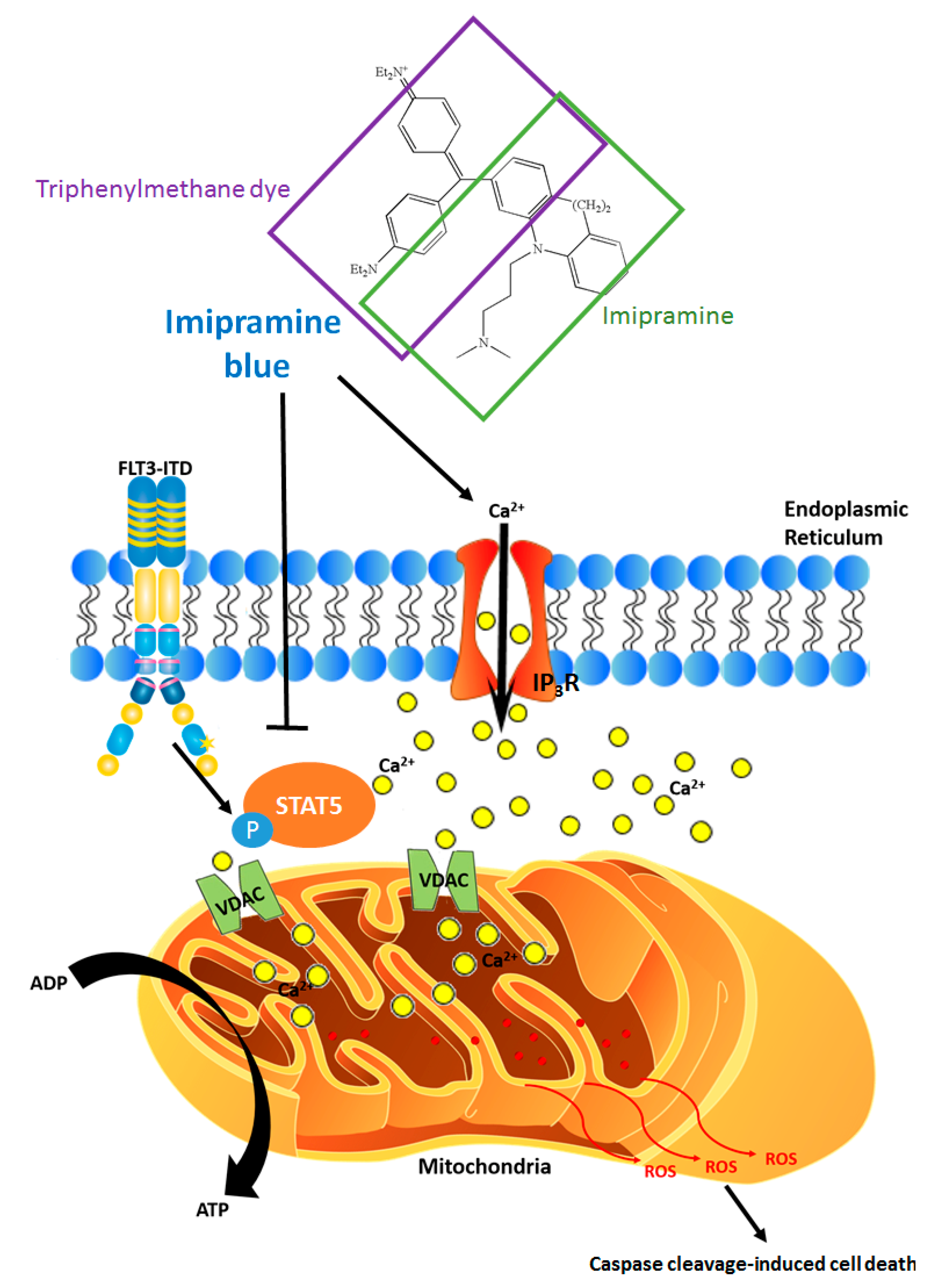
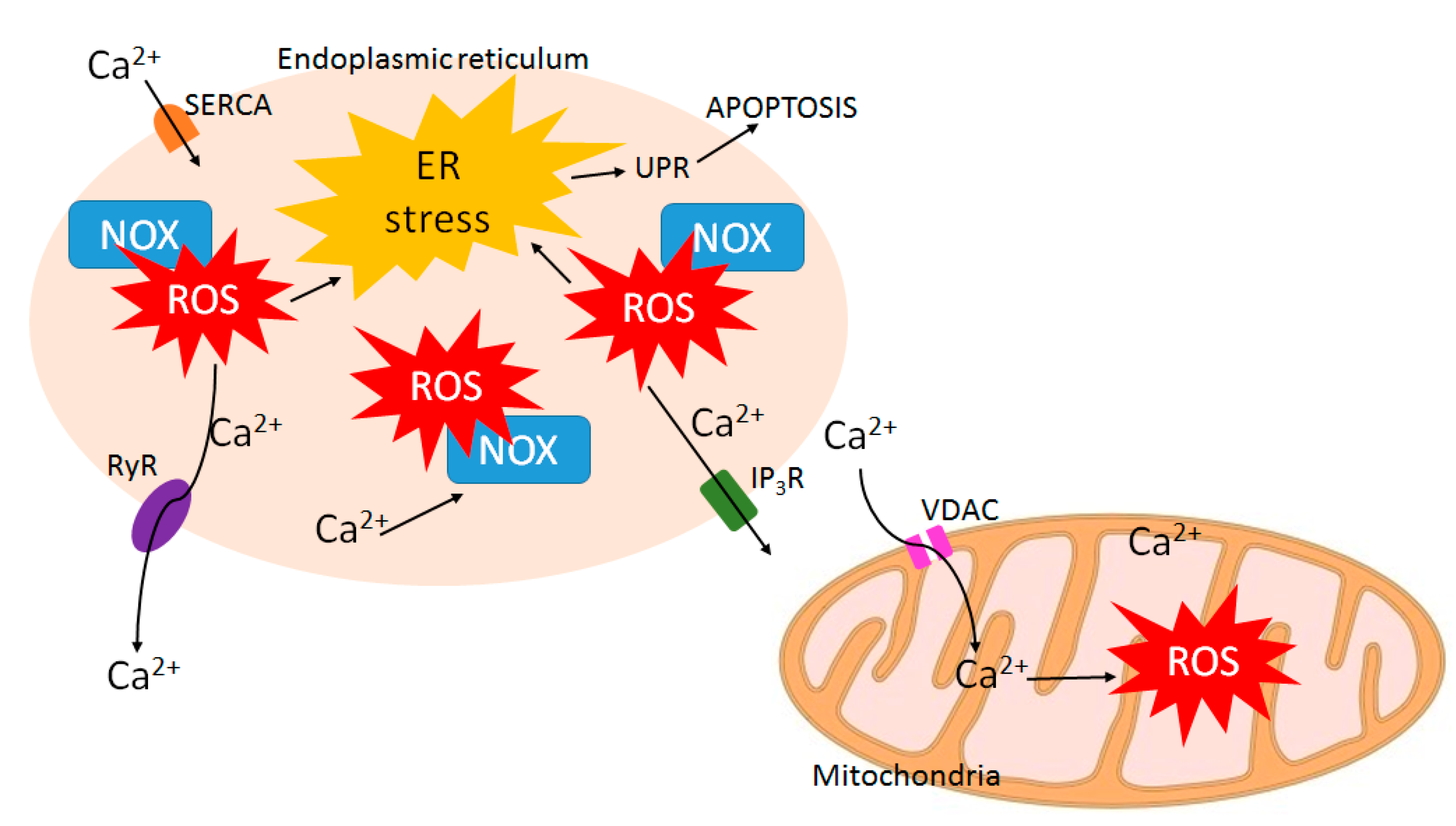
8. ROS-Calcium Connection and Potential for ROS-Increasing Cancer Therapies

Calcium plays an important role in cell signaling as a second messenger and calcium signaling serves essential functions in many cellular processes including cell proliferation, gene transcription, and apoptosis [74,75]. Considering its importance in determining cell fate, calcium signaling is often altered and remodeled in cancer cells facilitating tumor progression [74]. Interestingly, ROS signaling and calcium signaling are closely related and share complex bidirectional interactions between each other [76] (Figure 4).
On one hand, an increase of the intracellular calcium level can induce ROS production by activating enzymes that are involved in ROS formation. Those enzymes include NOX, which can be stimulated by calcium-dependent proteins [77]. Calcium also increases ROS generation by stimulating the Krebs cycle and oxidative phosphorylation in mitochondria [78].
On the other hand, ROS can regulate calcium signaling by modulating a variety of calcium channels. The sarcoplasmic/endoplasmic reticulum (SR/ER) is the major site of calcium storage in eukaryotic cells [79]. The SR/ER membranes are embedded with calcium release channels such as ryanodine receptors (RyR) and inositol 1,4,5-trisphosphate receptors (IP3R). Both RyR and IP3R channel activities can be enhanced by ROS, either through direct oxidation or by regulation of NOX [76]. Therefore, ROS increase calcium release from the SR/ER and elevate the intracellular calcium level. Such calcium release from the ER is an indicator of ER stress. ER stress is caused by perturbation of ER homeostasis. Such condition activates the unfolded protein response (UPR) to re-establish homeostasis. However, although initially serving as a protective mechanism, the UPR is toxic to cells when it is prolonged and will eventually lead to mitochondrial apoptosis [80].
Therefore, increased Calcium-ROS has the potential for use as a drug therapy. Notably, in addition to the ROS quenching role of IB, it can also induce calcium release from the ER and drive cell death. Therefore, IB functions in a dosage-dependent manner, with lower doses effectively increasing intracellular calcium and higher doses suppressing ROS and STAT5 tyrosine phosphorylation. These differences might come into play in scenarios where either bulk leukemia or LSCs are being targeted since STAT5 is a major regulator of LSC self-renewal [50,81].

9. Exploiting the ROS-STAT5 Kinome to Identify New Therapeutic Targets

The STAT5 whole “kinome” that links ROS with STAT5 phosphorylation, as well as downstream ROS and calcium-dependent gene expression, may be exploited to identify potential therapeutic targets of value for high risk AML. One such starting point could be calcium-regulated kinases. The ER is the largest membrane-bound organelle in eukaryotic cells and is the largest calcium store. Calcium release is known to target leukemia cells [82] but specificity for RTK-driven AML has not been reported. Diverse anti-depressants are structurally lipophilic amines that cross the blood-brain barrier efficiently and modulate phospholipids in neurons [83,84]. They have also been implicated as chemosensitizers [85,86] and possibly could be chemopreventative [87]. Due to their lipophilic properties, some compounds can efficiently accumulate in the ER or mitochondria where they quench ROS and suppress STAT5 tyrosine phosphorylation. Commercially available ER-tracker and Mito-tracker dyes have been selected for these basic properties [69]. Calcium and ROS have an intimate and complex connection and many drug targets require careful study since a calcium overload could lead to side effects that are widely known for anti-depressant drugs.
Two important calcium-regulated genes, calcium/calmodulin-dependent protein kinase II (CAMKII) [88] and a nuclear factor of activated T-cells 1 (NFATc1) [89], are expressed in leukemia cells that are characterized by high levels of ROS. NFATc1 is a DNA-binding transcription factor causative for FLT3-ITD positive AML that is resistant to the kinase inhibitor sorafenib [89]. This function is believed to be mediated through its effects on driving Ras oncogene expression. CAMKII is believed to function through the regulation of critical signaling pathways involved in STAT3 transcriptional activation [88]. In contrast to the potential pro-leukemic roles of calcium, high risk AML may also be characterized by increased calcium storage, facilitating a “primed state” whereby perturbation of ER calcium storage might be especially effective at inducing apoptosis. In leukemia, the unfolded protein response (UPR) is a protective mechanism against ER stress that is regulated by XBP1 and IRE1. However, in presence of prolonged or severe ER stress, the pro-survival signal of the UPR turns into a toxic signal that induces mitochondrial apoptosis as shown in Figure 3 [80]. While XBP1 is expressed as a splice variant (XBP1s) in 16% of AML, the functional role of this splicing event is not well characterized. XBP1s is not associated with FLT3-ITD mutation and confers favorable prognosis [90]. Therefore, while unlikely that the UPR is involved in the initial drug sensitivity, it may be induced by IB or pimozide (PIM).
Calcium-release inducing therapeutic approaches hold promise for selectively targeting cancer cells. It was recently demonstrated that AML selective cytotoxicity in vivo could be achieved with the SERCA antagonist curcumin, a natural product found in turmeric and curry powders [82]. Similarly, it has been shown that thapsigargin (TG), the most potent SERCA antagonist could be effectively dosed in vivo in a mouse model of T-ALL. It was found through a complementary genetic screen that both SERCA and Notch [91] are targets of TG. Despite these advances in demonstrating the proof-of-principle for SERCA inhibition in cancer, there remain significant barriers to implementing this strategy and optimizing beyond pre-clinical studies. There are numerous natural products that are ER or SERCA targeted to be considered [92] for applications in AML but also myeloproliferative neoplasms driven by JAK2 and MPL mutants. However, because of the key role of calcium oscillations in muscle and brain function, these approaches come with significant risk for serious side effects. There has been some progress in this area in recent years and it might be able to overcome limitations through the use of novel drug delivery.
Calcium-inhibiting therapeutic approaches are characterized by pimozide (PIM), an FDA approved antipsychotic drug of the diphenylbutylpiperidine class and acts as an antagonist of the D2, D3, and D4 dopamine receptor and the 5-hydroxytryptamine receptor [93]. Pimozide is known to inhibit T-type voltage-gated calcium channels [94], which can promote cancer cell proliferation [95]. PIM was identified as a STAT5 inhibitor by a high throughput screen based on STAT transcriptional activity and it decreased the survival of CML cells resistant tyrosine kinase inhibitors [96]. PIM is able to have a combinatorial effect with TKI midostaurin and sunitinib in the inhibition of induction of apoptosis in FLT3-driven AML [97]. Recently, we showed that PIM inhibited pSTAT5 at a dose larger than 5 µM in the MV4-11 cell line. Additional, reduced expression of STAT5 target genes and selective induction of apoptosis was observed. Using a sub-optimal dose of IB and pimozide, this combination was highly synergistic and selective in FLT3/ITD+ cell lines, including those with FLT3 point mutations, with little effect on FLT3/ITD negative cell lines or on CD34+ cord blood cells. Furthermore, this combination was also selective and synergistic for 32D cells transduced with a c-KIT D814V mutant [73]. In contrast, no synergy has been observed between TG and pimozide (K.D.B., unpublished observations), suggesting that the mechanism of action of IB and TG may be unique.

10. STAT5 in Normal Hematopoiesis—Potential Side Effects of STAT5 Inhibition

STAT5 regulates normal lympho-myeloid development through activation downstream of early-acting cytokines, their receptors, and JAKs and plays a critical role in stem cell self-renewal; both in normal and leukemic stem cells. The greatest challenge to direct targeting of STAT5 signaling is to find the potential therapeutic window for leukemia cells while having minimal cytotoxicity to normal cells. STAT5 can be activated by the upstream tyrosine kinase. Therefore, TKIs could be initially effective to inhibit STAT5 activation but could have significant long-term off-target toxicity and the TKI resistance usually occurs with further increased activation. Recently, a few STAT5-specific inhibitors such as 13a [98], Stafib-2 [99], and AC-4-130 [100] have been reported to target the STAT5 SH2 domain. AC-4-130 treatment can inhibit proliferation and clonogenic growth in both AML cell lines and primary AML patient cells with IC50 between 1.6 to 4.9 µM but less toxicity against healthy CD34+ cells with IC50 between 6.3 to 7.6 µM [100]. It also shows synergistic treatment effects with TKI although whether the therapeutic window is wide enough to spare normal healthy donors still needs to be further investigated. As STAT5 activation promotes an overall elevation of ROS, targeting ROS could provide additional treatment avenues, especially in leukemia characterized by persistent STAT5 phosphorylation. ROS-targeting, in combination with TKI or STAT5 specific inhibitors, might further improve therapeutic benefits in leukemia. However, since ROS is closely aligned with mitochondrial function, the cellular phenotypes associated with STAT5 inhibition may need to be monitored closely. Autophagy is a protective mechanism that is induced when oxidative phosphorylation is inhibited and thus this could be a target for combination therapy.

11. Conclusions and Perspectives

It is an exciting time for molecularly targeted therapies directed at transcription factors, which make up prime targets for eradicating bulk leukemia and LSC populations. New agents have been described with in vitro and in vivo efficacy for direct and indirect targeting of STAT5. Indirect approaches rely on the liberation of inhibitory phosphatases or other negative regulators that normally comprise a negative feedback loop. Dysregulation of calcium-ROS in AML is also a vulnerability that remains to be fully exploited due to the difficulty of targeting these signaling intermediates without non-hematopoietic toxicity. Each approach presents its own strengths and weaknesses. The normal role of STAT5 in hematopoiesis has been extensively characterized during the past 20 years, showing virtually ubiquitous roles in all hematopoietic lineages as well as regulatory T-cells required to prevent severe autoimmunity. Due to this extensive connection of STAT5 in normal and leukemic hematopoiesis, innovative new approaches will be required for safe and effective long-term cures based on STAT5 inhibition. If STAT5 targeted therapies can be harnessed in a safe and targeted manner, they will be applicable for a wide range of pSTAT5+ leukemias as well as some solid tumors and thus have a significant potential for positive impact on future patient care.

Author Contributions

The individual contributions of the authors are as follows: Conceptualization, K.D.B.; resources, K.D.B.; data curation, T.M.; Z.W.; writing-original draft preparation, T.M.; Z.W.; K.D.B.; writing-review and editing, T.M.; Z.W.; K.D.B.; visualization, T.M.; K.D.B.; supervision, Z.W.; K.D.B.; project administration, K.D.B.; funding acquisition, K.D.B.

Funding

This research was funded by R01DK059380, the Cure Childhood Cancer Foundation, the Rally Foundation for Childhood Cancer Research, and the Truth 365.

Acknowledgments

The authors acknowledge the support of the Aflac Cancer and Blood Disorders Center of Children’s Healthcare of Atlanta and Emory University School of Medicine.

Conflicts of Interest

K.D.B. has a financial interest in the Valhalla Scientific Editing Service. The funding sponsors had no role in the design of the study; in the collection, analyses, or interpretation of data; in the writing of the manuscript, and in the decision to publish the results.

References

  1. Thannickal, V.J.; Fanburg, B.L. Reactive oxygen species in cell signaling. Am. J. Physiol. Lung Cell. Mol. Physiol. 2000, 279, L1005–L1028. [Google Scholar] [CrossRef] [PubMed]
  2. Ray, P.D.; Huang, B.W.; Tsuji, Y. Reactive oxygen species (ROS) homeostasis and redox regulation in cellular signaling. Cell. Signal. 2012, 24, 981–990. [Google Scholar] [CrossRef] [PubMed] [Green Version]
  3. Schumacker, P.T. Reactive oxygen species in cancer cells: Live by the sword, die by the sword. Cancer Cell 2006, 10, 175–176. [Google Scholar] [CrossRef] [PubMed]
  4. Jayavelu, A.K.; Moloney, J.N.; Bohmer, F.D.; Cotter, T.G. NOX-driven ROS formation in cell transformation of FLT3-ITD-positive AML. Exp. Hematol. 2016, 44, 1113–1122. [Google Scholar] [CrossRef] [PubMed] [Green Version]
  5. Hole, P.S.; Zabkiewicz, J.; Munje, C.; Newton, Z.; Pearn, L.; White, P.; Marquez, N.; Hills, R.K.; Burnett, A.K.; Tonks, A.; et al. Overproduction of NOX-derived ROS in AML promotes proliferation and is associated with defective oxidative stress signaling. Blood 2013, 122, 3322–3330. [Google Scholar] [CrossRef] [PubMed] [Green Version]
  6. Touyz, R.M. Reactive oxygen species and angiotensin II signaling in vascular cells—Implications in cardiovascular disease. Braz. J. Med. Boil. Res. 2004, 37, 1263–1273. [Google Scholar] [CrossRef]
  7. Hensley, K.; Robinson, K.A.; Gabbita, S.P.; Salsman, S.; Floyd, R.A. Reactive oxygen species, cell signaling, and cell injury. Free Radic. Boil. Med. 2000, 28, 1456–1462. [Google Scholar] [CrossRef]
  8. Simon, A.R.; Rai, U.; Fanburg, B.L.; Cochran, B.H. Activation of the JAK-STAT pathway by reactive oxygen species. Am. J. Physiol. 1998, 275, C1640–C1652. [Google Scholar] [CrossRef] [PubMed] [Green Version]
  9. Yu, H.; Jove, R. The STATs of cancer—New molecular targets come of age. Nat. Rev. Cancer 2004, 4, 97–105. [Google Scholar] [CrossRef] [PubMed]
  10. Constantinescu, S.N.; Girardot, M.; Pecquet, C. Mining for JAK-STAT mutations in cancer. Trends Biochem. Sci. 2008, 33, 122–131. [Google Scholar] [CrossRef]
  11. Rani, A.; Murphy, J.J. STAT5 in cancer and immunity. J. Interferon Cytokine Res. 2016, 36, 226–237. [Google Scholar] [CrossRef] [PubMed]
  12. Wittig, I.; Groner, B. Signal transducer and activator of transcription 5 (STAT5), a crucial regulator of immune and cancer cells. Curr. Drug Targets Immune Endocr. Metab. Disord. 2005, 5, 449–463. [Google Scholar] [CrossRef]
  13. Shuai, K.; Halpern, J.; ten Hoeve, J.; Rao, X.; Sawyers, C.L. Constitutive activation of STAT5 by the Bcr-Abl oncogene in chronic myelogenous leukemia. Oncogene 1996, 13, 247–254. [Google Scholar] [PubMed]
  14. Huang, M.; Dorsey, J.F.; Epling-Burnette, P.K.; Nimmanapalli, R.; Landowski, T.H.; Mora, L.B.; Niu, G.; Sinibaldi, D.; Bai, F.; Kraker, A.; et al. Inhibition of Bcr-Abl kinase activity by PD180970 blocks constitutive activation of STAT5 and growth of cml cells. Oncogene 2002, 21, 8804–8816. [Google Scholar] [CrossRef] [PubMed]
  15. Spiekermann, K.; Bagrintseva, K.; Schwab, R.; Schmieja, K.; Hiddemann, W. Overexpression and constitutive activation of FLT3 induces STAT5 activation in primary acute myeloid leukemia blast cells. Clin. Cancer Res. 2003, 9, 2140–2150. [Google Scholar] [PubMed]
  16. Harir, N.; Boudot, C.; Friedbichler, K.; Sonneck, K.; Kondo, R.; Martin-Lanneree, S.; Kenner, L.; Kerenyi, M.; Yahiaoui, S.; Gouilleux-Gruart, V.; et al. Oncogenic kit controls neoplastic mast cell growth through a STAT5/PI3-kinase signaling cascade. Blood 2008, 112, 2463–2473. [Google Scholar] [CrossRef] [PubMed]
  17. Wilbanks, A.M.; Mahajan, S.; Frank, D.A.; Druker, B.J.; Gilliland, D.G.; Carroll, M. Tel/PDGFbetar fusion protein activates STAT1 and STAT5: A common mechanism for transformation by tyrosine kinase fusion proteins. Exp. Hematol. 2000, 28, 584–593. [Google Scholar] [CrossRef]
  18. Quintas-Cardama, A.; Verstovsek, S. Molecular pathways: JAK/STAT pathway: Mutations, inhibitors, and resistance. Clin. Cancer Res. 2013, 19, 1933–1940. [Google Scholar] [CrossRef] [PubMed]
  19. Silva, C.M. Role of STATs as downstream signal transducers in Src family kinase-mediated tumorigenesis. Oncogene 2004, 23, 8017–8023. [Google Scholar] [CrossRef] [PubMed] [Green Version]
  20. Ozawa, Y.; Williams, A.H.; Estes, M.L.; Matsushita, N.; Boschelli, F.; Jove, R.; List, A.F. Src family kinases promote AML cell survival through activation of signal transducers and activators of transcription (STAT). Leuk. Res. 2008, 32, 893–903. [Google Scholar] [CrossRef] [PubMed]
  21. Rajala, H.L.; Eldfors, S.; Kuusanmaki, H.; van Adrichem, A.J.; Olson, T.; Lagstrom, S.; Andersson, E.I.; Jerez, A.; Clemente, M.J.; Yan, Y.; et al. Discovery of somatic STAT5b mutations in large granular lymphocytic leukemia. Blood 2013, 121, 4541–4550. [Google Scholar] [CrossRef] [PubMed]
  22. Rajala, H.L.; Porkka, K.; Maciejewski, J.P.; Loughran, T.P., Jr.; Mustjoki, S. Uncovering the pathogenesis of large granular lymphocytic leukemia-novel STAT3 and STAT5b mutations. Ann. Med. 2014, 46, 114–122. [Google Scholar] [CrossRef] [PubMed]
  23. Kucuk, C.; Jiang, B.; Hu, X.; Zhang, W.; Chan, J.K.; Xiao, W.; Lack, N.; Alkan, C.; Williams, J.C.; Avery, K.N.; et al. Activating mutations of STAT5b and STAT3 in lymphomas derived from γδ-T or NK cells. Nat. Commun. 2015, 6, 6025. [Google Scholar] [CrossRef] [PubMed] [Green Version]
  24. Roberti, A.; Dobay, M.P.; Bisig, B.; Vallois, D.; Boechat, C.; Lanitis, E.; Bouchindhomme, B.; Parrens, M.C.; Bossard, C.; Quintanilla-Martinez, L.; et al. Type II enteropathy-associated T-cell lymphoma features a unique genomic profile with highly recurrent SETD2 alterations. Nat. Commun. 2016, 7, 12602. [Google Scholar] [CrossRef] [PubMed] [Green Version]
  25. Ma, X.; Wen, L.; Wu, L.; Wang, Q.; Yao, H.; Wang, Q.; Ma, L.; Chen, S. Rare occurrence of a STAT5b N642H mutation in adult T-cell acute lymphoblastic leukemia. Cancer Genet. 2015, 208, 52–53. [Google Scholar] [CrossRef] [PubMed]
  26. Bandapalli, O.R.; Schuessele, S.; Kunz, J.B.; Rausch, T.; Stutz, A.M.; Tal, N.; Geron, I.; Gershman, N.; Izraeli, S.; Eilers, J.; et al. The activating STAT5B N642H mutation is a common abnormality in pediatric T-cell acute lymphoblastic leukemia and confers a higher risk of relapse. Haematologica 2014, 99, e188–e192. [Google Scholar] [CrossRef] [PubMed]
  27. McKinney, M.; Moffitt, A.B.; Gaulard, P.; Travert, M.; De Leval, L.; Nicolae, A.; Raffeld, M.; Jaffe, E.S.; Pittaluga, S.; Xi, L.; et al. The genetic basis of hepatosplenic T-cell lymphoma. Cancer Discov. 2017, 7, 369–379. [Google Scholar] [CrossRef] [PubMed]
  28. Pham, H.T.T.; Maurer, B.; Prchal-Murphy, M.; Grausenburger, R.; Grundschober, E.; Javaheri, T.; Nivarthi, H.; Boersma, A.; Kolbe, T.; Elabd, M. STAT5B N642H is a driver mutation for T cell neoplasia. J. Clin. Investig. 2018, 128, 387–401. [Google Scholar] [CrossRef] [PubMed]
  29. Bourgeais, J.; Gouilleux-Gruart, V.; Gouilleux, F. Oxidative metabolism in cancer: A STAT affair? JAKSTAT 2013, 2, e25764. [Google Scholar] [CrossRef] [PubMed]
  30. Warsch, W.; Grundschober, E.; Berger, A.; Gille, L.; Cerny-Reiterer, S.; Tigan, A.S.; Hoelbl-Kovacic, A.; Valent, P.; Moriggl, R.; Sexl, V. STAT5 triggers BCR-ABL1 mutation by mediating ROS production in chronic myeloid leukaemia. Oncotarget 2012, 3, 1669–1687. [Google Scholar] [CrossRef] [PubMed] [Green Version]
  31. Sallmyr, A.; Fan, J.; Rassool, F.V. Genomic instability in myeloid malignancies: Increased reactive oxygen species (ROS), DNA double strand breaks (DSBs) and error-prone repair. Cancer Lett. 2008, 270, 1–9. [Google Scholar] [CrossRef] [PubMed]
  32. Bedard, K.; Krause, K.H. The NOX family of ROS-generating NADPH oxidases: Physiology and pathophysiology. Physiol. Rev. 2007, 87, 245–313. [Google Scholar] [CrossRef] [PubMed]
  33. Jayavelu, A.K.; Muller, J.P.; Bauer, R.; Bohmer, S.A.; Lassig, J.; Cerny-Reiterer, S.; Sperr, W.R.; Valent, P.; Maurer, B.; Moriggl, R.; et al. NOX4-driven ROS formation mediates PTP inactivation and cell transformation in FLT3ITD-positive AML cells. Leukemia 2016, 30, 473–483. [Google Scholar] [CrossRef] [PubMed]
  34. Woolley, J.F.; Naughton, R.; Stanicka, J.; Gough, D.R.; Bhatt, L.; Dickinson, B.C.; Chang, C.J.; Cotter, T.G. H2O2 production downstream of FLT3 is mediated by p22phox in the endoplasmic reticulum and is required for STAT5 signalling. PLoS ONE 2012, 7, e34050. [Google Scholar] [CrossRef] [PubMed]
  35. Bourgeais, J.; Ishac, N.; Medrzycki, M.; Brachet-Botineau, M.; Desbourdes, L.; Gouilleux-Gruart, V.; Pecnard, E.; Rouleux-Bonnin, F.; Gyan, E.; Domenech, J.; et al. Oncogenic STAT5 signaling promotes oxidative stress in chronic myeloid leukemia cells by repressing antioxidant defenses. Oncotarget 2017, 8, 41876–41889. [Google Scholar] [CrossRef] [PubMed]
  36. Casetti, L.; Martin-Lanneree, S.; Najjar, I.; Plo, I.; Auge, S.; Roy, L.; Chomel, J.C.; Lauret, E.; Turhan, A.G.; Dusanter-Fourt, I. Differential contributions of STAT5a and STAT5b to stress protection and tyrosine kinase inhibitor resistance of chronic myeloid leukemia stem/progenitor cells. Cancer Res. 2013, 73, 2052–2058. [Google Scholar] [CrossRef] [PubMed]
  37. Iiyama, M.; Kakihana, K.; Kurosu, T.; Miura, O. Reactive oxygen species generated by hematopoietic cytokines play roles in activation of receptor-mediated signaling and in cell cycle progression. Cell. Signal. 2006, 18, 174–182. [Google Scholar] [CrossRef] [PubMed]
  38. Zhao, S.; Sedwick, D.; Wang, Z. Genetic alterations of protein tyrosine phosphatases in human cancers. Oncogene 2015, 34, 3885–3894. [Google Scholar] [CrossRef] [PubMed]
  39. Chiarugi, P.; Cirri, P. Redox regulation of protein tyrosine phosphatases during receptor tyrosine kinase signal transduction. Trends Biochem. Sci. 2003, 28, 509–514. [Google Scholar] [CrossRef]
  40. Ostman, A.; Bohmer, F.D. Regulation of receptor tyrosine kinase signaling by protein tyrosine phosphatases. Trends Cell Boil. 2001, 11, 258–266. [Google Scholar] [CrossRef]
  41. Duhe, R.J. Redox regulation of Janus kinase: The elephant in the room. JAK-STAT 2013, 2, e26141. [Google Scholar] [CrossRef] [PubMed]
  42. Duhe, R.J.; Evans, G.A.; Erwin, R.A.; Kirken, R.A.; Cox, G.W.; Farrar, W.L. Nitric oxide and thiol redox regulation of janus kinase activity. Proc. Natl. Acad. Sci. USA 1998, 95, 126–131. [Google Scholar] [CrossRef] [PubMed]
  43. Klco, J.M.; Miller, C.A.; Griffith, M.; Petti, A.; Spencer, D.H.; Ketkar-Kulkarni, S.; Wartman, L.D.; Christopher, M.; Lamprecht, T.L.; Helton, N.M.; et al. Association between mutation clearance after induction therapy and outcomes in acute myeloid leukemia. JAMA 2015, 314, 811–822. [Google Scholar] [CrossRef] [PubMed]
  44. Grimwade, D.; Hills, R.K.; Moorman, A.V.; Walker, H.; Chatters, S.; Goldstone, A.H.; Wheatley, K.; Harrison, C.J.; Burnett, A.K. Refinement of cytogenetic classification in acute myeloid leukemia: Determination of prognostic significance of rare recurring chromosomal abnormalities among 5876 younger adult patients treated in the united kingdom medical research council trials. Blood 2010, 116, 354–365. [Google Scholar] [CrossRef] [PubMed]
  45. Harrison, C.J.; Hills, R.K.; Moorman, A.V.; Grimwade, D.J.; Hann, I.; Webb, D.K.; Wheatley, K.; de Graaf, S.S.; van den Berg, E.; Burnett, A.K.; et al. Cytogenetics of childhood acute myeloid leukemia: United kingdom medical research council treatment trials AML 10 and 12. J. Clin. Oncol. 2010, 28, 2674–2681. [Google Scholar] [CrossRef] [PubMed]
  46. Tyner, J.W.; Erickson, H.; Deininger, M.W.; Willis, S.G.; Eide, C.A.; Levine, R.L.; Heinrich, M.C.; Gattermann, N.; Gilliland, D.G.; Druker, B.J.; et al. High-throughput sequencing screen reveals novel, transforming ras mutations in myeloid leukemia patients. Blood 2009, 113, 1749–1755. [Google Scholar] [CrossRef] [PubMed]
  47. Heuser, M.; Sly, L.M.; Argiropoulos, B.; Kuchenbauer, F.; Lai, C.; Weng, A.; Leung, M.; Lin, G.; Brookes, C.; Fung, S.; et al. Modelling the functional heterogeneity of leukemia stem cells: Role of STAT5 in leukemia stem cell self-renewal. Blood 2009, 114, 3983–3993. [Google Scholar] [CrossRef] [PubMed]
  48. Sallmyr, A.; Fan, J.; Datta, K.; Kim, K.T.; Grosu, D.; Shapiro, P.; Small, D.; Rassool, F. Internal tandem duplication of FLT3 (FLT3/ITD) induces increased ros production, DNA damage, and misrepair: Implications for poor prognosis in AML. Blood 2008, 111, 3173–3182. [Google Scholar] [CrossRef] [PubMed]
  49. Bunting, K.D. STAT5 signaling in normal and pathologic hematopoiesis. Front. Biosci. 2007, 12, 2807–2820. [Google Scholar] [CrossRef] [PubMed]
  50. Schepers, H.; van, G.D.; Wierenga, A.T.; Eggen, B.J.; Schuringa, J.J.; Vellenga, E. STAT5 is required for long-term maintenance of normal and leukemic human stem/progenitor cells. Blood 2007, 110, 2880–2888. [Google Scholar] [CrossRef] [PubMed] [Green Version]
  51. Warsch, W.; Kollmann, K.; Eckelhart, E.; Fajmann, S.; Cerny-Reiterer, S.; Holbl, A.; Gleixner, K.V.; Dworzak, M.; Mayerhofer, M.; Hoermann, G.; et al. High STAT5 levels mediate imatinib resistance and indicate disease progression in chronic myeloid leukemia. Blood 2011, 117, 3409–3420. [Google Scholar] [CrossRef] [PubMed] [Green Version]
  52. Zhou, J.; Bi, C.; Janakakumara, J.V.; Liu, S.C.; Chng, W.J.; Tay, K.G.; Poon, L.F.; Xie, Z.; Palaniyandi, S.; Yu, H.; et al. Enhanced activation of STAT pathways and overexpression of survivin confer resistance to FLT3 inhibitors and could be therapeutic targets in AML. Blood 2009, 113, 4052–4062. [Google Scholar] [CrossRef] [PubMed] [Green Version]
  53. Weisberg, E.; Boulton, C.; Kelly, L.M.; Manley, P.; Fabbro, D.; Meyer, T.; Gilliland, D.G.; Griffin, J.D. Inhibition of mutant FLT3 receptors in leukemia cells by the small molecule tyrosine kinase inhibitor pkc412. Cancer Cell 2002, 1, 433–443. [Google Scholar] [CrossRef]
  54. Starr, P. Midostaurin the first targeted therapy to improve survival in AML: Potentially practice-changing. Am. Health Drug Benefits 2016, 9, 1–21. [Google Scholar] [PubMed]
  55. Choudhary, C.; Brandts, C.; Schwable, J.; Tickenbrock, L.; Sargin, B.; Ueker, A.; Bohmer, F.D.; Berdel, W.E.; Muller-Tidow, C.; Serve, H. Activation mechanisms of STAT5 by oncogenic FLT3-ITD. Blood 2007, 110, 370–374. [Google Scholar] [CrossRef] [PubMed]
  56. Rocnik, J.L.; Okabe, R.; Yu, J.C.; Giese, N.; Schenkein, D.P.; Gilliland, D.G. Roles of tyrosine 589 and 591 in STAT5 activation and transformation mediated by FLT3-ITD. Blood 2006, 108, 1339–1345. [Google Scholar] [CrossRef] [PubMed] [Green Version]
  57. Schmidt-Arras, D.; Bohmer, S.A.; Koch, S.; Muller, J.P.; Blei, L.; Cornils, H.; Bauer, R.; Korasikha, S.; Thiede, C.; Bohmer, F.D. Anchoring of FLT3 in the endoplasmic reticulum alters signaling quality. Blood 2009, 113, 3568–3576. [Google Scholar] [CrossRef] [PubMed] [Green Version]
  58. Choudhary, C.; Olsen, J.V.; Brandts, C.; Cox, J.; Reddy, P.N.; Bohmer, F.D.; Gerke, V.; Schmidt-Arras, D.E.; Berdel, W.E.; Muller-Tidow, C.; et al. Mislocalized activation of oncogenic RTKs switches downstream signaling outcomes. Mol. Cell 2009, 36, 326–339. [Google Scholar] [CrossRef] [PubMed]
  59. Obata, Y.; Toyoshima, S.; Wakamatsu, E.; Suzuki, S.; Ogawa, S.; Esumi, H.; Abe, R. Oncogenic kit signals on endolysosomes and endoplasmic reticulum are essential for neoplastic mast cell proliferation. Nat. Commun. 2014, 5, 5715. [Google Scholar] [CrossRef] [PubMed]
  60. Wang, Z.; Li, G.; Tse, W.; Bunting, K.D. Conditional deletion of STAT5 in adult mouse hematopoietic stem cells causes loss of quiescence and permits efficient nonablative stem cell replacement. Blood 2009, 113, 4856–4865. [Google Scholar] [CrossRef] [PubMed] [Green Version]
  61. Couldrey, C.; Bradley, H.L.; Bunting, K.D. A STAT5 modifier locus on murine chromosome 7 modulates engraftment of hematopoietic stem cells during steady-state hematopoiesis. Blood 2005, 105, 1476–1483. [Google Scholar] [CrossRef] [PubMed] [Green Version]
  62. Bradley, H.L.; Couldrey, C.; Bunting, K.D. Hematopoietic-repopulating defects from STAT5-deficient bone marrow are not fully accounted for by loss of thrombopoietin responsiveness. Blood 2004, 103, 2965–2972. [Google Scholar] [CrossRef] [PubMed] [Green Version]
  63. Bunting, K.D.; Bradley, H.L.; Hawley, T.S.; Moriggl, R.; Sorrentino, B.P.; Ihle, J.N. Reduced lymphomyeloid repopulating activity from adult bone marrow and fetal liver of mice lacking expression of STAT5. Blood 2002, 99, 479–487. [Google Scholar] [CrossRef] [PubMed] [Green Version]
  64. Miaczynska, M. Effects of membrane trafficking on signaling by receptor tyrosine kinases. Cold Spring Harb. Perspect. Boil. 2013, 5, a009035. [Google Scholar] [CrossRef] [PubMed]
  65. Monteith, G.R.; Davis, F.M.; Roberts-Thomson, S.J. Calcium channels and pumps in cancer: Changes and consequences. J. Biol. Chem. 2012, 287, 31666–31673. [Google Scholar] [CrossRef] [PubMed]
  66. Rimessi, A.; Patergnani, S.; Bonora, M.; Wieckowski, M.R.; Pinton, P. Mitochondrial Ca(2+) remodeling is a prime factor in oncogenic behavior. Front. Oncol. 2015, 5, 143. [Google Scholar] [CrossRef] [PubMed]
  67. Munson, J.M.; Fried, L.; Rowson, S.A.; Bonner, M.Y.; Karumbaiah, L.; Diaz, B.; Courtneidge, S.A.; Knaus, U.G.; Brat, D.J.; Arbiser, J.L.; et al. Anti-invasive adjuvant therapy with imipramine blue enhances chemotherapeutic efficacy against glioma. Sci. Transl. Med. 2012, 4, 127ra136. [Google Scholar] [CrossRef] [PubMed]
  68. Perry, B.N.; Govindarajan, B.; Bhandarkar, S.S.; Knaus, U.G.; Valo, M.; Sturk, C.; Carrillo, C.O.; Sohn, A.; Cerimele, F.; Dumont, D.; et al. Pharmacologic blockade of angiopoietin-2 is efficacious against model hemangiomas in mice. J. Investig. Dermatol. 2006, 126, 2316–2322. [Google Scholar] [CrossRef] [PubMed]
  69. Colston, J.; Horobin, R.W.; Rashid-Doubell, F.; Pediani, J.; Johal, K.K. Why fluorescent probes for endoplasmic reticulum are selective: An experimental and QSAR-modelling study. Biotech. Histochem. 2003, 78, 323–332. [Google Scholar] [CrossRef] [PubMed]
  70. Kazmi, F.; Hensley, T.; Pope, C.; Funk, R.S.; Loewen, G.J.; Buckley, D.B.; Parkinson, A. Lysosomal sequestration (trapping) of lipophilic amine (cationic amphiphilic) drugs in immortalized human hepatocytes (Fa2N-4 cells). Drug Metab. Dispos. Boil. Fate Chem. 2013, 41, 897–905. [Google Scholar] [CrossRef] [PubMed]
  71. Quintero, J.L.; Arenas, M.I.; Garcia, D.E. The antidepressant imipramine inhibits m current by activating a phosphatidylinositol 4,5-bisphosphate (PIP2)-dependent pathway in rat sympathetic neurones. Br. J. Pharmacol. 2005, 145, 837–843. [Google Scholar] [CrossRef] [PubMed]
  72. Fukuda, H.; Nishida, A.; Saito, H.; Shimizu, M.; Yamawaki, S. Imipramine stimulates phospholipase C activity in rat brain. Neurochem. Int. 1994, 25, 567–571. [Google Scholar] [CrossRef]
  73. Metts, J.; Bradley, H.L.; Wang, Z.; Shah, N.P.; Kapur, R.; Arbiser, J.L.; Bunting, K.D. Imipramine blue sensitively and selectively targets FLT3-ITD positive acute myeloid leukemia cells. Sci. Rep. 2017, 7, 4447. [Google Scholar] [CrossRef] [PubMed]
  74. Stewart, T.A.; Yapa, K.T.; Monteith, G.R. Altered calcium signaling in cancer cells. Biochim. Biophys. Acta 2015, 1848, 2502–2511. [Google Scholar] [CrossRef] [PubMed] [Green Version]
  75. Skupin, A.; Thurley, K. Calcium signaling: From single channels to pathways. Adv. Exp. Med. Boil. 2012, 740, 531–551. [Google Scholar]
  76. Gorlach, A.; Bertram, K.; Hudecova, S.; Krizanova, O. Calcium and ROS: A mutual interplay. Redox Boil. 2015, 6, 260–271. [Google Scholar] [CrossRef] [PubMed]
  77. Gordeeva, A.V.; Zvyagilskaya, R.A.; Labas, Y.A. Cross-talk between reactive oxygen species and calcium in living cells. Biochem. Biokhimiia 2003, 68, 1077–1080. [Google Scholar] [CrossRef]
  78. Brookes, P.S.; Yoon, Y.; Robotham, J.L.; Anders, M.W.; Sheu, S.S. Calcium, ATP, and ROS: A mitochondrial love-hate triangle. Am. J. Physiol. Cell Physiol. 2004, 287, C817–C833. [Google Scholar] [CrossRef] [PubMed]
  79. Koch, G.L. The endoplasmic reticulum and calcium storage. Bioessays News Rev. Mol. Cell. Dev. Boil. 1990, 12, 527–531. [Google Scholar] [CrossRef] [PubMed]
  80. Verfaillie, T.; Garg, A.D.; Agostinis, P. Targeting ER stress induced apoptosis and inflammation in cancer. Cancer Lett. 2013, 332, 249–264. [Google Scholar] [CrossRef] [PubMed]
  81. Schepers, H.; Wierenga, A.T.; Vellenga, E.; Schuringa, J.J. STAT5-mediated self-renewal of normal hematopoietic and leukemic stem cells. JAKSTAT 2012, 1, 13–22. [Google Scholar] [CrossRef] [PubMed]
  82. Pesakhov, S.; Nachliely, M.; Barvish, Z.; Aqaqe, N.; Schwartzman, B.; Voronov, E.; Sharoni, Y.; Studzinski, G.P.; Fishman, D.; Danilenko, M. Cancer-selective cytotoxic Ca2+ overload in acute myeloid leukemia cells and attenuation of disease progression in mice by synergistically acting polyphenols curcumin and carnosic acid. Oncotarget 2016, 7, 31847–31861. [Google Scholar] [CrossRef] [PubMed]
  83. Aboukhatwa, M.A.; Undieh, A.S. Antidepressant stimulation of CDP-diacylglycerol synthesis does not require monoamine reuptake inhibition. BMC Neurosci. 2010, 11, 10. [Google Scholar] [CrossRef] [PubMed]
  84. Tyeryar, K.R.; Vongtau, H.O.; Undieh, A.S. Diverse antidepressants increase CDP-diacylglycerol production and phosphatidylinositide resynthesis in depression-relevant regions of the rat brain. BMC Neurosci. 2008, 9, 12. [Google Scholar] [CrossRef] [PubMed]
  85. Schenk, T.; Chen, W.C.; Gollner, S.; Howell, L.; Jin, L.; Hebestreit, K.; Klein, H.U.; Popescu, A.C.; Burnett, A.; Mills, K.; et al. Inhibition of the lsd1 (KDM1A) demethylase reactivates the all-trans-retinoic acid differentiation pathway in acute myeloid leukemia. Nat. Med. 2012, 18, 605–611. [Google Scholar] [CrossRef] [PubMed]
  86. Amit, B.H.; Gil-Ad, I.; Taler, M.; Bar, M.; Zolokov, A.; Weizman, A. Proapoptotic and chemosensitizing effects of selective serotonin reuptake inhibitors on T cell lymphoma/leukemia (Jurkat) in vitro. Eur. Neuropsychopharmacol. J. Eur. Coll. Neuropsychopharmacol. 2009, 19, 726–734. [Google Scholar] [CrossRef] [PubMed]
  87. Walker, A.J.; Card, T.; Bates, T.E.; Muir, K. Tricyclic antidepressants and the incidence of certain cancers: A study using the GPRD. Br. J. Cancer 2011, 104, 193–197. [Google Scholar] [CrossRef] [PubMed]
  88. Si, J.; Collins, S.J. Activated Ca2+/calmodulin-dependent protein kinase IIgamma is a critical regulator of myeloid leukemia cell proliferation. Cancer Res. 2008, 68, 3733–3742. [Google Scholar] [CrossRef] [PubMed]
  89. Metzelder, S.K.; Michel, C.; von Bonin, M.; Rehberger, M.; Hessmann, E.; Inselmann, S.; Solovey, M.; Wang, Y.; Sohlbach, K.; Brendel, C.; et al. NFATc1 as a therapeutic target in FLT3-ITD-positive AML. Leukemia 2015, 29, 1470–1477. [Google Scholar] [CrossRef] [PubMed]
  90. Schardt, J.A.; Weber, D.; Eyholzer, M.; Mueller, B.U.; Pabst, T. Activation of the unfolded protein response is associated with favorable prognosis in acute myeloid leukemia. Clin. Cancer Res. 2009, 15, 3834–3841. [Google Scholar] [CrossRef] [PubMed]
  91. Roti, G.; Carlton, A.; Ross, K.N.; Markstein, M.; Pajcini, K.; Su, A.H.; Perrimon, N.; Pear, W.S.; Kung, A.L.; Blacklow, S.C.; et al. Complementary genomic screens identify SERCA as a therapeutic target in NOTCH1 mutated cancer. Cancer Cell 2013, 23, 390–405. [Google Scholar] [CrossRef] [PubMed]
  92. Pereira, D.M.; Valentao, P.; Correia-da-Silva, G.; Teixeira, N.; Andrade, P.B. Translating endoplasmic reticulum biology into the clinic: A role for ER-targeted natural products? Nat. Prod. Rep. 2015, 32, 705–722. [Google Scholar] [CrossRef] [PubMed]
  93. Colvin, C.L.; Tankanow, R.M. Pimozide: Use in tourette’s syndrome. Drug Intell. Clin. Pharm. 1985, 19, 421–424. [Google Scholar] [PubMed]
  94. Santi, C.M.; Cayabyab, F.S.; Sutton, K.G.; McRory, J.E.; Mezeyova, J.; Hamming, K.S.; Parker, D.; Stea, A.; Snutch, T.P. Differential inhibition of T-type calcium channels by neuroleptics. J. Neurosci. 2002, 22, 396–403. [Google Scholar] [CrossRef] [PubMed]
  95. Panner, A.; Cribbs, L.L.; Zainelli, G.M.; Origitano, T.C.; Singh, S.; Wurster, R.D. Variation of T-type calcium channel protein expression affects cell division of cultured tumor cells. Cell Calcium 2005, 37, 105–119. [Google Scholar] [CrossRef] [PubMed]
  96. Nelson, E.A.; Walker, S.R.; Weisberg, E.; Bar-Natan, M.; Barrett, R.; Gashin, L.B.; Terrell, S.; Klitgaard, J.L.; Santo, L.; Addorio, M.R.; et al. The STAT5 inhibitor pimozide decreases survival of chronic myelogenous leukemia cells resistant to kinase inhibitors. Blood 2011, 117, 3421–3429. [Google Scholar] [CrossRef] [PubMed] [Green Version]
  97. Nelson, E.A.; Walker, S.R.; Xiang, M.; Weisberg, E.; Bar-Natan, M.; Barrett, R.; Liu, S.; Kharbanda, S.; Christie, A.L.; Nicolais, M.; et al. The STAT5 inhibitor pimozide displays efficacy in models of acute myelogenous leukemia driven by FLT3 mutations. Genes Cancer 2012, 3, 503–511. [Google Scholar] [CrossRef] [PubMed]
  98. Cumaraswamy, A.A.; Lewis, A.M.; Geletu, M.; Todic, A.; Diaz, D.B.; Cheng, X.R.; Brown, C.E.; Laister, R.C.; Muench, D.; Kerman, K.; et al. Nanomolar-potency small molecule inhibitor of STAT5 protein. ACS Med. Chem. Lett. 2014, 5, 1202–1206. [Google Scholar] [CrossRef] [PubMed]
  99. Elumalai, N.; Berg, A.; Rubner, S.; Blechschmidt, L.; Song, C.; Natarajan, K.; Matysik, J.; Berg, T. Rational development of Stafib-2: A selective, nanomolar inhibitor of the transcription factor STAT5b. Sci. Rep. 2017, 7, 819. [Google Scholar] [CrossRef] [PubMed]
  100. Wingelhofer, B.; Maurer, B.; Heyes, E.C.; Cumaraswamy, A.A.; Berger-Becvar, A.; de Araujo, E.D.; Orlova, A.; Freund, P.; Ruge, F.; Park, J.; et al. Pharmacologic inhibition of STAT5 in acute myeloid leukemia. Leukemia 2018, 32, 1135–1146. [Google Scholar] [CrossRef] [PubMed] [Green Version]
Figure 1. Signal transducer and activator of transcription-5 (STAT5) activation in normal and leukemic hematopoiesis—two faces of the same molecule. Different growth factors (left) normally activate STAT5 to drive hematopoietic differentiation in many lymphoid and myeloid lineages. In contrast, activating mutations (right), including some in STAT5b, result in persistent phosphorylation and transcriptional activation, which drives lymphoid and myeloid hematologic malignancies as well as myeloproliferative neoplasms.
Cancers 10 00359 g001
Figure 2. STAT5 cooperates with reactive oxygen species (ROS) in a feedforward and feedback manner. Close connections between STAT5 and ROS provide leverage for therapeutic targeting. Inhibition of ROS permits liberation of inhibitory phosphatases which remove the phosphate from activated STAT5 thus causing de-activation.
Cancers 10 00359 g002
Figure 3. Non-canonical STAT5 activation in leukemia and therapeutic targeting using novel endoplasmic reticulum (ER)-targeted chimeric drugs. FLT3 mutants are defective in glycosylation and are trapped on the ER, near the surrounding areas of high local concentrations of ROS, which conveniently leads to favorable conditions for phosphorylation of STAT5. Drugs that are selectively trapped in the ER-lysosome compartments can suppress local ROS. An example is shown of Imipramine Blue, a chimeric drug that combines a triphenylmethane backbone (purple) with imipramine (green), an FDA approved anti-depressant. Agents with this potential are ideal candidates for leukemia selective indirect targeting of STAT5. Alternatively, drugs that induce calcium release from the ER or increase mitochondrial sensitivity to released calcium have the potential to induce mitochondrial outer membrane permeabilization and caspase cleavage-induced cell death.
Cancers 10 00359 g003
Figure 4. Calcium-ROS connection in controlling ER-mitochondria cross-talk. ER-resident NADPH oxidase (NOX) produce ROS, which induces calcium release and ER stress, leading to the unfolded protein response (UPR). This response can subsequently cause apoptosis. Calcium release also leads to mitochondrial ROS production and this second pathway can also lead to apoptosis through mitochondrial outer membrane permeabilization (MOMP). The mechanism of IB-induced cell death has previously been shown to involve calcium release from the ER, MOMP, and caspase-induced cell death. In contrast, TG-induced cell death involves calcium release via inhibition of the sarco(endo)plasmic reticulum calcium ATPase (SERCA) as well as potent induction of ER-stress and UPR. It will be important to understand how IB and TG differ mechanistically in regard to their anti-leukemic properties.
Cancers 10 00359 g004

