Magnetic Particles: Their Applications from Sample Preparations to Biosensing Platforms


Abstract
1. Introduction
2. Sample Preparations Using Magnetic Particles
2.1. MP-Based Drug Extraction from Biological Samples
2.2. MP-Based DNA/RNA Extraction from Biological Samples
2.3. MP-Based Protein/Peptide Isolation from Biological Samples
3. Biosensing Devices Using Magnetic Particles
3.1. Optical Biosensing Devices
3.1.1. Colorimetric Biosensing Devices
3.1.2. Fluorescent Biosensing Devices
3.1.3. Surface Plasmon Resonance Biosensing Devices
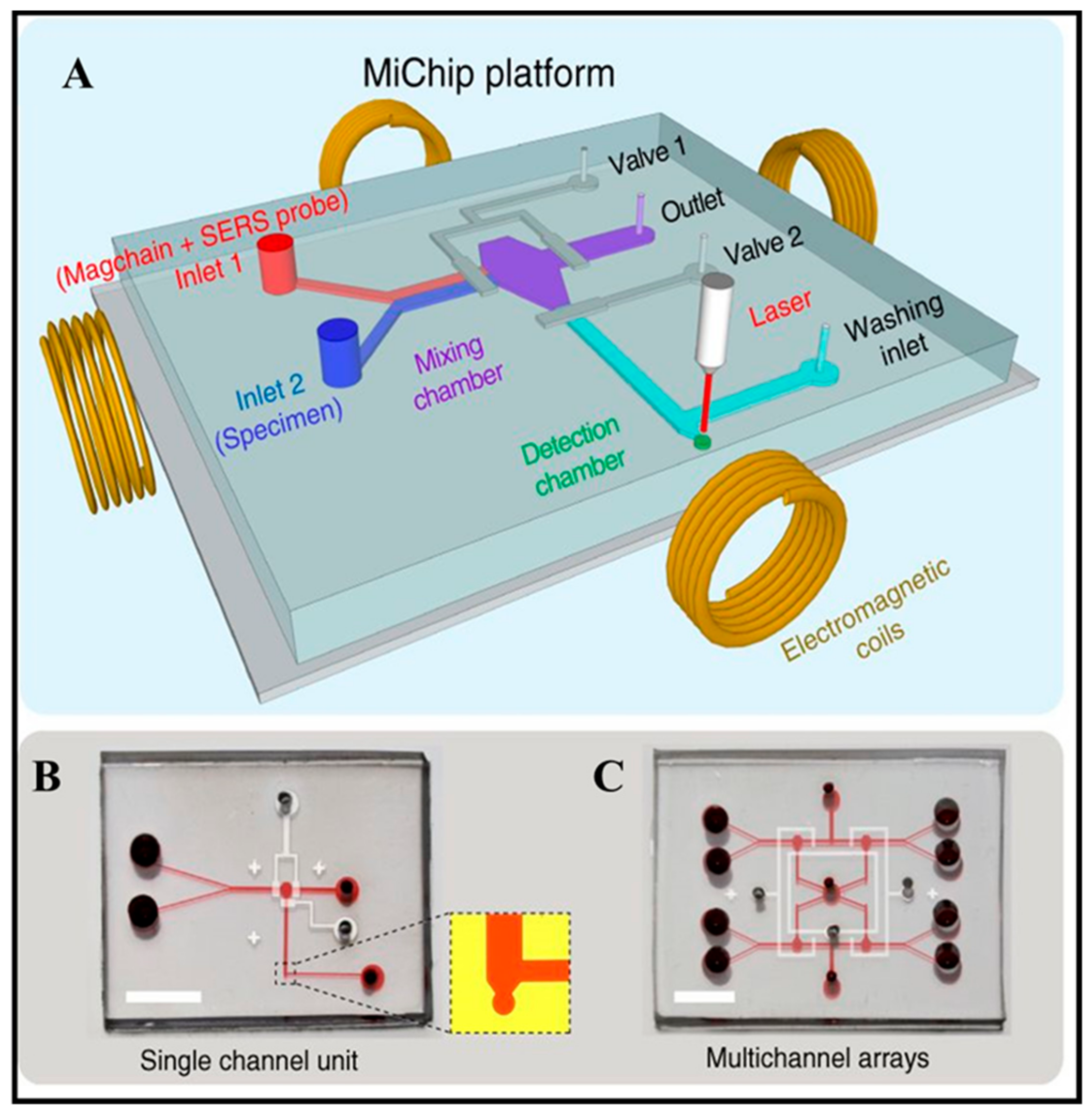
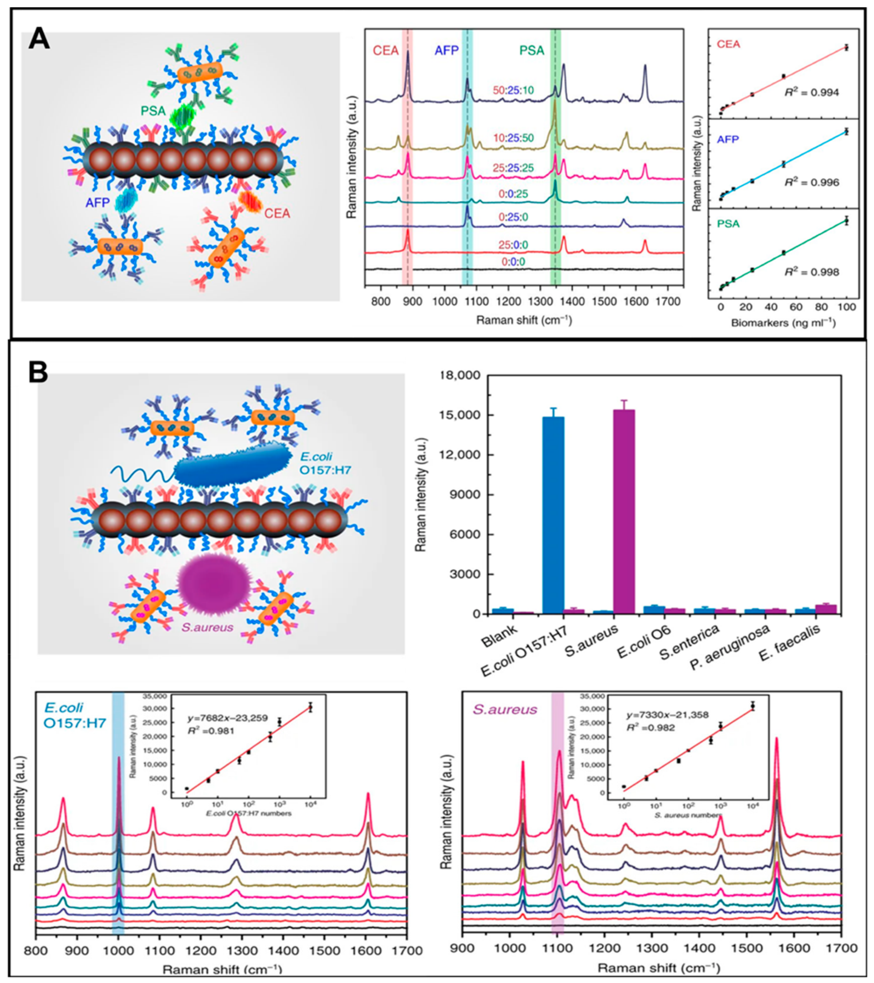
3.1.4. Surface-Enhanced Raman Scattering Biosensing Devices
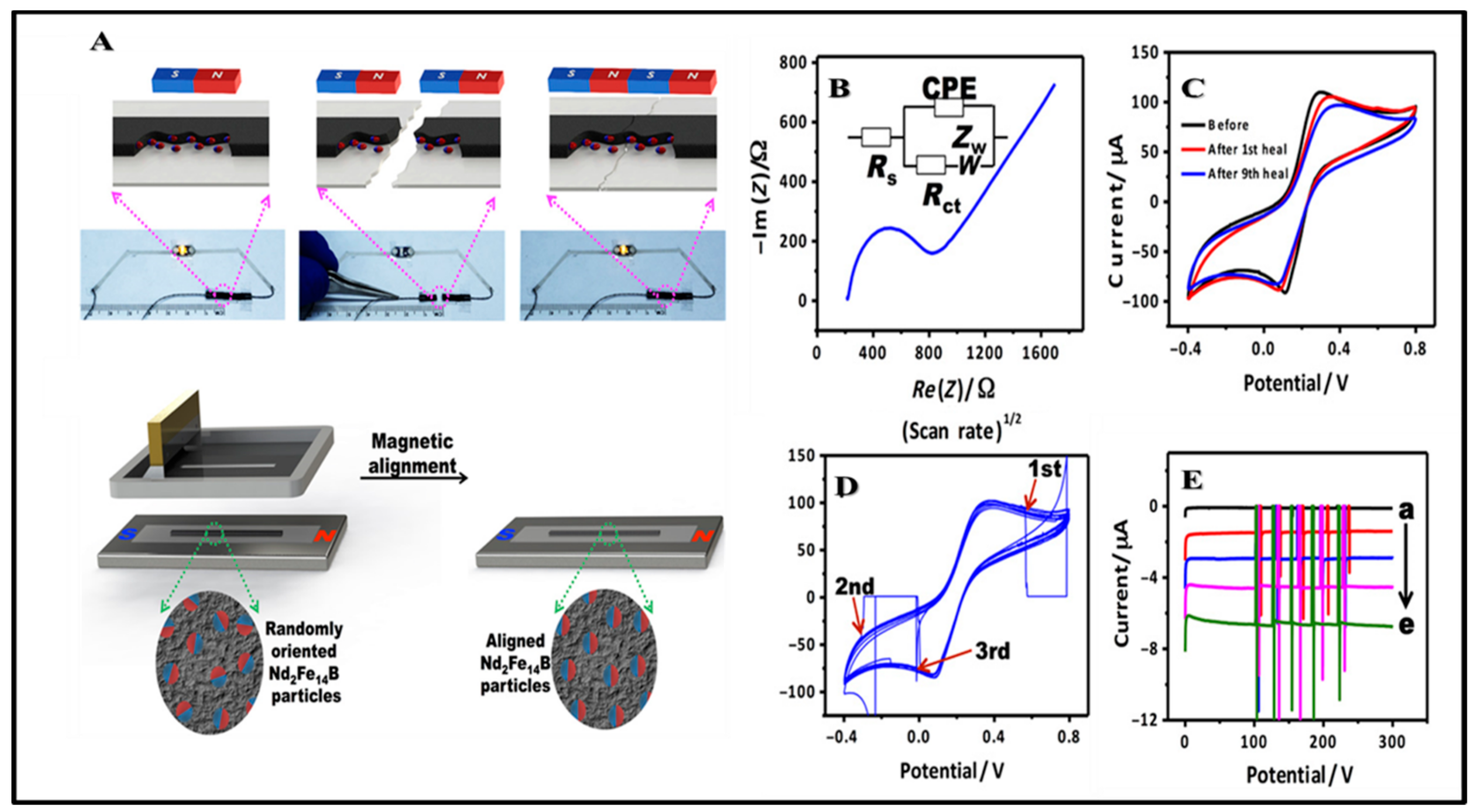
3.2. Electrochemical Biosensing Devices
3.2.1. Potentiometric Biosensing Devices
3.2.2. Conductometric Biosensing Devices
3.2.3. Amperometric Biosensing Devices
3.2.4. Impedimetric Biosensing Devices
4. Magnetic Phenomena-Based Bioassays
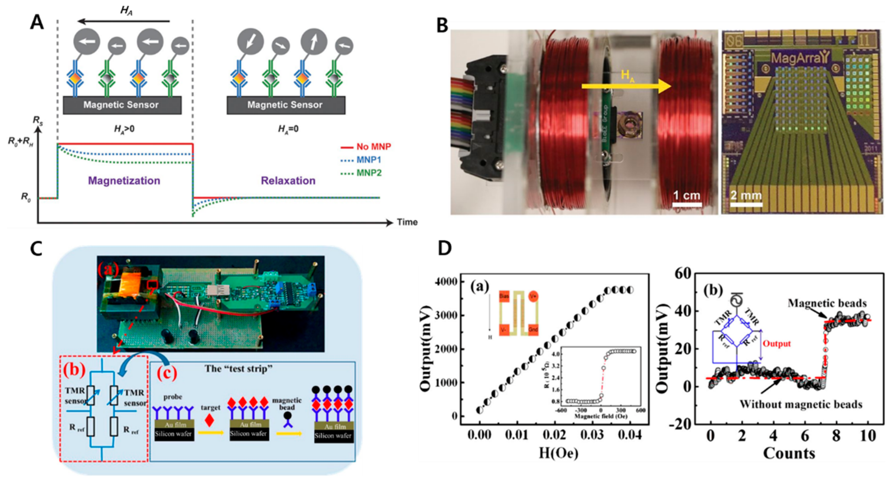
4.1. GMR-Based Bioassay
4.2. MTJ-Based Bioassay
4.3. MPS-Based Bioassay
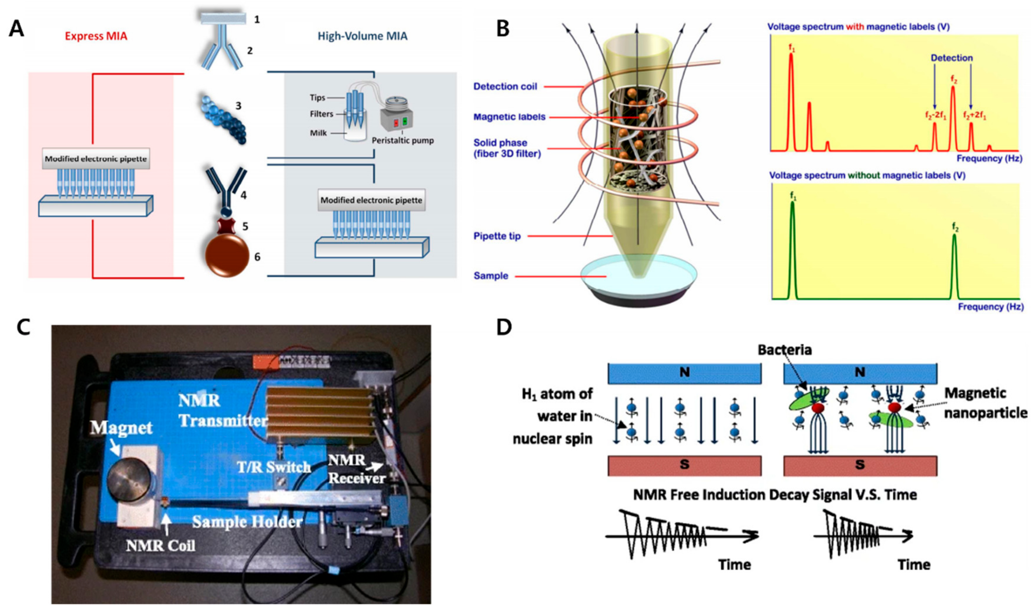
4.4. NMR-Based Bioassay
5. Concluding Remarks and Future Perspectives
Author Contributions
Funding
Conflicts of Interest
References
- Wu, W.; Wu, Z.; Yu, T.; Jiang, C.; Kim, W.S. Recent progress on magnetic iron oxide nanoparticles: Synthesis, surface functional strategies and biomedical applications. Sci. Technol. Adv. Mater. 2015, 16, 023501. [Google Scholar] [CrossRef]
- Mahdavi, M.; Ahmad, M.B.; Haron, M.J.; Namvar, F.; Nadi, B.; Rahman, M.Z.; Amin, J. Synthesis, surface modification and characterisation of biocompatible magnetic iron oxide nanoparticles for biomedical applications. Molecules 2013, 18, 7533–7548. [Google Scholar] [CrossRef]
- Silva, S.M.; Tavallaie, R.; Sandiford, L.; Tilley, R.D.; Gooding, J.J. Gold coated magnetic nanoparticles: From preparation to surface modification for analytical and biomedical applications. Chem. Commun. 2016, 52, 7528–7540. [Google Scholar] [CrossRef]
- Yang, H.M.; Park, C.W.; Ahn, T.; Jung, B.; Seo, B.K.; Park, J.H.; Kim, J.D. A direct surface modification of iron oxide nanoparticles with various poly (amino acids) for use as magnetic resonance probes. J. Colloid Interface Sci. 2013, 391, 158–167. [Google Scholar] [CrossRef]
- Yang, P.; Xu, H.; Zhang, Z.; Yang, L.; Kuang, H.; Aguilar, Z.P. Surface modification affect the biodistribution and toxicity characteristics of iron oxide magnetic nanoparticles in rats. IET Nanobiotechnology 2018, 12, 562–568. [Google Scholar] [CrossRef]
- Iravani, S. Bio-Based Synthesis of Magnetic Nanoparticles and Their Applications. In Magnetic Nanostructures: Environmental and Agricultural Applications; Abd-Elsalam, K.A., Mohamed, M.A., Prasad, R., Eds.; Springer International Publishing: Berlin, Germany, 2019; pp. 13–31. [Google Scholar]
- Elrahman, A.A.A.; Mansour, F.R. Targeted magnetic iron oxide nanoparticles: Preparation, functionalization and biomedical application. J. Drug Deliv. Sci. Technol. 2019, 52, 702–712. [Google Scholar] [CrossRef]
- Kotchoni, S.O.; Gachomo, E.W. A rapid and hazardous reagent free protocol for genomic DNA extraction suitable for genetic studies in plants. Mol. Boil. Rep. 2009, 36, 1633–1636. [Google Scholar] [CrossRef] [PubMed]
- Yi, D.K.; Selvan, S.T.; Lee, S.S.; Papaefthymiou, G.C.; Kundaliya, D.; Ying, J.Y. Silica-coated nanocomposites of magnetic nanoparticles and quantum dots. J. Am. Chem. Soc. 2005, 127, 4990–4991. [Google Scholar] [CrossRef] [PubMed]
- Elrefai, A.L.; Yoshida, T.; Enpuku, K. Magnetic parameters evaluation of magnetic nanoparticles for use in biomedical applications. J. Magn. Magn. Mater. 2019, 474, 522–527. [Google Scholar] [CrossRef]
- Xianyu, Y.; Wang, Q.; Chen, Y. Magnetic particles-enabled biosensors for point-of-care testing. TrAC Trends Anal. Chem. 2018, 106, 213–224. [Google Scholar] [CrossRef]
- Wu, K.; Su, D.; Liu, J.; Saha, R.; Wang, J.-P. Magnetic nanoparticles in nanomedicine: A review of recent advances. Nanotechnology 2019, 30, 502003. [Google Scholar] [CrossRef] [PubMed]
- Vallabani, N.V.S.; Singh, S.; Karakoti, A.S. Magnetic Nanoparticles: Current Trends and Future Aspects in Diagnostics and Nanomedicine. Curr. Drug Metab. 2019, 20, 457–472. [Google Scholar] [CrossRef] [PubMed]
- Ha, Y.; Ko, S.; Kim, I.; Huang, Y.; Mohanty, K.; Huh, C.; Maynard, J.A. Recent Advances Incorporating Superparamagnetic Nanoparticles into Immunoassays. ACS Appl. Nano Mater. 2018, 1, 512–521. [Google Scholar] [CrossRef] [PubMed]
- Heidari, H.; Limouei-Khosrowshahi, B. Magnetic solid phase extraction with carbon-coated Fe3O4 nanoparticles coupled to HPLC-UV for the simultaneous determination of losartan, carvedilol, and amlodipine besylate in plasma samples. J. Chromatogr. B 2019, 1114, 24–30. [Google Scholar] [CrossRef] [PubMed]
- Asgharinezhad, A.A.; Ebrahimzadeh, H.; Mirbabaei, F.; Mollazadeh, N.; Shekari, N. Dispersive micro-solid-phase extraction of benzodiazepines from biological fluids based on polyaniline/magnetic nanoparticles composite. Anal. Chim. Acta 2014, 844, 80–89. [Google Scholar] [CrossRef]
- Feng, Z.; Yang, R.; Du, B. Fast SPE and HPLC–DAD determination of brucine in human urine using multi-walled carbon nanotubes modified with magnetic nanoparticles. J. Anal. Chem. 2017, 72, 862–869. [Google Scholar] [CrossRef]
- Chen, L.; Zhang, M.; Fu, F.; Li, J.; Lin, Z. Facile synthesis of magnetic covalent organic framework nanobeads and application to magnetic solid-phase extraction of trace estrogens from human urine. J. Chromatogr. A 2018, 1567, 136–146. [Google Scholar] [CrossRef]
- Mirzapour, H.; Panahi, H.A.P.A.; Moniri, E.M.; Feizbakhsh, A.F. Magnetic nanoparticles modified with organic dendrimers containing methyl methacrylate and ethylene diamine for the microextraction of rosuvastatin. Microchim. Acta 2018, 185, 440. [Google Scholar] [CrossRef]
- Ji, W.-H.; Guo, Y.-S.; Wang, X.; Guo, D.-S. A water-compatible magnetic molecularly imprinted polymer for the selective extraction of risperidone and 9-hydroxyrisperidone from human urine. Talanta 2018, 181, 392–400. [Google Scholar] [CrossRef]
- Zhang, R.; Wang, S.; Yang, Y.; Deng, Y.; Li, D.; Su, P.; Yang, Y. Modification of polydopamine-coated Fe3O4 nanoparticles with multi-walled carbon nanotubes for magnetic-μ-dispersive solid-phase extraction of antiepileptic drugs in biological matrices. Anal. Bioanal. Chem. 2018, 410, 3779–3788. [Google Scholar] [CrossRef]
- Zhou, Z.; Kadam, U.; Irudayaraj, J. One-step Genome DNA extraction by Salicylic Acid Coated Magnetic Nanoparticles. Anal. Biochem. 2013, 442, 249–252. [Google Scholar] [CrossRef] [PubMed]
- Saiyed, Z.M.; Ramchand, C.N.; Telang, S.D. Isolation of genomic DNA using magnetic nanoparticles as a solid-phase support. J. Physics: Condens. Matter 2008, 20, 204153. [Google Scholar] [CrossRef] [PubMed]
- Shan, Z.; Zhou, Z.; Chen, H.; Zhang, Z.; Zhou, Y.; Wen, A.; Oakes, K.D.; Servos, M.R. PCR-ready human DNA extraction from urine samples using magnetic nanoparticles. J. Chromatogr. B 2012, 881, 63–68. [Google Scholar] [CrossRef] [PubMed]
- Odabaşi, M.; Denizli, A.; Odabaşı, M. Polyhydroxyethylmethacrylate-based magnetic DNA-affinity beads for anti-DNA antibody removal from systemic lupus erythematosus patient plasma. J. Chromatogr. B 2001, 760, 137–148. [Google Scholar] [CrossRef]
- Meng, Z.-H.; Lin, B.; Xie, Y.-H.; Zhang, K.-Q. Cloning and expression of fused Fc-binding protein (SPA-SPG) and its application in purification of IgG. Prog. Biochem. Biophys. 2004, 31, 146–149. [Google Scholar]
- Bhagwati, S.; Ghatpande, A.; Leung, B. Identification of Two Nuclear Proteins Which Bind to RNA CUG Repeats: Significance for Myotonic Dystrophy. Biochem. Biophys. Res. Commun. 1996, 228, 55–62. [Google Scholar] [CrossRef]
- Zollner, T.C.A.; Zollner, R.D.L.; De Cuyper, M.; Santana, M.H.A. Adsorption of Isotype “E” Antibodies on Affinity Magnetoliposomes. J. Dispers. Sci. Technol. 2003, 24, 615–622. [Google Scholar] [CrossRef]
- Zeng, G.; Gao, L.; Xia, T.; Tencomnao, T.; Yu, R.K. Characterization of the 5′-flanking fragment of the human GM3-synthase gene. Biochim. Biophys. Acta (BBA)-Gene Struct. Expr. 2003, 1625, 30–35. [Google Scholar] [CrossRef]
- Blank, M.; Weinschenk, T.; Priemer, M.; Schluesener, H. Systematic evolution of a DNA aptamer binding to rat brain tumor microvessels selective targeting of endothelial regulatory protein pigpen. J. Biol. Chem. 2001, 276, 16464–16468. [Google Scholar] [CrossRef]
- Chakraborty, A.; White, S.M.; Schaefer, T.S.; Ball, E.D.; Dyer, K.F.; Tweady, D.J. Granulocyte Colony-Stimulating Factor Activation of Stat3alpha and Stat3beta in Immature Normal and Leukemic Human Myeloid Cells. Blood 1996, 88, 2442–2449. [Google Scholar] [CrossRef]
- Müller-Schulte, D.; Brunner, H. Novel magnetic microspheres on the basis of poly (vinyl alcohol) as affinity medium for quantitative detection of glycated haemoglobin. J. Chromatogr. A 1995, 711, 53–60. [Google Scholar] [CrossRef]
- Ji, Z.; Pinon, D.I.; Miller, L. Development of Magnetic Beads for Rapid and Efficient Metal-Chelate Affinity Purifications. Anal. Biochem. 1996, 240, 197–201. [Google Scholar] [CrossRef] [PubMed]
- Lu, H.; Yi, G.; Zhao, S.; Chen, D.; Guo, L.-H.; Cheng, J. Synthesis and characterization of multi-functional nanoparticles possessing magnetic, up-conversion fluorescence and bio-affinity properties. J. Mater. Chem. 2004, 14, 1336. [Google Scholar] [CrossRef]
- Tiikkaja, M.; Aro, A.L.; Alanko, T.; Lindholm, H.; Sistonen, H.; Hartikainen, J.E.; Toivonen, L.; Juutilainen, J.; Hietanen, M. Electromagnetic interference with cardiac pacemakers and implantable cardioverter-defibrillators from low-frequency electromagnetic fields in vivo. Europace 2013, 15, 388–394. [Google Scholar] [CrossRef] [PubMed]
- Wang, C.; Yuan, H.; Duan, Z.; Xiao, D. Integrated multi-ISE arrays with improved sensitivity, accuracy and precision. Sci. Rep. 2017, 7, 44771. [Google Scholar] [CrossRef] [PubMed]
- Nima, Z.A.; Watanabe, F.; Jamshidi-Parsian, A.; Sarimollaoglu, M.; Nedosekin, D.A.; Han, M.; Watts, J.A.; Biris, A.S.; Zharov, V.P.; Galanzha, E.I. Bioinspired magnetic nanoparticles as multimodal photoacoustic, photothermal and photomechanical contrast agents. Sci. Rep. 2019, 9, 887. [Google Scholar] [CrossRef]
- Coleman, B.; Coarsey, C.; Asghar, W. Cell phone based colorimetric analysis for point-of-care settings. Analyst 2019, 144, 1935–1947. [Google Scholar] [CrossRef]
- Kangas, M.J.; Burks, R.M.; Atwater, J.; Lukowicz, R.M.; Williams, P.; Holmes, A.E. Colorimetric Sensor Arrays for the Detection and Identification of Chemical Weapons and Explosives. Crit. Rev. Anal. Chem. 2017, 47, 138–153. [Google Scholar] [CrossRef]
- Yetisen, A.K.; Akram, M.S.; Lowe, C.R. Paper-based microfluidic point-of-care diagnostic devices. Lab Chip 2013, 13, 2210–2251. [Google Scholar] [CrossRef]
- Reboud, J.; Xu, G.; Garrett, A.; Adriko, M.; Yang, Z.; Tukahebwa, E.M.; Rowell, C.; Cooper, J. Paper-based microfluidics for DNA diagnostics of malaria in low resource underserved rural communities. Proc. Natl. Acad. Sci. 2019, 116, 4834–4842. [Google Scholar] [CrossRef]
- Shin, J.; Choi, S.; Yang, J.-S.; Song, J.; Choi, J.-S.; Jung, H.-I. Smart Forensic Phone: Colorimetric analysis of a bloodstain for age estimation using a smartphone. Sensors Actuators B Chem. 2017, 243, 221–225. [Google Scholar] [CrossRef]
- Jung, Y.; Kim, J.; Awofeso, O.; Kim, H.; Regnier, F.; Bae, E. Smartphone-based colorimetric analysis for detection of saliva alcohol concentration. Appl. Opt. 2015, 54, 9183–9189. [Google Scholar] [CrossRef] [PubMed]
- Srisa-Art, M.; Boehle, K.E.; Geiss, B.J.; Henry, C.S. Highly Sensitive Detection of Salmonella typhimurium Using a Colorimetric Paper-Based Analytical Device Coupled with Immunomagnetic Separation. Anal. Chem. 2018, 90, 1035–1043. [Google Scholar] [CrossRef] [PubMed]
- Kim, M.S.; Kweon, S.H.; Cho, S.; An, S.S.A.; Kim, M.I.; Doh, J.; Lee, J. Pt-Decorated Magnetic Nanozymes for Facile and Sensitive Point-of-Care Bioassay. ACS Appl. Mater. Interfaces 2017, 9, 35133–35140. [Google Scholar] [CrossRef]
- Wang, D.B.; Tian, B.; Zhang, Z.P.; Wang, X.Y.; Fleming, J.; Bi, L.J.; Yang, R.F.; Zhang, X.E. Detection of Bacillus anthracis spores by super-paramagnetic lateral-flow immunoassays based on “Road Closure”. Biosens. Bioelectron. 2015, 67, 608–614. [Google Scholar] [CrossRef]
- Suaifan, G.; Alhogail, S.; Zourob, M. Paper-based magnetic nanoparticle-peptide probe for rapid and quantitative colorimetric detection of Escherichia coli O157:H7. Biosens. Bioelectron. 2017, 92, 702–708. [Google Scholar] [CrossRef]
- Guo, R.; Wang, S.; Huang, F.; Chen, Q.; Li, Y.; Liao, M.; Lin, J. Rapid detection of Salmonella Typhimurium using magnetic nanoparticle immunoseparation, nanocluster signal amplification and smartphone image analysis. Sensors Actuators B Chem. 2019, 284, 134–139. [Google Scholar] [CrossRef]
- Huang, Y.; Wu, T.; Wang, F.; Li, K.; Qian, L.; Zhang, X.; Liu, G. Magnetized Carbon Nanotube Based Lateral Flow Immunoassay for Visual Detection of Complement Factor B. Molecules 2019, 24, 2759. [Google Scholar] [CrossRef]
- Russell, S.M.; Alba-Patino, A.; Borges, M.; de la Rica, R. Multifunctional motion-to-color janus transducers for the rapid detection of sepsis biomarkers in whole blood. Biosens. Bioelectron. 2019, 140, 111346. [Google Scholar] [CrossRef]
- Li, F.; Li, F.; Luo, D.; Lai, W.; Xiong, Y.; Xu, H. Biotin-exposure-based immunomagnetic separation coupled with nucleic acid lateral flow biosensor for visibly detecting viable Listeria monocytogenes. Anal. Chim. Acta 2018, 1017, 48–56. [Google Scholar] [CrossRef]
- Quinn, M.K.; Gnan, N.; James, S.; Ninarello, A.; Sciortino, F.; Zaccarelli, E.; McManus, J.J. How fluorescent labelling alters the solution behaviour of proteins. Phys. Chem. Chem. Phys. 2015, 17, 31177–31187. [Google Scholar] [CrossRef] [PubMed]
- Prendergast, F.G.; Mann, K.G. Chemical and physical properties of aequorin and the green fluorescent protein isolated from Aequorea forskalea. Biochemistry 1978, 17, 3448–3453. [Google Scholar] [CrossRef] [PubMed]
- Zeng, Y.; Zhu, J.; Wang, J.; Parasuraman, P.; Busi, S.; Nauli, S.M.; Wang, Y.X.J.; Pala, R.; Liu, G. Functional probes for cardiovascular molecular imaging. Quant. Imaging Med. Surg. 2018, 8, 838–852. [Google Scholar] [CrossRef] [PubMed]
- Kim, S.E.; Jo, S.D.; Kwon, K.C.; Won, Y.Y.; Lee, J. Genetic Assembly of Double-Layered Fluorescent Protein Nanoparticles for Cancer Targeting and Imaging. Adv. Sci. 2017, 4, 1600471. [Google Scholar] [CrossRef]
- Lim, M.C.; Kim, Y.R. Analytical Applications of Nanomaterials in Monitoring Biological and Chemical Contaminants in Food. J. Microbiol. Biotechnol. 2016, 26, 1505–1516. [Google Scholar] [CrossRef]
- Li, C. A targeted approach to cancer imaging and therapy. Nat. Mater. 2014, 13, 110–115. [Google Scholar] [CrossRef]
- Reslova, N.; Michna, V.; Kasny, M.; Mikel, P.; Kralik, P. xMAP Technology: Applications in Detection of Pathogens. Front. Microbiol. 2017, 8, 55. [Google Scholar] [CrossRef]
- Burnham, C.-A.D.; Doern, C.; Binder, S.R. Viral Diseases 1, The Immunoassay Handbook; Elsevier: Amsterdam, The Netherlands, 2013; pp. 919–927. [Google Scholar]
- Doseeva, V.; Colpitts, T.; Gao, G.; Woodcock, J.; Knezevic, V. Performance of a multiplexed dual analyte immunoassay for the early detection of non-small cell lung cancer. J. Transl. Med. 2015, 13, 55. [Google Scholar] [CrossRef]
- Breen, E.J.; Tan, W.; Khan, A. The Statistical Value of Raw Fluorescence Signal in Luminex xMAP Based Multiplex Immunoassays. Sci. Rep. 2016, 6, 26996. [Google Scholar] [CrossRef]
- Verbarg, J.; Hadass, O.; Olivo, P.D.; Danielli, A. High sensitivity detection of a protein biomarker interleukin-8 utilizing a magnetic modulation biosensing system. Sensors Actuators B Chem. 2017, 241, 614–618. [Google Scholar] [CrossRef]
- Chunyk, A.G.; Joyce, A.; Fischer, S.K.; Dysinger, M.; Mikulskis, A.; Jeromin, A.; Lawrence-Henderson, R.; Baker, D.; Yeung, D. A Multi-site In-depth Evaluation of the Quanterix Simoa from a User’s Perspective. AAPS J. 2017, 20, 10. [Google Scholar] [CrossRef] [PubMed]
- Poorbaugh, J.; Samanta, T.; Bright, S.W.; Sissons, S.E.; Chang, C.Y.; Oberoi, P.; MacDonald, A.J.; Martin, A.P.; Cox, K.L.; Benschop, R.J. Measurement of IL-21 in human serum and plasma using ultrasensitive MSD S-PLEX(R) and Quanterix SiMoA methodologies. J. Immunol. Methods 2019, 466, 9–16. [Google Scholar] [CrossRef] [PubMed]
- Anker, J.N.; Behrend, C.J.; Huang, H.; Kopelman, R. Magnetically-modulated optical nanoprobes (MagMOONs) and systems. J. Magn. Magn. Mater. 2005, 293, 655–662. [Google Scholar] [CrossRef]
- Nguyen, K.T.; Anker, J.N. Detecting De-gelation through Tissue Using Magnetically Modulated Optical Nanoprobes (MagMOONs). Sensors Actuators B Chem. 2014, 205, 313–321. [Google Scholar] [CrossRef]
- Wang, S.; Zheng, L.; Cai, G.; Liu, N.; Liao, M.; Li, Y.; Zhang, X.; Lin, J. A microfluidic biosensor for online and sensitive detection of Salmonella typhimurium using fluorescence labeling and smartphone video processing. Biosens. Bioelectron. 2019, 140, 111333. [Google Scholar] [CrossRef]
- Zhang, Y.; Zhang, L.; Yang, L.; Vong, C.I.; Chan, K.-F.; Wu, W.K.K.; Kwong, T.N.Y.; Lo, N.W.; Ip, M.; Wong, S.H.; et al. Real-time tracking of fluorescent magnetic spore–based microrobots for remote detection of C. diff toxins. Sci. Adv. 2019, 5, eaau9650. [Google Scholar] [CrossRef]
- Burg, S.; Cohen, M.; Margulis, M.; Roth, S.; Danielli, A. Magnetically aggregated biosensors for sensitive detection of biomarkers at low concentrations. Appl. Phys. Lett. 2019, 115, 103702. [Google Scholar] [CrossRef]
- Zeng, S.; Baillargeat, D.; Ho, H.P.; Yong, K.T. Nanomaterials enhanced surface plasmon resonance for biological and chemical sensing applications. Chem. Soc. Rev. 2014, 43, 3426–3452. [Google Scholar] [CrossRef]
- Zhou, X.; Li, X.; Li, S.; An, G.-W.; Cheng, T. Magnetic Field Sensing Based on SPR Optical Fiber Sensor Interacting With Magnetic Fluid. IEEE Trans. Instrum. Meas. 2019, 68, 234–239. [Google Scholar] [CrossRef]
- Jiang, J.; Wang, X.; Li, S.; Ding, F.; Li, N.; Meng, S.; Li, R.; Qi, J.; Liu, Q.; Liu, G.L. Plasmonic nano-arrays for ultrasensitive bio-sensing. Nanophotonics 2018, 7, 1517–1531. [Google Scholar] [CrossRef]
- Reiner, A.T.; Fossati, S.; Dostalek, J. Biosensor platform for parallel surface plasmon-enhanced epifluorescence and surface plasmon resonance detection. Sensors Actuators B Chem. 2018, 257, 594–601. [Google Scholar] [CrossRef]
- Liu, Y.; Liu, Q.; Chen, S.; Cheng, F.; Wang, H.; Peng, W. Surface Plasmon Resonance Biosensor Based on Smart Phone Platforms. Sci. Rep. 2015, 5, 12864. [Google Scholar] [CrossRef] [PubMed]
- Pang, Y.; Wan, N.; Shi, L.; Wang, C.; Sun, Z.; Xiao, R.; Wang, S. Dual-recognition surface-enhanced Raman scattering(SERS)biosensor for pathogenic bacteria detection by using vancomycin-SERS tags and aptamer-Fe3O4@Au. Anal. Chim. Acta 2019, 1077, 288–296. [Google Scholar] [CrossRef] [PubMed]
- Ilkhani, H.; Hughes, T.; Li, J.; Zhong, C.J.; Hepel, M. Nanostructured SERS-electrochemical biosensors for testing of anticancer drug interactions with DNA. Biosens. Bioelectron. 2016, 80, 257–264. [Google Scholar] [CrossRef]
- Fleischmann, M.; Hendra, P.J.; McQuillan, A.J. Raman spectra of pyridine adsorbed at a silver electrode. Chem. Phys. Lett. 1974, 26, 163–166. [Google Scholar] [CrossRef]
- Xiong, Q.; Lim, C.Y.; Ren, J.; Zhou, J.; Pu, K.; Chan-Park, M.B.; Mao, H.; Lam, Y.C.; Duan, H. Magnetic nanochain integrated microfluidic biochips. Nat. Commun. 2018, 9, 1743. [Google Scholar] [CrossRef]
- Zhang, H.; Ma, X.; Liu, Y.; Duan, N.; Wu, S.; Wang, Z.; Xu, B. Gold nanoparticles enhanced SERS aptasensor for the simultaneous detection of Salmonella typhimurium and Staphylococcus aureus. Biosens. Bioelectron. 2015, 74, 872–877. [Google Scholar] [CrossRef]
- Mohammadniaei, M.; Nguyen, H.V.; Tieu, M.V.; Lee, M.H. 2D Materials in Development of Electrochemical Point-of-Care Cancer Screening Devices. Micromachines 2019, 10, 662. [Google Scholar] [CrossRef]
- Wu, H.; Gao, W.; Yin, Z. Materials, Devices and Systems of Soft Bioelectronics for Precision Therapy. Adv. Healthc. Mater. 2017, 6, 1700017. [Google Scholar] [CrossRef]
- Bandodkar, A.J.; Mohan, V.; López, C.S.; Ramírez, J.; Wang, J. Self-Healing Inks for Autonomous Repair of Printable Electrochemical Devices. Adv. Electron. Mater. 2015, 1, 1500289. [Google Scholar] [CrossRef]
- Sempionatto, J.R.; Martin, A.; García-Carmona, L.; Barfidokht, A.; Kurniawan, J.F.; Moreto, J.R.; Tang, G.; Shin, A.; Liu, X.; Escarpa, A.; et al. Skin-worn Soft Microfluidic Potentiometric Detection System. Electroanalysis 2019, 31, 239–245. [Google Scholar] [CrossRef]
- Dobbelaere, T.; Vereecken, P.M.; Detavernier, C. A USB-controlled potentiostat/galvanostat for thin-film battery characterization. HardwareX 2017, 2, 34–49. [Google Scholar] [CrossRef]
- Guilbault, G.G.; Montalvo, J.G. Urea-specific enzyme electrode. J. Am. Chem. Soc. 1969, 91, 2164–2165. [Google Scholar] [CrossRef]
- Liu, C.; Neale, Z.G.; Cao, G. Understanding electrochemical potentials of cathode materials in rechargeable batteries. Mater. Today 2016, 19, 109–123. [Google Scholar] [CrossRef]
- Ainla, A.; Mousavi, M.P.S.; Tsaloglou, M.N.; Redston, J.; Bell, J.G.; Fernandez-Abedul, M.T.; Whitesides, G.M. Open-Source Potentiostat for Wireless Electrochemical Detection with Smartphones. Anal. Chem. 2018, 90, 6240–6246. [Google Scholar] [CrossRef]
- Canovas, R.; Parrilla, M.; Blondeau, P.; Andrade, F.J. A novel wireless paper-based potentiometric platform for monitoring glucose in blood. Lab Chip 2017, 17, 2500–2507. [Google Scholar] [CrossRef]
- Liu, F.; Kc, P.; Zhang, G.; Zhe, J. In situ single cell detection via microfluidic magnetic bead assay. PLoS ONE 2017, 12, e0172697. [Google Scholar] [CrossRef]
- Dzyadevych, S.; Jaffrezic-Renault, N. Conductometric biosensors. Biological Identification 2014, 6, 153–193. [Google Scholar]
- Soldatkin, O.O.; Burdak, O.S.; Sergeyeva, T.A.; Arkhypova, V.M.; Dzyadevych, S.V.; Soldatkin, A.P. Acetylcholinesterase-based conductometric biosensor for determination of aflatoxin B1. Sensors Actuators B Chem. 2013, 188, 999–1003. [Google Scholar] [CrossRef]
- Saiapina, O.Y.; Pyeshkova, V.M.; Soldatkin, O.O.; Melnik, V.G.; Kurç, B.A.; Walcarius, A.; Dzyadevych, S.V.; Jaffrezic-Renault, N. Conductometric enzyme biosensors based on natural zeolite clinoptilolite for urea determination. Mater. Sci. Eng. C 2011, 31, 1490–1497. [Google Scholar] [CrossRef]
- Chou, J.C.; Wu, C.Y.; Lin, S.H.; Kuo, P.Y.; Lai, C.H.; Nien, Y.H.; Wu, Y.X.; Lai, T.Y. The Analysis of the Urea Biosensors Using Different Sensing Matrices via Wireless Measurement System & Microfluidic Measurement System. Sensors 2019, 19, 3004. [Google Scholar]
- Tieu, M.V.; Go, A.; Park, Y.J.; Nguyen, H.V.; Hwang, S.Y.; Lee, M.-H. Highly Sensitive ELISA Using Membrane-Based Microwave-Mediated Electrochemical Immunoassay for Thyroid-Stimulating Hormone Detection. IEEE Sensors J. 2019, 19, 9826–9831. [Google Scholar] [CrossRef]
- Mistry, K.K.; Layek, K.; Chell, T.N.; Chaudhuri, C.R.; Saha, H. Design and development of an amperometric immunosensor based on screen-printed electrodes. Anal. Methods 2016, 8, 3096–3101. [Google Scholar] [CrossRef]
- Bandodkar, A.J.; López, C.S.; Mohan, A.M.V.; Yin, L.; Kumar, R.; Wang, J. All-printed magnetically self-healing electrochemical devices. Sci. Adv. 2016, 2, e1601465. [Google Scholar] [CrossRef] [PubMed]
- Ducote, M.; Vinson, B.; Hogquist, S. Electrochemical Impedance Spectroscopy (EIS) as a Tool for Pathogen Detection. J. Biosens. Bioelectron. 2016, 7, 224. [Google Scholar]
- Baleg, A.A.; Jahed, N.; Yonkeu, A.L.D.; Njomo, N.; Mbambisa, G.; Molapo, K.M.; Fuku, X.G.; Fomo, G.; Makelane, H.; Tsegaye, A.; et al. Impedimetry and microscopy of electrosynthetic poly (propylene imine)-co-polypyrrole conducting dendrimeric star copolymers. Electrochimica Acta 2014, 128, 448–457. [Google Scholar] [CrossRef]
- Yagati, A.K.; Chavan, S.G.; Baek, C.; Lee, M.H.; Min, J. Label-Free Impedance Sensing of Aflatoxin B(1) with Polyaniline Nanofibers/Au Nanoparticle Electrode Array. Sensors 2018, 18, 1320. [Google Scholar] [CrossRef]
- Spain, E.; Carrara, S.; Adamson, K.; Ma, H.; O’Kennedy, R.; de Cola, L.; Forster, R.J. Cardiac Troponin I: Ultrasensitive Detection Using Faradaic Electrochemical Impedance. ACS Omega 2018, 3, 17116–17124. [Google Scholar] [CrossRef]
- Wang, Y.; Ye, Z.; Ping, J.; Jing, S.; Ying, Y. Development of an aptamer-based impedimetric bioassay using microfluidic system and magnetic separation for protein detection. Biosens. Bioelectron. 2014, 59, 106–111. [Google Scholar] [CrossRef]
- Kongsuphol, P.; Ng, H.H.; Pursey, J.P.; Arya, S.K.; Wong, C.C.; Stulz, E.; Park, M.K. EIS-based biosensor for ultra-sensitive detection of TNF-alpha from non-diluted human serum. Biosens. Bioelectron. 2014, 61, 274–279. [Google Scholar] [CrossRef]
- Chen, Q.; Lin, J.; Gan, C.; Wang, Y.; Wang, D.; Xiong, Y.; Lai, W.; Li, Y.; Wang, M. A sensitive impedance biosensor based on immunomagnetic separation and urease catalysis for rapid detection of Listeria monocytogenes using an immobilization-free interdigitated array microelectrode. Biosens. Bioelectron. 2015, 74, 504–511. [Google Scholar] [CrossRef] [PubMed]
- Rackus, D.G.; Dryden, M.D.; Lamanna, J.; Zaragoza, A.; Lam, B.; Kelley, S.O.; Wheeler, A.R. A digital microfluidic device with integrated nanostructured microelectrodes for electrochemical immunoassays. Lab Chip 2015, 15, 3776–3784. [Google Scholar] [CrossRef] [PubMed]
- Shafiee, H.; Asghar, W.; Inci, F.; Yuksekkaya, M.; Jahangir, M.; Zhang, M.H.; Durmus, N.G.; Gurkan, U.A.; Kuritzkes, D.R.; Demirci, U. Paper and flexible substrates as materials for biosensing platforms to detect multiple biotargets. Sci. Rep. 2015, 5, 8719. [Google Scholar] [CrossRef] [PubMed]
- Torrente-Rodriguez, R.M.; Campuzano, S.; Montiel, V.R.; Gamella, M.; Pingarron, J.M. Electrochemical bioplatforms for the simultaneous determination of interleukin (IL)-8 mRNA and IL-8 protein oral cancer biomarkers in raw saliva. Biosens. Bioelectron. 2016, 77, 543–548. [Google Scholar] [CrossRef]
- Wang, D.; Chen, Q.; Huo, H.; Bai, S.; Cai, G.; Lai, W.; Lin, J. Efficient separation and quantitative detection of Listeria monocytogenes based on screen-printed interdigitated electrode, urease and magnetic nanoparticles. Food Control. 2017, 73, 555–561. [Google Scholar] [CrossRef]
- Angulo-Ibanez, A.; Eletxigerra, U.; Lasheras, X.; Campuzano, S.; Merino, S. Electrochemical tropomyosin allergen immunosensor for complex food matrix analysis. Anal. Chim. Acta 2019, 1079, 94–102. [Google Scholar] [CrossRef]
- Pereira-Barros, M.A.; Barroso, M.F.; Martin-Pedraza, L.; Vargas, E.; Benede, S.; Villalba, M.; Rocha, J.M.; Campuzano, S.; Pingarron, J.M. Direct PCR-free electrochemical biosensing of plant-food derived nucleic acids in genomic DNA extracts. Application to the determination of the key allergen Sola l 7 in tomato seeds. Biosens. Bioelectron. 2019, 137, 171–177. [Google Scholar] [CrossRef]
- Toldra, A.; Alcaraz, C.; Diogene, J.; O’Sullivan, C.K.; Campas, M. Detection of Ostreopsis cf. ovata in environmental samples using an electrochemical DNA-based biosensor. Sci. Total. Environ. 2019, 689, 655–661. [Google Scholar] [CrossRef]
- Ruiz-Vega, G.; Garcia-Berrocoso, T.; Montaner, J.; Baldrich, E. Paper microfluidics on screen-printed electrodes for simple electrochemical magneto-immunosensor performance. Sensors Actuators B: Chem. 2019, 298, 298. [Google Scholar] [CrossRef]
- Binasch, G.; Grünberg, P.; Saurenbach, F.; Zinn, W. Enhanced magnetoresistance in layered magnetic structures with antiferromagnetic interlayer exchange. Phys. Rev. B 1989, 39, 4828–4830. [Google Scholar] [CrossRef]
- Krishna, V.D.; Wu, K.; Perez, A.M.; Wang, J.-P. Giant Magnetoresistance-based Biosensor for Detection of Influenza A Virus. Front. Microbiol. 2016, 7, 156. [Google Scholar] [CrossRef] [PubMed]
- Wang, W.; Wang, Y.; Tu, L.; Feng, Y.; Klein, T.; Wang, J.-P. Magnetoresistive performance and comparison of supermagnetic nanoparticles on giant magnetoresistive sensor-based detection system. Sci. Rep. 2014, 4, 5716. [Google Scholar] [CrossRef] [PubMed]
- Hall, D.A.; Gaster, R.S.; Lin, T.; Osterfeld, S.J.; Han, S.; Murmann, B.; Wang, S.X. GMR biosensor arrays: A system perspective. Biosens. Bioelectron. 2010, 25, 2051–2057. [Google Scholar] [CrossRef]
- Qiu, W.; Chang, L.; Liang, Y.-C.; Litvinov, J.; Guo, J.; Chen, Y.-T.; Vu, B.; Kourentzi, K.; Xu, S.; Lee, T.R.; et al. Spin-Valve based magnetoresistive nanoparticle detector for applications in biosensing. Sensors Actuators A Phys. 2017, 265, 174–180. [Google Scholar] [CrossRef]
- Wu, K.; Klein, T.; Krishna, V.D.; Su, D.; Perez, A.M.; Wang, J.-P. Portable GMR Handheld Platform for the Detection of Influenza A Virus. ACS Sensors 2017, 2, 1594–1601. [Google Scholar] [CrossRef] [PubMed]
- Gupta, S.; Kakkar, V. DARPin based GMR Biosensor for the detection of ESAT-6 Tuberculosis Protein. Tuberculosis 2019, 118, 101852. [Google Scholar] [CrossRef] [PubMed]
- Zhu, F.; Li, D.; Ding, Q.; Lei, C.; Ren, L.; Ding, X.; Sun, X. 2D magnetic MoS2–Fe3O4 hybrid nanostructures for ultrasensitive exosome detection in GMR sensor. Biosens. Bioelectron. 2020, 147, 111787. [Google Scholar] [CrossRef]
- Klein, T.; Wang, W.; Yu, L.; Wu, K.; Boylan, K.L.M.; Vogel, R.I.; Skubitz, A.P.N.; Wang, J.-P. Development of a multiplexed giant magnetoresistive biosensor array prototype to quantify ovarian cancer biomarkers. Biosens. Bioelectron. 2019, 126, 301–307. [Google Scholar] [CrossRef]
- Gao, Y.; Huo, W.; Zhang, L.; Lian, J.; Tao, W.; Song, C.; Tang, J.; Shi, S.; Gao, Y. Multiplex measurement of twelve tumor markers using a GMR multi-biomarker immunoassay biosensor. Biosens. Bioelectron. 2019, 123, 204–210. [Google Scholar] [CrossRef]
- Huang, C.-C.; Zhou, X.; Hall, D.A. Giant Magnetoresistive Biosensors for Time-Domain Magnetorelaxometry: A Theoretical Investigation and Progress Toward an Immunoassay. Sci. Rep. 2017, 7, 45493. [Google Scholar] [CrossRef]
- Yuasa, S.; Nagahama, T.; Fukushima, A.; Suzuki, Y.; Ando, K. Giant room-temperature magnetoresistance in single-crystal Fe/MgO/Fe magnetic tunnel junctions. Nat. Mater. 2004, 3, 868–871. [Google Scholar] [CrossRef] [PubMed]
- Parkin, S.S.P.; Kaiser, C.; Panchula, A.; Rice, P.M.; Hughes, B.; Samant, M.; Yang, S.-H. Giant tunnelling magnetoresistance at room temperature with MgO (100) tunnel barriers. Nat. Mater. 2004, 3, 862–867. [Google Scholar] [CrossRef] [PubMed]
- Negulescu, B.; Lacour, D.; Montaigne, F.; Gerken, A.; Paul, J.; Spetter, V.; Marien, J.; Duret, C.; Hehn, M. Wide range and tunable linear magnetic tunnel junction sensor using two exchange pinned electrodes. Appl. Phys. Lett. 2009, 95, 112502. [Google Scholar] [CrossRef]
- Shen, W.; Schrag, B.D.; Carter, M.J.; Xie, J.; Xu, C.; Sun, S.; Xiao, G. Detection of DNA labeled with magnetic nanoparticles using MgO-based magnetic tunnel junction sensors. J. Appl. Phys. 2008, 103, 07A306. [Google Scholar] [CrossRef]
- Wu, Y.; Liu, Y.; Zhan, Q.; Liu, J.P.; Li, R.-W. Rapid detection of Escherichia coli O157:H7 using tunneling magnetoresistance biosensor. AIP Adv. 2017, 7, 056658. [Google Scholar] [CrossRef]
- Mu, X.-H.; Liu, H.-F.; Tong, Z.-Y.; Du, B.; Liu, S.; Liu, B.; Liu, Z.-W.; Gao, C.; Wang, J.; Dong, H. A new rapid detection method for ricin based on tunneling magnetoresistance biosensor. Sensors Actuators B Chem. 2019, 284, 638–649. [Google Scholar] [CrossRef]
- Wu, K.; Su, D.; Saha, R.; Wong, D.; Wang, J.-P. Magnetic particle spectroscopy-based bioassays: Methods, applications, advances, and future opportunities. J. Phys. D Appl. Phys. 2019, 52, 173001. [Google Scholar] [CrossRef]
- Orlov, A.V.; Khodakova, J.A.; Nikitin, M.P.; Shepelyakovskaya, A.O.; Brovko, F.A.; Laman, A.G.; Grishin, E.V.; Nikitin, P.I. Magnetic Immunoassay for Detection of Staphylococcal Toxins in Complex Media. Anal. Chem. 2013, 85, 1154–1163. [Google Scholar] [CrossRef]
- Biederer, S.; Knopp, T.; Sattel, T.F.; Lüdtke-Buzug, K.; Gleich, B.; Weizenecker, J.; Borgert, J.; Buzug, T.M. Magnetization response spectroscopy of superparamagnetic nanoparticles for magnetic particle imaging. J. Phys. D Appl. Phys. 2009, 42, 205007. [Google Scholar] [CrossRef]
- Wu, K.; Liu, J.; Su, D.; Saha, R.; Wang, J.-P. Magnetic Nanoparticle Relaxation Dynamics-Based Magnetic Particle Spectroscopy for Rapid and Wash-Free Molecular Sensing. ACS Appl. Mater. Interfaces 2019, 11, 22979–22986. [Google Scholar] [CrossRef]
- Zohra, I.; Fauzi, A.; Jawad, M. Uses of Nuclear Magnetic Resonance Spectroscopy Technique in Pharmaceutical Analysis: A Review. Int. J. Curr. Pharm. Rev. Res. 2017, 8, 79–84. [Google Scholar] [CrossRef]
- Luo, Y.; Alocilja, E.C. Portable nuclear magnetic resonance biosensor and assay for a highly sensitive and rapid detection of foodborne bacteria in complex matrices. J. Boil. Eng. 2017, 11, 14. [Google Scholar] [CrossRef] [PubMed]
- Hash, S.; Martinez-Viedma, M.P.; Fung, F.; Han, J.E.; Yang, P.; Wong, C.; Doraisamy, L.; Menon, S.; Lightner, D. Nuclear magnetic resonance biosensor for rapid detection of Vibrio parahaemolyticus. Biomed. J. 2019, 42, 187–192. [Google Scholar] [CrossRef] [PubMed]
- Khramtsov, P.; Kropaneva, M.; Bochkova, M.; Timganova, V.; Zamorina, S.; Rayev, M. Solid-phase nuclear magnetic resonance immunoassay for the prostate-specific antigen by using protein-coated magnetic nanoparticles. Microchim. Acta 2019, 186, 768. [Google Scholar] [CrossRef] [PubMed]
- Zhao, Y.; Li, Y.; Jiang, K.; Wang, J.; White, W.L.; Yang, S.; Lu, J. Rapid detection of Listeria monocytogenes in food by biofunctionalized magnetic nanoparticle based on nuclear magnetic resonance. Food Control. 2017, 71, 110–116. [Google Scholar] [CrossRef]
- Huber, S.; Min, C.; Staat, C.; Oh, J.; Castro, C.M.; Haase, A.; Weissleder, R.; Gleich, B.; Lee, H. Multichannel digital heteronuclear magnetic resonance biosensor. Biosens. Bioelectron. 2019, 126, 240–248. [Google Scholar] [CrossRef]
- Zou, D.; Jin, L.; Wu, B.; Hu, L.; Chen, X.; Huang, G.; Zhang, J. Rapid detection of Salmonella in milk by biofunctionalised magnetic nanoparticle cluster sensor based on nuclear magnetic resonance. Int. Dairy J. 2019, 91, 82–88. [Google Scholar] [CrossRef]
- Wu, B.; Yang, T.; Zou, D.; Jin, L.; Liang, X.; Li, T.; Huang, G.; Zhang, J. Nuclear magnetic resonance biosensor based on streptavidin–biotin system and poly-l-lysine macromolecular targeted gadolinium probe for rapid detection of Salmonella in milk. Int. Dairy J. 2020, 102, 104594. [Google Scholar] [CrossRef]


| Purified Protein/Peptide | Source | Magnetic Carrier | Affinity Ligand | Further Details | Ref. |
|---|---|---|---|---|---|
| Anti-DNA antibody | Systemic lupus erythematosus patient plasma | Magnetic poly(2-hydroxyethyl-methacrylate) beads | DNA | Desorption with 1 M NBaSCN solution | [25] |
| Immunoglobulin G | Blood serum | Carboxyl-terminated magnetic particles | MproteinAG (fused-Fc-binding protein staphylococcal A and G ) | - | [26] |
| IgE antibodies | Allergic patients sera | Magnetoliposomes | Antigenic proteins | - | [27] |
| CUG binding proteins (RNA binding protein1) | Human myoblasts or fibroblasts | Dynabeads M-280 streptavidin | Biotinylated(CUG)10 | Elution with 1 M NaCl | [28] |
| DNA-binding proteins | HeLa nuclear extracts | Dynabeads M-280 streptavidin | Biotin-labelled DNA fragment | Elution with 2 M NaCl | [29] |
| Pigpen protein | Endothelial cells | Magnetic streptavidin beads | Biotinylated aptamer | Elution with 1 M NaCl | [30] |
| Transcription proteins | Human myeloid cells | Dynabeads M-280 streptavidin | Biotinylated serum inducible element | Elution with high salt buffer | [31] |
| Glycated heamoglobin | Human blood | Magnetic poly(vinyl alcohol) beads | m-Aminophenyl-boronic acid | Elution with sorbitol | [32] |
| (His)6-Ala-Tyr-Gly | Synthetic peptide | Dynabeads M-280 tosylactivated | Aminocaproic nitrilotriacetic acid charged with Ni+ | Elution with imidazole solution | [33] |
| Types of Detection Platform | Target | Magnetic Biomaterial | Linear Range | Limit of Detection (LOD) | Advantageousness | Detection Time | Ref. |
|---|---|---|---|---|---|---|---|
| Paper-based analytical device (PAD) | Bacteria (S. typhimurium) | Magnetic beads | 102 to 108 colony forming unit (CFU) mL−1 | 102 CFU mL−1 | Simple, rapid and sensitive without pre-enrichment sample | 90 min | [42] |
| Lateral-flow immunochro-matographic assays (LFIA) | Antigen (hCG) | Fe3O4−Pt/ core−shell nanoparticles | 0 to 1 ng mL−1 | 0.039 ng mL−1 | Sensitive more than conventional Au-LFIA | - | [43] |
| Lateral-flow immunochro-matographic assays (LFIA) | Bacteria (B. anthracis) | Super-paramagnetic particles | 2.0 × 10³ to 1.0 × 10⁶ CFU mL−1 | 7.0 × 10³ CFU mL−1 | Sensitive, specificity, cost and ease of operation | 20 min | [44] |
| Paper-based MNP-gold sensing assays | Bacteria (Escherichia coli O157:H7) | Magnetic nanoparticles | 1.21 × 101 to 1.21 × 10⁶ CFU mL−1 | 12 CFU mL−1 | Simple to perform, low cost, rapid without pre-enrichment sample | Less than 30s | [45] |
|
Paper-based analytical devices | Antigen (PCT) | Janus particles | 1 to 20 ng mL−1 | 2 ng mL−1 | Not required any pre-enrichment sample | 13 min | [46] |
| Lateral-flow immunochro-matographic assays (LFIA) | Antigen (CFB) | Magnetized Carbon Nanotube | 5 to 100 ng mL−1 | 5 ng mL−1 | Rapid and low-cost tool | 30 min | [47] |
| Peroxide strip based assay analyzed by Smartphone | Bacteria (S. typhimurium) | Immunomagnetic nanoparticles | 101 to 105 CFU mL−1 | 1.6 × 101 CFU mL−1 | Simple, portable and low-cost method for rapid detection | 45 min | [48] |
|
Nucleic acid lateral flow (NALF) assay | Nucleic acid (L. monocytogenes) | Immunomagnetic nanoparticles | 100 to 107 CFU mL−1 | 3.5 × 10³ CFU mL−1 | Excellent viable capability for viable L. monocytogenes | 6 h | [49] |
| Device Concepts | Target | Magnetic Biomaterial | Detection Range | LOD | Advantageousness | Detection Time | Detection Method | Ref. |
|---|---|---|---|---|---|---|---|---|
| Microfluidic device | Human umbilical vein endothelial cells (HUVECs) | Magnetic beads | 13 to 100 cell µL−1 | - | Simple device and easy operation | - | Potentio-metry | [89] |
| Wireless measurement system and microfluidic measurement system | Urea | Magnetic beads | 0 to 50 mV (mg/dl) | 4.780 mV (mg/dl) −1 for wireless measurement and 5.582 mV (mg/dl) −1 for microfluidic measurement | Reliability measurement | - | Conducto-metry | [93] |
| An immobilization- free interdigitated array microelectrode | Bacteria (L monocytogenes) | Immunomagnetic nanoparticles | 3.0 × 101 to 3.0 × 10⁴ CFU mL−1 | 300 CFU mL−1 | Simple, low-cost and sensitive method for rapid screening | - | Impedi-metry | [103] |
| Digital microfluidics - Nanostructured microelectrodes (DMF-NME) device | Rubella virus (RV) | Magnetic beads | 0 to 250 IU mL−1 | 0.07 IU mL−1 | Sensitive device with the small size | 30 min | Ampero-metry | [104] |
| Flexible film -based devices | Viruses (HIV) | Magnetic beads | 106 to 108 copies mL−1 | 106 copies mL−1 | Sensitive, robust, portable, and inexpensive device | - | Impedi-metry | [105] |
| Screen-printed car- bon electrodes (SPdCEs) | Antigen (IL-8 protein) and IL-8 mRNA | Magnetic beads | - | 0.21 nM for IL-8 mRNA and 72.4 pg mL−1 for IL-8 protein | Detectable in undiluted saliva samples and confirm test with commercial ELISA Kit | 5 h | Ampero-metry | [106] |
| Screen-printed interdigitated electrode (SPIE) | Bacteria (L monocytogenes) | Immuno-magnetic nanoparticles | - | 1.6 x 10³ CFU mL−1 | Simple, low cost and good specificity | 3 h | Impedi-metry | [107] |
| Screen Printed Carbon Electrodes (SPCEs) | Antigen (TPM) | Magnetic particles | 0 to 218.7 ng mL−1 | 47 pg mL−1 | High sensitivity and specificity | Under 3 h | Ampero-metry | [108] |
| Screen Printed Carbon Electrodes (SPCEs) | Nucleic acid (Sola l 7) | Magnetic beads | - | 0.2 pM | Highly sensitive without pre-amplification sample, simple handling, low cost and safety | 90 min | Ampero-metry | [109] |
| Screen Printed Carbon Electrodes (SPCEs) | Nucleic acid (Ostreopsis cf. ovata) | Maleimide-coated magnetic beads | - | 9 pg µL−1 | Sensitivity, specificity, storage stability and good correlation with other molecular methods | - | Ampero-metry | [110] |
| Paper microfluidics on screen-printed electrodes | Antigen (MMP-9) | Magnetic beads | 30 pg mL−1 and 2 ng mL−1 | 0.01 ng mL−1 | Simplify manipulation, providing fast, simple and sensitive assay formats | 10 min | Ampero-metry | [111] |
© 2020 by the authors. Licensee MDPI, Basel, Switzerland. This article is an open access article distributed under the terms and conditions of the Creative Commons Attribution (CC BY) license (http://creativecommons.org/licenses/by/4.0/).
Share and Cite
Kim, S.-E.; Tieu, M.V.; Hwang, S.Y.; Lee, M.-H. Magnetic Particles: Their Applications from Sample Preparations to Biosensing Platforms. Micromachines 2020, 11, 302. https://doi.org/10.3390/mi11030302
Kim S-E, Tieu MV, Hwang SY, Lee M-H. Magnetic Particles: Their Applications from Sample Preparations to Biosensing Platforms. Micromachines. 2020; 11(3):302. https://doi.org/10.3390/mi11030302
Chicago/Turabian StyleKim, Seong-Eun, My Van Tieu, Sei Young Hwang, and Min-Ho Lee. 2020. "Magnetic Particles: Their Applications from Sample Preparations to Biosensing Platforms" Micromachines 11, no. 3: 302. https://doi.org/10.3390/mi11030302
APA StyleKim, S.-E., Tieu, M. V., Hwang, S. Y., & Lee, M.-H. (2020). Magnetic Particles: Their Applications from Sample Preparations to Biosensing Platforms. Micromachines, 11(3), 302. https://doi.org/10.3390/mi11030302