Sex-Dependent Determinants of Uremic Toxicity in Chronic Kidney Disease



Abstract
1. Introduction
2. Sex Differences in CKD-Associated Uremic Toxicity
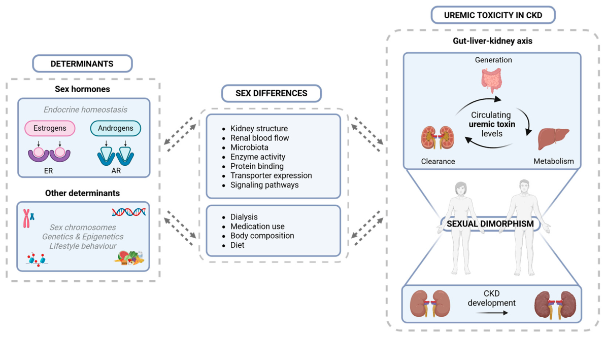
2.1. Sex Hormone Biology and Their Effects on Kidney Structure and Function
2.2. Differential Effects of Sex Hormones on Kidney Health
2.3. Sex Hormones and Their Influence on Organ Crosstalk in CKD
3. Sex Differences in UT Biology
3.1. Classification and Origin of UTs
3.2. Sex-Specific UT Profiles: What Do We Know?
3.3. The Role of Sex in UT Generation and Metabolism: Inter-Organ and Inter-Organism Crosstalk
3.3.1. The Gut and Its Microbiome
3.3.2. Liver Metabolism
3.3.3. Plasma Protein Binding
3.3.4. Kidney Clearance
3.3.5. Immune-Mediated Mechanisms
3.4. Sex Differences in Uremic Toxicity
3.4.1. Multi-Organ Toxicity and the Role of Sex
Nephrotoxicity
Cardiotoxicity
Musculoskeletal Toxicity
Neurotoxicity
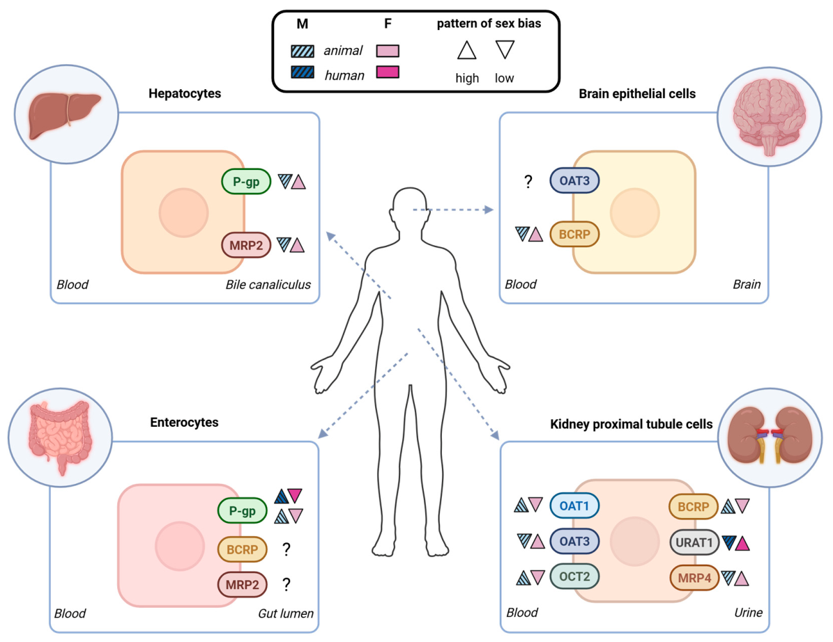
3.4.2. Sex Differences in Membrane Transporter Proteins
Kidney
Gut
Liver
Nervous System
3.4.3. UTs as Ligands and Signaling Molecules and the Role of Sex
4. Clinical Considerations of Sex-Dependent Uremic Toxicity in CKD
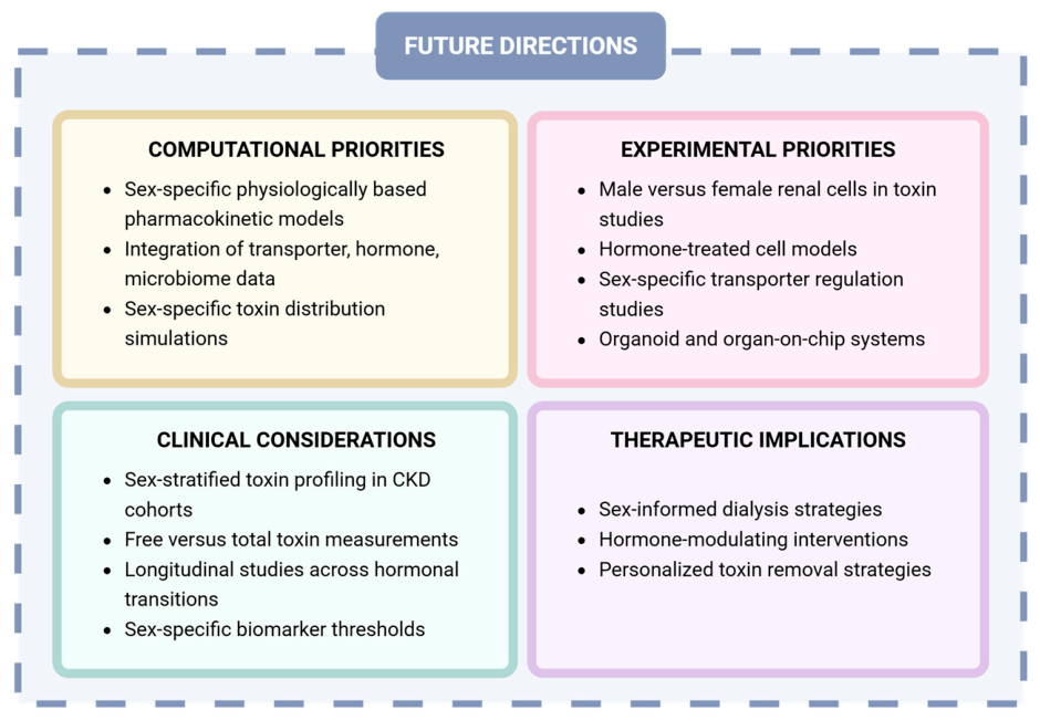
5. Research Gaps and Future Directions
5.1. Improving Our Understanding of Sex-Dependent Determinants of Uremic Toxicity in CKD
5.1.1. Computational Modeling of Sex Differences in Uremic Toxicity
5.1.2. Sex-Aware Experimental Models to Study UT Handling
5.2. Prospects for Future Research: Sex-Aware Uremic Toxicity in CKD
6. Conclusions
Author Contributions
Funding
Institutional Review Board Statement
Informed Consent Statement
Data Availability Statement
Conflicts of Interest
Abbreviations
| ABC | ATP-binding cassette |
| ADMA | Asymmetric dimethylarginine |
| AhR | Aryl hydrocarbon receptor |
| AR | Androgen receptor |
| B2M | β2-microglobulin |
| BBB | Blood–brain barrier |
| BCRP | Breast cancer resistance protein |
| CKD | Chronic kidney disease |
| CKD-MBD | Mineral bone disorder |
| CMPF | 3-carboxy-4-methyl-5-propyl-2-furanpropionate |
| CNS | Central nervous system |
| CV | Cardiovascular |
| CVD | Cardiovascular disease |
| CYP450 | Cytochrome P450 |
| eGFR | Estimated glomerular filtration rate |
| ER | Estrogen receptor |
| ESKD | End-stage kidney disease |
| EUTox | European uremic toxin work group |
| HA | Hippuric acid |
| HSA | Human serum albumin |
| IAA | Indole-3-acetic acid |
| IL-1β | Interleukin-1β |
| IL-6 | Interleukin-6 |
| IxS | Indoxyl sulfate |
| MATE | Multidrug and toxin extrusion protein |
| mRNA | Messenger ribonucleic acid |
| MRP | Multidrug resistance-associated proteins |
| MW | Molecular weight |
| NF-κB | Nuclear factor kappa B |
| NO | Nitric oxide |
| Nx | Nephrectomy |
| OAT1 | Organic anion transporter 1 |
| OATP | Organic anion-transporting polypeptides |
| OCT | Organic cation transporter |
| PBUT | Protein-bound uremic toxin |
| pCG | p-cresyl glucuronide |
| pCS | p-cresyl sulfate |
| P-gp | P-glycoprotein |
| PTC | Proximal tubular cells |
| RAAS | Renin–angiotensin–aldosterone system |
| ROS | Reactive oxygen species |
| RSST | Remote sensing and signaling theory |
| SHBG | Sex hormone-binding globulin |
| SULT | Sulfotransferase |
| TMAO | Trimethylamine N-oxide |
| TNF-α | tumor necrosis factor-α |
| UA | Uric acid |
| UDP | Uridine diphosphate |
| UGT | UDP-glucuronosyltransferase |
| URAT | Urate transporter |
| UT | Uremic toxin |
| VC | Vascular calcification |
References
- Lohia, S.; Vlahou, A.; Zoidakis, J. Microbiome in Chronic Kidney Disease (CKD): An Omics Perspective. Toxins 2022, 14, 176. [Google Scholar] [CrossRef] [Scilit]
- Bikbov, B.; Purcell, C.A.; Levey, A.S.; Smith, M.; Abdoli, A.; Abebe, M.; Adebayo, O.M.; Afarideh, M.; Agarwal, S.K.; Agudelo-Botero, M.; et al. Global, regional, and national burden of chronic kidney disease, 1990–2017: A systematic analysis for the Global Burden of Disease Study 2017. Lancet 2020, 395, 709–733. [Google Scholar] [CrossRef] [Scilit]
- Pina-Beltran, B.; Dimitrov, D.; McKay, N.; Giot, M.; Zdráhal, Z.; Potěšil, D.; Pustka, V.; Peinado-Izaguerri, J.; Saez-Rodriguez, J.; Poitevin, S.; et al. Unveiling the role of sex in the metabolism of indoxyl sulfate and apixaban. Sci. Rep. 2025, 15, 6075. [Google Scholar] [CrossRef] [Scilit]
- Andrassy, K.M. Comments on ‘KDIGO 2012 Clinical Practice Guideline for the Evaluation and Management of Chronic Kidney Disease’. Kidney Int. 2013, 84, 622–623. [Google Scholar] [CrossRef] [Scilit]
- National Kidney Foundation. K/DOQI clinical practice guidelines for chronic kidney disease: Evaluation, classification, and stratification. Am. J. Kidney Dis. 2002, 39, S1–S266. [Google Scholar]
- Webster, A.C.; Nagler, E.V.; Morton, R.L.; Masson, P. Chronic Kidney Disease. Lancet 2017, 389, 1238–1252. [Google Scholar] [CrossRef] [Scilit]
- Shlipak, M.G.; Tummalapalli, S.L.; Boulware, L.E.; Grams, M.E.; Ix, J.H.; Jha, V.; Kengne, A.P.; Madero, M.; Mihaylova, B.; Tangri, N.; et al. The case for early identification and intervention of chronic kidney disease: Conclusions from a Kidney Disease: Improving Global Outcomes (KDIGO) Controversies Conference. Kidney Int. 2021, 99, 34–47. [Google Scholar] [CrossRef] [Scilit]
- Evenepoel, P.; Meijers, B.K.I.; Bammens, B.R.M.; Verbeke, K. Uremic toxins originating from colonic microbial metabolism. Kidney Int. 2009, 76, S12–S19. [Google Scholar] [CrossRef] [Scilit]
- Vanholder, R.; Argilés, A.; Baurmeister, U.; Brunet, P.; Clark, W.; Cohen, G.; De Deyn, P.P.; Deppisch, R.; Descamps-Latscha, B.; Henle, T.; et al. Uremic toxicity: Present state of the art. Int. J. Artif. Organs 2001, 24, 695–725. [Google Scholar] [CrossRef] [Scilit]
- Vanholder, R.; Pletinck, A.; Schepers, E.; Glorieux, G. Biochemical and Clinical Impact of Organic Uremic Retention Solutes: A Comprehensive Update. Toxins 2018, 10, 33. [Google Scholar] [CrossRef] [Scilit]
- Gryp, T.; De Paepe, K.; Vanholder, R.; Kerckhof, F.M.; Van Biesen, W.; Van de Wiele, T.; Verbeke, F.; Speeckaert, M.; Joossens, M.; Couttenye, M.M.; et al. Gut microbiota generation of protein-bound uremic toxins and related metabolites is not altered at different stages of chronic kidney disease. Kidney Int. 2020, 97, 1230–1242. [Google Scholar] [CrossRef] [Scilit]
- Poesen, R.; Evenepoel, P.; de Loor, H.; Kuypers, D.; Augustijns, P.; Meijers, B. Metabolism, Protein Binding, and Renal Clearance of Microbiota-Derived p-Cresol in Patients with CKD. Clin. J. Am. Soc. Nephrol. 2016, 11, 1136–1144. [Google Scholar] [CrossRef] [Scilit] [PubMed]
- Sarnak, M.J.; Levey, A.S.; Schoolwerth, A.C.; Coresh, J.; Culleton, B.; Hamm, L.L.; McCullough, P.A.; Kasiske, B.L.; Kelepouris, E.; Klag, M.J.; et al. Kidney disease as a risk factor for development of cardiovascular disease: A statement from the American Heart Association Councils on Kidney in Cardiovascular Disease, High Blood Pressure Research, Clinical Cardiology, and Epidemiology and Prevention. Hypertension 2003, 42, 1050–1065. [Google Scholar] [CrossRef] [Scilit]
- Glorieux, G.; Burtey, S.; Evenepoel, P.; Jankowski, J.; Koppe, L.; Masereeuw, R.; Vanholder, R. A guide to uraemic toxicity. Nat. Rev. Nephrol. 2026, 22, 50–68. [Google Scholar] [CrossRef] [Scilit]
- Hung, S.C.; Kuo, K.L.; Wu, C.C.; Tarng, D.C. Indoxyl Sulfate: A Novel Cardiovascular Risk Factor in Chronic Kidney Disease. J. Am. Heart Assoc. 2017, 6, e005022. [Google Scholar] [CrossRef] [Scilit]
- Lekawanvijit, S. Cardiotoxicity of Uremic Toxins: A Driver of Cardiorenal Syndrome. Toxins 2018, 10, 352. [Google Scholar] [CrossRef] [Scilit]
- Zhao, Y.; Yang, K.; Nguyen, C.M.; Wu, H.; Liu, H.; Velez, L.M.; Kim, J.K.; Seldin, M.; Lau, W.L. Sex-specific cardiac dysfunction in mice with chronic kidney disease. Nephrol. Dial. Transplant. 2025, 40, 1865–1875. [Google Scholar] [CrossRef] [Scilit]
- Lu, Y.C.; Wu, C.C.; Tsai, I.T.; Hung, W.C.; Lee, T.L.; Hsuan, C.F.; Yu, T.H.; Wei, C.T.; Chung, F.M.; Lee, Y.J.; et al. Associations among total p-cresylsulfate, indoxyl sulfate and hippuric acid levels with hemodialysis quality indicators in maintenance hemodialysis patients. Clin. Chim. Acta 2021, 516, 83–91. [Google Scholar] [CrossRef] [Scilit] [PubMed]
- Faucher, Q.; van der Made, T.K.; De Lange, E.; Masereeuw, R. Blood-brain barrier perturbations by uremic toxins: Key contributors in chronic kidney disease-induced neurological disorders? Eur. J. Pharm. Sci. 2023, 187, 106462. [Google Scholar] [CrossRef] [Scilit]
- Schoots, A.C.; De Vries, P.M.; Thiemann, R.; Hazejager, W.A.; Visser, S.L.; Oe, P.L. Biochemical and neurophysiological parameters in hemodialyzed patients with chronic renal failure. Clin. Chim. Acta 1989, 185, 91–107. [Google Scholar] [CrossRef] [Scilit] [PubMed]
- Hamed, S.A. Neurologic conditions and disorders of uremic syndrome of chronic kidney disease: Presentations, causes, and treatment strategies. Expert Rev. Clin. Pharmacol. 2019, 12, 61–90. [Google Scholar] [CrossRef] [Scilit] [PubMed]
- Barreto, F.C.; Barreto, D.V.; Liabeuf, S.; Drüeke, T.B.; Massy, Z.A. Effects of uremic toxins on vascular and bone remodeling. Semin. Dial. 2009, 22, 433–437. [Google Scholar] [CrossRef] [Scilit]
- Wu, I.W.; Hsu, K.H.; Lee, C.C.; Sun, C.Y.; Hsu, H.J.; Tsai, C.J.; Tzen, C.Y.; Wang, Y.C.; Lin, C.Y.; Wu, M.S. p-Cresyl sulphate and indoxyl sulphate predict progression of chronic kidney disease. Nephrol. Dial. Transplant. 2011, 26, 938–947. [Google Scholar] [CrossRef] [Scilit]
- Massy, Z.A.; Chesnaye, N.C.; Larabi, I.A.; Dekker, F.W.; Evans, M.; Caskey, F.J.; Torino, C.; Porto, G.; Szymczak, M.; Drechsler, C.; et al. The relationship between uremic toxins and symptoms in older men and women with advanced chronic kidney disease. Clin. Kidney J. 2022, 15, 798–807. [Google Scholar] [CrossRef] [Scilit]
- Ito, S.; Yoshida, M. Protein-bound uremic toxins: New culprits of cardiovascular events in chronic kidney disease patients. Toxins 2014, 6, 665–678. [Google Scholar] [CrossRef] [Scilit]
- Magnani, S.; Atti, M. Uremic Toxins and Blood Purification: A Review of Current Evidence and Future Perspectives. Toxins 2021, 13, 246. [Google Scholar] [CrossRef] [Scilit]
- Vanholder, R.C.; Eloot, S.; Glorieux, G.L.R.L. Future Avenues to Decrease Uremic Toxin Concentration. Am. J. Kidney Dis. 2016, 67, 664–676. [Google Scholar] [CrossRef] [Scilit]
- Ahmed, S.; Sparidans, R.W.; Vernooij, R.W.M.; Mihaila, S.M.; Broekhuizen, R.; Goldschmeding, R.; Nguyen, T.Q.; Masereeuw, R.; Gerritsen, K.G.F. Protein-bound uremic toxin clearance as biomarker of kidney tubular function in diabetic kidney disease. Sci. Rep. 2025, 15, 23406. [Google Scholar] [CrossRef] [Scilit]
- Ahmed, S.; Chen, S.X.L.; Lucas, J.P.F.E.; Knoppert, S.N.; Harwood, R.; Faria, J.; Besseling, P.J.; Sparidans, R.W.; Broekhuizen, R.; Westphal, K.G.C.; et al. Indoxyl sulfate as a potential tubular function marker across kidney disease models. Am. J. Physiol. Ren. Physiol. 2025, 329, F160–F177. [Google Scholar] [CrossRef] [Scilit]
- Wulczyn, K.E.; Shafi, T.; Anderson, A.; Rincon-Choles, H.; Clish, C.B.; Denburg, M.; Feldman, H.I.; He, J.; Hsu, C.Y.; Kelly, T.; et al. Metabolites Associated With Uremic Symptoms in Patients With CKD: Findings From the Chronic Renal Insufficiency Cohort (CRIC) Study. Am. J. Kidney Dis. 2024, 84, 49–61.e41. [Google Scholar] [CrossRef] [Scilit]
- Short, K.M.; Smyth, I.M. A morphological investigation of sexual and lateral dimorphism in the developing metanephric kidney. Sci. Rep. 2015, 5, 15209. [Google Scholar] [CrossRef] [Scilit]
- Steiger, S.; Li, L.; Bruchfeld, A.; Stevens, K.I.; Moran, S.M.; Floege, J.; Caravaca-Fontán, F.; Mirioglu, S.; Teng, O.Y.K.; Frangou, E.; et al. Sex dimorphism in kidney health and disease: Mechanistic insights and clinical implication. Kidney Int. 2025, 107, 51–67. [Google Scholar] [CrossRef] [Scilit]
- Murphy, D.; McCulloch, C.E.; Lin, F.; Banerjee, T.; Bragg-Gresham, J.L.; Eberhardt, M.S.; Morgenstern, H.; Pavkov, M.E.; Saran, R.; Powe, N.R.; et al. Trends in Prevalence of Chronic Kidney Disease in the United States. Ann. Intern. Med. 2016, 165, 473–481. [Google Scholar] [CrossRef] [Scilit]
- Kitai, Y.; Toriu, N.; Yoshikawa, T.; Sahara, Y.; Kinjo, S.; Shimizu, Y.; Sato, Y.; Oguchi, A.; Yamada, R.; Kondo, M.; et al. Female sex hormones inversely regulate acute kidney disease susceptibility throughout life. Kidney Int. 2025, 107, 68–83. [Google Scholar] [CrossRef] [Scilit]
- Carrero, J.J.; Hecking, M.; Chesnaye, N.C.; Jager, K.J. Sex and gender disparities in the epidemiology and outcomes of chronic kidney disease. Nat. Rev. Nephrol. 2018, 14, 151–164. [Google Scholar] [CrossRef] [Scilit]
- Valdivielso, J.M.; Jacobs-Cachá, C.; Soler, M.J. Sex hormones and their influence on chronic kidney disease. Curr. Opin. Nephrol. Hypertens. 2019, 28, 1–9. [Google Scholar] [CrossRef] [Scilit]
- Balafa, O.; Fernandez-Fernandez, B.; Ortiz, A.; Dounousi, E.; Ekart, R.; Ferro, C.J.; Mark, P.B.; Valdivielso, J.M.; Del Vecchio, L.; Mallamaci, F. Sex disparities in mortality and cardiovascular outcomes in chronic kidney disease. Clin. Kidney J. 2024, 17, sfae044. [Google Scholar] [CrossRef] [Scilit]
- Ricardo, A.C.; Yang, W.; Sha, D.; Appel, L.J.; Chen, J.; Krousel-Wood, M.; Manoharan, A.; Steigerwalt, S.; Wright, J.; Rahman, M.; et al. Sex-Related Disparities in CKD Progression. J. Am. Soc. Nephrol. 2019, 30, 137–146. [Google Scholar] [CrossRef] [Scilit]
- Joseph, S.; Nicolson, T.J.; Hammons, G.; Word, B.; Green-Knox, B.; Lyn-Cook, B. Expression of drug transporters in human kidney: Impact of sex, age, and ethnicity. Biol. Sex Differ. 2015, 6, 4. [Google Scholar] [CrossRef] [Scilit]
- Zou, W.; Shi, B.; Zeng, T.; Zhang, Y.; Huang, B.; Ouyang, B.; Cai, Z.; Liu, M. Drug Transporters in the Kidney: Perspectives on Species Differences, Disease Status, and Molecular Docking. Front. Pharmacol. 2021, 12, 746208. [Google Scholar] [CrossRef] [Scilit]
- Veser, C.; Carlier, A.; Dubois, V.; Mihăilă, S.M.; Swapnasrita, S. Embracing sex-specific differences in engineered kidney models for enhanced biological understanding of kidney function. Biol. Sex Differ. 2024, 15, 99. [Google Scholar] [CrossRef] [Scilit]
- Poulsen, C.G.; Kjaergaard, K.D.; Peters, C.D.; Jespersen, B.; Jensen, J.D. Quality of life development during initial hemodialysis therapy and association with loss of residual renal function. Hemodial. Int. 2017, 21, 409–421. [Google Scholar] [CrossRef] [Scilit]
- Kotanko, P.; Thijssen, S.; Kitzler, T.; Wystrychowski, G.; Sarkar, S.R.; Zhu, F.; Gotch, F.; Levin, N.W. Size matters: Body composition and outcomes in maintenance hemodialysis patients. Blood Purif. 2007, 25, 27–30. [Google Scholar] [CrossRef] [Scilit]
- Ha, S.; Son, M.; Kim, J.; Kim, D.; Kim, M.J.; Yoo, J.; Kim, B.M.; Kim, D.; Chung, H.Y.; Chung, K.W. Gender Differences in Adenine Diet-Induced Kidney Toxicity: The Impact of 17β-Estradiol on Renal Inflammation and Fibrosis. Int. J. Mol. Sci. 2025, 26, 1358. [Google Scholar] [CrossRef] [Scilit]
- Cozzolino, M.; Magagnoli, L.; Ciceri, P. From Physicochemical Classification to Multidimensional Insights: A Comprehensive Review of Uremic Toxin Research. Toxins 2025, 17, 295. [Google Scholar] [CrossRef] [Scilit]
- Evenepoel, P.; Poesen, R.; Meijers, B. The gut-kidney axis. Pediatr. Nephrol. 2017, 32, 2005–2014. [Google Scholar] [CrossRef] [Scilit]
- Yang, T.; Richards, E.M.; Pepine, C.J.; Raizada, M.K. The gut microbiota and the brain-gut-kidney axis in hypertension and chronic kidney disease. Nat. Rev. Nephrol. 2018, 14, 442–456. [Google Scholar] [CrossRef] [Scilit]
- Bush, K.T.; Wu, W.; Lun, C.; Nigam, S.K. The drug transporter OAT3 (SLC22A8) and endogenous metabolite communication via the gut-liver-kidney axis. J. Biol. Chem. 2017, 292, 15789–15803. [Google Scholar] [CrossRef] [Scilit]
- Lowenstein, J.; Nigam, S.K. Uremic Toxins in Organ Crosstalk. Front. Med. 2021, 8, 592602. [Google Scholar] [CrossRef] [Scilit]
- Calcaterra, V.; Rossi, V.; Massini, G.; Regalbuto, C.; Hruby, C.; Panelli, S.; Bandi, C.; Zuccotti, G. Precocious puberty and microbiota: The role of the sex hormone-gut microbiome axis. Front. Endocrinol. 2022, 13, 1000919. [Google Scholar] [CrossRef] [Scilit]
- Clotet-Freixas, S.; Zaslaver, O.; Kotlyar, M.; Pastrello, C.; Quaile, A.T.; McEvoy, C.M.; Saha, A.D.; Farkona, S.; Boshart, A.; Zorcic, K.; et al. Sex differences in kidney metabolism may reflect sex-dependent outcomes in human diabetic kidney disease. Sci. Transl. Med. 2024, 16, eabm2090. [Google Scholar] [CrossRef] [Scilit]
- Li, G.; Xu, Z.; Yang, H.; Zhang, D.; Liu, B.; Song, Y.; Li, Q.; Zhang, Y.; Zhou, H.; Wang, Y. Sex-specific mechanisms in the pathogenesis and progression of chronic kidney disease. Autoimmun. Rev. 2026, 25, 103938. [Google Scholar] [CrossRef] [Scilit]
- Ljubojevic, M.; Herak-Kramberger, C.M.; Hagos, Y.; Bahn, A.; Endou, H.; Burckhardt, G.; Sabolic, I. Rat renal cortical OAT1 and OAT3 exhibit gender differences determined by both androgen stimulation and estrogen inhibition. Am. J. Physiol. Ren. Physiol. 2004, 287, F124–F138. [Google Scholar] [CrossRef] [Scilit]
- Looijer-van Langen, M.; Hotte, N.; Dieleman, L.A.; Albert, E.; Mulder, C.; Madsen, K.L. Estrogen receptor-β signaling modulates epithelial barrier function. Am. J. Physiol. Gastrointest. Liver Physiol. 2011, 300, G621–G626. [Google Scholar] [CrossRef] [Scilit]
- Menon, R.; Watson, S.E.; Thomas, L.N.; Allred, C.D.; Dabney, A.; Azcarate-Peril, M.A.; Sturino, J.M. Diet complexity and estrogen receptor β status affect the composition of the murine intestinal microbiota. Appl. Environ. Microbiol. 2013, 79, 5763–5773. [Google Scholar] [CrossRef] [Scilit]
- Lichtenecker, D.C.K.; Argeri, R.; Castro, C.H.d.M.; Dias-da-Silva, M.R.; Gomes, G.N. Cross-sex testosterone therapy modifies the renal morphology and function in female rats and might underlie increased systolic pressure. Clin. Exp. Pharmacol. Physiol. 2021, 48, 978–986. [Google Scholar] [CrossRef] [Scilit]
- Oh, E.S.; Steele, C.N.; You, Z.; Nowak, K.L.; Jovanovich, A.J. Sex hormones and the risk of cardiovascular disease and mortality in male and female patients with chronic kidney disease: A systematic review and meta-analysis. Physiol. Rep. 2022, 10, e15490. [Google Scholar] [CrossRef] [Scilit]
- Shin, J.H.; Park, Y.H.; Sim, M.; Kim, S.A.; Joung, H.; Shin, D.M. Serum level of sex steroid hormone is associated with diversity and profiles of human gut microbiome. Res. Microbiol. 2019, 170, 192–201. [Google Scholar] [CrossRef] [Scilit]
- Zeginiadou, T.; Kolias, S.; Kouretas, D.; Antonoglou, O. Nonlinear binding of sex steroids to albumin and sex hormone binding globulin. Eur. J. Drug Metab. Pharmacokinet. 1997, 22, 229–235. [Google Scholar] [CrossRef] [Scilit]
- van Eeghen, S.A.; Pyle, L.; Narongkiatikhun, P.; Choi, Y.J.; Obeid, W.; Parikh, C.R.; Vosters, T.G.; van Valkengoed, I.G.; Krebber, M.M.; Touw, D.J.; et al. Unveiling mechanisms underlying kidney function changes during sex hormone therapy. J. Clin. Investig. 2025, 135, e190850. [Google Scholar] [CrossRef] [Scilit]
- Cobo, G.; Hecking, M.; Port, F.K.; Exner, I.; Lindholm, B.; Stenvinkel, P.; Carrero, J.J. Sex and gender differences in chronic kidney disease: Progression to end-stage renal disease and haemodialysis. Clin. Sci. 2016, 130, 1147–1163. [Google Scholar] [CrossRef] [Scilit]
- Hammes, S.R.; Levin, E.R. Impact of estrogens in males and androgens in females. J. Clin. Investig. 2019, 129, 1818–1826. [Google Scholar] [CrossRef] [Scilit]
- Almeida, M.; Laurent, M.R.; Dubois, V.; Claessens, F.; O’Brien, C.A.; Bouillon, R.; Vanderschueren, D.; Manolagas, S.C. Estrogens and Androgens in Skeletal Physiology and Pathophysiology. Physiol. Rev. 2017, 97, 135–187. [Google Scholar] [CrossRef] [Scilit]
- Laurent, M.; Antonio, L.; Sinnesael, M.; Dubois, V.; Gielen, E.; Classens, F.; Vanderschueren, D. Androgens and estrogens in skeletal sexual dimorphism. Asian J. Androl. 2014, 16, 213–222. [Google Scholar] [CrossRef] [Scilit]
- Soldin, S.J.; Soldin, O.P. Steroid hormone analysis by tandem mass spectrometry. Clin. Chem. 2009, 55, 1061–1066. [Google Scholar] [CrossRef] [Scilit]
- Sabolić, I.; Asif, A.R.; Budach, W.E.; Wanke, C.; Bahn, A.; Burckhardt, G. Gender differences in kidney function. Pflug. Arch. 2007, 455, 397–429. [Google Scholar] [CrossRef] [Scilit]
- Bennett, N.C.; Rajandram, R.; Ng, K.L.; Gobe, G.C. Evaluation of steroid hormones and their receptors in development and progression of renal cell carcinoma. J. Kidney Cancer VHL 2014, 1, 17–25. [Google Scholar] [CrossRef] [Scilit]
- Conte, C.; Antonelli, G.; Melica, M.E.; Tarocchi, M.; Romagnani, P.; Peired, A.J. Role of Sex Hormones in Prevalent Kidney Diseases. Int. J. Mol. Sci. 2023, 24, 8244. [Google Scholar] [CrossRef] [Scilit]
- Sharma, P.K.; Thakur, M.K. Estrogen receptor alpha expression in mice kidney shows sex differences during aging. Biogerontology 2004, 5, 375–381. [Google Scholar] [CrossRef] [Scilit]
- Wilson, C.M.; McPhaul, M.J. A and B forms of the androgen receptor are expressed in a variety of human tissues. Mol. Cell. Endocrinol. 1996, 120, 51–57. [Google Scholar] [CrossRef] [Scilit]
- Antus, B.; Yao, Y.; Song, E.; Liu, S.; Lutz, J.; Heemann, U. Opposite effects of testosterone and estrogens on chronic allograft nephropathy. Transpl. Int. 2002, 15, 494–501. [Google Scholar] [CrossRef] [Scilit]
- Kang, D.H.; Yu, E.S.; Yoon, K.I.; Johnson, R. The impact of gender on progression of renal disease: Potential role of estrogen-mediated vascular endothelial growth factor regulation and vascular protection. Am. J. Pathol. 2004, 164, 679–688. [Google Scholar] [CrossRef] [Scilit]
- Quinkler, M.; Bumke-Vogt, C.; Meyer, B.; Bähr, V.; Oelkers, W.; Diederich, S. The human kidney is a progesterone-metabolizing and androgen-producing organ. J. Clin. Endocrinol. Metab. 2003, 88, 2803–2809. [Google Scholar] [CrossRef] [Scilit]
- Lu, H.; Lei, X.; Klaassen, C. Gender differences in renal nuclear receptors and aryl hydrocarbon receptor in 5/6 nephrectomized rats. Kidney Int. 2006, 70, 1920–1928. [Google Scholar] [CrossRef] [Scilit] [PubMed]
- Laouari, D.; Vergnaud, P.; Hirose, T.; Zaidan, M.; Rabant, M.; Nguyen, C.; Burtin, M.; Legendre, C.; Codogno, P.; Friedlander, G.; et al. The sexual dimorphism of kidney growth in mice and humans. Kidney Int. 2022, 102, 78–95. [Google Scholar] [CrossRef] [Scilit] [PubMed]
- McDonough, A.A.; Harris, A.N.; Xiong, L.I.; Layton, A.T. Sex differences in renal transporters: Assessment and functional consequences. Nat. Rev. Nephrol. 2024, 20, 21–36. [Google Scholar] [CrossRef] [Scilit]
- Conte, C.; Angelotti, M.L.; Mazzinghi, B.; Melica, M.E.; Antonelli, G.; Carangelo, G.; Landini, S.; Raglianti, V.; Ravaglia, F.; Cirillo, L.; et al. Estrogen-regulated renal progenitors determine pregnancy adaptation and preeclampsia. Science 2025, 389, 1016–1023. [Google Scholar] [CrossRef] [Scilit]
- Veiras, L.C.; Girardi, A.C.C.; Curry, J.; Pei, L.; Ralph, D.L.; Tran, A.; Castelo-Branco, R.C.; Pastor-Soler, N.; Arranz, C.T.; Yu, A.S.L.; et al. Sexual Dimorphic Pattern of Renal Transporters and Electrolyte Homeostasis. J. Am. Soc. Nephrol. 2017, 28, 3504–3517. [Google Scholar] [CrossRef] [Scilit]
- Layton, A.T.; Sullivan, J.C. Recent advances in sex differences in kidney function. Am. J. Physiol. Ren. Physiol. 2019, 316, F328–F331. [Google Scholar] [CrossRef] [Scilit]
- Li, Q.; McDonough, A.A.; Layton, H.E.; Layton, A.T. Functional implications of sexual dimorphism of transporter patterns along the rat proximal tubule: Modeling and analysis. Am. J. Physiol. Ren. Physiol. 2018, 315, F692–F700. [Google Scholar] [CrossRef] [Scilit]
- Baylis, C. Sexual dimorphism: The aging kidney, involvement of nitric oxide deficiency, and angiotensin II overactivity. J. Gerontol. A Biol. Sci. Med. Sci. 2012, 67, 1365–1372. [Google Scholar] [CrossRef] [Scilit]
- Melsom, T.; Norvik, J.V.; Enoksen, I.T.; Stefansson, V.; Mathisen, U.D.; Fuskevåg, O.M.; Jenssen, T.G.; Solbu, M.D.; Eriksen, B.O. Sex Differences in Age-Related Loss of Kidney Function. J. Am. Soc. Nephrol. 2022, 33, 1891–1902. [Google Scholar] [CrossRef] [Scilit]
- Saastad, V.; Rinde, L.B.; Enoksen, I.T.; Porrini, E.; Eriksen, B.O.; Melsom, T. Sex Differences in Kidney Function Decline in the Healthy General Population. Nephron 2025, 150, 167–172. [Google Scholar] [CrossRef] [Scilit]
- Hilliard, L.M.; Sampson, A.K.; Brown, R.D.; Denton, K.M. The “his and hers” of the renin-angiotensin system. Curr. Hypertens. Rep. 2013, 15, 71–79. [Google Scholar] [CrossRef] [Scilit]
- Ho, L.T.; Sprague, S.M. Women and CKD-mineral and bone disorder. Adv. Chronic Kidney Dis. 2013, 20, 423–426. [Google Scholar] [CrossRef] [Scilit]
- Curtis, L.M. Sex and Gender Differences in AKI. Kidney360 2024, 5, 160–167. [Google Scholar] [CrossRef] [Scilit]
- Wei, Q.; Wang, M.H.; Dong, Z. Differential gender differences in ischemic and nephrotoxic acute renal failure. Am. J. Nephrol. 2005, 25, 491–499. [Google Scholar] [CrossRef] [Scilit] [PubMed]
- Dousdampanis, P.; Trigka, K.; Fourtounas, C.; Bargman, J.M. Role of testosterone in the pathogenesis, progression, prognosis and comorbidity of men with chronic kidney disease. Ther. Apher. Dial. 2014, 18, 220–230. [Google Scholar] [CrossRef] [Scilit] [PubMed]
- Nwia, S.M.; Leite, A.P.O.; Li, X.C.; Zhuo, J.L. Sex differences in the renin-angiotensin-aldosterone system and its roles in hypertension, cardiovascular, and kidney diseases. Front. Cardiovasc. Med. 2023, 10, 1198090. [Google Scholar] [CrossRef] [Scilit]
- Mauvais-Jarvis, F.; Lindsey, S.H. Metabolic benefits afforded by estradiol and testosterone in both sexes: Clinical considerations. J. Clin. Investig. 2024, 134, e180073. [Google Scholar] [CrossRef] [Scilit] [PubMed]
- Yeo, J.K.; Koo, H.S.; Yu, J.; Park, M.G. Effects of Testosterone Treatment on Quality of Life in Patients With Chronic Kidney Disease. Am. J. Mens. Health 2020, 14, 1557988320917258. [Google Scholar] [CrossRef] [Scilit]
- Simpson, E.R. Aromatization of androgens in women: Current concepts and findings. Fertil. Steril. 2002, 77, 6–10. [Google Scholar] [CrossRef] [Scilit]
- van der Burgh, A.C.; Aribas, E.; Ikram, M.A.; Kavousi, M.; Neggers, S.J.C.M.M.; Hoorn, E.J.; Chaker, L. Sex Differences in the Association Between Serum Testosterone and Kidney Function in the General Population. Kidney Int. Rep. 2023, 8, 1342–1351. [Google Scholar] [CrossRef] [Scilit]
- van der Burgh, A.C.; Khan, S.R.; Neggers, S.; Hoorn, E.J.; Chaker, L. The role of serum testosterone and dehydroepiandrosterone sulfate in kidney function and clinical outcomes in chronic kidney disease: A systematic review and meta-analysis. Endocr. Connect. 2022, 11, e220061. [Google Scholar] [CrossRef] [Scilit]
- Edey, M.M. Male Sexual Dysfunction and Chronic Kidney Disease. Front. Med. 2017, 4, 32. [Google Scholar] [CrossRef] [Scilit]
- Hajdu, A.; Rona, G. The protective effect of estrogens against spontaneous pancratic islet and renal changes in aging male rats. Experientia 1971, 27, 956–957. [Google Scholar] [CrossRef] [Scilit] [PubMed]
- Silbiger, S.R.; Neugarten, J. The impact of gender on the progression of chronic renal disease. Am. J. Kidney Dis. 1995, 25, 515–533. [Google Scholar] [CrossRef] [Scilit] [PubMed]
- Ahmed, S.B.; Culleton, B.F.; Tonelli, M.; Klarenbach, S.W.; Macrae, J.M.; Zhang, J.; Hemmelgarn, B.R. Oral estrogen therapy in postmenopausal women is associated with loss of kidney function. Kidney Int. 2008, 74, 370–376. [Google Scholar] [CrossRef] [Scilit]
- Agarwal, M.; Selvan, V.; Freedman, B.I.; Liu, Y.; Wagenknecht, L.E. The relationship between albuminuria and hormone therapy in postmenopausal women. Am. J. Kidney Dis. 2005, 45, 1019–1025. [Google Scholar] [CrossRef] [Scilit] [PubMed]
- Kattah, A.G.; Suarez, M.L.G.; Milic, N.; Kantarci, K.; Zeydan, B.; Mosley, T.; Turner, S.T.; Ware, E.B.; Kardia, S.L.R.; Garovic, V.D. Hormone therapy and urine protein excretion: A multiracial cohort study, systematic review, and meta-analysis. Menopause 2018, 25, 625–634. [Google Scholar] [CrossRef] [Scilit]
- Krupka, E.; Curtis, S.; Ferguson, T.; Whitlock, R.; Askin, N.; Millar, A.C.; Dahl, M.; Fung, R.; Ahmed, S.B.; Tangri, N.; et al. The Effect of Gender-Affirming Hormone Therapy on Measures of Kidney Function: A Systematic Review and Meta-Analysis. Clin. J. Am. Soc. Nephrol. 2022, 17, 1305–1315. [Google Scholar] [CrossRef] [Scilit]
- Zimmerman, M.A.; Ogola, B.O.; Wilkinson, M.M.; Visniauskas, B.; De Miguel, C.; Daniel, J.M.; Lindsey, S.H. Medroxyprogesterone opposes estradiol-induced renal damage in midlife ovariectomized Long Evans rats. Menopause 2020, 27, 1411–1419. [Google Scholar] [CrossRef] [Scilit]
- Mineiro, R.; Santos, C.; Gonçalves, I.; Lemos, M.; Cavaco, J.E.B.; Quintela, T. Regulation of ABC transporters by sex steroids may explain differences in drug resistance between sexes. J. Physiol. Biochem. 2023, 79, 467–487. [Google Scholar] [CrossRef] [Scilit]
- Connelly, P.J.; Casey, H.; Montezano, A.C.; Touyz, R.M.; Delles, C. Sex steroids receptors, hypertension, and vascular ageing. J. Hum. Hypertens. 2022, 36, 120–125. [Google Scholar] [CrossRef] [Scilit]
- Wierman, M.E. Sex steroid effects at target tissues: Mechanisms of action. Adv. Physiol. Educ. 2007, 31, 26–33. [Google Scholar] [CrossRef] [Scilit]
- Neugarten, J.; Golestaneh, L. Influence of Sex on the Progression of Chronic Kidney Disease. Mayo Clin. Proc. 2019, 94, 1339–1356. [Google Scholar] [CrossRef] [Scilit]
- Piani, F.; Melena, I.; Tommerdahl, K.L.; Nokoff, N.; Nelson, R.G.; Pavkov, M.E.; van Raalte, D.H.; Cherney, D.Z.; Johnson, R.J.; Nadeau, K.J.; et al. Sex-related differences in diabetic kidney disease: A review on the mechanisms and potential therapeutic implications. J. Diabetes Complicat. 2021, 35, 107841. [Google Scholar] [CrossRef] [Scilit]
- Mitchell, T.; De Miguel, C.; Gohar, E.Y. Sex differences in redox homeostasis in renal disease. Redox Biol. 2020, 31, 101489. [Google Scholar] [CrossRef] [Scilit]
- Li, L.; Ju, H.; Jin, H.; Chen, H.; Sun, M.; Zhou, Z. Low Testosterone Level and Risk of Adverse Clinical Events among Male Patients with Chronic Kidney Disease: A Systematic Review and Meta-Analysis of Cohort Studies. J. Healthc. Eng. 2022, 2022, 3630429. [Google Scholar] [CrossRef] [Scilit]
- Grossmann, M.; Hoermann, R.; Ng Tang Fui, M.; Zajac, J.D.; Ierino, F.L.; Roberts, M.A. Sex steroids levels in chronic kidney disease and kidney transplant recipients: Associations with disease severity and prediction of mortality. Clin. Endocrinol. 2015, 82, 767–775. [Google Scholar] [CrossRef] [Scilit]
- Shiraki, N.; Nakashima, A.; Doi, S.; Carrero, J.J.; Sugiya, N.; Ueno, T.; Stenvinkel, P.; Kohno, N.; Masaki, T. Low serum testosterone is associated with atherosclerosis in postmenopausal women undergoing hemodialysis. Clin. Exp. Nephrol. 2014, 18, 499–506. [Google Scholar] [CrossRef] [Scilit]
- Dhindsa, S.; Miller, M.G.; McWhirter, C.L.; Mager, D.E.; Ghanim, H.; Chaudhuri, A.; Dandona, P. Testosterone concentrations in diabetic and nondiabetic obese men. Diabetes Care 2010, 33, 1186–1192. [Google Scholar] [CrossRef] [Scilit]
- Zitzmann, M. Testosterone deficiency and chronic kidney disease. J. Clin. Transl. Endocrinol. 2024, 37, 100365. [Google Scholar] [CrossRef] [Scilit]
- Doumouchtsis, K.K.; Perrea, D.N.; Doumouchtsis, S.K. The impact of sex hormone changes on bone mineral deficit in chronic renal failure. Endocr. Res. 2009, 34, 90–99. [Google Scholar] [CrossRef] [Scilit]
- Christakou, C.D.; Diamanti-Kandarakis, E. Role of androgen excess on metabolic aberrations and cardiovascular risk in women with polycystic ovary syndrome. Womens Health 2008, 4, 583–594. [Google Scholar] [CrossRef] [Scilit]
- Stamellou, E.; Sterzer, V.; Alam, J.; Roumeliotis, S.; Liakopoulos, V.; Dounousi, E. Sex-Specific Differences in Kidney Function and Blood Pressure Regulation. Int. J. Mol. Sci. 2024, 25, 8637. [Google Scholar] [CrossRef] [Scilit]
- Yamada, S.; Nakano, T. Role of Chronic Kidney Disease (CKD)-Mineral and Bone Disorder (MBD) in the Pathogenesis of Cardiovascular Disease in CKD. J. Atheroscler. Thromb. 2023, 30, 835–850. [Google Scholar] [CrossRef] [Scilit]
- van den Beld, A.W.; de Jong, F.H.; Grobbee, D.E.; Pols, H.A.; Lamberts, S.W. Measures of bioavailable serum testosterone and estradiol and their relationships with muscle strength, bone density, and body composition in elderly men. J. Clin. Endocrinol. Metab. 2000, 85, 3276–3282. [Google Scholar] [CrossRef] [Scilit]
- Adeli, A.; Swahn, E.; Lind, L.; Soderberg, S.; Blomberg, A.; Engström, G.; Östgren, C.J.; Jernberg, T.; Bergström, G.; Settergren, M.; et al. Sex differences in the prevalence and risk factors for aortic valve calcification in the general population. Heart 2026. Epub ahead of printing. [Google Scholar] [CrossRef] [Scilit]
- Shen, Y.; Yu, C. The Bone-Vascular Axis: A Key Player in Chronic Kidney Disease Associated Vascular Calcification. Kidney Dis. 2024, 10, 545–557. [Google Scholar] [CrossRef] [Scilit]
- Evenepoel, P.; Stenvinkel, P.; Shanahan, C.; Pacifici, R. Inflammation and gut dysbiosis as drivers of CKD–MBD. Nat. Rev. Nephrol. 2023, 19, 646–657. [Google Scholar] [CrossRef] [Scilit]
- Li, J.Y.; Chassaing, B.; Tyagi, A.M.; Vaccaro, C.; Luo, T.; Adams, J.; Darby, T.M.; Weitzmann, M.N.; Mulle, J.G.; Gewirtz, A.T.; et al. Sex steroid deficiency-associated bone loss is microbiota dependent and prevented by probiotics. J. Clin. Investig. 2016, 126, 2049–2063. [Google Scholar] [CrossRef] [Scilit]
- Zhao, Y.; Tran, T.; Fang, C.; Paganini-Hill, A.; Dulkanchainun, M.; Mai, E.; Eprem, L.; Cribbs, D.; Fisher, M.; Lau, W.L. Gut dysbiosis and brain microhemorrhages in young vs. aged mice with chronic kidney disease. Sci. Rep. 2025, 15, 32910. [Google Scholar] [CrossRef] [Scilit]
- David, K.; Narinx, N.; De Loor, J.; Antonio, L.; Vanderschueren, D.; Claessens, F.; Decallonne, B.; Evenepoel, P. #4184 Male Chronic Kidney Disease Is a State of Premature Testicular Aging. Nephrol. Dial. Transplant. 2023, 38, gfad063c_4184. [Google Scholar] [CrossRef] [Scilit]
- Rosner, M.H.; Reis, T.; Husain-Syed, F.; Vanholder, R.; Hutchison, C.; Stenvinkel, P.; Blankestijn, P.J.; Cozzolino, M.; Juillard, L.; Kashani, K.; et al. Classification of Uremic Toxins and Their Role in Kidney Failure. Clin. J. Am. Soc. Nephrol. 2021, 16, 1918–1928. [Google Scholar] [CrossRef] [Scilit]
- Glorieux, G.; Nigam, S.K.; Vanholder, R.; Verbeke, F. Role of the Microbiome in Gut-Heart-Kidney Cross Talk. Circ. Res. 2023, 132, 1064–1083. [Google Scholar] [CrossRef] [Scilit]
- Vanholder, R.; De Smet, R.; Glorieux, G.; Argilés, A.; Baurmeister, U.; Brunet, P.; Clark, W.; Cohen, G.; De Deyn, P.P.; Deppisch, R.; et al. Review on uremic toxins: Classification, concentration, and interindividual variability. Kidney Int. 2003, 63, 1934–1943. [Google Scholar] [CrossRef] [Scilit]
- Spicher, P.; Brazier, F.; Laville, S.M.; Liabeuf, S.; Kamel, S.; Culot, M.; Bodeau, S. Transporter-Mediated Interactions Between Uremic Toxins and Drugs: A Hidden Driver of Toxicity in Chronic Kidney Disease. Int. J. Mol. Sci. 2025, 26, 6328. [Google Scholar] [CrossRef] [Scilit]
- Leong, S.C.; Sirich, T.L. Indoxyl Sulfate-Review of Toxicity and Therapeutic Strategies. Toxins 2016, 8, 358. [Google Scholar] [CrossRef] [Scilit]
- Vanholder, R.; Glorieux, G.; Argiles, A.; Burtey, S.; Cohen, G.; Duranton, F.; Koppe, L.; Massy, Z.A.; Ortiz, A.; Masereeuw, R.; et al. Metabolomics to Identify Unclassified Uremic Toxins: A Comprehensive Literature Review. Kidney Med. 2025, 7, 100955. [Google Scholar] [CrossRef] [Scilit]
- Mishima, E.; Fukuda, S.; Mukawa, C.; Yuri, A.; Kanemitsu, Y.; Matsumoto, Y.; Akiyama, Y.; Fukuda, N.N.; Tsukamoto, H.; Asaji, K.; et al. Evaluation of the impact of gut microbiota on uremic solute accumulation by a CE-TOFMS-based metabolomics approach. Kidney Int. 2017, 92, 634–645. [Google Scholar] [CrossRef] [Scilit] [PubMed]
- Konturek, P.C.; Haziri, D.; Brzozowski, T.; Hess, T.; Heyman, S.; Kwiecien, S.; Konturek, S.J.; Koziel, J. Emerging role of fecal microbiota therapy in the treatment of gastrointestinal and extra-gastrointestinal diseases. J. Physiol. Pharmacol. 2015, 66, 483–491. [Google Scholar]
- Viaene, L.; Thijs, L.; Jin, Y.; Liu, Y.; Gu, Y.; Meijers, B.; Claes, K.; Staessen, J.; Evenepoel, P. Heritability and clinical determinants of serum indoxyl sulfate and p-cresyl sulfate, candidate biomarkers of the human microbiome enterotype. PLoS ONE 2014, 9, e79682. [Google Scholar] [CrossRef] [Scilit]
- Lin, C.J.; Wu, V.; Wu, P.C.; Wu, C.J. Meta-Analysis of the Associations of p-Cresyl Sulfate (PCS) and Indoxyl Sulfate (IS) with Cardiovascular Events and All-Cause Mortality in Patients with Chronic Renal Failure. PLoS ONE 2015, 10, e0132589. [Google Scholar] [CrossRef] [Scilit]
- Lin, C.J.; Chen, H.H.; Pan, C.F.; Chuang, C.K.; Wang, T.J.; Sun, F.J.; Wu, C.J. p-Cresylsulfate and indoxyl sulfate level at different stages of chronic kidney disease. J. Clin. Lab. Anal. 2011, 25, 191–197. [Google Scholar] [CrossRef] [Scilit]
- Arumugam, M.; Raes, J.; Pelletier, E.; Le Paslier, D.; Yamada, T.; Mende, D.R.; Fernandes, G.R.; Tap, J.; Bruls, T.; Batto, J.M.; et al. Enterotypes of the human gut microbiome. Nature 2011, 473, 174–180. [Google Scholar] [CrossRef] [Scilit]
- Kim, K.; Anderson, E.M.; Thome, T.; Lu, G.; Salyers, Z.R.; Cort, T.A.; O’Malley, K.A.; Scali, S.T.; Ryan, T.E. Skeletal myopathy in CKD: A comparison of adenine-induced nephropathy and 5/6 nephrectomy models in mice. Am. J. Physiol. Ren. Physiol. 2021, 321, F106–F119. [Google Scholar] [CrossRef] [Scilit]
- Hukriede, N.A.; Soranno, D.E.; Sander, V.; Perreau, T.; Starr, M.C.; Yuen, P.S.T.; Siskind, L.J.; Hutchens, M.P.; Davidson, A.J.; Burmeister, D.M.; et al. Experimental models of acute kidney injury for translational research. Nat. Rev. Nephrol. 2022, 18, 277–293. [Google Scholar] [CrossRef] [Scilit]
- Liang, J.; Liu, Y. Animal Models of Kidney Disease: Challenges and Perspectives. Kidney360 2023, 4, 1479–1493. [Google Scholar] [CrossRef] [Scilit] [PubMed]
- Packialakshmi, B.; Stewart, I.J.; Burmeister, D.M.; Chung, K.K.; Zhou, X. Large animal models for translational research in acute kidney injury. Ren. Fail. 2020, 42, 1042–1058. [Google Scholar] [CrossRef] [Scilit] [PubMed]
- Nemours, S.; Castro, L.; Ribatallada-Soriano, D.; Semidey, M.E.; Aranda, M.; Ferrer, M.; Sanchez, A.; Morote, J.; Cantero-Recasens, G.; Meseguer, A. Temporal and sex-dependent gene expression patterns in a renal ischemia–reperfusion injury and recovery pig model. Sci. Rep. 2022, 12, 6926. [Google Scholar] [CrossRef] [Scilit] [PubMed]
- Vanholder, R.; Nigam, S.K.; Burtey, S.; Glorieux, G. What If Not All Metabolites from the Uremic Toxin Generating Pathways Are Toxic? A Hypothesis. Toxins 2022, 14, 221. [Google Scholar] [CrossRef] [Scilit]
- Lu, Y.; Meng, L.; Wang, X.; Zhang, Y.; Zhang, C.; Zhang, M. The Non-Traditional Cardiovascular Culprits in Chronic Kidney Disease: Mineral Imbalance and Uremic Toxin Accumulation. Int. J. Mol. Sci. 2025, 26, 7938. [Google Scholar] [CrossRef] [Scilit]
- Rysz, J.; Franczyk, B.; Ławiński, J.; Olszewski, R.; Ciałkowska-Rysz, A.; Gluba-Brzózka, A. The Impact of CKD on Uremic Toxins and Gut Microbiota. Toxins 2021, 13, 252. [Google Scholar] [CrossRef] [Scilit]
- Vanholder, R.; Glorieux, G. Gut-Derived Metabolites and Chronic Kidney Disease: The Forest (F)or the Trees? Clin. J. Am. Soc. Nephrol. 2018, 13, 1311–1313. [Google Scholar] [CrossRef] [Scilit]
- Dou, L.; Bertrand, E.; Cerini, C.; Faure, V.; Sampol, J.; Vanholder, R.; Berland, Y.; Brunet, P. The uremic solutes p-cresol and indoxyl sulfate inhibit endothelial proliferation and wound repair. Kidney Int. 2004, 65, 442–451. [Google Scholar] [CrossRef] [Scilit]
- Liabeuf, S.; Glorieux, G.; Lenglet, A.; Diouf, M.; Schepers, E.; Desjardins, L.; Choukroun, G.; Vanholder, R.; Massy, Z.A. Does p-cresylglucuronide have the same impact on mortality as other protein-bound uremic toxins? PLoS ONE 2013, 8, e67168. [Google Scholar] [CrossRef] [Scilit]
- Vanholder, R.; Schepers, E.; Pletinck, A.; Nagler, E.V.; Glorieux, G. The uremic toxicity of indoxyl sulfate and p-cresyl sulfate: A systematic review. J. Am. Soc. Nephrol. 2014, 25, 1897–1907. [Google Scholar] [CrossRef] [Scilit] [PubMed]
- Hwang, I.K.; Yoo, K.Y.; Li, H.; Park, O.K.; Lee, C.H.; Choi, J.H.; Jeong, Y.G.; Lee, Y.L.; Kim, Y.M.; Kwon, Y.G.; et al. Indole-3-propionic acid attenuates neuronal damage and oxidative stress in the ischemic hippocampus. J. Neurosci. Res. 2009, 87, 2126–2137. [Google Scholar] [CrossRef] [Scilit] [PubMed]
- Auld, F.; Maschauer, E.L.; Morrison, I.; Skene, D.J.; Riha, R.L. Evidence for the efficacy of melatonin in the treatment of primary adult sleep disorders. Sleep Med. Rev. 2017, 34, 10–22. [Google Scholar] [CrossRef] [Scilit]
- Chyan, Y.J.; Poeggeler, B.; Omar, R.A.; Chain, D.G.; Frangione, B.; Ghiso, J.; Pappolla, M.A. Potent neuroprotective properties against the Alzheimer beta-amyloid by an endogenous melatonin-related indole structure, indole-3-propionic acid. J. Biol. Chem. 1999, 274, 21937–21942. [Google Scholar] [CrossRef] [Scilit] [PubMed]
- Glorieux, G.; Gryp, T.; Perna, A. Gut-Derived Metabolites and Their Role in Immune Dysfunction in Chronic Kidney Disease. Toxins 2020, 12, 245. [Google Scholar] [CrossRef] [Scilit]
- Shen, Y.; Fan, N.; Ma, S.X.; Cheng, X.; Yang, X.; Wang, G. Gut Microbiota Dysbiosis: Pathogenesis, Diseases, Prevention, and Therapy. MedComm 2025, 6, e70168. [Google Scholar] [CrossRef] [Scilit]
- Krukowski, H.; Valkenburg, S.; Vich Vila, A.; Maciel, L.F.; Vázquez-Castellanos, J.F.; Gryp, T.; Joossens, M.; Van Biesen, W.; Verbeke, F.; Derrien, M.; et al. Host factors dictate gut microbiome alterations in chronic kidney disease more strongly than kidney function. Nat. Microbiol. 2026, 11, 664–677. [Google Scholar] [CrossRef] [Scilit]
- Tsuji, K.; Uchida, N.; Nakanoh, H.; Fukushima, K.; Haraguchi, S.; Kitamura, S.; Wada, J. The Gut–Kidney Axis in Chronic Kidney Diseases. Diagnostics 2025, 15, 21. [Google Scholar] [CrossRef] [Scilit]
- Anders, H.J.; Andersen, K.; Stecher, B. The intestinal microbiota, a leaky gut, and abnormal immunity in kidney disease. Kidney Int. 2013, 83, 1010–1016. [Google Scholar] [CrossRef] [Scilit]
- Huang, Y.; Xin, W.; Xiong, J.; Yao, M.; Zhang, B.; Zhao, J. The Intestinal Microbiota and Metabolites in the Gut-Kidney-Heart Axis of Chronic Kidney Disease. Front. Pharmacol. 2022, 13, 837500. [Google Scholar] [CrossRef] [Scilit]
- Kim, Y.S.; Unno, T.; Kim, B.Y.; Park, M.S. Sex Differences in Gut Microbiota. World J. Mens Health 2020, 38, 48–60. [Google Scholar] [CrossRef] [Scilit]
- Koren, O.; Goodrich, J.K.; Cullender, T.C.; Spor, A.; Laitinen, K.; Bäckhed, H.K.; Gonzalez, A.; Werner, J.J.; Angenent, L.T.; Knight, R.; et al. Host remodeling of the gut microbiome and metabolic changes during pregnancy. Cell 2012, 150, 470–480. [Google Scholar] [CrossRef] [Scilit]
- Sinha, T.; Vich Vila, A.; Garmaeva, S.; Jankipersadsing, S.A.; Imhann, F.; Collij, V.; Bonder, M.J.; Jiang, X.; Gurry, T.; Alm, E.J.; et al. Analysis of 1135 gut metagenomes identifies sex-specific resistome profiles. Gut Microbes 2019, 10, 358–366. [Google Scholar] [CrossRef] [Scilit]
- Cox-York, K.A.; Sheflin, A.M.; Foster, M.T.; Gentile, C.L.; Kahl, A.; Koch, L.G.; Britton, S.L.; Weir, T.L. Ovariectomy results in differential shifts in gut microbiota in low versus high aerobic capacity rats. Physiol. Rep. 2015, 3, e12488. [Google Scholar] [CrossRef] [Scilit]
- Org, E.; Mehrabian, M.; Parks, B.W.; Shipkova, P.; Liu, X.; Drake, T.A.; Lusis, A.J. Sex differences and hormonal effects on gut microbiota composition in mice. Gut Microbes 2016, 7, 313–322. [Google Scholar] [CrossRef] [Scilit]
- Lombardo, M.; Feraco, A.; Armani, A.; Camajani, E.; Gorini, S.; Strollo, R.; Padua, E.; Caprio, M.; Bellia, A. Gender differences in body composition, dietary patterns, and physical activity: Insights from a cross-sectional study. Front. Nutr. 2024, 11, 1414217. [Google Scholar] [CrossRef] [Scilit]
- Zhang, P.; Fang, J.; Li, G.; Zhang, L.; Lai, X.; Xu, L.; Liu, L.; Xiong, Y.; Li, L.; Zhang, T.; et al. Sex Differences in Fecal Microbiota Correlation With Physiological and Biochemical Indices Associated With End-Stage Renal Disease Caused by Immunoglobulin a Nephropathy or Diabetes. Front. Microbiol. 2021, 12, 752393. [Google Scholar] [CrossRef] [Scilit]
- Lin, T.-Y.; Wu, W.-K.; Hung, S.-C. High interindividual variability of indoxyl sulfate production identified by an oral tryptophan challenge test. npj Biofilms Microbiomes 2025, 11, 15. [Google Scholar] [CrossRef] [Scilit]
- James, P.; Stanford, J.; Dixit, O.V.A.; Nicdao, M.A.; McWhinney, B.; Sud, K.; Ryan, M.; Read, S.; Ahlenstiel, G.; Lambert, K.; et al. Associations Between Uraemic Toxins and Gut Microbiota in Adults Initiating Peritoneal Dialysis. Toxins 2025, 17, 334. [Google Scholar] [CrossRef] [Scilit]
- Matz-Soja, M.; Berg, T.; Kietzmann, T. Sex-related variations in liver homeostasis and disease: From zonation dynamics to clinical implications. J. Hepatol. 2026, 84, 181–193. [Google Scholar] [CrossRef] [Scilit]
- Waxman, D.J.; Pampori, N.A.; Ram, P.A.; Agrawal, A.K.; Shapiro, B.H. Interpulse interval in circulating growth hormone patterns regulates sexually dimorphic expression of hepatic cytochrome P450. Proc. Natl. Acad. Sci. USA 1991, 88, 6868–6872. [Google Scholar] [CrossRef] [Scilit]
- Yan, Z.; Zhong, H.M.; Maher, N.; Torres, R.; Leo, G.C.; Caldwell, G.W.; Huebert, N. Bioactivation of 4-methylphenol (p-cresol) via cytochrome P450-mediated aromatic oxidation in human liver microsomes. Drug Metab. Dispos. 2005, 33, 1867–1876. [Google Scholar] [CrossRef] [Scilit]
- Behal, M.; McCalla, Z.; Wang, X. Sex differences in hepatic enzymes and transporters involved in pharmacokinetics. Liver Res. 2025, 9, 253–272. [Google Scholar] [CrossRef] [Scilit]
- Yang, L.; Li, Y.; Hong, H.; Chang, C.W.; Guo, L.W.; Lyn-Cook, B.; Shi, L.; Ning, B. Sex Differences in the Expression of Drug-Metabolizing and Transporter Genes in Human Liver. J. Drug Metab. Toxicol. 2012, 3, 1000119. [Google Scholar] [CrossRef]
- Rong, Y.; Kiang, T.K.L. Mechanisms of Metabolism Interaction Between p-Cresol and Mycophenolic Acid. Toxicol. Sci. 2020, 173, 267–279. [Google Scholar] [CrossRef] [Scilit] [PubMed]
- Rong, Y.; Kiang, T.K.L. Characterizations of Human UDP-Glucuronosyltransferase Enzymes in the Conjugation of p-Cresol. Toxicol. Sci. 2020, 176, 285–296. [Google Scholar] [CrossRef] [Scilit]
- Kojima, M.; Degawa, M. Sex differences in the constitutive gene expression of sulfotransferases and UDP-glucuronosyltransferases in the pig liver: Androgen-mediated regulation. Drug Metab. Pharmacokinet. 2014, 29, 192–197. [Google Scholar] [CrossRef] [Scilit]
- Buckley, D.B.; Klaassen, C.D. Tissue- and gender-specific mRNA expression of UDP-glucuronosyltransferases (UGTs) in mice. Drug Metab. Dispos. 2007, 35, 121–127. [Google Scholar] [CrossRef] [Scilit] [PubMed]
- Hou, H.; Horikawa, M.; Narita, Y.; Jono, H.; Kakizoe, Y.; Izumi, Y.; Kuwabara, T.; Mukoyama, M.; Saito, H. Suppression of Indoxyl Sulfate Accumulation Reduces Renal Fibrosis in Sulfotransferase 1a1-Deficient Mice. Int. J. Mol. Sci. 2023, 24, 1329. [Google Scholar] [CrossRef] [Scilit] [PubMed]
- Rong, Y.; Kiang, T.K.L. Characterization of human sulfotransferases catalyzing the formation of p-cresol sulfate and identification of mefenamic acid as a potent metabolism inhibitor and potential therapeutic agent for detoxification. Toxicol. Appl. Pharmacol. 2021, 425, 115553. [Google Scholar] [CrossRef] [Scilit]
- Klaassen, C.D.; Liu, L.; Dunn, R.T., II. Regulation of sulfotransferase mRNA expression in male and female rats of various ages. Chem.-Biol. Interact. 1998, 109, 299–313. [Google Scholar] [CrossRef] [Scilit]
- D’Agostino, G.D.; Chaudhari, S.N.; Devlin, A.S. Host-microbiome orchestration of the sulfated metabolome. Nat. Chem. Biol. 2024, 20, 410–421. [Google Scholar] [CrossRef] [Scilit]
- Perna, A.F.; Glorieux, G.; Zacchia, M.; Trepiccione, F.; Capolongo, G.; Vigorito, C.; Anishchenko, E.; Ingrosso, D. The role of the intestinal microbiota in uremic solute accumulation: A focus on sulfur compounds. J. Nephrol. 2019, 32, 733–740. [Google Scholar] [CrossRef] [Scilit]
- Duranton, F.; Cohen, G.; De Smet, R.; Rodriguez, M.; Jankowski, J.; Vanholder, R.; Argiles, A. Normal and pathologic concentrations of uremic toxins. J. Am. Soc. Nephrol. 2012, 23, 1258–1270. [Google Scholar] [CrossRef] [Scilit]
- Cunha, R.S.D.; Azevedo, C.A.B.; Falconi, C.A.; Ruiz, F.F.; Liabeuf, S.; Carneiro-Ramos, M.S.; Stinghen, A.E.M. The Interplay between Uremic Toxins and Albumin, Membrane Transporters and Drug Interaction. Toxins 2022, 14, 177. [Google Scholar] [CrossRef] [Scilit] [PubMed]
- Klammt, S.; Wojak, H.-J.; Mitzner, A.; Koball, S.; Rychly, J.; Reisinger, E.C.; Mitzner, S. Albumin-binding capacity (ABiC) is reduced in patients with chronic kidney disease along with an accumulation of protein-bound uraemic toxins. Nephrol. Dial. Transplant. 2011, 27, 2377–2383. [Google Scholar] [CrossRef] [Scilit]
- Lim, Y.J.; Sidor, N.A.; Tonial, N.C.; Che, A.; Urquhart, B.L. Uremic Toxins in the Progression of Chronic Kidney Disease and Cardiovascular Disease: Mechanisms and Therapeutic Targets. Toxins 2021, 13, 142. [Google Scholar] [CrossRef] [Scilit]
- Weaving, G.; Batstone, G.F.; Jones, R.G. Age and sex variation in serum albumin concentration: An observational study. Ann. Clin. Biochem. 2016, 53, 106–111. [Google Scholar] [CrossRef] [Scilit] [PubMed]
- Adams, S.V.; Rivara, M.; Streja, E.; Cheung, A.K.; Arah, O.A.; Kalantar-Zadeh, K.; Mehrotra, R. Sex Differences in Hospitalizations with Maintenance Hemodialysis. J. Am. Soc. Nephrol. 2017, 28, 2721–2728. [Google Scholar] [CrossRef] [Scilit]
- Narinx, N.; David, K.; Walravens, J.; Vermeersch, P.; Claessens, F.; Fiers, T.; Lapauw, B.; Antonio, L.; Vanderschueren, D. Role of sex hormone-binding globulin in the free hormone hypothesis and the relevance of free testosterone in androgen physiology. Cell. Mol. Life Sci. 2022, 79, 543. [Google Scholar] [CrossRef] [Scilit]
- Oettl, K.; Stauber, R.E. Physiological and pathological changes in the redox state of human serum albumin critically influence its binding properties. Br. J. Pharmacol. 2007, 151, 580–590. [Google Scholar] [CrossRef] [Scilit]
- Neugarten, J.; Kasiske, B.; Silbiger, S.R.; Nyengaard, J.R. Effects of sex on renal structure. Nephron 2002, 90, 139–144. [Google Scholar] [CrossRef] [Scilit] [PubMed]
- Toth-Manikowski, S.M.; Yang, W.; Appel, L.; Chen, J.; Deo, R.; Frydrych, A.; Krousel-Wood, M.; Rahman, M.; Rosas, S.E.; Sha, D.; et al. Sex Differences in Cardiovascular Outcomes in CKD: Findings From the CRIC Study. Am. J. Kidney Dis. 2021, 78, 200–209.e1. [Google Scholar] [CrossRef] [Scilit]
- Kattah, A.G.; Garovic, V.D. Understanding sex differences in progression and prognosis of chronic kidney disease. Ann. Transl. Med. 2020, 8, 897. [Google Scholar] [CrossRef] [Scilit]
- Mutsaers, H.A.; Wilmer, M.J.; Reijnders, D.; Jansen, J.; van den Broek, P.H.; Forkink, M.; Schepers, E.; Glorieux, G.; Vanholder, R.; van den Heuvel, L.P.; et al. Uremic toxins inhibit renal metabolic capacity through interference with glucuronidation and mitochondrial respiration. Biochim. Biophys. Acta 2013, 1832, 142–150. [Google Scholar] [CrossRef] [Scilit] [PubMed]
- Mutsaers, H.A.; van den Heuvel, L.P.; Ringens, L.H.; Dankers, A.C.; Russel, F.G.; Wetzels, J.F.; Hoenderop, J.G.; Masereeuw, R. Uremic toxins inhibit transport by breast cancer resistance protein and multidrug resistance protein 4 at clinically relevant concentrations. PLoS ONE 2011, 6, e18438. [Google Scholar] [CrossRef] [Scilit]
- Buckley, D.B.; Klaassen, C.D. Mechanism of gender-divergent UDP-glucuronosyltransferase mRNA expression in mouse liver and kidney. Drug Metab. Dispos. 2009, 37, 834–840. [Google Scholar] [CrossRef] [Scilit]
- Liang, J.; Qiu, Y.; Fu, T.; Li, J.; Yang, J.; Tong, Y. The Gut-Kidney Axis in Uric Acid Nephropathy: Microbiota, Metabolic Crosstalk, and Translational Prospects. J. Multidiscip. Healthc. 2025, 18, 8111–8132. [Google Scholar] [CrossRef] [Scilit]
- Grant, C.J.; Harrison, L.E.; Hoad, C.L.; Marciani, L.; Gowland, P.A.; McIntyre, C.W. Patients with chronic kidney disease have abnormal upper gastro-intestinal tract digestive function: A study of uremic enteropathy. J. Gastroenterol. Hepatol. 2017, 32, 372–377. [Google Scholar] [CrossRef] [Scilit]
- Liabeuf, S.; Pepin, M.; Franssen, C.F.M.; Viggiano, D.; Carriazo, S.; Gansevoort, R.T.; Gesualdo, L.; Hafez, G.; Malyszko, J.; Mayer, C.; et al. Chronic kidney disease and neurological disorders: Are uraemic toxins the missing piece of the puzzle? Nephrol. Dial. Transplant. 2021, 37, ii33–ii44. [Google Scholar] [CrossRef] [Scilit]
- Adamczak, M.; Surma, S. Metabolic Acidosis in Patients with CKD: Epidemiology, Pathogenesis, and Treatment. Kidney Dis. 2021, 7, 452–467. [Google Scholar] [CrossRef] [Scilit] [PubMed]
- Owada, S.; Goto, S.; Bannai, K.; Hayashi, H.; Nishijima, F.; Niwa, T. Indoxyl sulfate reduces superoxide scavenging activity in the kidneys of normal and uremic rats. Am. J. Nephrol. 2008, 28, 446–454. [Google Scholar] [CrossRef] [Scilit]
- Motojima, M.; Hosokawa, A.; Yamato, H.; Muraki, T.; Yoshioka, T. Uremic toxins of organic anions up-regulate PAI-1 expression by induction of NF-kappaB and free radical in proximal tubular cells. Kidney Int. 2003, 63, 1671–1680. [Google Scholar] [CrossRef] [Scilit]
- Shimizu, H.; Bolati, D.; Adijiang, A.; Muteliefu, G.; Enomoto, A.; Nishijima, F.; Dateki, M.; Niwa, T. NF-κB plays an important role in indoxyl sulfate-induced cellular senescence, fibrotic gene expression, and inhibition of proliferation in proximal tubular cells. Am. J. Physiol. Cell Physiol. 2011, 301, C1201–C1212. [Google Scholar] [CrossRef] [Scilit] [PubMed]
- Shimizu, H.; Bolati, D.; Higashiyama, Y.; Nishijima, F.; Shimizu, K.; Niwa, T. Indoxyl sulfate upregulates renal expression of MCP-1 via production of ROS and activation of NF-κB, p53, ERK, and JNK in proximal tubular cells. Life Sci. 2012, 90, 525–530. [Google Scholar] [CrossRef] [Scilit]
- Gryp, T.; Vanholder, R.; Vaneechoutte, M.; Glorieux, G. p-Cresyl Sulfate. Toxins 2017, 9, 52. [Google Scholar] [CrossRef] [Scilit]
- Han, H.; Zhu, J.; Zhu, Z.; Ni, J.; Du, R.; Dai, Y.; Chen, Y.; Wu, Z.; Lu, L.; Zhang, R. p-Cresyl sulfate aggravates cardiac dysfunction associated with chronic kidney disease by enhancing apoptosis of cardiomyocytes. J. Am. Heart Assoc. 2015, 4, e001852. [Google Scholar] [CrossRef] [Scilit] [PubMed]
- Mutsaers, H.A.; Caetano-Pinto, P.; Seegers, A.E.; Dankers, A.C.; van den Broek, P.H.; Wetzels, J.F.; van den Brand, J.A.; van den Heuvel, L.P.; Hoenderop, J.G.; Wilmer, M.J.; et al. Proximal tubular efflux transporters involved in renal excretion of p-cresyl sulfate and p-cresyl glucuronide: Implications for chronic kidney disease pathophysiology. Toxicol. Vitr. 2015, 29, 1868–1877. [Google Scholar] [CrossRef] [Scilit]
- Yao, H.; Zhao, H.; Du, Y.; Zhang, Y.; Li, Y.; Zhu, H. Sex-related differences in SIRT3-mediated mitochondrial dynamics in renal ischemia/reperfusion injury. Transl. Res. 2024, 270, 1–12. [Google Scholar] [CrossRef] [Scilit] [PubMed]
- Ricardo, S.D.; Zhang, T.; McArdle, Z.; Moore, B.K.; di Muzio, A.; Denton, K.M.; Widdop, R.E. Sex-dependent differences in the progression of renal injury and fibrosis following ischemic acute kidney injury. Clin. Sci. 2026, 140, 275–290. [Google Scholar] [CrossRef] [Scilit] [PubMed]
- Hyndman, D.; Liu, S.; Miner, J.N. Urate Handling in the Human Body. Curr. Rheumatol. Rep. 2016, 18, 34. [Google Scholar] [CrossRef] [Scilit]
- Falconi, C.A.; Junho, C.; Fogaça-Ruiz, F.; Vernier, I.C.S.; da Cunha, R.S.; Stinghen, A.E.M.; Carneiro-Ramos, M.S. Uremic Toxins: An Alarming Danger Concerning the Cardiovascular System. Front. Physiol. 2021, 12, 686249. [Google Scholar] [CrossRef] [Scilit]
- Lekawanvijit, S.; Adrahtas, A.; Kelly, D.J.; Kompa, A.R.; Wang, B.H.; Krum, H. Does indoxyl sulfate, a uraemic toxin, have direct effects on cardiac fibroblasts and myocytes? Eur. Heart J. 2010, 31, 1771–1779. [Google Scholar] [CrossRef] [Scilit]
- Lekawanvijit, S.; Kompa, A.R.; Manabe, M.; Wang, B.H.; Langham, R.G.; Nishijima, F.; Kelly, D.J.; Krum, H. Chronic kidney disease-induced cardiac fibrosis is ameliorated by reducing circulating levels of a non-dialysable uremic toxin, indoxyl sulfate. PLoS ONE 2012, 7, e41281. [Google Scholar] [CrossRef] [Scilit]
- Siroen, M.P.; Teerlink, T.; Nijveldt, R.J.; Prins, H.A.; Richir, M.C.; van Leeuwen, P.A. The clinical significance of asymmetric dimethylarginine. Annu. Rev. Nutr. 2006, 26, 203–228. [Google Scholar] [CrossRef] [Scilit]
- Aron-Wisnewsky, J.; Clément, K. The gut microbiome, diet, and links to cardiometabolic and chronic disorders. Nat. Rev. Nephrol. 2016, 12, 169–181. [Google Scholar] [CrossRef] [Scilit]
- Chen, M.L.; Zhu, X.H.; Ran, L.; Lang, H.D.; Yi, L.; Mi, M.T. Trimethylamine-N-Oxide Induces Vascular Inflammation by Activating the NLRP3 Inflammasome Through the SIRT3-SOD2-mtROS Signaling Pathway. J. Am. Heart Assoc. 2017, 6, e006347. [Google Scholar] [CrossRef] [Scilit]
- Chinnappa, S.; Tu, Y.K.; Yeh, Y.C.; Glorieux, G.; Vanholder, R.; Mooney, A. Association between Protein-Bound Uremic Toxins and Asymptomatic Cardiac Dysfunction in Patients with Chronic Kidney Disease. Toxins 2018, 10, 520. [Google Scholar] [CrossRef] [Scilit]
- Filipska, I.; Winiarska, A.; Knysak, M.; Stompór, T. Contribution of Gut Microbiota-Derived Uremic Toxins to the Cardiovascular System Mineralization. Toxins 2021, 13, 274. [Google Scholar] [CrossRef] [Scilit]
- Hobson, S.; Qureshi, A.R.; Ripswedan, J.; Wennberg, L.; de Loor, H.; Ebert, T.; Söderberg, M.; Evenepoel, P.; Stenvinkel, P.; Kublickiene, K. Phenylacetylglutamine and trimethylamine N-oxide: Two uremic players, different actions. Eur. J. Clin. Investig. 2023, 53, e14074. [Google Scholar] [CrossRef] [Scilit]
- Opdebeeck, B.; Maudsley, S.; Azmi, A.; De Maré, A.; De Leger, W.; Meijers, B.; Verhulst, A.; Evenepoel, P.; D’Haese, P.C.; Neven, E. Indoxyl Sulfate and p-Cresyl Sulfate Promote Vascular Calcification and Associate with Glucose Intolerance. J. Am. Soc. Nephrol. 2019, 30, 751–766. [Google Scholar] [CrossRef] [Scilit]
- Xue, B.; Johnson, A.K.; Hay, M. Sex differences in angiotensin II- and aldosterone-induced hypertension: The central protective effects of estrogen. Am. J. Physiol. Regul. Integr. Comp. Physiol. 2013, 305, R459–R463. [Google Scholar] [CrossRef] [Scilit]
- Reddy, Y.S.; Kiranmayi, V.S.; Bitla, A.R.; Krishna, G.S.; Rao, P.V.; Sivakumar, V. Nitric oxide status in patients with chronic kidney disease. Indian J. Nephrol. 2015, 25, 287–291. [Google Scholar] [CrossRef] [Scilit] [PubMed]
- Reckelhoff, J.F.; Hennington, B.S.; Moore, A.G.; Blanchard, E.J.; Cameron, J. Gender differences in the renal nitric oxide (NO) system: Dissociation between expression of endothelial NO synthase and renal hemodynamic response to NO synthase inhibition. Am. J. Hypertens. 1998, 11, 97–104. [Google Scholar] [CrossRef] [Scilit] [PubMed]
- Kapil, V.; Rathod, K.S.; Khambata, R.S.; Bahra, M.; Velmurugan, S.; Purba, A.; Watson, D.S.; Barnes, M.R.; Wade, W.G.; Ahluwalia, A. Sex differences in the nitrate-nitrite-NO• pathway: Role of oral nitrate-reducing bacteria. Free Radic. Biol. Med. 2018, 126, 113–121. [Google Scholar] [CrossRef] [Scilit]
- Woodward, H.J.; Zhu, D.; Hadoke, P.W.F.; MacRae, V.E. Regulatory Role of Sex Hormones in Cardiovascular Calcification. Int. J. Mol. Sci. 2021, 22, 4620. [Google Scholar] [CrossRef] [Scilit]
- Hung, K.-C.; Yao, W.-C.; Liu, Y.-L.; Yang, H.-J.; Liao, M.-T.; Chong, K.; Peng, C.-H.; Lu, K.-C. The Potential Influence of Uremic Toxins on the Homeostasis of Bones and Muscles in Chronic Kidney Disease. Biomedicines 2023, 11, 2076. [Google Scholar] [CrossRef] [Scilit] [PubMed]
- Anaya, J.M.; Bollag, W.B.; Hamrick, M.W.; Isales, C.M. The Role of Tryptophan Metabolites in Musculoskeletal Stem Cell Aging. Int. J. Mol. Sci. 2020, 21, 6670. [Google Scholar] [CrossRef] [Scilit] [PubMed]
- Yamada, S.; Giachelli, C.M. Vascular calcification in CKD-MBD: Roles for phosphate, FGF23, and Klotho. Bone 2017, 100, 87–93. [Google Scholar] [CrossRef] [Scilit]
- Cheung, W.W.; Zheng, R.; Hao, S.; Wang, Z.; Gonzalez, A.; Zhou, P.; Hoffman, H.M.; Mak, R.H. The role of IL-1 in adipose browning and muscle wasting in CKD-associated cachexia. Sci. Rep. 2021, 11, 15141. [Google Scholar] [CrossRef] [Scilit]
- Wang, X.H.; Mitch, W.E.; Price, S.R. Pathophysiological mechanisms leading to muscle loss in chronic kidney disease. Nat. Rev. Nephrol. 2022, 18, 138–152. [Google Scholar] [CrossRef] [Scilit]
- Enoki, Y.; Watanabe, H.; Arake, R.; Sugimoto, R.; Imafuku, T.; Tominaga, Y.; Ishima, Y.; Kotani, S.; Nakajima, M.; Tanaka, M.; et al. Indoxyl sulfate potentiates skeletal muscle atrophy by inducing the oxidative stress-mediated expression of myostatin and atrogin-1. Sci. Rep. 2016, 6, 32084. [Google Scholar] [CrossRef] [Scilit]
- Sato, E.; Mori, T.; Mishima, E.; Suzuki, A.; Sugawara, S.; Kurasawa, N.; Saigusa, D.; Miura, D.; Morikawa-Ichinose, T.; Saito, R.; et al. Metabolic alterations by indoxyl sulfate in skeletal muscle induce uremic sarcopenia in chronic kidney disease. Sci. Rep. 2016, 6, 36618. [Google Scholar] [CrossRef] [Scilit]
- Haizlip, K.M.; Harrison, B.C.; Leinwand, L.A. Sex-based differences in skeletal muscle kinetics and fiber-type composition. Physiology 2015, 30, 30–39. [Google Scholar] [CrossRef] [Scilit]
- Bredella, M.A. Sex Differences in Body Composition. Adv. Exp. Med. Biol. 2017, 1043, 9–27. [Google Scholar] [CrossRef] [Scilit]
- Tayama, Y.; Ogawa, T.; Konishi, Y.; Takahashi, K.; Fujii, K.; Shintani, A.; Hasegawa, H. Sex differences in the association between muscle mass or strength and nutrition status in chronic hemodialysis patients. Ren. Replace. Ther. 2025, 11, 39. [Google Scholar] [CrossRef] [Scilit]
- Stenvinkel, P.; Barany, P.; Chung, S.H.; Lindholm, B.; Heimbürger, O. A comparative analysis of nutritional parameters as predictors of outcome in male and female ESRD patients. Nephrol. Dial. Transplant. 2002, 17, 1266–1274. [Google Scholar] [CrossRef] [Scilit]
- Kumchev, E.P.; Tzvetkova, S.B.; Enchev, E.D.; Yaneva, M.P.; Dimitrova, R.H.; Botushanova, A.D.; Dimitrakov, D.J. Influence of age, sex and body weight on renal osteodystrophy in predialysis patients with chronic renal failure. Folia Med. 2000, 42, 28–33. [Google Scholar]
- Cheung, A.K.; Rocco, M.V.; Yan, G.; Leypoldt, J.K.; Levin, N.W.; Greene, T.; Agodoa, L.; Bailey, J.; Beck, G.J.; Clark, W.; et al. Serum beta-2 microglobulin levels predict mortality in dialysis patients: Results of the HEMO study. J. Am. Soc. Nephrol. 2006, 17, 546–555. [Google Scholar] [CrossRef] [Scilit]
- Locatelli, F.; Gauly, A.; Czekalski, S.; Hannedouche, T.; Jacobson, S.H.; Loureiro, A.; Martin-Malo, A.; Papadimitriou, M.; Passlick-Deetjen, J.; Ronco, C.; et al. The MPO Study: Just a European HEMO Study or something very different? Blood Purif. 2008, 26, 100–104. [Google Scholar] [CrossRef] [Scilit] [PubMed]
- Tonkovic, U.; Bogicevic, M.; Manzar, A.; Andrejic, N.; Sic, A.; Atanaskovic, M.; Gajić, S.; Bontić, A.; Ksiazek, S.H.; Mijušković, A.; et al. Neurological Manifestations of Hemolytic Uremic Syndrome: A Comprehensive Review. Brain Sci. 2025, 15, 717. [Google Scholar] [CrossRef] [Scilit] [PubMed]
- Koek, W.N.H.; Campos-Obando, N.; van der Eerden, B.C.J.; de Rijke, Y.B.; Ikram, M.A.; Uitterlinden, A.G.; van Leeuwen, J.; Zillikens, M.C. Age-dependent sex differences in calcium and phosphate homeostasis. Endocr. Connect. 2021, 10, 273–282. [Google Scholar] [CrossRef] [Scilit] [PubMed]
- Franco, Á.O.; Starosta, R.T.; Roriz-Cruz, M. The specific impact of uremic toxins upon cognitive domains: A review. J. Bras. Nefrol. 2019, 41, 103–111. [Google Scholar] [CrossRef] [Scilit]
- Makhammajanov, Z.; Gaipov, A.; Myngbay, A.; Bukasov, R.; Aljofan, M.; Kanbay, M. Tubular toxicity of proteinuria and the progression of chronic kidney disease. Nephrol. Dial. Transplant. 2023, 39, 589–599. [Google Scholar] [CrossRef] [Scilit]
- Masereeuw, R.; Verhaar, M.C. Innovations in approaches to remove uraemic toxins. Nat. Rev. Nephrol. 2020, 16, 552–553. [Google Scholar] [CrossRef] [Scilit]
- Breljak, D.; Brzica, H.; Sweet, D.H.; Anzai, N.; Sabolic, I. Sex-dependent expression of Oat3 (Slc22a8) and Oat1 (Slc22a6) proteins in murine kidneys. Am. J. Physiol. Ren. Physiol. 2013, 304, F1114–F1126. [Google Scholar] [CrossRef] [Scilit]
- Lu, H.; Klaassen, C. Gender differences in mRNA expression of ATP-binding cassette efflux and bile acid transporters in kidney, liver, and intestine of 5/6 nephrectomized rats. Drug Metab. Dispos. 2008, 36, 16–23. [Google Scholar] [CrossRef] [Scilit] [PubMed]
- Jansen, J.; Fedecostante, M.; Wilmer, M.J.; Peters, J.G.; Kreuser, U.M.; van den Broek, P.H.; Mensink, R.A.; Boltje, T.J.; Stamatialis, D.; Wetzels, J.F.; et al. Bioengineered kidney tubules efficiently excrete uremic toxins. Sci. Rep. 2016, 6, 26715. [Google Scholar] [CrossRef] [Scilit] [PubMed]
- Wu, W.; Bush, K.T.; Nigam, S.K. Key Role for the Organic Anion Transporters, OAT1 and OAT3, in the in vivo Handling of Uremic Toxins and Solutes. Sci. Rep. 2017, 7, 4939. [Google Scholar] [CrossRef] [Scilit] [PubMed]
- Yanai, H.; Katsuyama, H.; Hakoshima, M.; Adachi, H. Urate Transporter 1 Can Be a Therapeutic Target Molecule for Chronic Kidney Disease and Diabetic Kidney Disease: A Retrospective Longitudinal Study. Biomedicines 2023, 11, 567. [Google Scholar] [CrossRef] [Scilit]
- Masereeuw, R.; Mutsaers, H.A.M.; Toyohara, T.; Abe, T.; Jhawar, S.; Sweet, D.H.; Lowenstein, J. The Kidney and Uremic Toxin Removal: Glomerulus or Tubule? Semin. Nephrol. 2014, 34, 191–208. [Google Scholar] [CrossRef] [Scilit]
- Dalla, C.; Pavlidi, P.; Sakelliadou, D.G.; Grammatikopoulou, T.; Kokras, N. Sex Differences in Blood-Brain Barrier Transport of Psychotropic Drugs. Front. Behav. Neurosci. 2022, 16, 844916. [Google Scholar] [CrossRef] [Scilit]
- Urakami, Y.; Nakamura, N.; Takahashi, K.; Okuda, M.; Saito, H.; Hashimoto, Y.; Inui, K. Gender differences in expression of organic cation transporter OCT2 in rat kidney. FEBS Lett. 1999, 461, 339–342. [Google Scholar] [CrossRef] [Scilit]
- Slitt, A.; Cherrington, N.; Hartley, D.; Leazer, T.; Klaassen, C. Tissue Distribution and Renal Developmental Changes in Rat Organic Cation Transporter mRNA levels. Drug Metab. Dispos. 2002, 30, 212–219. [Google Scholar] [CrossRef] [Scilit]
- Alnouti, Y.; Petrick, J.S.; Klaassen, C.D. Tissue Distribution and Ontogeny of Organic Cation Transporters in Mice. Drug Metab. Dispos. 2006, 34, 477–482. [Google Scholar] [CrossRef] [Scilit]
- Maher, J.M.; Cheng, X.; Tanaka, Y.; Scheffer, G.L.; Klaassen, C.D. Hormonal regulation of renal multidrug resistance-associated proteins 3 and 4 (Mrp3 and Mrp4) in mice. Biochem. Pharmacol. 2006, 71, 1470–1478. [Google Scholar] [CrossRef] [Scilit] [PubMed]
- Tanaka, Y.; Slitt, A.L.; Leazer, T.M.; Maher, J.M.; Klaassen, C.D. Tissue distribution and hormonal regulation of the breast cancer resistance protein (Bcrp/Abcg2) in rats and mice. Biochem. Biophys. Res. Commun. 2005, 326, 181–187. [Google Scholar] [CrossRef] [Scilit] [PubMed]
- Imai, Y.; Ishikawa, E.; Asada, S.; Sugimoto, Y. Estrogen-mediated post transcriptional down-regulation of breast cancer resistance protein/ABCG2. Cancer Res. 2005, 65, 596–604. [Google Scholar] [CrossRef] [Scilit] [PubMed]
- Takiue, Y.; Hosoyamada, M.; Kimura, M.; Saito, H. The effect of female hormones upon urate transport systems in the mouse kidney. Nucleosides Nucleot. Nucl. Acids 2011, 30, 113–119. [Google Scholar] [CrossRef] [Scilit]
- Rezzani, R.; Franco, C.; Hardeland, R.; Rodella, L.F. Thymus-Pineal Gland Axis: Revisiting Its Role in Human Life and Ageing. Int. J. Mol. Sci. 2020, 21, 8806. [Google Scholar] [CrossRef] [Scilit]
- Xiong, L.; Liu, J.; Han, S.Y.; Koppitch, K.; Guo, J.J.; Rommelfanger, M.; Miao, Z.; Gao, F.; Hallgrimsdottir, I.B.; Pachter, L.; et al. Direct androgen receptor control of sexually dimorphic gene expression in the mammalian kidney. Dev. Cell 2023, 58, 2338–2358.e5. [Google Scholar] [CrossRef] [Scilit]
- Lauriola, M.; Zadora, W.; Farré, R.; Meijers, B. Intestinal transport of organic food compounds and drugs: A scoping review on the alterations observed in chronic kidney disease. Clin. Nutr. ESPEN 2024, 64, 461–482. [Google Scholar] [CrossRef] [Scilit]
- Naud, J.; Michaud, J.; Boisvert, C.; Desbiens, K.; Leblond, F.A.; Mitchell, A.; Jones, C.; Bonnardeaux, A.; Pichette, V. Down-regulation of intestinal drug transporters in chronic renal failure in rats. J. Pharmacol. Exp. Ther. 2007, 320, 978–985. [Google Scholar] [CrossRef] [Scilit]
- Veau, C.; Leroy, C.; Banide, H.; Auchère, D.; Tardivel, S.; Farinotti, R.; Lacour, B. Effect of chronic renal failure on the expression and function of rat intestinal P-glycoprotein in drug excretion. Nephrol. Dial. Transplant. 2001, 16, 1607–1614. [Google Scholar] [CrossRef] [Scilit]
- Tsujimoto, M.; Hatozaki, D.; Shima, D.; Yokota, H.; Furukubo, T.; Izumi, S.; Yamakawa, T.; Minegaki, T.; Nishiguchi, K. Influence of serum in hemodialysis patients on the expression of intestinal and hepatic transporters for the excretion of pravastatin. Ther. Apher. Dial. 2012, 16, 580–587. [Google Scholar] [CrossRef] [Scilit] [PubMed]
- Tatosian, D.A.; Yee, K.L.; Zhang, Z.; Mostoller, K.; Paul, E.; Sutradhar, S.; Larson, P.; Chhibber, A.; Wen, J.; Wang, Y.J.; et al. A Microdose Cocktail to Evaluate Drug Interactions in Patients with Renal Impairment. Clin. Pharmacol. Ther. 2021, 109, 403–415. [Google Scholar] [CrossRef] [Scilit]
- Yano, H.; Tamura, Y.; Kobayashi, K.; Tanemoto, M.; Uchida, S. Uric acid transporter ABCG2 is increased in the intestine of the 5/6 nephrectomy rat model of chronic kidney disease. Clin. Exp. Nephrol. 2014, 18, 50–55. [Google Scholar] [CrossRef] [Scilit] [PubMed]
- Mai, Y.; Dou, L.; Yao, Z.; Madla, C.M.; Gavins, F.K.H.; Taherali, F.; Yin, H.; Orlu, M.; Murdan, S.; Basit, A.W. Quantification of P-Glycoprotein in the Gastrointestinal Tract of Humans and Rodents: Methodology, Gut Region, Sex, and Species Matter. Mol. Pharm. 2021, 18, 1895–1904. [Google Scholar] [CrossRef] [Scilit]
- Madla, C.M.; Qin, Y.; Gavins, F.K.H.; Liu, J.; Dou, L.; Orlu, M.; Murdan, S.; Mai, Y.; Basit, A.W. Sex Differences in Intestinal P-Glycoprotein Expression in Wistar versus Sprague Dawley Rats. Pharmaceutics 2022, 14, 1030. [Google Scholar] [CrossRef] [Scilit] [PubMed]
- Gutmann, H.; Hruz, P.; Zimmermann, C.; Beglinger, C.; Drewe, J. Distribution of breast cancer resistance protein (BCRP/ABCG2) mRNA expression along the human GI tract. Biochem. Pharmacol. 2005, 70, 695–699. [Google Scholar] [CrossRef] [Scilit]
- Mai, Y.; Gavins, F.K.H.; Dou, L.; Liu, J.; Taherali, F.; Alkahtani, M.E.; Murdan, S.; Basit, A.W.; Orlu, M. A Non-Nutritive Feeding Intervention Alters the Expression of Efflux Transporters in the Gastrointestinal Tract. Pharmaceutics 2021, 13, 1789. [Google Scholar] [CrossRef] [Scilit]
- Naud, J.; Michaud, J.; Beauchemin, S.; Hébert, M.J.; Roger, M.; Lefrancois, S.; Leblond, F.A.; Pichette, V. Effects of chronic renal failure on kidney drug transporters and cytochrome P450 in rats. Drug Metab. Dispos. 2011, 39, 1363–1369. [Google Scholar] [CrossRef] [Scilit]
- Santana Machado, T.; Poitevin, S.; Paul, P.; McKay, N.; Jourde-Chiche, N.; Legris, T.; Mouly-Bandini, A.; Dignat-George, F.; Brunet, P.; Masereeuw, R.; et al. Indoxyl Sulfate Upregulates Liver P-Glycoprotein Expression and Activity through Aryl Hydrocarbon Receptor Signaling. J. Am. Soc. Nephrol. 2018, 29, 906–918. [Google Scholar] [CrossRef] [Scilit]
- Rost, D.; Kopplow, K.; Gehrke, S.; Mueller, S.; Friess, H.; Ittrich, C.; Mayer, D.; Stiehl, A. Gender-specific expression of liver organic anion transporters in rat. Eur. J. Clin. Investig. 2005, 35, 635–643. [Google Scholar] [CrossRef] [Scilit]
- Suzuki, T.; Zhao, Y.L.; Nadai, M.; Naruhashi, K.; Shimizu, A.; Takagi, K.; Takagi, K.; Hasegawa, T. Gender-related differences in expression and function of hepatic P-glycoprotein and multidrug resistance-associated protein (Mrp2) in rats. Life Sci. 2006, 79, 455–461. [Google Scholar] [CrossRef] [Scilit]
- Swann, J.R.; Spitzer, S.O.; Diaz Heijtz, R. Developmental Signatures of Microbiota-Derived Metabolites in the Mouse Brain. Metabolites 2020, 10, 172. [Google Scholar] [CrossRef] [Scilit]
- Mahringer, A.; Fricker, G. ABC transporters at the blood-brain barrier. Expert Opin. Drug Metab. Toxicol. 2016, 12, 499–508. [Google Scholar] [CrossRef] [Scilit]
- Ohtsuki, S.; Tomi, M.; Hata, T.; Nagai, Y.; Hori, S.; Mori, S.; Hosoya, K.; Terasaki, T. Dominant expression of androgen receptors and their functional regulation of organic anion transporter 3 in rat brain capillary endothelial cells; comparison of gene expression between the blood-brain and -retinal barriers. J. Cell. Physiol. 2005, 204, 896–900. [Google Scholar] [CrossRef] [Scilit] [PubMed]
- Curran, C.S.; Kopp, J.B. Aryl Hydrocarbon Receptor Mechanisms Affecting Chronic Kidney Disease. Front. Pharmacol. 2022, 13, 782199. [Google Scholar] [CrossRef] [Scilit]
- Madella, A.M.; Van Bergenhenegouwen, J.; Garssen, J.; Masereeuw, R.; Overbeek, S.A. Microbial-Derived Tryptophan Catabolites, Kidney Disease and Gut Inflammation. Toxins 2022, 14, 645. [Google Scholar] [CrossRef] [Scilit]
- Mo, Y.; Lu, Z.; Wang, L.; Ji, C.; Zou, C.; Liu, X. The Aryl Hydrocarbon Receptor in Chronic Kidney Disease: Friend or Foe? Front. Cell Dev. Biol. 2020, 8, 589752. [Google Scholar] [CrossRef] [Scilit]
- Shinde, R.; McGaha, T.L. The Aryl Hydrocarbon Receptor: Connecting Immunity to the Microenvironment. Trends Immunol. 2018, 39, 1005–1020. [Google Scholar] [CrossRef] [Scilit]
- Dou, L.; Sallée, M.; Cerini, C.; Poitevin, S.; Gondouin, B.; Jourde-Chiche, N.; Fallague, K.; Brunet, P.; Calaf, R.; Dussol, B.; et al. The cardiovascular effect of the uremic solute indole-3 acetic acid. J. Am. Soc. Nephrol. 2015, 26, 876–887. [Google Scholar] [CrossRef] [Scilit] [PubMed]
- Gondouin, B.; Cerini, C.; Dou, L.; Sallée, M.; Duval-Sabatier, A.; Pletinck, A.; Calaf, R.; Lacroix, R.; Jourde-Chiche, N.; Poitevin, S.; et al. Indolic uremic solutes increase tissue factor production in endothelial cells by the aryl hydrocarbon receptor pathway. Kidney Int. 2013, 84, 733–744. [Google Scholar] [CrossRef] [Scilit] [PubMed]
- Dou, L.; Poitevin, S.; Sallée, M.; Addi, T.; Gondouin, B.; McKay, N.; Denison, M.S.; Jourde-Chiche, N.; Duval-Sabatier, A.; Cerini, C.; et al. Aryl hydrocarbon receptor is activated in patients and mice with chronic kidney disease. Kidney Int. 2018, 93, 986–999. [Google Scholar] [CrossRef] [Scilit]
- Kolachalama, V.B.; Shashar, M.; Alousi, F.; Shivanna, S.; Rijal, K.; Belghasem, M.E.; Walker, J.; Matsuura, S.; Chang, G.H.; Gibson, C.M.; et al. Uremic Solute-Aryl Hydrocarbon Receptor-Tissue Factor Axis Associates with Thrombosis after Vascular Injury in Humans. J. Am. Soc. Nephrol. 2018, 29, 1063–1072. [Google Scholar] [CrossRef] [Scilit] [PubMed]
- On Behalf Of The Stop-As Investigators; Candellier, A.; Issa, N.; Grissi, M.; Brouette, T.; Avondo, C.; Gomila, C.; Blot, G.; Gubler, B.; Touati, G.; et al. Indoxyl-sulfate activation of the AhR- NF-κB pathway promotes interleukin-6 secretion and the subsequent osteogenic differentiation of human valvular interstitial cells from the aortic valve. J. Mol. Cell. Cardiol. 2023, 179, 18–29. [Google Scholar] [CrossRef] [Scilit]
- Bobot, M.; Thomas, L.; Moyon, A.; Fernandez, S.; McKay, N.; Balasse, L.; Garrigue, P.; Brige, P.; Chopinet, S.; Poitevin, S.; et al. Uremic Toxic Blood-Brain Barrier Disruption Mediated by AhR Activation Leads to Cognitive Impairment during Experimental Renal Dysfunction. J. Am. Soc. Nephrol. 2020, 31, 1509–1521. [Google Scholar] [CrossRef] [Scilit]
- Bobot, M.; Guedj, E.; Resseguier, N.; Faraut, J.; Garrigue, P.; Nail, V.; Hache, G.; Gonzalez, S.; McKay, N.; Vial, R.; et al. Increased Blood-Brain Barrier Permeability and Cognitive Impairment in Patients With ESKD. Kidney Int. Rep. 2024, 9, 2988–2995. [Google Scholar] [CrossRef] [Scilit]
- Haque, N.; Tischkau, S.A. Sexual Dimorphism in Adipose-Hypothalamic Crosstalk and the Contribution of Aryl Hydrocarbon Receptor to Regulate Energy Homeostasis. Int. J. Mol. Sci. 2022, 23, 7679. [Google Scholar] [CrossRef] [Scilit]
- Liabeuf, S.; Cheddani, L.; Massy, Z.A. Uremic Toxins and Clinical Outcomes: The Impact of Kidney Transplantation. Toxins 2018, 10, 229. [Google Scholar] [CrossRef] [Scilit] [PubMed]
- Dai, L.; Massy, Z.A.; Stenvinkel, P.; Chesnaye, N.C.; Larabi, I.A.; Alvarez, J.C.; Caskey, F.J.; Torino, C.; Porto, G.; Szymczak, M.; et al. The association between TMAO, CMPF, and clinical outcomes in advanced chronic kidney disease: Results from the European QUALity (EQUAL) Study. Am. J. Clin. Nutr. 2022, 116, 1842–1851. [Google Scholar] [CrossRef] [Scilit]
- Barreto, F.C.; Barreto, D.V.; Liabeuf, S.; Meert, N.; Glorieux, G.; Temmar, M.; Choukroun, G.; Vanholder, R.; Massy, Z.A. Serum indoxyl sulfate is associated with vascular disease and mortality in chronic kidney disease patients. Clin. J. Am. Soc. Nephrol. 2009, 4, 1551–1558. [Google Scholar] [CrossRef] [Scilit] [PubMed]
- Liabeuf, S.; Barreto, D.V.; Barreto, F.C.; Meert, N.; Glorieux, G.; Schepers, E.; Temmar, M.; Choukroun, G.; Vanholder, R.; Massy, Z.A. Free p-cresylsulphate is a predictor of mortality in patients at different stages of chronic kidney disease. Nephrol. Dial. Transplant. 2010, 25, 1183–1191. [Google Scholar] [CrossRef] [Scilit]
- Meijers, B.K.; Claes, K.; Bammens, B.; de Loor, H.; Viaene, L.; Verbeke, K.; Kuypers, D.; Vanrenterghem, Y.; Evenepoel, P. p-Cresol and cardiovascular risk in mild-to-moderate kidney disease. Clin. J. Am. Soc. Nephrol. 2010, 5, 1182–1189. [Google Scholar] [CrossRef] [Scilit]
- Reyes, M.; Benet, L.Z. Effects of uremic toxins on transport and metabolism of different biopharmaceutics drug disposition classification system xenobiotics. J. Pharm. Sci. 2011, 100, 3831–3842. [Google Scholar] [CrossRef] [Scilit]
- Arakawa, H.; Kato, Y. Emerging Roles of Uremic Toxins and Inflammatory Cytokines in the Alteration of Hepatic Drug Disposition in Patients with Kidney Dysfunction. Drug Metab. Dispos. 2023, 51, 1127–1135. [Google Scholar] [CrossRef] [Scilit]
- André, C.; Choukroun, G.; Bennis, Y.; Kamel, S.; Lemaire-Hurtel, A.S.; Masmoudi, K.; Bodeau, S.; Liabeuf, S. Potential interactions between uraemic toxins and drugs: An application in kidney transplant recipients treated with calcineurin inhibitors. Nephrol. Dial. Transplant. 2022, 37, 2284–2292. [Google Scholar] [CrossRef] [Scilit]
- Xiong, J.; Wang, M.; Wang, J.; Yang, K.; Shi, Y.; Zhang, J.; Zhang, B.; Zhang, L.; Zhao, J. Geriatric nutrition risk index is associated with renal progression, cardiovascular events and all-cause mortality in chronic kidney disease. J. Nephrol. 2020, 33, 783–793. [Google Scholar] [CrossRef] [Scilit] [PubMed]
- Thompson, L.E.; Joy, M.S. Endogenous markers of kidney function and renal drug clearance processes of filtration, secretion, and reabsorption. Curr. Opin. Toxicol. 2022, 31, 100344. [Google Scholar] [CrossRef] [Scilit] [PubMed]
- Risso, M.A.; Sallustio, S.; Sueiro, V.; Bertoni, V.; Gonzalez-Torres, H.; Musso, C.G. The Importance of Tubular Function in Chronic Kidney Disease. Int. J. Nephrol. Renov. Dis. 2019, 12, 257–262. [Google Scholar] [CrossRef] [Scilit] [PubMed]
- Rhee, E.P.; Guallar, E.; Hwang, S.; Kim, N.; Tonelli, M.; Moe, S.M.; Himmelfarb, J.; Thadhani, R.I.; Powe, N.R.; Shafi, T. Prevalence and Persistence of Uremic Symptoms in Incident Dialysis Patients. Kidney360 2020, 1, 86–92. [Google Scholar] [CrossRef] [Scilit]
- Hu, J.-R.; Myint, L.; Levey, A.S.; Coresh, J.; Inker, L.A.; Grams, M.E.; Guallar, E.; Hansen, K.D.; Rhee, E.P.; Shafi, T. A metabolomics approach identified toxins associated with uremic symptoms in advanced chronic kidney disease. Kidney Int. 2022, 101, 369–378. [Google Scholar] [CrossRef] [Scilit]
- Lin, W.; Chen, Y.; Unadkat, J.D.; Zhang, X.; Wu, D.; Heimbach, T. Applications, Challenges, and Outlook for PBPK Modeling and Simulation: A Regulatory, Industrial and Academic Perspective. Pharm. Res. 2022, 39, 1701–1731. [Google Scholar] [CrossRef] [Scilit]
- Lahane, G.P.; Dhar, A.; Bhat, A. Model Systems of Chronic Kidney Disease: A Detailed Overview and Recent Advances. J. Biochem. Mol. Toxicol. 2025, 39, e70302. [Google Scholar] [CrossRef] [Scilit] [PubMed]
- Malik, P.R.V.; Yeung, C.H.T.; Ismaeil, S.; Advani, U.; Djie, S.; Edginton, A.N. A Physiological Approach to Pharmacokinetics in Chronic Kidney Disease. J. Clin. Pharmacol. 2020, 60, S52–S62. [Google Scholar] [CrossRef] [Scilit]
- Dubinsky, S.; Malik, P.; Hajducek, D.M.; Edginton, A. Determining the Effects of Chronic Kidney Disease on Organic Anion Transporter1/3 Activity Through Physiologically Based Pharmacokinetic Modeling. Clin. Pharmacokinet. 2022, 61, 997–1012. [Google Scholar] [CrossRef] [Scilit]
- Hsueh, C.H.; Hsu, V.; Zhao, P.; Zhang, L.; Giacomini, K.M.; Huang, S.M. PBPK Modeling of the Effect of Reduced Kidney Function on the Pharmacokinetics of Drugs Excreted Renally by Organic Anion Transporters. Clin. Pharmacol. Ther. 2018, 103, 485–492. [Google Scholar] [CrossRef] [Scilit]
- Scotcher, D.; Jones, C.R.; Galetin, A.; Rostami-Hodjegan, A. Delineating the Role of Various Factors in Renal Disposition of Digoxin through Application of Physiologically Based Kidney Model to Renal Impairment Populations. J. Pharmacol. Exp. Ther. 2017, 360, 484–495. [Google Scholar] [CrossRef] [Scilit]
- Tan, S.P.F.; Willemin, M.E.; Snoeys, J.; Shen, H.; Rostami-Hodjegan, A.; Scotcher, D.; Galetin, A. Development of 4-Pyridoxic Acid PBPK Model to Support Biomarker-Informed Evaluation of OAT1/3 Inhibition and Effect of Chronic Kidney Disease. Clin. Pharmacol. Ther. 2023, 114, 1243–1253. [Google Scholar] [CrossRef] [Scilit] [PubMed]
- Chang, S.Y.; Huang, W.; Chapron, A.; Quiñones, A.J.L.; Wang, J.; Isoherranen, N.; Shen, D.D.; Kelly, E.J.; Himmelfarb, J.; Yeung, C.K. Incorporating Uremic Solute-mediated Inhibition of OAT1/3 Improves PBPK Prediction of Tenofovir Renal and Systemic Disposition in Patients with Severe Kidney Disease. Pharm. Res. 2023, 40, 2597–2606. [Google Scholar] [CrossRef] [Scilit]
- Pak, Y.A.; Posada, M.M.; Bacon, J.; Long, A.; Annes, W.; Witcher, J.; Mitchell, M.; Tirona, R.G.; Hall, S.D.; Hillgren, K.M. Prediction of the Renal Organic Anion Transporter 1 (OAT1)- Mediated Drug Interactions for LY404039, the Active Metabolite of Pomaglumetad Methionil. Pharm. Res. 2023, 40, 2499–2511. [Google Scholar] [CrossRef] [Scilit]
- Asano, S.; Galetin, A.; Tomita, Y.; Giacomini, K.M.; Chu, X.; Yang, X.; Nakamura, T.; Kusuhara, H.; Sugiyama, Y. Predicting OCT2/MATEs-Mediated Drug Interactions in Healthy Volunteers and Patients with Chronic Kidney Disease: Insights from Extended Clearance Concept, Endogenous Biomarkers, and In Vitro Inhibition Studies (Perspectives from the International Transporter Consortium). Clin. Pharmacol. Ther. 2025, 118, 994–1014. [Google Scholar] [CrossRef] [Scilit]
- King, J.; Mihaila, S.M.; Ahmed, S.; Truckenmüller, R.; Giselbrecht, S.; Masereeuw, R.; Carlier, A. The Influence of OAT1 Density and Functionality on Indoxyl Sulfate Transport in the Human Proximal Tubule: An Integrated Computational and In Vitro Study. Toxins 2021, 13, 674. [Google Scholar] [CrossRef] [Scilit] [PubMed]
- Hu, R.; McDonough, A.A.; Layton, A.T. Functional implications of the sex differences in transporter abundance along the rat nephron: Modeling and analysis. Am. J. Physiol. Ren. Physiol. 2019, 317, F1462–F1474. [Google Scholar] [CrossRef] [Scilit] [PubMed]
- Hu, R.; McDonough, A.A.; Layton, A.T. Sex differences in solute transport along the nephrons: Effects of Na(+) transport inhibition. Am. J. Physiol. Ren. Physiol. 2020, 319, F487–F505. [Google Scholar] [CrossRef] [Scilit] [PubMed]
- Hu, R.; McDonough, A.A.; Layton, A.T. Sex differences in solute and water handling in the human kidney: Modeling and functional implications. iScience 2021, 24, 102667. [Google Scholar] [CrossRef] [Scilit] [PubMed]
- Burrowes, K.S.; Ruppage, M.; Lowry, A.; Zhao, D. Sex matters: The frequently overlooked importance of considering sex in computational models. Front. Physiol. 2023, 14, 1186646. [Google Scholar] [CrossRef] [Scilit]
- Bhargava, A.; Arnold, A.P.; Bangasser, D.A.; Denton, K.M.; Gupta, A.; Hilliard Krause, L.M.; Mayer, E.A.; McCarthy, M.; Miller, W.L.; Raznahan, A.; et al. Considering Sex as a Biological Variable in Basic and Clinical Studies: An Endocrine Society Scientific Statement. Endocr. Rev. 2021, 42, 219–258. [Google Scholar] [CrossRef] [Scilit]
- Castro-Aldrete, L.; Einsiedler, M.; Cuní-López, C.; Vanhaelen, Q.; Silvestri, A.; Ferretti, M.T.; de Gennaro, M.E.; Putignano, G.; Guix, M.; Marino, N.; et al. Modelling sex differences of neurological disorders in vitro. Nat. Rev. Bioeng. 2026, 4, 112–133. [Google Scholar] [CrossRef] [Scilit]
- Sex as a biological variable in biomedical engineering. Nat. Rev. Bioeng. 2026, 4, 99–100. [CrossRef] [Scilit]
- Ilatovskaya, D.V.; Ogola, B.; Faulkner, J.L.; Mamenko, M.; Taylor, E.B.; Dent, E.; Ryan, M.J.; Sullivan, J.C. Guidelines for sex-specific considerations to improve rigor in renal research and how we got there. Am. J. Physiol. Ren. Physiol. 2025, 328, F204–F217. [Google Scholar] [CrossRef] [Scilit]
- Soranno, D.E.; Awdishu, L.; Bagshaw, S.M.; Basile, D.; Bell, S.; Bihorac, A.; Bonventre, J.; Brendolan, A.; Claure-Del Granado, R.; Collister, D.; et al. The role of sex and gender in acute kidney injury-consensus statements from the 33rd Acute Disease Quality Initiative. Kidney Int. 2025, 107, 606–616. [Google Scholar] [CrossRef] [Scilit]
- Caetano-Pinto, P.; Stahl, S.H. Renal Organic Anion Transporters 1 and 3 In Vitro: Gone but Not Forgotten. Int. J. Mol. Sci. 2023, 24, 5419. [Google Scholar] [CrossRef] [Scilit]
- Pou Casellas, C.; Jansen, K.; Rookmaaker, M.B.; Clevers, H.; Verhaar, M.C.; Masereeuw, R. Regulation of solute carriers oct2 and OAT1/3 in the kidney: A phylogenetic, ontogenetic, and cell dynamic perspective. Physiol. Rev. 2022, 102, 993–1024. [Google Scholar] [CrossRef] [Scilit] [PubMed]
- Kim, Y.K.; Nam, S.A.; Yang, C.W. Applications of kidney organoids derived from human pluripotent stem cells. Korean J. Intern. Med. 2018, 33, 649–659. [Google Scholar] [CrossRef] [Scilit]
- Nakanoh, H.; Tsuji, K.; Fukushima, K.; Uchida, N.; Haraguchi, S.; Kitamura, S.; Wada, J. Kidney Organoids: Current Advances and Applications. Life 2025, 15, 1680. [Google Scholar] [CrossRef] [Scilit] [PubMed]
- Rizki-Safitri, A.; Traitteur, T.; Morizane, R. Bioengineered Kidney Models: Methods and Functional Assessments. Function 2021, 2, zqab026. [Google Scholar] [CrossRef] [Scilit]
- Ashammakhi, N.; Wesseling-Perry, K.; Hasan, A.; Elkhammas, E.; Zhang, Y.S. Kidney-on-a-chip: Untapped opportunities. Kidney Int. 2018, 94, 1073–1086. [Google Scholar] [CrossRef] [Scilit]
- Zommiti, M.; Connil, N.; Tahrioui, A.; Groboillot, A.; Barbey, C.; Konto-Ghiorghi, Y.; Lesouhaitier, O.; Chevalier, S.; Feuilloley, M.G.J. Organs-on-Chips Platforms Are Everywhere: A Zoom on Biomedical Investigation. Bioengineering 2022, 9, 646. [Google Scholar] [CrossRef] [Scilit] [PubMed]


Disclaimer/Publisher’s Note: The statements, opinions and data contained in all publications are solely those of the individual author(s) and contributor(s) and not of MDPI and/or the editor(s). MDPI and/or the editor(s) disclaim responsibility for any injury to people or property resulting from any ideas, methods, instructions or products referred to in the content. |
© 2026 by the authors. Licensee MDPI, Basel, Switzerland. This article is an open access article distributed under the terms and conditions of the Creative Commons Attribution (CC BY) license.
Share and Cite
Nobus, O.; Carlier, A.; Mihăilă, S.M.; Dubois, V. Sex-Dependent Determinants of Uremic Toxicity in Chronic Kidney Disease. Toxins 2026, 18, 242. https://doi.org/10.3390/toxins18060242
Nobus O, Carlier A, Mihăilă SM, Dubois V. Sex-Dependent Determinants of Uremic Toxicity in Chronic Kidney Disease. Toxins. 2026; 18(6):242. https://doi.org/10.3390/toxins18060242
Chicago/Turabian StyleNobus, Oriana, Aurélie Carlier, Silvia M. Mihăilă, and Vanessa Dubois. 2026. "Sex-Dependent Determinants of Uremic Toxicity in Chronic Kidney Disease" Toxins 18, no. 6: 242. https://doi.org/10.3390/toxins18060242
APA StyleNobus, O., Carlier, A., Mihăilă, S. M., & Dubois, V. (2026). Sex-Dependent Determinants of Uremic Toxicity in Chronic Kidney Disease. Toxins, 18(6), 242. https://doi.org/10.3390/toxins18060242