Article

Development of a Novel Magnetic-Bead-Based Automated Strategy for Efficient and Low-Cost Sample Preparation for Ochratoxin A Detection Using Mycotoxin–Albumin Interaction

1
Key Laboratory of Grain Information Processing and Control, Henan University of Technology, Ministry of Education, Zhengzhou 450001, China
2
Henan Key Laboratory of Grain Photoelectric Detection and Control, Henan University of Technology, Zhengzhou 450001, China
3
College of Information Science and Engineering, Henan University of Technology, Zhengzhou 450001, China
4
Academy of National Food and Strategic Reserves Administration, Beijing 102600, China
5
College of Food Science and Engineering, Henan University of Technology, Zhengzhou 450001, China
*
Authors to whom correspondence should be addressed.
These authors contributed equally to this work.
Toxins 2023, 15(4), 270; https://doi.org/10.3390/toxins15040270
Submission received: 17 February 2023 / Revised: 28 March 2023 / Accepted: 4 April 2023 / Published: 5 April 2023

Abstract

:
The mycotoxin ochratoxin A (OTA) is toxic to humans and frequently contaminates wine and beer. Antibodies are essential recognition probes for the detection of OTA. However, they have several drawbacks, such as high costs and difficulty in preparation. In this study, a novel magnetic-bead-based automated strategy for efficient and low-cost OTA sample preparation was developed. Human serum albumin, which is an economical and stable receptor based on the mycotoxin–albumin interaction, was adapted and validated to replace conventional antibodies to capture OTA in the sample. Ultra-performance liquid chromatography–fluorescence detection was used in combination with this preparation method for efficient detection. The effects of different conditions on this method were investigated. The recovery of OTA samples spiked at three different concentrations ranged from 91.2% to 102.1%, and the relative standard deviations (RSDs) were 1.2%–8.2% in wine and beer. For red wine and beer samples, the LODs were 0.37 and 0.15 µg/L, respectively. This reliable method overcomes the drawbacks of conventional methods and offers significant application prospects.
Key Contribution: A novel magnetic-bead-based automated strategy for efficient and low-cost OTA sample preparation was developed.

1. Introduction

Ochratoxin is a secondary metabolite of Aspergillus ochraceus, Aspergillus carbonarius, and Penicillium verrucosum [1]. Ochratoxin A (OTA) is the most toxic and common ochratoxin and can lead to severe nephrotoxic, carcinogenic, teratogenic, mutagenic, genotoxic, and immunotoxic effects [2,3,4]. The International Agency for Research on Cancer has classified OTA as a Group 2B potential human carcinogen [5]. OTA is predominantly found in cereals, grapes, beer, wine, dried fruit, cocoa, coffee, and other foods [6,7]. Beer and wine are widely consumed beverages and are among the largest sources of daily OTA intake in many countries [8,9]. Hence, many countries and regions have set very low maximum limits (MLs) of OTA to reduce the risk of OTA contamination [6]. For example, the MLs of OTA in wine, grape juice, and grape drinks are 2 µg/kg in the European Union (EU) [10], and the current national standard ML of OTA in wine and other alcoholic beverages in China (GB 2761-2017) is 2 µg/kg. Considering the significant adverse effects of OTA on human health, an accurate and efficient analytical method is essential to ensure that food products are safe from OTA contamination.
The currently used methods for OTA detection include high-performance liquid chromatography–fluorescence detection (HPLC-FLD) [11,12], ultra-high-performance liquid chromatography–fluorescence detection (UPLC-FLD) [13,14,15], or liquid chromatography–tandem mass spectrometry (LC-MS/MS) [16]. These are considered to be reliable and sensitive detection methods; however; actual samples cannot be analyzed without undergoing some preparation steps to eliminate interference from the complex matrix. Common preparation methods include solid-phase extraction (SPE) [17,18], immunoaffinity chromatography (IAC) [19,20,21], dispersive liquid–liquid extraction [22,23], QuEChERS [24,25], and matrix solid-phase dispersion (MSPD) [16,26]. OTA samples are most often prepared by IAC owing to its high selectivity and stability; however, the expensive and complex antibody preparation process and large differences between batches limit the application of IAC [27]. Thus, a novel sample preparation method is highly desirable. In particular, aptamers [28,29,30], molecularly imprinted polymers [31,32,33], and a novel receptor with antibody-like properties have been employed in applications ranging from separation systems to sensors.
Albumin is a water-soluble macromolecule with excellent biocompatibility. Many studies have been conducted on the interactions between xenobiotics and albumin. Poór M et al. calculated the binding constant of the OTA–albumin interaction using spectroscopic techniques and molecular modeling [34,35,36,37,38]. Human serum albumin has a higher binding constant (5.2 × 10⁶ M−1) [37], whereas that of bovine serum albumin (BSA) is slightly lower (3.2 × 10⁵ M−1) [38]. As these albumins are easy to obtain and their preparation cost is significantly lower than that of antibodies, albumin is a promising alternative to antibodies for the determination of OTA with a high affinity, low cost, and good biocompatibility.
In this study, a novel magnetic-bead-based strategy for efficient and low-cost OTA sample preparation was developed using the mycotoxin–albumin interaction. An evaluation of the binding performance of OTA with HSA, BSA, and ovalbumin (OVA) revealed HSA as the optimal recognition probe, demonstrating its ability to replace antibodies for immobilization in magnetic beads. In addition, the entire sample preparation process was automatically completed via a magnetic-bead-based sample preparation platform developed in our previous study [15,39,40,41,42]. Coupled with ultra-performance liquid chromatography–fluorescence detection (UPLC-FLD), this novel method was successfully applied for the routine detection of OTA in beverages.

2. Results and Discussion

2.1. Adsorption Ability of the Three Types of Albumin–Magnetic Beads

The adsorption ability of different albumins, as identification probes, for OTA, was investigated by preparing three types of albumin–magnetic beads coupled with different albumins (HSA, BSA, and OVA). The recovery and maximum adsorption capacity of the albumin–magnetic beads for OTA were evaluated. The results show that the recovery of all albumin–magnetic beads is higher than 70% (Figure 1a) and that HSA has the strongest recognition ability for OTA, with a recovery >95%. In addition, the maximum adsorption capacity of HSA and OVA is >80 ng, whereas the maximum adsorption capacity of BSA is approximately 60 ng. Therefore, HSA is the optimal identification probe for albumins common to OTA. The adsorption ability of albumin in the magnetic beads was consistent with the binding constant of OTA–albumin in the solution.

2.2. Optimization of the Coupling Amount of HSA–Magnetic Beads

The coupling ratio of HSA and magnetic beads was optimized to achieve a high coupling efficiency of HSA with magnetic beads (Figure 1b). A series of ratios of HSA and magnetic beads (0.5:1, 1:1, 1.5:1, 2:1, and 2.5:1) was investigated. The maximum adsorption capacity of OTA was increased by increasing the coupling ratio from 0.5:1 to 1:1, indicating that the surface of the magnetic beads was not saturated. When the coupling ratio increased from 1:1 to 2.5:1, the adsorption capacity stabilized, indicating that the HSA on the magnetic bead surface was saturated. The recovery was the highest (>90%) when HSA and magnetic beads were coupled at a ratio of 1:1. Therefore, 1:1 was deemed the most suitable coupling ratio for HSA and magnetic beads.

2.3. The Principle of This Automated System

The principle of this automated system is illustrated in Figure 2. HSA was coupled to carboxylated magnetic beads via a condensation reaction between the carboxyl and amino groups to form HSA–magnetic beads (Step 1). OTA was then captured by the HAS–magnetic beads in the beer or wine sample to form OTA –HSA–magnetic beads (Step 2), which were transferred to the cleaning solution and washed twice to remove unwanted species (Steps 3 and 4). The OTA–HSA–magnetic beads were then transferred to the eluent using a magnetic stick and magnetic stick coat, after which the OTA was released into the eluent using organic reagents, forming an eluent solution containing only OTA (Step 5). Finally, UPLC-FLD was used to quantitatively detect the eluted OTA.

2.4. Optimization of the Sample Clean-Up Method

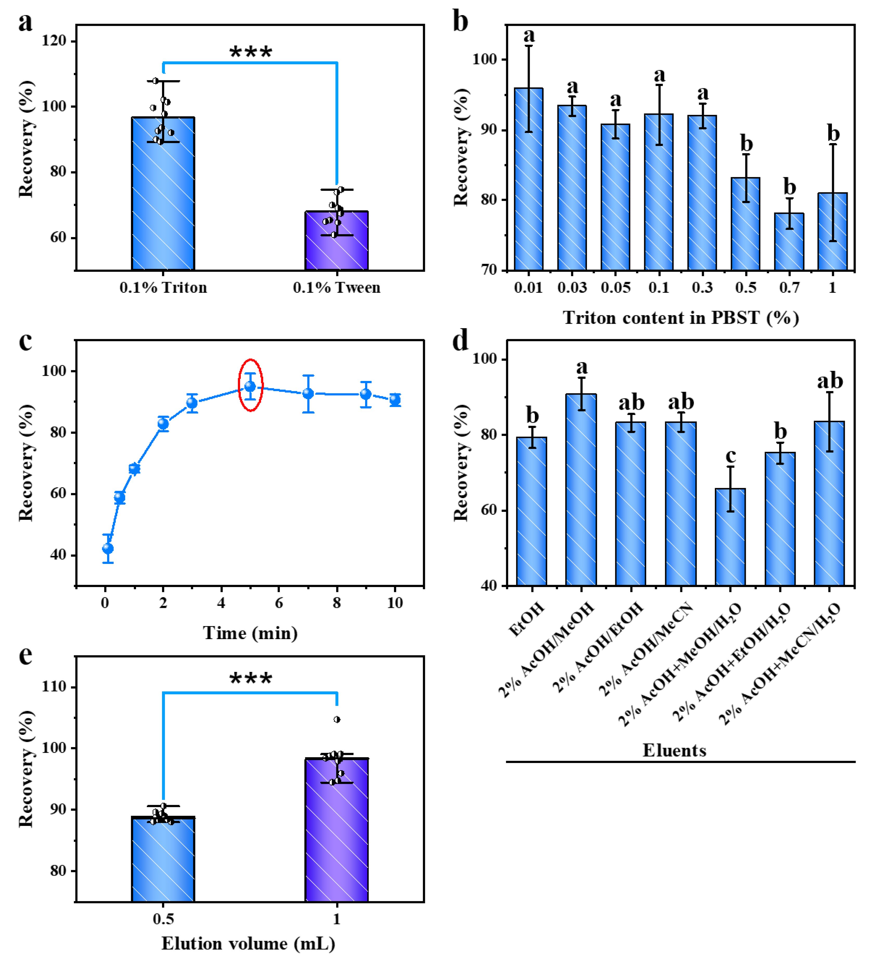
To increase the contact area between the HSA–magnetic beads and sample and, thereby, improve the efficiency of OTA capture by HSA, the surfactant and its content were optimized (Figure 3a,b). Figure 3a shows that the recovery of PBST containing 0.1% Triton is above 95%, whereas the recovery of PBST containing 0.1% Tween is significantly different, at less than 80%. Therefore, Triton can promote the capture of OTA by HSA more effectively than Tween. Figure 3b shows that when the content of Triton in PBST is within 0.3%, the OTA recovery is higher than 90%. Therefore, the content of Triton in PBST can be controlled within 0.3%.
The reaction time was optimized to complete OTA detection in the shortest time. As shown in Figure 3c, the recovery of OTA gradually increases when the reaction time in the reaction well is increased. With a reaction time of 5 min, the recovery was closest to 1, whereas the recovery decreased slightly with longer reaction times. Therefore, 5 min was determined to be the optimal reaction time.
The eluate and elution volume were optimized to achieve the complete release of OTA, and the results are shown in Figure 3d,e, respectively. Based on Figure 3d, the OTA recovery was the highest when 2% acetic acid in methanol was used as the eluent. Therefore, 2% acetic acid in methanol was deemed the optimal eluent. Figure 3e shows that the recovery of OTA is close to 100% when the elution volume is 1 mL. Therefore, 1 mL was considered the optimal elution volume.
Considering the issue of the alcohol tolerance of HSA (to avoid the mechanism interference effect), the loading quantities of beer and red wine samples were optimized prior to sample detection (Figure 4a,b). Figure 4a shows that the recovery gradually decreases with an increase in the beer loading volume. When the beer loading volume was 5 mL, the recovery was less than 75%. However, when the beer loading volume was 2.5 mL, the recovery was as high as 95%. Therefore, 2.5 mL was determined to be the maximum loading volume for beer. As shown in Figure 4b, the recovery gradually decreases with increasing red wine loading volume. When the red wine loading volume was 2.5 mL, the recovery was below 50%. However, when the beer loading volume was 1 mL, the recovery was as high as 95%. Therefore, 1 mL was determined to be the maximum loading volume for red wine. The retention time, peak shape, and peak area in the UPLC-FLD fluorescence spectra of the spiked samples of beer and red wine prepared under optimized conditions were essentially identical to those of the OTA standard solution of the same concentration (Figure 4c). Moreover, no peaks corresponding to impurities were observed in the vicinity of the target peak, which indicates the high purification efficacy of the technique.

2.5. Analytical Performance

The calibration curve was established by preparing a series of working solutions (0.125, 0.25, 0.5, 1.25, 2.5, 5, 10, and 20 ng/mL) to satisfy the requirements of an OTA analysis. The calibration curve (Figure S1) was constructed by plotting the peak areas (y) versus the injected concentrations of the standard solution (x), yielding the following regression equation: y = 4870.7x + 423.01 and a correlation coefficient (R2) of 0.9999, indicating excellent linearity.
The sensitivity of this method was estimated using the limit of detection (LOD) and limit of quantification (LOQ), which were calculated from the spiked blank samples at different concentrations of OTA. The two lowest concentrations, which produced 3-fold and 10-fold signal-to-noise (S/N) ratios, were considered to be the LOD and LOQ, respectively. The LOD and LOQ for red wine samples were 0.37 and 1.23 µg/L, respectively. For beer samples, the LOD and LOQ were 0.15 and 0.49 µg/L, respectively, which were lower than the MLs for OTA set for wine and other alcoholic beverages in China and the EU.
The specificity of HSA–magnetic beads for OTA was assessed by comparing the recovery of 13 common mycotoxins, including DON, 15-AcDON, 3-AcDON, 3G-DON, AFB1, AFB2, AFG1, AFG2, HT-2, NIV, ST, T-2, and ZEN, with that of OTA. The results are presented in Table 1. Among the common mycotoxins, all toxins except ST were not detected, and the recovery of ST was <1.5%, whereas the recovery of OTA reached 111.8%. These results indicated that HSA has good specificity for OTA.
The applicability of this method to real complex samples was investigated by preparing and analyzing real samples spiked at three different levels of OTA (low, medium, and high). The low-, medium-, and high-spiked recoveries of beer and red wine are listed in Table 2. The recoveries of beer and wine ranged from 91.2% to 102.1%, whereas the RSD was 1.2%–8.2%, indicating good accuracy and precision.

2.6. Detection of Actual Samples

Twenty-six brands of beer and wine, randomly purchased from supermarkets, were analyzed. As listed in Table 3, the amounts of OTA determined by this method were in good agreement with those obtained by the conventional IAC method (Figure S2, R2 = 0.9995), with no bias effect arising from the various types of sample matrices.

3. Conclusions

This study successfully established a novel magnetic-bead-based automated strategy for efficient and low-cost OTA sample preparation. The experimental data showed that the UPLC-FLD method has high sensitivity, accuracy, and precision. Although the binding capacity of HSA is weaker than that of conventional antibodies, further protein modification studies could improve its capacity for binding to OTA and other contaminants, leading to a low-cost receptor.

4. Materials and Methods

4.1. Experimental Materials

Human serum albumin (HSA), bovine serum albumin (BSA) and ovalbumin (OVA), Sodium azide, glycine, Tween-20, Triton X-100, and morpholinoethanesulfonic acid (MES) were purchased from Sigma-Aldrich (St. Louis, MO, USA). Carboxyl magnetic beads (10 mg/mL) were purchased from Kangyuan Techbio Biologicals (Beijing, China). Ultra-pure water used for the preparation of all the reagents was obtained from a Milli-Q purification system (Bedford, MA, USA). A standard solution of ochratoxin A (OTA), aflatoxin B1 (AFB1), aflatoxin B2 (AFB2), aflatoxin G1 (AFG1), aflatoxin G2 (AFG2), HT-2 toxin (HT-2), nivalenol (NIV), sterigmatocystin (ST), zearalenone (ZEN), T-2 toxin (T-2), Deoxynivalenol (DON), deoxynivalenol-3-glucoside (3G-DON), 3-acetyl-deoxynivalenol (3-AcDON), and 15-acetyl-deoxynivalenol (15-AcDON) were purchased from Biopure (Tulln, Austria) and were stored at −18 °C. Working solutions were prepared by dilution with HPLC-grade methanol and then stored in vials at 4 °C and renewed weekly. HPLC-grade methanol (MeOH) and Acetic acid (HAC) were purchased from Fisher Scientific (Atlanta, GA, USA).

4.2. Instrument and Analytical Conditions

Automated reaction conditions: an appropriate mixing frequency, mixing amplitude, and reaction time can ensure adequate reaction, washing, and elution of the samples. To prevent solvent spatter during mixing, the mixing amplitude was set to 80%. Other conditions, such as the reaction time, are listed in Table 4. At the end of the treatment, the eluate from the five wells was collected for UPLC-FLD testing.
UPLC-FLD analysis conditions: OTA terminal detection was performed using a Waters ACQUITY UPLC H-class system (Waters, Manchester, UK) consisting of a vacuum degasser, an auto-sampler, a binary pump, and a BEH reverse-phase C18 column (2.1 × 100 mm, 1.7 μm) for separation. The mobile phase consisted of acetic acid:water:methanol (1:51:48, v/v/v) at a flow rate of 0.3 mL/min. The excitation and emission wavelengths of the fluorescence detector were 333 and 460 nm, respectively. The column temperature was maintained at 40 °C; the sample temperature was maintained at 10 °C, and the sample injection volume was 10 μL. The Waters EMPOWER3 software 7.20.00.00 was used to acquire and analyze the data.

4.3. Coupling of Magnetic Beads to Albumin

First, 2 mg of protein was dissolved in 2 mL of MES to form a protein dilution. Magnetic beads (0.5 mL) were washed twice with absolute ethanol (0.5 mL) using a strong magnet. Next, 0.5 mL protein dilution was added, and the solution was incubated for 3 h. After magnetic separation, 1 mL of 2% glycine solution was added directly and blocked for 3 h. The solution was then washed twice with 1 mL 0.1% PBST and twice with 1 mL PBS. Finally, the volume was fixed in PBS (0.5 mL) and stored at 4 °C until further use.

4.4. Identification Ability of Magnetic Beads

The protein–magnetic beads were coupled with HSA, BSA, or OVA at ratios of 1 mg:1 mL. Subsequently, 50 µL of protein–magnetic beads were added to Well 2, and 4 ng (or 100 ng) of OTA standard was added to Well 1 and diluted to 5 mL with PBST. After processing using an automated magneto-controlled preprocessing platform, the eluent in Well 5 was collected for detection via HPLC-FLD.

4.5. Optimization of Conditions

4.5.1. Optimization of the Protein/Magnetic Bead Coupling Ratio and Loading Volume

The magnetic beads (0.1, 0.2, 0.3, 0.4, and 0.5 mL) were coupled with 0.2 mg of HSA to form HSA–magnetic beads and added to Well 2. Subsequently, 4 ng (or 200 ng) of OTA standard was added to Well 1 and diluted to 5 mL with PBST. After processing using an automated magneto-controlled preprocessing platform, the eluent in Well 5 was collected for detection via HPLC-FLD. The optimum ratio of protein-to-magnetic beads was investigated. HSA–magnetic beads (50, 100, and 200 µL; HSA:magnetic beads = 1 mg:1 mL) were then collected, and 4 ng of OTA was added to Well 1 and eluted with 1 mL of 2% acetic acid in methanol. The effect of the loading volume of the HSA–magnetic beads on recovery was then investigated.

4.5.2. Optimization of the Elution Solvent and Volume

HSA–magnetic beads (100 µL) were added to Well 2, and 8 ng of OTA was added to Well 1 and eluted with different solvents to investigate the optimal eluent of HSA–magnetic beads. HSA–magnetic beads (50 µL) were added to Well 2, and 4 ng of OTA was added to Well 1, and the eluent volume of HSA beads was investigated with different volumes of 2% acetic acid in methanol.

4.5.3. Optimization of the Sample Extract

OTA (8 ng) was added to Well 1 and then diluted to 5 mL with a PBST solution containing 0.1% Tween and 0.1% Triton. Finally, 1 mL of 2% acetic acid in methanol solution was eluted to determine the optimal surfactant. The OTA in Well 1 was diluted to 10 mL with PBST solutions containing different surfactant concentrations, and the other conditions were maintained unchanged to investigate the optimal proportion of surfactant. Similarly, 8 ng of OTA was added to Well 1 and diluted with a PBST solution containing 0.1% Triton to 5, 7, 8, 9, and 10 mL. The effect of different volumes of the reaction solution on the OTA recovery was investigated.

4.5.4. Optimization of the Capture Time

The mixing time in Well 1 was set to 0.1, 0.5, 1, 2, 3, 4, 5, 7, and 9 min, and the other conditions were maintained unchanged. The effect of different capture times on the OTA recovery was investigated.

4.5.5. Optimization of the Beer Loading Volume

OTA (10 ng) was added to Well 1, and 2.5, 3, 3.5, 4, 4.5, and 5 mL of beer were added. When the volume was <10 mL, the beads were diluted to 10 mL with PBST, while the other conditions remained unchanged. The ability of the HSA–magnetic beads to tolerate beer matrices was also investigated.

4.5.6. Optimization of the Loading Volume of Red Wine Samples

OTA (10 ng) was added to Well 1, and 0.5, 1, 2.5, 5, 7.5, and 10 mL red wine were then added. When the volume was <10 mL, the beads were diluted to 10 mL with PBST, while the other conditions remained unchanged. The ability of the HSA–magnetic beads to tolerate red wine matrices was also investigated.

4.6. Sample Treatment

The recovery of beer and red wine samples was determined using low, medium, and high spikes (final OTA concentrations of 1, 2, and 4 µg/L in the samples), and the other conditions were unchanged.

4.7. Investigation of the Detection Performance

The linearity was determined using eight concentrations of OTA (0.125, 0.25, 0.5, 1.25, 2.5, 5, 10, and 20 µg/L), and the regression equation and coefficient (R2) were calculated by plotting the peak area (y) against the concentration of OTA (x). The LOD and LOQ were estimated in triplicate using a serially diluted OTA standard solution, which produced a chromatogram peak with S/N ratios of 3 and 10, respectively. The selectivity was investigated using 13 common mycotoxins: DON, 15-AcDON, 3-AcDON, 3G-DON, AFB1, AFB2, AFG1, AFG2, HT-2, NIV, ST, T-2, and ZEN.

4.8. Manual IAC Clean-Up Procedure

The following IAC clean-up procedures were based on the appropriate ISO standard [43] with some modifications. In particular, an aliquot (4 mL) of the sample solution extracted from the obtained sample was diluted with the PBST solution (26 mL). The diluted extract was centrifuged at 8000× g for 5 min before the conduction of the IAC of OTA at a flow rate of approximately 1–2 drops/s and thereafter washed with a solution containing PBST (10 mL) and distilled water (10 mL) at a flow rate of 2 drops/s. After elution, the OTA eluted with the elution solvent (1 mL) was transferred to an LC vial.

Supplementary Materials

The following supporting information can be downloaded at: https://www.mdpi.com/article/10.3390/toxins15040270/s1, Figure S1. The calibration curve, Figure S2. Comparison on the determination of OTA in real samples between this method and IAC.

Author Contributions

Conceptualization, J.Y., M.Z. and H.B.; methodology, J.Y., J.C. and H.B.; software, M.Z.; validation, H.M., Y.Z. and S.W.; formal analysis, H.L.; investigation, J.Y.; writing—original draft preparation, J.Y. and M.Z.; writing—review and editing, J.Y. and M.Z.; supervision, Y.Z. and H.M.; project administration, J.Y. and Y.Z.; funding acquisition, J.Y. and S.W. All authors have read and agreed to the published version of the manuscript.

Funding

This research was funded by the Open Fund Project of Key Laboratory of Grain Information Processing & Control, Ministry of Education, Henan University of Technology (KFJJ-2020-102), the Talent Project of National Food and Strategic Reserves Administration (QN 2022202, LJ2022201), and Young Elite Scientists Sponsorship Program by CAST (2021QNRC001).

Institutional Review Board Statement

Not applicable.

Informed Consent Statement

Not applicable.

Data Availability Statement

Not applicable.

Conflicts of Interest

The authors declare no conflict of interest.

References

  1. Duarte, S.; Pena, A.; Lino, C. A review on ochratoxin A occurrence and effects of processing of cereal and cereal derived food products. Food Microbiol. 2010, 27, 187–198. [Google Scholar] [CrossRef]
  2. Yang, D.; Wang, Z.; Jiang, X.; Fang, W.; Song, H.; Zhang, X. Rapid and sensitive detection of ochratoxin A by using a quantum dots-based immunochromatographic assay. Mycosystema 2019, 38, 1003–1013. [Google Scholar]
  3. Ringot, D.; Chango, A.; Schneider, Y.-J.; Larondelle, Y. Toxicokinetics and toxicodynamics of ochratoxin A, an update. Chem.-Biol. Interact. 2006, 159, 18–46. [Google Scholar] [CrossRef]
  4. Heussner, A.H.; Bingle, L.E. Comparative ochratoxin toxicity: A review of the available data. Toxins 2015, 7, 4253–4282. [Google Scholar] [CrossRef] [Green Version]
  5. Wei, M.; He, X.; Xie, Y. A novel signal-on fluorescent aptasensor for ochratoxin A detection based on RecJ f exonuclease-induced signal amplification. J. Chin. Chem. Soc. Taipei 2020, 67, 1247–1253. [Google Scholar] [CrossRef]
  6. Li, X.; Ma, W.; Ma, Z.; Zhang, Q.; Li, H. The occurrence and contamination level of ochratoxin A in plant and animal-derived food commodities. Molecules 2021, 26, 6928. [Google Scholar] [CrossRef] [PubMed]
  7. Soto, J.B.; Fernandez-Franzon, M.; Ruiz, M.-J.; Juan-Garcia, A. Presence of ochratoxin A (OTA) mycotoxin in alcoholic drinks from southern European countries: Wine and beer. J. Agric. Food Chem. 2014, 62, 7643–7651. [Google Scholar] [CrossRef]
  8. Schabo, D.C.; Alvarenga, V.O.; Schaffner, D.W.; Magnani, M. A worldwide systematic review, meta-analysis, and health risk assessment study of mycotoxins in beers. Compr. Rev. Food Sci. Food Saf. 2021, 20, 5742–5764. [Google Scholar] [CrossRef] [PubMed]
  9. Leal, T.; Abrunhosa, L.; Domingues, L.; Venâncio, A.; Oliveira, C. BSA-based sample clean-up columns for ochratoxin A determination in wine: Method development and validation. Food Chem. 2019, 300, 125204. [Google Scholar] [CrossRef] [PubMed] [Green Version]
  10. The Commission of the European Communities. Commission Regulation (EC) No.1881/2006 of 19 December 2006 Setting Maximum Levels for Certain Contaminants in Foodstuffs. 2006. Available online: https://eur-lex.europa.eu/LexUriServ/LexUriServ.do?uri=OJ:L:2006:364:0005:0024:EN:PDF (accessed on 21 March 2023).
  11. Dimzoska, B.S.; Musliu, Z.H.; Uzunov, R.; Angeleska, A.; Blagoevska, K.; Nikolovska, R.C.; Ilievska, G.; Stojkovic, E.D. Application of reliable cost-effective strategy for analysis of mycotoxins in corn-based foods with HPLC-FLD after multi-toxin immunoaffinity clean-up. Maced. J. Chem. Chem. Eng. 2022, 41, 77–88. [Google Scholar]
  12. Afsah-Hejri, L.; Jinap, S.; Mirhosseini, H. Ochratoxin A quantification: Newly developed HPLC conditions. Food Control. 2012, 23, 113–119. [Google Scholar] [CrossRef]
  13. Mikulíková, R.; Běláková, S.; Benešová, K.; Svoboda, Z. Study of ochratoxin A content in South Moravian and foreign wines by the UPLC method with fluorescence detection. Food Chem. 2012, 133, 55–59. [Google Scholar] [CrossRef]
  14. Gentile, F.; La Torre, G.L.; Potortì, A.G.; Saitta, M.; Alfa, M.; Dugo, G. Organic wine safety: UPLC-FLD determination of Ochratoxin A in Southern Italy wines from organic farming and winemaking. Food Control. 2016, 59, 20–26. [Google Scholar] [CrossRef]
  15. Ye, J.; Xuan, Z.; Zhang, B.; Wu, Y.; Li, L.; Wang, S.; Xie, G.; Wang, S. Automated analysis of ochratoxin A in cereals and oil by immunoaffinity magnetic beads coupled to UPLC-FLD. Food Control. 2019, 104, 57–62. [Google Scholar] [CrossRef]
  16. Rubert, J.; Soler, C.; Mañes, J. Evaluation of matrix solid-phase dispersion (MSPD) extraction for multi-mycotoxin determination in different flours using LC–MS/MS. Talanta 2011, 85, 206–215. [Google Scholar] [CrossRef] [PubMed]
  17. Zhou, S.N.; Lai, E.P.; Miller, J.D. Analysis of wheat extracts for ochratoxin A by molecularly imprinted solid-phase extraction and pulsed elution. Anal. Bioanal. Chem. 2004, 378, 1903–1906. [Google Scholar] [CrossRef]
  18. Huertas-Pérez, J.F.; Arroyo-Manzanares, N.; García-Campaña, A.M.; Gámiz-Gracia, L. Solid phase extraction as sample treatment for the determination of Ochratoxin A in foods: A review. Crit. Rev. Food Sci. Nutr. 2017, 57, 3405–3420. [Google Scholar] [CrossRef]
  19. Belli, N.; Marin, S.; Sanchis, V.; Ramos, A. Ochratoxin A (OTA) in wines, musts and grape juices: Occurrence, regulations and methods of analysis. Food Sci. Technol. Int. 2002, 8, 325–335. [Google Scholar] [CrossRef]
  20. Garcia-Villanova, R.J.; Cordón, C.; Paramás, A.M.G.; Aparicio, P.; Rosales, M.E.G. Simultaneous immunoaffinity column cleanup and HPLC analysis of aflatoxins and ochratoxin A in Spanish bee pollen. J. Agric. Food Chem. 2004, 52, 7235–7239. [Google Scholar] [CrossRef]
  21. Trucksess, M.; Weaver, C.; Oles, C.; D’Ovidio, K.; Rader, J. Determination of aflatoxins and ochratoxin A in ginseng and other botanical roots by immunoaffinity column cleanup and liquid chromatography with fluorescence detection. J. AOAC Int. 2006, 89, 624–630. [Google Scholar] [CrossRef] [Green Version]
  22. Karami-Osboo, R.; Miri, R.; Javidnia, K.; Kobarfard, F.; AliAbadi, M.H.S.; Maham, M. A validated dispersive liquid-liquid microextraction method for extraction of ochratoxin A from raisin samples. J. Food Sci. Technol. 2015, 52, 2440–2445. [Google Scholar] [CrossRef] [PubMed] [Green Version]
  23. Arroyo-Manzanares, N.; Gámiz-Gracia, L.; García-Campaña, A.M. Determination of ochratoxin A in wines by capillary liquid chromatography with laser induced fluorescence detection using dispersive liquid–liquid microextraction. Food Chem. 2012, 135, 368–372. [Google Scholar] [CrossRef]
  24. Paíga, P.; Morais, S.; Oliva-Teles, T.; Correia, M.; Delerue-Matos, C.; Duarte, S.C.; Pena, A.; Lino, C.M. Extraction of ochratoxin A in bread samples by the QuEChERS methodology. Food Chem. 2012, 135, 2522–2528. [Google Scholar] [CrossRef] [PubMed] [Green Version]
  25. Juan, C.; Mañes, J.; Font, G.; Juan-García, A. Determination of mycotoxins in fruit berry by-products using QuEChERS extraction method. Lwt 2017, 86, 344–351. [Google Scholar] [CrossRef]
  26. Rubert, J.; Soler, C.; Mañes, J. Optimization of Matrix Solid-Phase Dispersion method for simultaneous extraction of aflatoxins and OTA in cereals and its application to commercial samples. Talanta 2010, 82, 567–574. [Google Scholar] [CrossRef]
  27. Toh, S.Y.; Citartan, M.; Gopinath, S.C.; Tang, T.-H. Aptamers as a replacement for antibodies in enzyme-linked immunosorbent assay. Biosens. Bioelectron. 2015, 64, 392–403. [Google Scholar] [CrossRef]
  28. Guo, J.-X.; Pan, L.-M.; Wang, M.-C.; Chen, L.-J.; Zhao, X. Exogenous Interference and Autofluorescence-free Ratiometric Aptasensor for Detection of OTA Based on Dual-Colored Persistent Luminescence Nanoparticles. Food Chem. 2023, 413, 135611. [Google Scholar] [CrossRef] [PubMed]
  29. Zhu, D.; Huang, T.; Zhou, Q.; Yang, Z.; Liu, B.; Li, M.; Li, C.; Chen, J.-X.; Dai, Z.; Chen, J. A label-free fluorescent aptasensor based on a novel exponential rolling circle amplification for highly sensitive Ochratoxin A detection. Food Chem. 2023, 410, 135427. [Google Scholar] [CrossRef]
  30. Zheng, M.; Liu, H.; Ye, J.; Ni, B.; Xie, Y.; Wang, S. Target-responsive aptamer-cross-linked hydrogel sensors for the visual quantitative detection of aflatoxin B1 using exonuclease I-Triggered target cyclic amplification. Food Chem. X 2022, 15, 100395. [Google Scholar] [CrossRef]
  31. Ye, J.; Chen, Y.; Liu, Z. A boronate affinity sandwich assay: An appealing alternative to immunoassays for the determination of glycoproteins. Angew. Chem. Int. Ed. 2014, 53, 10386–10389. [Google Scholar] [CrossRef]
  32. Hu, M.; Huang, P.; Suo, L.; Wu, F. Polydopamine-based molecularly imprinting polymers on magnetic nanoparticles for recognition and enrichment of ochratoxins prior to their determination by HPLC. Microchim. Acta 2018, 185, 300. [Google Scholar] [CrossRef]
  33. Pichon, V.; Combès, A. Selective tools for the solid-phase extraction of Ochratoxin A from various complex samples: Immunosorbents, oligosorbents, and molecularly imprinted polymers. Anal. Bioanal. Chem. 2016, 408, 6983–6999. [Google Scholar] [CrossRef] [Green Version]
  34. Poór, M.; Kunsági-Máté, S.; Bencsik, T.; Petrik, J.; Vladimir-Knežević, S.; Kőszegi, T. Flavonoid aglycones can compete with Ochratoxin A for human serum albumin: A new possible mode of action. Int. J. Biol. Macromol. 2012, 51, 279–283. [Google Scholar] [CrossRef] [PubMed]
  35. Poór, M.; Kunsági-Máté, S.; Czibulya, Z.; Li, Y.; Peles-Lemli, B.; Petrik, J.; Vladimir-Knežević, S.; Kőszegi, T. Fluorescence spectroscopic investigation of competitive interactions between ochratoxin A and 13 drug molecules for binding to human serum albumin. Luminescence 2013, 28, 726–733. [Google Scholar] [CrossRef]
  36. Poór, M.; Li, Y.; Matisz, G.; Kiss, L.; Kunsági-Máté, S.; Kőszegi, T. Quantitation of species differences in albumin–ligand interactions for bovine, human and rat serum albumins using fluorescence spectroscopy: A test case with some Sudlow’s site I ligands. J. Lumin. 2014, 145, 767–773. [Google Scholar] [CrossRef]
  37. Il’ichev, Y.V.; Perry, J.L.; Rüker, F.; Dockal, M.; Simon, J.D. Interaction of ochratoxin A with human serum albumin. Binding sites localized by competitive interactions with the native protein and its recombinant fragments. Chem.-Biol. Interact. 2002, 141, 275–293. [Google Scholar] [CrossRef]
  38. Chu, F.S. A comparative study of the interaction of ochratoxins with bovine serum albumin. Biochem. Pharmacol. 1974, 23, 1105–1113. [Google Scholar] [PubMed]
  39. Xuan, Z.; Liu, H.; Ye, J.; Li, L.; Tian, W.; Wang, S. Reliable and disposable quantum dot–based electrochemical immunosensor for aflatoxin B 1 simplified analysis with automated magneto-controlled pretreatment system. Anal. Bioanal. Chem. 2020, 412, 7615–7625. [Google Scholar] [CrossRef]
  40. Chen, J.; Ye, J.; Li, L.; Wu, Y.; Liu, H.; Xuan, Z.; Chen, M.; Wang, S. One-step automatic sample pretreatment for rapid, simple, sensitive, and efficient determination of aflatoxin M1 in milk by immunomagnetic beads coupled to liquid chromatography-tandem mass spectrometry. Food Control 2022, 137, 108927. [Google Scholar] [CrossRef]
  41. Sarwat, A.; Rauf, W.; Majeed, S.; De Boevre, M.; De Saeger, S.; Iqbal, M. LC-MS/MS based appraisal of multi-mycotoxin co-occurrence in poultry feeds from different regions of Punjab, Pakistan. Food Addit. Contam. Part B 2022, 15, 106–122. [Google Scholar] [CrossRef] [PubMed]
  42. Xuan, Z.; Ye, J.; Zhang, B.; Li, L.; Wu, Y.; Wang, S. An automated and high-throughput immunoaffinity magnetic bead-based sample clean-up platform for the determination of aflatoxins in grains and oils using UPLC-FLD. Toxins 2019, 11, 583. [Google Scholar] [CrossRef] [PubMed] [Green Version]
  43. ISO 15141, I.; Cereals and Cereal Products-Determination of Ochratoxin A-High Performance Liquid Chromatographic Method with Immunoaffinity Column Cleanup and Fluorescence Detection. ISO International Standard: Geneva, Switzerland, 2018; Volume 5.
Figure 1. Screening for albumin (a); optimization of the ratio of magnetic beads to HSA (b). Blue bars represent recovery, and the red line represents the adsorption capacity. Letters denote different significant differences (ANOVA, p < 0.05).
Toxins 15 00270 g001
Figure 2. Clean-up procedure used by the automated system (a); synthesis of HSA–magnetic beads (b); HSA–magnetic beads capture OTA (c); removal of unwanted species (this step is performed twice) (d,e); release of OTA (f).
Toxins 15 00270 g002
Figure 3. Optimization of the surfactant (a) and Triton content in PBST (b) in the reaction well (Well 1); optimization of the capture time (c) in the reaction well (Well 1); optimization of the eluate (d) and elution volume (e) in the elution/collection well (Well 5). Error bars were obtained from three parallel experiments and represent the standard deviation of recovery (as (bd)). Black dots represent parallel data points (as (a,e)). *** Data are statistically different (ANOVA, p < 0.001). Letters represent different significant differences (ANOVA, p < 0.05). Red circles denote the optimal conditions.
Toxins 15 00270 g003
Figure 4. Optimization of the loading volume for beer (a) and wine (b); UPLC-FLD chromatograms of beer and red wine samples at optimal loading volume (c). Red circles indicate the optimal conditions.
Toxins 15 00270 g004
Table 1. Comparison of the recovery of 13 common mycotoxins with that of OTA.
ToxinsSpiking Concentration (µg/L)Recovery (%)RSD (%)
DON30.00NDND
15-AcDON4.00NDND
3-AcDON8.00NDND
3G-DON5.00NDND
AFB10.20NDND
AFB20.20NDND
AFG10.20NDND
AFG20.20NDND
HT-22.00NDND
NIV40.00NDND
ST0.201.10.2
T-20.16NDND
ZEN4.00NDND
OTA1.00111.80.04
Table 2. Recovery of beer and wine at different spiking levels (n = 3) (RSDs: relative standard deviations).
Spiked Level (µg/L)BeerWine
Average
Recovery (%)
RSD (%)Average
Recovery (%)
RSD (%)
1101.48.2102.12.9
291.22.996.42.2
497.01.2100.42.7
Table 3. Results of real wine and beer sample test.
BrandConcentrations (µg/L)
This MethodReference IAC Method
RIO 3° Cocktail White Peach Brandy BeerNDND
Qingyi Pineapple Beer NDND
Corona BeerNDND
Brave Rudolf Rock Beer0.160.16
Kaiser Yellow Beer NDND
Tsingtao Augerta Beer NDND
Kingway King Of Golden Wheat BeerNDND
Qingdao Bingchun BeerNDND
Tsingtao Laoshan Beer NDND
Barreker Wheat Beer NDND
Qingyi Wheat King BeerNDND
Tsingtao Whole Wheat Whitebeer NDND
Hoegaarden Whitebeer NDND
Hoegaarden Amber Orange BeerNDND
Budweiser Blackgold BeerNDND
Budweiser Supreme BeerNDND
Yanjing U8 Beer NDND
Beijing Yanjing Brewery NDND
Yanjing Boutique Beer0.260.20
Weijixiong Craft BeerNDND
Harbin Beer Chunshuang Pale Lager 0.170.26
Hoegaarden Rosee BeerNDND
Fort Shengfei Reserve Dry Red Wine 1.251.35
Changbai Mountain Ice-Red Wine NDND
Home-made Red wine (1)7.897.56
Home-made Red wine (2)3.263.17
Table 4. The sequence of events in an automated clean-up program.
StepWellMixing Time/minMixing Frequency/HzVolume/mL
Transfer21.06.50.8
Reaction15.01.510.0
Wash 131.06.51.0
Wash 241.06.51.0
Elution/Collection51.06.51.0
Disclaimer/Publisher’s Note: The statements, opinions and data contained in all publications are solely those of the individual author(s) and contributor(s) and not of MDPI and/or the editor(s). MDPI and/or the editor(s) disclaim responsibility for any injury to people or property resulting from any ideas, methods, instructions or products referred to in the content.