Article

Phytochemical Analysis and Toxicity Assessment of Bouea Macrophylla Yoghurt

by
Rusydatul Nabila Mahmad Rusli
1,
Ruth Naomi
1,
Muhammad Dain Yazid
2,
Hashim Embong
3,
Kokilavani Perumal
1,
Fezah Othman
4,
Azmiza Syawani Jasni
5,
Siti Hadizah Jumidil
1,
Santhra Segaran Balan
6,
Azrina Zainal Abidin
6,
Khairul Kamilah Abdul Kadir
7,
Hasnah Bahari
1,* and
Zainul Amiruddin Zakaria
8,*
1
Department of Human Anatomy, Faculty of Medicine and Health Sciences, Universiti Putra Malaysia, Serdang 43400, Malaysia
2
Centre for Tissue Engineering and Regenerative Medicine, Faculty of Medicine, Universiti Kebangsaan Malaysia, Kuala Lumpur 56000, Malaysia
3
Department of Emergency Medicine, Faculty of Medicine, Universiti Kebangsaan Malaysia, Kuala Lumpur 56000, Malaysia
4
Department of Biomedical Sciences, Faculty of Medicine and Health Sciences, Universiti Putra Malaysia, Serdang 43400, Malaysia
5
Department of Medical Microbiology and Parasitology, Faculty of Medicine and Health Sciences, Universiti Putra Malaysia, Serdang 43400, Malaysia
6
Faculty of Health and Life Sciences, Management and Science University, Shah Alam 40100, Malaysia
7
Bahagian Inovasi dan Komersialisasi (ICD), Institut Penyelidikan Perhutanan Malaysia (FRIM), Kuala Lumpur 52109, Malaysia
8
Department of Biomedical Sciences, Faculty of Medicine and Health Sciences, Universiti Malaysia Sabah, Kota Kinabalu 88400, Malaysia
*
Authors to whom correspondence should be addressed.
Toxins 2023, 15(2), 125; https://doi.org/10.3390/toxins15020125
Submission received: 3 October 2022 / Revised: 23 December 2022 / Accepted: 4 January 2023 / Published: 3 February 2023
(This article belongs to the Special Issue Toxicity of Plant Toxins in Medicinal Herbs: What’s New?)

Abstract

:
The Bouea macrophylla fruit is native to Malaysia and is known for its many beneficial effects on one’s health. Probiotics are well-known for their roles as anti-inflammatory, antioxidant, and anti-tumour properties due to their widespread use. As a result, the purpose of this study was to incorporate the ethanolic extract of Bouea macrophylla into yoghurt and then assess the rodents for any toxicological effects. According to the findings of the nutritional analysis, each 100 mL serving of the newly formulated yoghurt contains 3.29 g of fat, 5.79 g of carbohydrates, 2.92 g of total protein, and 2.72 g of sugar. The ability of the newly developed yoghurt to stimulate the growth of Lactobacilli was demonstrated by the fact that the peak intensity of Lactobacillus species was measured at 1.2 × 10⁶ CFU/g while the titratable acidity of the lactic acid was measured at 0.599 CFU/g. In order to carry out the toxicological evaluation, forty-eight male Sprague Dawley (SD) rats were utilized. Oral administration of single doses of 2000 mg/kg over the course of 14 days was used for the study of acute toxicity. Subacute toxicity was studied by giving animals Bouea macrophylla yoghurt (BMY) at repeated doses of 50, 250, 500, and 1000 mg/kg/day over a period of 28 days, while the control group was given normal saline. The results of the acute toxicity test revealed that rats treated with increasing doses up to a maximum of 2000 mg/kg exhibited no signs of toxicity. After an additional 14 days without treatment, acute toxicity of a single dose (2000 mg/kg) of BMY did not show any treatment-related toxicity in any of the rats that were observed. According to the data from the subacute toxicity study, there were no differences between the treated groups and the control groups in terms of food and water intake, body weight, plasma biochemistry (AST, ALT, ALP, and creatinine), haematological products, or organ weights. The architecture of the liver, heart, and kidney were all found to be normal upon histological examination. This indicates that oral consumption of BMY did not result in any negative effects being manifested in the rodents.
Key Contribution: This is the first toxicological study of tropical fruit integrated with yoghurt that was found free from toxicity with high content of phenolic and flavonoid compounds.

Graphical Abstract

1. Introduction

Nowadays, the usage of plants and fruits as alternative supplements has become prominent due to their lesser toxicity and fewer side effects. In addition, the intake of fresh fruits has been shown to play a significant role in reducing the risk of developing chronic diseases, which is a fact that is widely recognised. A tropical fruit tree native to Southeast Asia, Bouea macrophylla, is also known as Gandaria mango or Plum mango. It is grown commercially in the ASEAN regions (Malaysia, Thailand, and Indonesia). These plants were reported to have beneficial effects, including the fruit, leaves, and seeds of the fruit. The leaves are commonly consumed raw as a salad in Malaysia and other parts of Asia, whereas the fruits are typically used in the preparation of jam, drinks, and ‘rojak’ [1]. It has been demonstrated that this fruit contains high levels of antioxidant compounds such as flavonoids, polyphenols, vitamin C, and other phytochemical constituents [2]. These mixtures might be answerable for the pharmacological potential of Bouea macrophylla as an antioxidant [1], antimicrobial, anticancer [3], antihyperglycemic, and antiphotoaging [4] agent. Consumption of Bouea macrophylla fruit can also increase the β-carotene concentration in the blood [5]. However, data on this fruit are still limited and the safety efficacy, particularly in toxicological studies, is still under-explored [5].
Recently, probiotics have drawn the attention of researchers due to their anti-inflammatory, antioxidant, and anti-tumour properties [6]. Probiotics are living microorganisms, which, when integrated into yoghurt, are speculated to improve the composition of gut microbiota. In light of the fact that probiotics play an important part in maintaining overall body health and there is a pressing need to find a more value-added and long-term application for Bouea macrophylla, the product was therefore developed to combine the positive effects of probiotics found in yoghurt with the pharmacological properties of Bouea macrophylla. However, the dosage and safety of consuming natural extracts are not guaranteed, as there is no published data available regarding the toxicity of Bouea macrophylla [5], specifically when it is combined with yoghurt. Hence, the purpose of this research is to investigate the acute and subacute oral toxicity of Bouea macrophylla yoghurt (BMY) in male Sprague Dawley (SD) rats.

2. Results

2.1. Nutritional Analysis of Bouea macrophylla Yoghurt

The nutritional profile of the BMY was expressed as g/100 g of fresh weight without any food preservative, food flavouring and food colourant. Table 1 shows the nutritional profile of BMY formulation.

2.2. Microbiological Analysis of Bouea macrophylla Yoghurt

Based on our microbiological analysis, the pH value of BMY during seven days of storage at 4 °C was recorded at 4.51. Our study shows that there were two species of live microorganisms contained in the BMY (as seen in Table 2).

2.3. Phytochemical Compound of Ethanolic Boeau macrophylla

The screening and identification of phytochemicals of phenolic compounds and other compounds were conducted under negative ionisation and compounds shown in the Table 3 belong to different groups of phenolic, terpenoid, alkaloid, benzenoid, and other groups of the compound.

2.4. Acute Toxicity Assessment

The oral administration of a dose of BMY equal to 2000 mg/kg in SD rats did not result in any mortalities in the treated rats. The animals all behaved normally through the research project, and they all made it to the end of the 14-day experiment period alive. During the observation period, there were no distinguished clinical shifts that they exhibited. In terms of body weight, there was no noticeable difference between the control group and the group that received 2000 mg/kg BMY (Table 4). However, after oral administration of BMY, all of the animals showed a normal increase in body weight without a significant difference between the control group and the treated group. This was the case even though both groups had been given BMY. Based on relative organ weight, there was found to be no significant difference between the two groups (Table 5). Analyses involving haematological and serum biochemistry revealed that BMY did not result in any significant changes in any of the parameters (Table 6 and Table 7), demonstrating that it remains within the reference range. The histopathological examination of the liver and kidney in both the control group and the group that was treated with 2000 mg/kg BMY revealed no abnormalities, as can be seen in Figure 1 and Figure 2. As a direct consequence of this, the LD50 value for BMY was found to be greater than 2000 mg/kg of body weight.

2.5. Subacute Toxicity Assessment

2.5.1. Body Weight, Food and Water Intake

Table 8 presents the results of measurements that were taken to determine the average body weight of rats that were part of the subacute1 and subacute satellite2 groups. It was found that neither the subacute nor the subacute satellite group experienced any significant shifts in their body weight. Although there was no significant difference between the groups of animals after the oral administration of BMY, all of the animals showed a normal increase in their body weight. There was also no significant difference found in caloric intake, total food consumption, or water consumption for the sub-acute group that received repeated doses of SC, BMY 50, BMY 250, and BMY 1000 over the course of 28 days. Moreover, during the 42 days of subacute satellite groups (SSC and SST 1000), there was no significant difference observed in the rats’ calorie intake, total food consumption, or water intake (Table 9).

2.5.2. Relative Organ Weight

Table 10 displays the results of the organ relative weight measurements taken from SD rats that were assigned to the subacute1 or subacute satellite2 groups. The relative weight of the kidneys, liver, lungs, and heart shows that there were no significant differences (p < 0.05).

2.5.3. Haematology and Biochemistry Analysis

According to Table 11, the haematology parameters of the subacute groups1 differ significantly (p < 0.05) from one another, except for MCH, MCHC, and lymphocytes. Significant differences are detected in MCH value between rats from groups BMY50 and BMY1000 compared to the SC group. MCHC value also shows a significant increase in all groups supplemented with various doses of BMY compared to the SC group. Lymphocytes’ level also displays a significant decrement in BMY 50 and BMY 500 groups compared to the SC group. In the subacute satellite groups, on the other hand, no significant changes were seen in any of the haematology parameters. Table 12 presents the serum biochemical parameters that have been categorised as either renal parameters (such as sodium, potassium, chloride, urea, and creatinine) or liver parameters (such as total protein, albumin, globulin, alanine aminotransferase (ALT), aspartate aminotransferase (AST), and alkaline phosphatase (ALP)). Renal parameters for subacute groups1 show no significant changes. In contrast, the liver parameters of the subacute groups1 display a significant increase in the albumin level of both the BMY50 and BMY500 groups in comparison to the SC group. When compared to the SC group, the levels of globulin are found to be significantly decreased in the BMY 500 and BMY 1000 groups. The remaining liver parameters for the subacute group exhibited no significant difference. However, in subacute satellite groups, all renal and liver parameters show no significant changes.

2.5.4. Histopathological Changes

Figure 2 and Figure 3 illustrate the histological findings that were discovered in the kidney, liver, and heart sections of the subacute and subacute satellite groups, respectively. Compared with the SC group, the histopathological images of the kidney sections of rats supplemented with BMY 50, BMY 250, BMY 500, and BMY 1000 showed no abnormalities. Rats that were given BMY had a structure that was comparable to that of rats in group SC in terms of their glomeruli, tubules, and interstitial space. In all of the groups that were given BMY, there were no indications of inflammation, pyknotic nucleus, or congestion of white blood cells that could be seen. In the subacute satellite group for subacute toxicity, the SST 1000 showed kidney architecture that was comparable to that of the SSC group. There was no inflammation, cell congestion, or pyknotic nucleus in either of the groups. Histopathological examination of liver sections taken from the subacute control group as well as groups supplemented with various doses of BMY (50 mg, 250 mg, 500 mg, and 1000 mg) revealed a normal appearance of the central vein and hepatic sinusoids lined by endothelial cells with normal radiating hepatocytes. During the subacute satellite study, the architecture of the SST 1000 liver was found to be comparable with the SSC group. There was no evidence of inflammation or WBC congestion. Histopathology examination of heart tissue obtained from both subacute and subacute satellite groups revealed the presence of transverse striation of cardiac muscle cells, a central nucleus, epicardium, and mesothelial pavement cells.

3. Discussion

This study was divided into two phases. In the first phase, ethanol-extracted Boeau macrophylla (Figure 4) was incorporated into yoghurt and characterised for its nutritional composition, as well as its phytochemical identification. Based on the nutritional analysis conducted in this study, BMY can be classified under low-sugar yoghurt as it only has 2.72 g of sugar per 100 g. This is according to the cut-off point indicated by European Union (EU) regulations, where a product which contains <5 g of sugar per 100 g can be claimed to be low sugar [7]. Meanwhile, according to the Code of Federal Regulations (CFR) of the Food and Drug Administration of the United States, the composition of fat, protein, and carbohydrate of our formulated BMY meet the standard sets of yoghurt (fat >3.25; protein 4.4; carbohydrate ≥7.5) [8].
Based on the phytochemical screening of ethanolic Boeau macrophylla conducted in this study, most of the compounds identified belong to the phenolic and terpenoid groups. Across the compound list, the phenolic group with diverse sub-classes were detected and flavonoids were the dominant sub-classes discovered, with 32 compounds out of 66 compounds of the phenolic group. According to the previous study, flavonoids have long to be known to possess anticancer, antioxidant, anti-inflammatory, and antiviral properties [9,10]. Apart from flavonoids, tannins, and phenolic acid are other groups of sub-classes of the phenolic group that are primarily found in ethanolic Boeau macrophylla. Both tannins and phenolic acid have been reported to have great antioxidant properties [11]. Specifically, gallic acid, which is common phenolic acid found in this study, was reported to exhibit a nephroprotective effect [12]. In this study, the terpenoid group of compounds is also abundant in ethanolic Boeau macrophylla, which possesses various health benefits. For instance, the previous study demonstrated that terpenoids or isoprenoid display antidiabetic action in mice [13] and also exhibit cytotoxicity against tumour cells [14].
In the second part of this study project, the formulated BMY was put through its paces in terms of toxicity testing, which included testing for both acute and subacute oral toxicity. BMY can be considered nontoxic based on the acute toxicity classification method. The LD50 of BMY is greater than 2000 mg/kg of body weight, and a single dose of 2000 mg/kg per body weight did not result in any mortality, behavioural abnormalities, postural changes, or significant differences in the rats’ body weights. As shown in the haematology result (Table 6), the WBC was reduced in the 2000 mg/kg but was not significantly different to the control, which might be due to the suppressive effect of some components such as flavonoids and tannins in BMY on bone marrow [15]. There were no perceptible differences found in the relative weight of the organs or the biochemistry of the serum. This was supported by the findings of the histopathological section, which revealed that the kidney, liver, and heart all displayed normal histological characteristics.
Subacute studies provide information on dosage regimens, toxicity to vital organs, and adverse effects that may affect the average life span of an experimental animal. Rats in this study that were subjected to subacute oral toxicity for a period of 28 days showed no signs of mortality, and there were no noticeable changes in their behaviour, regardless of the different doses that were administered (50 mg, 250 mg, 500 mg, and 1000 mg). One of the crucial indications in evaluating an animal’s health status is body weight [16]. In this study, all rats exhibited normal increments of body weight without any significant differences detected. This indicates that BMY did not interfere with the normal metabolism of the experimented rats [17]. The findings of this study also indicate that there were no organ injuries either atrophy or hypertrophy or swell [18,19], since there were no significant differences in relative organ weight between rats supplemented with various doses of BMY and the subacute control group. This indication could be supported by a histopathological evaluation of vital organs that are influenced by metabolic reactions caused by toxicants [20]. Through these histopathological evaluation results, no significant damage was found in the three critical organs (liver, kidney, and heart).
In this present study, haematology parameters were analysed to assess for toxicity in BMY. The haematopoietic system is one of the organs that is most affected by toxic substances and is a good indicator of how healthy or sick a person or animal is [21]. Based on our study, all haematology parameters showed no significant changes except for MCH, MCHC, and lymphocytes. However, the values still lie within the normal reference range [22,23]. Remarkably, the WBCs (Table 11) were slightly reduced across BMY-administrated groups, but were not statistically significantly different to the control group (SC), which might be due to the suppressive effect on growth and differentiation factors in the bone marrow by some components such as flavonoids and tannins in BMY [15]. In satellite, a subacute toxicity study that serves as an adverse event assessment of BMY exposure showed that all haematology parameters did not exert any significant changes between the control group (SSC) and SST 1000. This suggests our formulated BMY did not alter the production of circulating RBC, WBC, or platelets.
In addition to haematology parameters, the utilisation of blood serum or plasma enzymes can function as a diagnostic tool for the detection of organ damage, cell damage, enzyme induction, activation, or inhibition [15]. It is possible to use some different blood parameters to evaluate the extent of the damage done to tissues, potential target organs, and organ function impairment. Renal parameters such as sodium, potassium, chloride, urea, and creatinine, as well as liver parameters such as total protein, albumin, globulin, alanine aminotransferase (ALT), aspartate aminotransferase (AST), and alkaline phosphatase (ALP), are essential in determining whether or not there has been a change in metabolic function involving the kidney and liver [24]. In this study, the subacute administration of BMY did not cause any damage to the kidney, as evidenced by the fact that renal parameters did not significantly change in either the control group or the various BMY-supplemented groups. These insignificant changes in renal parameters were also presented in both satellite subacute groups (SSC and SST 1000). Notably, the level of potassium (Table 12), was slightly increased in the control group (SC) compared across the BMY-administered groups. However, this increment was considered toxicologically irrelevant as this change is not significant statistically. On the other hand, in liver parameters, the level of albumin significantly increased in BMY 50 and BMY 500 groups compared to the SC group. Contrary to this, a significant decrease was detected in the level of globulin in BMY 500 and BMY 1000 compared to the SC group. Yet, the values still lie within the normal reference range [22]. Total protein ALT, AST, and ALP did not exhibit any significant changes across all groups either within subacute groups or within satellite subacute groups. Insignificant changes in ALT, AST, and ALP, as well as creatinine, serve as good indicators of liver and kidney functions [25].

4. Conclusions

In conclusion, as BMY produced no evidence of toxicity in both acute and subacute oral toxicity studies, we can conclude that formulated Bouea macrophylla yoghurt is non-toxic and tolerable up to 2000 mg/kg. The histology examination revealed no significant changes in the internal organs of the rats, such as the kidney, liver, and heart, in both the control and supplemented groups. To complete the safety profile of this newly formulated BMY, additional studies in future on sub-chronic toxicity, genotoxicity, and compound toxicity are required before proceeding with clinical trials.

5. Materials and Methods

5.1. Collection of Bouea macrophylla

Fresh fruits of Bouea macrophylla were obtained from Kelantan, Malaysia and sent for identification to the Institute of Bioscience, Universiti Putra Malaysia. (UPMSK3154117).

5.2. Extraction Bouea macrophylla Fruit

The flesh of the fruits was separated from the seed and peel. Water was removed from flesh of the fruit by using a fruit juice extractor. Approximately 100 g of ground Bouea macrophylla flesh were soaked in 300 mL of food-grade ethanol with a sonicator at 50 Hz with a 1 h interval for 3 days [26]. The mixture was then filtered with Whatman’s filter paper No. 1, and the leftover material was re-extracted twice. The filtrates were combined and then evaporated using a Source Turbo (turbo mode) for 1 h, 5K feet, 40 °C, followed by freeze drying, Labconco, Kansas City, MO. Bouea macrophylla flesh powdered ethanol extract was kept at −20 °C.

5.3. Preparation of Bouea macrophylla Yoghurt

The incorporation of yoghurt with Bouea macrophylla fruit extract was conducted based on Fidelis et al. 2021 [27]. In order to make yoghurt, 100 mL of fresh cow’s milk was brought to a temperature of 72 °C for thirty minutes before being lowered to 45 °C. Approximately, 0.06 g (0.06% w/w) of starter culture (Streptococcus salivarius subsp. thermophilus and Lactobacillus delbrueckii subsp. bulgaricus) were added. The mixture was incubated using a yoghurt maker at 40–45 °C for 7–10 h until pH dropped to 4.5–4.6 [27]. After that, the yoghurt was stabilised by cooling at 4 °C overnight. 2 g of Bouea macrophylla flesh powder extraction was added into 100 mL yoghurt to obtain an approximate final concentration of 2000 mg/100 mL (20 mg/mL).

5.4. Characterisation of Bouea Macrophylla Yoghurt

Nutritional analysis was carried out to determine the protein, energy, fat, sugar and carbohydrate content in the formulated Bouea macrophylla yoghurt (BMY). The protein content of the BMY was measured using the Kjeldahl method [28]. BMY yoghurt was placed into a Kjeldahl flask and digested with H2SO4 and a catalyst. Dilution with H2O and neutralisation with sodium thiosulfate to obtain a boric acid solution were then carried out. Then, hydrochloric acid was used to titrate the borate anions, which eventually formed nitrogen. The crude protein was calculated by multiplying with the conversion factor of 6.38 using the formula (1) below:
% N × 6.38 = % protein
The number of carbohydrates was determined by using phenol sulfuric acid, and the results were read off using a spectrophotometer at an absorbance of 490 nm [29]. In order to determine the amount of sugar present in the BMY, the refractometric method was applied. The presence of boundary lines in the refraction field was observed and recorded, and the refractive index was identified [30]. The amount of fat was determined by following the Garber protocol [31], and the amount of energy was determined by following the procedure outlined in the Malaysian food composition database [32]. The Formulas (2 and 3) used to calculate energy (kcal) are as follows:
Energy, kcal/100 g = [Fat] × 9 + [Protein] × 4 + [Carbohydrate] × 4
Energy, kJ/100 g = kcal × 4.184
The pH of the prepared yoghurt was determined using a benchtop pH metre. The microbiological analysis was also performed to determine the numbers of Lactobacillus spp., Bifidobacteria spp., lactic acid bacteria, and titratable acidity as lactic acid. Lactobacillus spp. and Bifidobacteria spp. were counted using the pour plate technique and serial dilutions in phosphate-buffer saline (1% PBS). After anaerobic incubation at 37 °C for 72 h, plate counts of Bifidobacteria spp. and Lactobacillus spp. were performed in Bifidobacterium agar and MRS agar (pH 6.2) containing 1 mg/L sorbitol, respectively. Results were expressed as colony-forming units per mL (CFU/mL) [33].

5.5. Phytochemical Screening

UHPLC was used to analyse the chemical components (ACQUITY UPLC I-Class system from Water Corp., Milford, MA, United States). Compounds were separated chromatographically using a column manufactured by Waters called ACQUITY UPLC HSS T3 (100 mm × 2.1 mm × 1.8 μm), which was kept at a temperature of 40 °C. Mobile phases A and B were used in a linear binary gradient with water (containing 0.1% formic acid) and acetonitrile (mobile phase B). During the run, the composition of the mobile phase was altered as follows: at 0 min, it contained 1% B; at 0.5 min, it contained 1% B; at 16.00 min, it contained 35% B; at 18.00 min, it contained 100% B; and at 20.00 min, it contained 1% B. The volume of injection was 1 μL, and the flow rate was set to 0.6 mL/min. The UHPLC system was connected to a hybrid mass spectrometer from Waters called a Vion IMS QTOF. This mass spectrometer was fully equipped with a Lock Spray ion source. The ion source was performed in the negative electron-spray ionisation (ESI) mode under the following particular operating conditions: the capillary voltage was 1.50 kV, the reference capillary voltage was 3.00 kV, the source temperature was 120 °C, the desolvation gas temperature was 550 °C, the desolvation gas flow was 800 L/h, and the cone gas flow was 50 L/h. Nitrogen with a purity greater than 99.5% was used for both the desolvation and the cone gas. The data were collected using the high-definition MSE (HDMSE) mode at a scan time resolution of 0.1 s over the range of m/z 50 to 1500. Consequently, in the course of the run, two separate scans with distinct collision energies (CE) were alternately acquired: a low-energy (LE) scan with a constant CE of 4 eV, and a high-energy (HE) scan in which the CE was ramped from 10 to 40 eV. Both scans were performed in the same manner. The collision-induced dissociation (CID) gas used was argon with a purity of 99.9999 per cent [34].

5.6. Experimental Animal

In this particular study, Sprague Dawley rats that had reached the age of five weeks and weighed between 150 and 200 g on average served as the subjects. Acclimatisation of the rats took place over the course of one week at a temperature range of 23–25 °C and a humidity level of 55–60%. The light/dark cycle was 12 h. The procedures that required the use of animals were carried out with the approval of the Animal Care and Use Committee of the Management and Science University (AE-MSU-073, Date of approval: 15th Feb 2017.

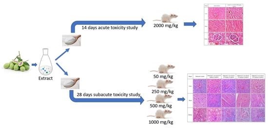
5.7. Acute Toxicity Study

The acute toxicity experiment was performed according to the protocol described in OECD 407 guidelines. Before the acute toxicity experiment, six male Sprague Dawley rats were split into two groups (n = 3). The first group (the controls) received water conveyance. The individuals in Group 2 were given an oral dose of BMY equal to 2000 mg/kg of body weight. After the administration of the substances under study, the rats were monitored at 1, 2, 4, and 6-h intervals for any changes in their general behaviour (including their skin, respiratory activity, tremors, salivation, lethargy, or sleep). Visual observation was carried out to monitor the somatomotor, nervous, circulatory, and respiratory systems. Physical changes, including those to the eyes, fur, skin, and mucous membranes, as well as changes to the behavioural pattern, were also observed. The rats were first observed after 24 h, and then they were observed once a day for the next 14 days. On days 1, 7, and 14, body weight was measured. On day 15, all rats fasted overnight before being anaesthetised and sacrificed. Studies were conducted to determine the LD50 or median lethal dose. Blood and organs were taken for collection.

5.8. Subacute Toxicity Study

A subacute toxicity experiment was performed according to the protocol described in OECD 407 guidelines. In this study, forty-two male rats in good health were used. A normal treatment group and a satellite treatment group comprised the two divisions of this study. In the normal treatment group, rats were put into five groups at random, with six rats in each group (n = 6). The groups were designated as subacute control (SC), BMY 50 (50 mg/kg), BMY 250 (250 mg/kg), BMY 500 (500 mg/kg), and BMY 1000 (1000 mg/kg), and were fed BMY orally for 28 days. While the rats were in the satellite treatment group, they were put into two treatment groups at random, with six rats in each group (n = 6). The two groups were called SSC (Satellite Control) and SST (1000 mg/kg), respectively. Scheme 1 below illustrates the experimental group of animals. The rats were given supplements by mouth for 28 days (for both normal treatment and satellite treatment groups), and then they had 14 days to rest and be watched without treatment (satellite treatment group only). For the evaluation of toxicokinetics, the satellite group was utilised to determine the level of drug exposure in animals that results in an adverse event [35,36]. The animals were monitored daily for their overall health as well as for any clinical signs of toxicity throughout the duration of the treatments. Variations in body weight were measured on days 0, 7, 14, 21, and 28 of the experiment, respectively. After the experiment, all of the rats were fasted for a full 24 h before being put under anaesthesia so that the cardiac puncture could be performed to collect blood [37]. Blood was collected in both EDTA and plain tubes.

5.9. Haematological and Serum Biochemical Analysis

Haematological and biochemical parameters were measured using an automated analyser, Cell Dyn 3700, Abbott Diagnostics, NJ, USA and Cobas C311, Roche Diagnostic, Basel, Switzerland, respectively. For the haematological studies, blood from an EDTA tube was used to measure the haematology parameter, including red blood cell count (RBC), haemoglobin concentration (Hb), haematocrit (HCT), mean corpuscular volume (MCV), mean corpuscular haemoglobin (MCH), mean corpuscular haemoglobin concentration (MCHC), platelets (Pt), and white blood cell count (WBC). For the plain tube, the serum was separated for further analysis of plasma aspartate transaminase (AST), alanine transaminase (ALT), alkaline phosphatase (ALP), total proteins, albumin, total and direct bilirubin, urea, creatinine, and ions (sodium, potassium, and chloride) [37].

5.10. Histopathology Analysis

All of the rats were sacrificed by inhaling carbon dioxide immediately following the cardiac puncture. A surgical procedure was performed to remove samples of organs and tissues of interest, such as the liver, kidneys, and heart. These samples were then weighed and examined macroscopically. After determining the relative organ weight and fixing organ samples in formalin, dehydrating them, and embedding them in paraffin, the results were measured. Subsequently, for histological examination, 5 µm thick sections of each sample were stained with hematoxylin/eosin [38,39]. Based on the pathological observations of these tissues, both a macroscopic and a microscopic basis were performed.

5.11. Statistical Analysis

The results for body weight, haematological and serum biochemical parameters, as well as relative organ weight, were all expressed as the mean ± SEM. The data were analysed with a one-way ANOVA, and the Tukey HSD Independent T-test was used to compare the results of the two different groups. Software developed by IBM Corp. in New York, NY, USA called IBM SPSS Statistic version 22.0, was used to carry out the statistical analyses.

Author Contributions

Conceptualization, R.N. and R.N.M.R.; methodology, M.D.Y. and R.N.M.R.; software, R.N.M.R.; validation, H.B., H.E. and Z.A.Z.; formal analysis, R.N.; investigation, R.N.M.R.; resources, K.P.; data curation, Z.A.Z.; writing—original draft preparation, R.N. and R.N.M.R.; writing—review and editing, R.N. and R.N.M.R.; visualization, F.O. and A.S.J.; supervision, H.B.; project administration, S.H.J., S.S.B., A.Z.A. and K.K.A.K.; funding acquisition, Z.A.Z. All authors have read and agreed to the published version of the manuscript.

Funding

This work was supported by the Ministry of Higher Education, Malaysia under the Fundamental Research Grant Scheme with the code project of FRGS/1/2020/SKK0/UPM/02/4 and reference number under Universiti Putra Malaysia 04-0L-20-2274FR.

Institutional Review Board Statement

The study was conducted in accordance with the Declaration of MSU and UPM. The animal study protocol was approved by the Ethics Committee of MSU (Animal Ethics No: AE-MSU-073, Date of Approval: 15th Februray 2017).

Informed Consent Statement

Not applicable.

Data Availability Statement

Not applicable.

Conflicts of Interest

The authors declare no conflict of interest.

References

  1. Rajan, N.S.; Bhat, R. Bioactive Compounds of Plum Mango (Bouea Microphylla Griffith). In Bioactive Compounds in Underutilized Fruits and Nuts; Murthy, H.N., Bapat, V.A., Eds.; Reference Series in Phytochemistry; Springer International Publishing: Cham, Switzerland, 2020; p. 14. ISBN 978-3-030-30181-1. [Google Scholar]
  2. Sukalingam, K. Preliminary phytochemical analysis and in vitro antioxidant properties of Malaysian. Trends Phytochem. Res. 2018, 2, 261–266. [Google Scholar]
  3. Dechsupa, N.; Kantapan, J.; Tungjai, M.; Intorasoot, S. Maprang “Bouea macrophylla Griffith” seeds: Proximate composition, HPLC fingerprint, and antioxidation, anticancer and antimicrobial properties of ethanolic seed extracts. Heliyon 2019, 5, e02052. [Google Scholar] [CrossRef]
  4. Cheong, Y.; Kim, C.; Kim, M.B.; Hwang, J.K. The anti-photoaging and moisturizing effects of Bouea macrophylla extract in UVB-irradiated hairless mice. Food Sci. Biotechnol. 2018, 27, 147–157. [Google Scholar] [CrossRef]
  5. Fu’adah, I.T.; Sumiwi, S.A.; Wilar, G. The Evolution of Pharmacological Activities Bouea macrophylla Griffith In Vivo and In Vitro Study: A Review. Pharmaceuticals 2022, 15, 238. [Google Scholar] [CrossRef] [PubMed]
  6. Yang, J.J.; Yu, D.; Xiang, Y.-B.; Blot, W.; White, E.; Robien, K.; Sinha, R.; Park, Y.; Takata, Y.; Lazovich, D.; et al. Association of Dietary Fiber and Yogurt Consumption with Lung Cancer Risk. JAMA Oncol. 2020, 6, e194107. [Google Scholar] [CrossRef] [PubMed]
  7. Zicari, G.; Carraro, E.; Bonetta, S. The Regulation (EC) N. 1924/2006 of the European Parliament and of the Council of 20 December 2006 on nutrition and health claims made on foods. Prog. Nutr. 2006, 9, 9–25. [Google Scholar]
  8. Surono, I.; Hosono, A. Fermented Milks. In Himalayan Fermented Foods; CRC Press: Boca Raton, FL, USA, 2009; pp. 95–116. [Google Scholar]
  9. Ullah, A.; Munir, S.; Badshah, S.L.; Khan, N.; Ghani, L.; Poulson, B.G.; Emwas, A.; Jaremko, M. Important Flavonoids and Their Role as a Therapeutic Agent. Molecules 2020, 25, 5243. [Google Scholar] [CrossRef]
  10. Zakaria, Z.A.; Kamisan, F.H.; Kek, T.L.; Salleh, M.Z. Hepatoprotective and antioxidant activities of Dicranopteris linearis leaf extract against paracetamol-induced liver intoxication in rats. Pharm. Biol. 2020, 58, 478–489. [Google Scholar] [CrossRef]
  11. Dutra, R.P.; de Barros Abreu, B.V.; Cunha, M.S.; Batista, M.C.A.; Torres, L.M.B.; Nascimento, F.R.F.; Ribeiro, M.N.S.; Guerra, R.N.M. Phenolic Acids, Hydrolyzable Tannins, and Antioxidant Activity of Geopropolis from the Stingless Bee Melipona fasciculata Smith. J. Agric. Food Chem. 2014, 62, 2549–2557. [Google Scholar] [CrossRef]
  12. Akomolafe, S.F.; Akinyemi, A.J.; Anadozie, S.O. Phenolic Acids (Gallic and Tannic Acids) Modulate Antioxidant Status and Cisplatin Induced Nephrotoxicity in Rats. Int. Sch. Res. Not. 2014, 2014, 984709. [Google Scholar] [CrossRef]
  13. Song, B.-R.; Alam, M.B.; Lee, S.-H. Terpenoid-Rich Extract of Dillenia indica L. Bark Displays Antidiabetic Action in Insulin-Resistant C2C12 Cells and STZ-Induced Diabetic Mice by Attenuation of Oxidative Stress. Antioxidants 2022, 11, 1227. [Google Scholar] [CrossRef]
  14. Menon, D.B.; Gopalakrishnan, V.K. Terpenoids Isolated From the Shoot of Plectranthus hadiensis Induces Apoptosis in Human Colon Cancer Cells Via the Mitochondria-Dependent Pathway. Nutr. Cancer 2015, 67, 697–705. [Google Scholar] [CrossRef] [PubMed]
  15. Nurul, S.A.S.; Hazilawati, H.; Mohd, R.S.; Mohd, F.H.R.; Noordin, M.M.; Norhaizan, M.E. Subacute Oral Toxicity Assesment of Ethanol Extract of Mariposa christia vespertilionis Leaves in Male Sprague Dawley Rats. Toxicol. Res. 2018, 34, 85–95. [Google Scholar] [CrossRef] [PubMed]
  16. Hilaly, J.E.; Israili, Z.H.; Lyoussi, B. Acute and chronic toxicological studies of Ajuga iva in experimental animals. J. Ethnopharmacol. 2004, 91, 43–50. [Google Scholar] [CrossRef]
  17. Raza, M.; Al-Shabanah, O.A.; El-Hadiyah, T.M.; Al-Majed, A.A. Effect of prolonged vigabatrin treatment on haematological and biochemical parameters in plasma, liver and kidney of Swiss albino mice. Sci. Pharm. 2002, 70, 135–145. [Google Scholar] [CrossRef]
  18. Amresh, G.; Singh, P.N.; Rao, C.V. Toxicological screening of traditional medicine Laghupatha (Cissampelos pareira) in experimental animals. J. Ethnopharmacol. 2008, 116, 454–460. [Google Scholar] [CrossRef]
  19. Mohamed, E.A.H.; Lim, C.P.; Ebrika, O.S.; Asmawi, M.Z.; Sadikun, A.; Yam, M.F. Toxicity evaluation of a standardised 50% ethanol extract of Orthosiphon stamineus. J. Ethnopharmacol. 2011, 133, 358–363. [Google Scholar] [CrossRef] [PubMed]
  20. Dybing, E.; Doe, J.; Groten, J.; Kleiner, J.; O’Brien, J.; Renwick, A.; Schlatter, J.; Steinberg, P.; Tritscher, A.; Walker, R.; et al. Hazard characterisation of chemicals in food and diet. Food Chem. Toxicol. 2002, 40, 237–282. [Google Scholar] [CrossRef] [PubMed]
  21. Adeneye, A.A.; Ajagbonna, O.P.; Adeleke, T.I.; Bello, S.O. Preliminary toxicity and phytochemical studies of the stem bark aqueous extract of Musanga cecropioides in rats. J. Ethnopharmacol. 2006, 105, 374–379. [Google Scholar] [CrossRef]
  22. Petterino, C.; Argentino-Storino, A. Clinical chemistry and haematology historical data in control Sprague-Dawley rats from pre-clinical toxicity studies. Exp. Toxicol. Pathol. 2006, 57, 213–219. [Google Scholar] [CrossRef]
  23. Delwatta, S.L.; Gunatilake, M.; Baumans, V.; Seneviratne, M.D.; Dissanayaka, M.L.B.; Batagoda, S.S.; Udagedara, A.H.; Walpola, P.B. Reference values for selected hematological, biochemical and physiological parameters of Sprague-Dawley rats at the Animal House, Faculty of Medicine, University of Colombo, Sri Lanka. Anim. Model. Exp. Med. 2018, 1, 250–254. [Google Scholar] [CrossRef]
  24. Porwal, M.; Khan, N.; Maheshwari, K. Evaluation of Acute and Subacute Oral Toxicity Induced by Ethanolic Extract of Marsdenia tenacissima Leaves in Experimental Rats. Sci. Pharm. 2017, 85, 29. [Google Scholar] [CrossRef] [Green Version]
  25. Olorunnisola, O.S.; Bradley, G.; Afolayan, A.J. Acute and sub-chronic toxicity studies of methanolic extract of Tulbaghia violacea rhizomes in Wistar rats. Afr. J. Biotechnol. 2012, 11, 14934–14940. [Google Scholar] [CrossRef]
  26. Kim, H.; Moon, J.Y.; Kim, H.; Lee, D.-S.; Cho, M.; Choi, H.-K.; Kim, Y.S.; Mosaddik, A.; Cho, S.K. Antioxidant and antiproliferative activities of mango (Mangifera indica L.) flesh and peel. Food Chem. 2010, 121, 429–436. [Google Scholar] [CrossRef]
  27. Fidelis, M.; Santos, J.S.; Escher, G.B.; Rocha, R.S.; Cruz, A.G.; Cruz, T.M.; Marques, M.B.; Nunes, J.B.; do Carmo, M.A.V.; de Almeida, L.A.; et al. Polyphenols of jabuticaba [Myrciaria jaboticaba (Vell.) O.Berg] seeds incorporated in a yoghurt model exert antioxidant activity and modulate gut microbiota of 1,2-dimethylhydrazine-induced colon cancer in rats. Food Chem. 2021, 334, 127565. [Google Scholar] [CrossRef]
  28. Chang, S.K.C. Protein Content, 4th ed.; Nielsen, S.S., Ed.; Food Analysis; Springer: New York, NY, USA, 2007; pp. 133–147. ISBN 978-0-47-085094-7. [Google Scholar]
  29. Nielsen, S.S. Phenol-Sulfuric Acid Method for Total Carbohydrates. In Food Analysis Laboratory Manual; Nielsen, S.S., Ed.; Food Science Texts Series; Springer: Boston, MA, USA, 2010; pp. 47–53. ISBN 978-1-4419-1462-0. [Google Scholar]
  30. Witschinski, F.; Demartini, D.; Kilian, J.; Dallago, R.M.; Rosa, C.D.; Cansian, R.L.; Valduga, E.; Steffens, J. Development and characterization of light yoghurt elaborated with Bifidobacterium animalis subsp. Lactis Bb-12 and fructooligosaccharides. Ciência Rural. 2018, 48. [Google Scholar] [CrossRef]
  31. Himed-Idir, H.; Mouhoubi, K.; Siar, E.; Boudries, H.; Mansouri, H.; Adjeroud, N.; Madani, K.; Boulekbache-Makhlouf, L. Effect of rosemary (Rosmarinus officinalis L.) supplementation on fresh cheese: Physicochemical properties, antioxidant potential, and sensory attributes. J. Food Process. Preserv. 2021, 45, e15057. [Google Scholar] [CrossRef]
  32. National Technical Working Group of Malaysian Food Composition Database. Protocol for Sampling and Methods of Analysis for Malaysian Food Composition Database. 2011; pp. 72–74. Available online: https://www.imr.gov.my/testlist/js/pdfjs/Protocol_Sampling_MY_FCD.pdf (accessed on 4 January 2023).
  33. Jovanović, M.; Petrović, M.; Miočinović, J.; Zlatanović, S.; Laličić Petronijević, J.; Mitić-Ćulafić, D.; Gorjanović, S. Bioactivity and Sensory Properties of Probiotic Yogurt Fortified with Apple Pomace Flour. Foods 2020, 9, 763. [Google Scholar]
  34. Zhang, Y.; Wang, C.; Yang, F.; Yang, Z.; Wang, F.; Sun, G. UHPLC-ESI-Q-TOF-MS/MS analysis, antioxidant activity combined fingerprints for quality consistency evaluation of compound liquorice tablets. RSC Adv. 2018, 8, 27661–27673. [Google Scholar] [CrossRef]
  35. Chapman, K.; Chivers, S.; Gliddon, D.; Mitchell, D.; Robinson, S.; Sangster, T.; Sparrow, S.; Spooner, N.; Wilson, A. Overcoming the barriers to the uptake of nonclinical microsampling in regulatory safety studies. Drug Discov. Today 2014, 19, 528–532. [Google Scholar] [CrossRef]
  36. Sparrow, S.S.; Robinson, S.; Bolam, S.; Bruce, C.; Danks, A.; Everett, D.; Fulcher, S.; Hill, R.E.; Palmer, H.; Scott, E.W.; et al. Opportunities to minimise animal use in pharmaceutical regulatory general toxicology: A cross-company review. Regul. Toxicol. Pharmacol. 2011, 61, 222–229. [Google Scholar] [CrossRef] [PubMed]
  37. Naomi, R.; Rusli, R.N.M.; Balan, S.S.; Othman, F.; Jasni, A.S.; Jumidil, S.H.; Bahari, H.; Yazid, M.D. E. tapos Yoghurt—A View from Nutritional Composition and Toxicological Evaluation. Foods 2022, 11, 1903. [Google Scholar] [CrossRef] [PubMed]
  38. Balan, S.S.; Abidin, A.Z.; Perumal, K.V.; Shafie, N.H.; Abdullah, M.A.; Jasni, A.S.; Kwaifa, I.K.; Bahari, H. Transgenerational Evaluation of Elateriospermum tapos Extracts on the Male Offspring of Obesity-Induced Sprague Dawley Rats. Sains Malays. 2021, 50, 3045–3057. [Google Scholar] [CrossRef]
  39. Kamsani, N.E.; Zakaria, Z.A.; Md Nasir, N.L.; Mohtarrudin, N.; Mohamad Alitheen, N.B. Safety Assessment of Methanol Extract of Melastoma malabathricum L. Leaves following the Subacute and Subchronic Oral Consumptions in Rats and Its Cytotoxic Effect against the HT29 Cancer Cell Line. Evid. Based Complement. Altern. Med. eCAM 2019, 2019, 5207958. [Google Scholar] [CrossRef] [PubMed] [Green Version]
Figure 1. A section on histology was included in the acute toxicity study. There is no evidence of an abnormal lesion or tubular dilation in the kidney histology. It appears that the renal corpuscle is normal. In the kidney histology section, there were no abnormalities found at all. The histology of the heart section reveals that the architecture of the heart is normal with no evidence of inflammation. The myocardium is denoted by M and the nucleus by N. Standard strands of hepatocytes (H), sinusoids (S), and the central vein (CV) are all visible in the section of the liver. Since there was no evidence of steatosis, lobular inflammation, or hepatocyte ballooning, the liver was given a score of 0 on the grading scale.
Toxins 15 00125 g001
Figure 2. Kidney, liver, and heart histology for subacute toxicity in the subacute control (SC) group and the BMY supplementation groups at varying doses (BMY 50, BMY 250, BMY 500, BMY 1000). There is no abnormal lesion or tubular dilation in the kidney’s histology. The renal corpuscle appears normal. The kidney histology section showed no abnormalities. Histological examination of a heart tissue sample revealed no evidence of inflammation or disruption to the heart’s normal architecture. The myocardium is denoted by M and nucleus by N. Hepatocytes (H), sinuses (S), and the central vein (CV) can all be seen in a typical liver section.
Toxins 15 00125 g002
Figure 3. Histology of liver, kidney and heart for subacute satellite control (SSC) and subacute satellite 1000 mg/kg (SST 1000). There is no abnormal lesion or tubular dilation in the kidney’s histology. The kidneys appear to have healthy corpuscles. The kidney histology section showed no abnormalities. Histological examination of a heart tissue sample revealed no evidence of inflammation or disruption to the heart’s normal architecture. The myocardium is denoted by M and nucleus by N. Hepatocytes (H), sinuses (S), and the central vein (CV) can all be seen in a typical liver section.
Toxins 15 00125 g003
Figure 4. (A) Fresh Bouea macrophylla fruit, (B) Bouea macrophylla fruit without seed, (C) Bouea macrophylla flesh powder.
Toxins 15 00125 g004
Scheme 1. Experimental group of animals in the subacute toxicity study.
Toxins 15 00125 sch001
Table 1. Nutritional analysis of BMY.
ParameterAmountUnit
Carbohydrate5.79 ± 0.01g/100 g
Protein2.92 ± 0.00g/100 g
Sugar2.72 ± 0.00g/100 g
Fat3.29 ± 0.01g/100 g
Energy64 ± 0.01kcal/100 g
Table 2. Microbiological analysis of BMY.
MicroorganismNumber of ColonyUnit
Lactobacillus spp.1.2 × 106CFU/g
Bifidobacteria2.0 × 1010CFU/g
Titratable acidity as lactic acid0.599g/100 g
CFU = colony forming unit; where results are expressed with an “E” this represents an exponential result to a base of 10.
Table 3. The phytochemical compound of ethanolic Boeau macrophylla under negative ionisation according to different groups of phenolic, terpenoid, alkaloid, benzenoid, carbohydrate, benzopyran, and carboxylic acid.
Component NameFormulaMolecular Weight (Da)Observed
Molecular Weight (Da)
Observed m/zMass Error
(ppm)
Observed RT
(min)
PHENOLIC
Flavonoid
1.2’’-O-AcetylrutinC29H32O17652.16395652.16070651.15340−5.00.73
2.5′-Methoxy-bilobetinC32H22O11582.11621582.11600581.10870−0.40.98
3.Cryptomerin BC32H22O10566.12130566.12180565.114500.900.83
4.Isolappaol CC30H34O10554.21520554.21330553.20600−3.56.84
5.Nevadensin-5-O-β-D-glucosideC24H26O12506.14243506.14210505.13480−0.612.11
6.SilybinC25H22O10482.12130482.12330481.116004.101.19
7.6-Methoxykaempferol-3-O-β-D-galactopyranosideC22H22O12478.11113478.11130477.104100.4012.71
8.Eriodictyol-7-O-β-D-methyl-glucuronopyranosideC22H22O12478.11113478.11120477.104000.208.24
9.Texifolin-3-O-β-D-glucopyranosideC21H22O12466.11113466.11130465.104000.406.62
10.6-Hydroxykaempferol-3-O-glucosideC21H20O12464.09548464.09570463.088400.509.23
11.HomoplantagininC22H22O11462.11621462.11580461.10860−0.8012.41
12.Rengyoside CC22H32O10456.19955456.19940455.19210−0.3013.97
13.Cinchonain ⅠbC24H20O9452.11073452.11060451.10340−0.209.99
14.Kaempferol-3-O-β-D-glucopyranosideC21H20O11448.10056448.10060447.093300.1010.29
15.SibiricaphenoneC20H28O11444.16316444.16280443.15550−0.87.57
16.Epicatechin gallate (Epicatechin-3-O-gallate)C22H18O10442.09000442.09000441.082700.009.04
17.5,7,4′-Trihydroxy-8-C-β-D-flavanone glucosideC21H22O10434.12130434.12130433.114000.008.70
18.Quercetin-3-O-α-L-arabinopyranosideC20H18O11434.08491434.08540433.078101.109.61
19.CnidimonalC23H16O7404.08960404.09140403.084204.507.32
20.Quercetin-3-sulphateC15H10O10S381.99947381.99850380.99120−2.701.98
21.Vitamin B2C17H20N4O6376.13828376.13800375.13070−0.706.97
22.(6aR,11aR)-2,8-Dihydroxy-3,4,9,10-tetramethoxypterocarpanC19H20O8376.11582376.11580375.108500.0014.26
23.Protosappanin CC16H14O6302.07904302.07870301.07140−1.304.74
24.QuercetinC15H10O7302.04265302.04160301.03430−3.4010.29
25.MedicagolC16H8O6296.03209296.03180295.02460−0.900.98
26.Cnidimol FC15H14O6290.07904290.07880289.07150−0.805.32
27.5,7,2’,5’-Tetrahydroxy-flavoneC15H10O6286.04774286.04750285.04030−0.708.70
28.GenisteinC15H12O5272.06847272.06810271.06090−1.208.70
29.Astragaline AC10H11NO4209.06881209.06820208.06090−2.904.69
30.NepitrinC22H22O12478.11113478.11130477.104000.307.33
31.2′′-O-GalloylhyperinC28H24O16616.10643616.10540615.09810−1.7010.63
32.Patuletin-3-O-glucosideC22H22O13494.10604494.10620493.098900.306.48
Tannin
33.1,2,3,4,6-Penta-O-galloyl-β-D-glucopyranosideC41H32O26940.11818940.12100939.113803.009.65
34.Tellimagrandin ⅡC41H30O26938.10253938.10510937.097802.709.65
35.CastalaginC41H26O26934.07123934.07180933.064500.609.65
36.1,2,3,6-Tetra-O-galloyl-β-D-glucopyranosideC34H28O22788.10722788.10900787.101702.307.38
37.Mallotinic acidC34H26O22786.09157786.09320785.085902.107.71
38.CasuariinC34H24O22784.07592784.07720783.069901.608.35
39.PedunculaginC34H24O22784.07592784.07680783.069601.208.57
40.1,2,6-Tri-O-galloyl-β-D-glucopyranosideC27H24O18636.09626636.09690635.089600.906.98
41.2,4,6-Tri-O-galloyl-β-D-glucoseC27H24O18636.09626636.09690635.089601.007.19
42.3,6-Di-O-Galloyl-β-D-glucoseC20H20O14484.08531484.08560483.078300.505.38
43.Glucosyringic acidC15H20O10360.10565360.10530359.09800−0.906.31
44.Ellagic acidC14H6O8302.00627302.00610300.99880−0.608.88
Phenylpropanoid
45.Tubuloside AC37H48O21828.26881828.27250827.265204.500.88
46.Suffruticosol AC42H32O9680.20463680.20340679.19610−1.900.56
Phenolic acid
47.1-Galloyl-glucoseC13H16O10332.07435332.07450331.067200.501.78
48.3-Ethoxy-4,5-dihydroxybenzoic acidC9H10O5198.05282198.05230197.04500−2.807.32
49.Methyl-3-hydroxy-4-methoxybenzoateC9H10O4182.05791182.05750181.05020−2.403.16
50.Gallic acidC7H6O5170.02152170.02110169.01380−2.401.78
51.Methyl gallateC8H8O5184.03717184.03670183.02940−2.604.95
52.m-Hydroxybenzoic acidC7H6O3138.03169138.03120137.02390−3.503.58
53.E-3,4,5-Trimethoxycinnamic acidC12H14O5238.08412238.08380237.07660−1.207.49
54.cis-Caffeic acidC9H8O4180.04226180.04180179.03450−2.605.37
55.E-p-Coumaric acidC9H8O3164.04734164.04700163.03970−2.003.91
56.1-O-Caffeoyl-β-D-glucopyranosideC15H18O9342.09508342.09610341.088802.905.37
Coumarin
57.Bergaptol-O-β-D-glucopyranosideC17H16O9364.07943364.07920363.07190−0.607.34
58.CichoriinC15H16O9340.07943340.07930339.07200−0.404.74
59.CalycanthosideC17H20O10384.10565384.10720383.099904.100.57
Lignan
60.(-)-Olivil-4’-O-β-D-glucopyranosideC26H34O12538.20503538.20520537.197900.308.88
61.(-)-Secoisolariciresinol-4-O-β-D-glucopyranosideC26H36O11524.22576524.22640523.219101.309.83
62.Indigoticoside AC26H34O11522.21011522.21040521.203200.609.02
Quinone
63.1,3,6-Trihydroxy-2-methylanthraquinone-3-O-β-D-glucopyranosideC21H20O10432.10565432.10570431.098400.1011.45
64.2,5-DimethoxybenzoquinoneC8H8O4168.04226168.04180167.03450−2.903.46
Xanthones
65.1-Hydroxy-2,3,4,5-tetramethoxyxanthoneC17H16O7332.08960332.08890331.08160−2.109.41
66.NeomangiferinC25H28O16584.13773584.13610583.12880−2.800.58
TERPENOID
67.Terrestrosin IC51H84O251096.530171096.52601095.51870−3.800.49
68.Escin ⅣgC53H84O231088.540341088.536601087.52930−3.500.49
69.Quinquenoside ⅢC50H84O19988.56068988.55630987.54900−4.509.62
70.Yemuoside YM2C31H42O16670.24729670.24750669.240200.308.59
71.Lucidumoside DC27H36O13568.21559568.21650567.209201.608.87
72.AsperulosideC18H22O11414.11621414.11810413.110804.600.57
73.DihydrobrusatolC26H34O11522.21011522.21030521.203000.3011.82
74.ReptosideC16H24O11392.13186392.13170391.12450−0.304.43
75.VogelosideC17H24O10388.13695388.13650387.12920−1.205.66
76.SecoxyloganinC17H24O11404.13186404.13150403.12430−0.802.74
77.ApocynosideⅠC19H30O8386.19407386.19380385.18660−0.606.75
78.SwerosideC16H22O9358.12638358.12600357.11870−1.105.07
79.GentiopicrosideC16H20O9356.11073356.11050355.10320−0.706.22
80.Thymol isobutyrateC14H20O2220.14633220.14580219.13860−2.2012.08
81.AcetylpaeoniflorinC25H30O12522.17373522.17450521.167201.5011.88
82.PseudosantoninC15H20O4264.13616264.13580263.12850−1.5012.08
83.3,5,7-TrihydroxychromoneC9H6O5194.02152194.02160193.014300.301.78
ALKALOID
84.DauricumineC19H24ClNO6397.12922397.12860396.12130−1.500.57
85.TryptophaneC11H12N2O2204.08988204.08930203.08200−2.903.94
86.ArginineC6H14N4O2174.11168174.11120173.10390−2.700.51
87.XanthosineC10H12N4O6284.07568284.07640283.069102.505.10
88.InosineC10H12N4O5268.08077268.08040267.07310−1.602.02
89.GuanineC5H5N5O151.04941151.04870150.04140−4.702.03
90.Mudanpioside EC24H30O13526.16864526.16620525.15890−4.705.26
91.EchinothiopheneC23H26O10S494.12467494.12710493.119804.802.42
92.2,3,5,4’-Tetrahydroxystilbene-2-O-β-D-glucopyranosideC20H22O9406.12638406.12640405.119100.0015.38
93.Benzyl alcohol xylopyranosyl(1→6)glucopyranosideC18H26O10402.15260402.15230401.14500−0.706.53
94.Cnideoside BC18H22O10398.12130398.12330397.116004.900.57
95.IsomaltoseC12H22O11342.11621342.11620341.10890−0.100.69
96.KoaburasideC14H20O9332.11073332.11070331.10340−0.103.46
BENZENOID
97.Sennoside BC42H38O20862.19564862.19810861.190802.803.14
98.Scroneoside BC23H26O11478.14751478.14740477.14010−0.3012.41
99.2,5-Dihydroxybenzeneacetic acid ethyl esterC10H12O4196.07356196.07300195.06570−3.005.07
100.m-Hydroxybenzoic acidC7H6O3138.03169138.03120137.02390−3.503.58
CARBOHYDRATE
101.MannotrioseC18H32O16504.16903504.16970503.162401.300.74
102.PentoseC5H10O5150.05282150.05220149.04500−4.000.70
103.GalactoseC6H12O6180.06339180.06290179.05560−2.600.69
BENZOPYRAN
104.NorbergeninC13H14O9314.06378314.06440313.057202.101.79
CARBOXYLIC ACID
105.2-(4-Hydroxybenzyl) malic acidC11H12O6240.06339240.06310239.05580−1.403.58
106.2-Hydroxy-1,2,3-propane tricarboxylic acid-2-methyl esterC7H10O7206.04265206.04230205.03510−1.500.72
107.Quinic acidC7H12O6192.06339192.06290191.05570−2.300.65
108.Shikimic acidC7H10O5174.05282174.05230173.04510−2.800.65
109.Phenylpropionic acidC9H11NO2165.07898165.07850164.07120−2.902.55
110.Mono-ethyl fumarateC6H8O4144.04226144.04170143.03440−3.800.69
111.Methylsuccinic acidC5H8O4132.04226132.04170131.03440−4.100.69
Table 4. Effect of BMY on the body weight (mean ± SEM) of rats at 14 days.
Week/GroupGroup
Control2000 mg/kg
1285.12 ± 10.41283.52 ± 15.13
2309.13 ± 10.58298.82 ± 8.10
n = 3. Values are expressed as mean ± standard error of the mean.
Table 5. The relative organ weight (mean ± SEM) of Sprague Dawley rats in acute oral toxicity study of BMY.
Parameter/GroupGroup
Control2000 mg/kg
Spleen0.37 ± 0.010.48 ± 0.05
Kidney1.35 ± 0.041.33 ± 0.28
Liver5.75 ± 0.336.37 ± 0.34
Lung1.12 ± 0.101.15 ± 0.09
Heart0.61 ± 0.000.66 ± 0.15
n = 3. Values are expressed as mean ± standard error of the mean.
Table 6. The haematology values (mean ± SEM) of SD rats in acute oral toxicity study of BMY.
Parameter/GroupGroup
Control2000 mg/kg
Hb (g/L)14.10 ± 1.0515.73 ± 0.120
RBC (1012/L)7.29 ± 0.577.98 ± 0.52
RDW (%)13.46 ± 0.4414.06 ± 0.31
PCV (%)43.33 ± 3.8447.33 ± 0.33
MCV (fL)59.33 ± 0.6659.66 ± 0.86
MCH (pg)19.33 ± 0.3319.66 ± 0.33
MCHC (g/dL)32.66 ± 0.6633.33 ± 0.33
WBC (109/L)11.33 ± 2.276.50 ± 0.47
Neutrophils (109/L)3.23 ± 0.831.46 ± 0.20
Lymphocytes (109/L)7.67 ± 1.684.67 ± 0.37
Monocytes (109/L)0.16 ± 0.670.16 ± 0.12
Eosinophils (109/L)0.24 ± 0.200.17 ± 0.55
Basophils (109/L)0.00 ± 0.000.00 ± 0.00
Platelet (109/L)936.67 ± 106.791054.33 ± 136.94
n = 3. Values are expressed as mean ± standard error of the mean.
Table 7. The serum biochemistry values (mean ± SEM) of SD rats in acute oral toxicity study of BMY.
Parameter/GroupGroup
Control2000 mg/kg
Sodium (mmol/L)141.67 ± 0.88142.00 ± 0.00
Potassium (mmol/L)6.33 ± 0.176.63 ± 0.08
Chloride (mmol/L)105.33 ± 1.20105.67 ± 2.33
Urea (mmol/L)12.03 ± 1.038.43 ± 0.79
Creatinine (mmol/L)29.67 ± 2.0229.00 ± 1.00
Total protein (g/L)59.00 ± 2.0864.00 ± 1.52
Albumin (g/L)34.67 ± 1.3338.00 ± 0.57
Globulin (g/L)24.33 ± 0.8826.00 ± 1.00
ALP (U/L)218.3 ± 28.15225.67 ± 30.20
AST (U/L)143.00 ± 11.00136.33 ± 5.23
ALT (U/L)36.00 ± 3.6038.67 ± 4.25
GGT(U/L)13.00 ± 0.0013.00 ± 0.00
n = 3. Values are expressed as mean ± standard error of the mean.
Table 8. Weekly body weight changes for subacute toxicity study. 1 The group was supplemented with BMY at various doses daily for 28 days in a subacute toxicity study. 2 Satellite group for subacute toxicity study which was given water vehicle or BMY at 1000 mg/kg daily for 28 days followed by no treatment for 14 days.
Week/
Group
Subacute 1Subacute Satellite 2
SCBMY 50BMY 250BMY 500BMY 1000SSCSST 1000
1250.00 ± 8.56275.00 ± 11.38261.83 ± 15.72272.50 ± 11.74268.67 ± 14.88274.83 ± 21.05263.83 ± 14.98
2352.50 ± 10.70330.00 ± 11.03312.50 ± 14.07315.00 ± 14.55320.00 ± 18.66326.67 ± 18.51315.83 ± 13.25
3360.00 ± 10.65351.67 ± 7.71351.67 ± 13.27340.83 ± 13.93355.00 ± 20.29364.17 ± 16.60355.83 ± 15.57
4361.67 ± 7.03366.67 ± 10.54364.17 ± 10.03360.00 ± 12.97368.33 ± 21.36388.33 ± 16.16387.50 ± 17.74
5-----415.83 ± 17.63426.67 ± 17.45
6-----428.33 ± 19.78431.67 ± 17.78
n = 6. Values are expressed as mean ± standard error of the mean.
Table 9. Total food intake, caloric intake and water intake for both subacute toxicity 1 and subacute satellite 2.
Subacute 1 Subacute Satellite 2
SCBMY 50BMY 250BMY 500BMY 1000SSCSST 1000
Total food consumption (g)737.30 ± 27.50746.67 ± 15.43747.83 ± 23.93735.00 ± 30.31732.67 ± 51.961146.25 ± 29.021155.00 ± 35.00
Calorie intake (KJ)9437.87 ± 353.079557.33 ± 197.549572.27 ± 306.409408.00 ± 224.009378.00 ± 665.114,672 ± 371.4614,784.00 ± 448.00
Total water intake (mL)886.67 ± 11.67875.00 ± 26.73880.83 ± 11.67898.33 ± 21.03892.50 ± 61.451627.50 ± 18.91685.83 ± 23.33
n = 6. Values are expressed as mean ± standard error of the mean.
Table 10. The relative organ weight for both subacute toxicity 1 and subacute satellite 2.
Parameter/GroupSubacute 1Subacute Satellite 2
SCBMY 50BMY 250BMY 500BMY 1000SSCSST 1000
Kidney0.74 ± 0.480.82 ± 0.260.82 ± 0.240.82 ± 0.300.81 ± 0.760.73 ± 0.310.75 ± 0.43
Liver3.77 ± 0.143.95 ± 0.103.78 ± 0.103.50 ± 0.283.63 ± 0.133.60 ± 0.183.94 ± 0.18
Lung0.57 ± 0.660.65 ± 0.430.66 ± 0.490.74 ± 0.420.74 ± 0.280.63 ± 0.020.68 ± 0.05
Heart0.32 ± 0.170.37 ± 0.080.36 ± 0.150.40 ± 0.310.39 ± 0.300.43 ± 0.070.40 ± 0.03
n = 6. Values are expressed as mean ± standard error of the mean.
Table 11. The haematology values for both subacute toxicity 1 and subacute satellite 2.
Parameter/GroupSubacute 1Subacute Satellite 2
SCBMY 50BMY 250BMY 500BMY 1000SSCSST 1000
Hb (g/L)15.53 ± 0.5616.22 ± 0.3917.10 ± 0.2416.12 ± 0.3717.00 ± 0.3916.9 ± 0.2916.7 ± 0.58
RBC (1012/L)8.85 ± 0.288.43 ± 0.258.95 ± 0.208.55 ± 0.178.77 ± 0.149.12 ± 0.289.17 ± 0.38
RDW (%)17.65 ± 0.4316.53 ± 0.4916.70 ± 0.3816.48 ± 0.5616.38 ± 0.2516.2 ± 0.6816.9 ± 0.51
PCV (%)57.33 ± 1.4855.50 ± 1.5758.50 ± 0.8954.50 ± 1.2656.17 ± 1.2555.00 ± 0.8953.9 ± 1.99
MCV (fL)62.67 ± 0.7166.00 ± 0.5865.33 ± 0.5663.67 ± 0.4964.00 ± 1.1060.7 ± 0.9858.70 ± 0.5
MCH (pg)18.00 ± 0.3119.33 ± 0.21 *18.83 ± 0.1719.00 ± 0.0019.20 ± 0.37 *18.00 ± 0.7018.30 ± 0.21
MCHC (g/dL)28.20 ± 0.2029.33 ± 0.21 *29.33 ± 0.21 *30.00 ± 0.00 *30.40 ± 0.24 *30.83 ± 0.3031.20 ± 0.17
WBC (109/L)21.28 ± 2.4512.17 ± 1.9514.37 ± 1.6610.53 ± 1.7914.85 ± 0.8911.43 ± 2.0517.10 ± 4.76
Neutrophils (109/L)3.10 ± 0.482.78 ± 0.922.67 ± 0.483.85 ± 1.572.47 ± 0.212.81 ± 0.282.30 ± 0.38
Lymphocytes (109/L)13.57 ± 0.618.23 ± 1.42 *10.60 ± 1.247.98 ± 1.10 *11.70 ± 0.767.40 ± 1.507.70 ± 1.83
Monocytes (109/L)0.67 ± 0.100.90 ± 0.210.88 ± 0.121.24 ± 0.400.50 ± 0.030.52 ± 0.110.86 ± 0.18
Eosinophils (109/L)0.20 ± 0.020.16 ± 0.020.20 ± 0.030.15 ± 0.030.14 ± 0.010.16 ± 0.040.17 ± 0.07
Basophils (109/L)0.13 ± 0.020.05 ± 0.020.05 ± 0.02164.35 ± 164.330.12 ± 0.080.05 ± 0.020.02 ± 0.02
Platelet (109/L)1036.83 ± 35.071115.00 ± 52.081084.33 ± 86.32854.17 ± 164.971227.00 ± 56.19982.90 ± 49.21060.90 ± 41.4
n = 6. Values are expressed as mean ± standard error of the mean. (*) indicate a significant difference at p < 0.05.
Table 12. The serum biochemistry values for both subacute toxicity 1 and subacute satellite 2.
Parameter/GroupSubacute 1Subacute Satellite 2
SCBMY 50BMY 250BMY 500BMY 1000SSCSST1000
Sodium (mmol/L)144.50 ± 0.56144.83 ± 0.40145.00 ± 0.82146.00 ± 0.83143.60 ± 0.40146.1 ± 0.65147.5 ± 0.72
Potassium (mmol/L)9.60 ± 0.254.20 ± 1.904.35 ± 1.946.51 ± 1.407.15 ± 1.452.87 ± 1.813.65 ± 1.66
Chloride (mmol/L)98.17 ± 0.7096.67 ± 0.8498.83 ± 0.4098.00 ± 0.3698.33 ± 0.6199 ± 0.52101.1 ± 0.48
Urea (mmol/L)7.02 ± 0.299.07 ± 0.687.73 ± 0.379.43 ± 0.918.57 ± 0.597.85 ± 0.326.35 ± 0.35
Creatinine (mmol/L)56.00 ± 2.4854.83 ± 3.7353.50 ± 1.1848.00 ± 2.7246.83 ± 1.2541.17 ± 7.6335.00 ± 2.31
Total protein (g/L)69.67 ± 0.8070.33 ± 1.4569.83 ± 0.6564.33 ± 4.1567.50 ± 1.6770.83 ± 1.3869.33 ± 2.10
Albumin (g/L)39.80 ± 0.6742.83 ± 0.70 *42.67 ± 0.6742.83 ± 0.60 *41.40 ± 0.8140.5 ± 0.9940.67 ± 0.95
Globulin (g/L)31.50 ± 1.4127.50 ± 0.8527.17 ± 0.9121.76 ± 4.13 *25.50 ± 1.02 *30.33 ± 1.0528.67 ± 1.23
ALP (U/L)199.00 ± 8.13200.50 ± 17.27175.83 ± 9.36202.00 ± 9.67160.00 ± 11.66147.3 ± 4.94128.67 ± 7.52
AST (U/L)118.83 ± 10.33102.50 ± 8.7698.33 ± 7.67103.33 ± 9.4985.17 ± 3.6699.83 ± 5.2682 ± 4.93
ALT (U/L)64.83 ±4.1346.83 ± 3.3253.83 ± 4.1858.50 ± 5.2755.330 ± 3.4052.17 ± 1.2543.83 ± 1.70
GGT (U/L)13.00 ± 0.0013.00 ± 0.0013.00 ± 0.0013.00 ± 0.0013.00 ± 0.0013.00 ± 0.0013.00 ± 0.00
n = 6. Values are expressed as mean ± standard error of the mean. (*) indicate a significant difference at p < 0.05.
Disclaimer/Publisher’s Note: The statements, opinions and data contained in all publications are solely those of the individual author(s) and contributor(s) and not of MDPI and/or the editor(s). MDPI and/or the editor(s) disclaim responsibility for any injury to people or property resulting from any ideas, methods, instructions or products referred to in the content.

