Communication

New Insights into the Potential Cytotoxic Role of Bacillus cytotoxicus Cytotoxin K-1

by
Klèma Marcel Koné
1,
Pauline Hinnekens
1,
Jelena Jovanovic
2,
Andreja Rajkovic
2 and
Jacques Mahillon
1,*
1
Laboratory of Food and Environmental Microbiology, Université Catholique de Louvain (UCLouvain), 1348 Louvain, Belgium
2
Department of Food Technology, Safety and Health, Research Group of Food Microbiology and Food Preservation, Faculty of Bioscience Engineering, Ghent University (UGent), 9000 Ghent, Belgium
*
Author to whom correspondence should be addressed.
Toxins 2021, 13(10), 698; https://doi.org/10.3390/toxins13100698
Submission received: 2 September 2021 / Revised: 21 September 2021 / Accepted: 24 September 2021 / Published: 1 October 2021
(This article belongs to the Special Issue The Effect of Microbial Toxins on Animal Health and Food Safety)

Abstract

:
The thermotolerant representative of the Bacillus cereus group, Bacillus cytotoxicus, reliably harbors the coding gene of cytotoxin K-1 (CytK-1). This protein is a highly cytotoxic variant of CytK toxin, initially recovered from a diarrheal foodborne outbreak that caused the death of three people. In recent years, the cytotoxicity of B. cytotoxicus has become controversial, with some strains displaying a high cytotoxicity while others show no cytotoxicity towards cell lines. In order to better circumscribe the potential pathogenic role of CytK-1, knockout (KO) mutants were constructed in two B. cytotoxicus strains, E8.1 and E28.3. The complementation of the cytK-1 KO mutation was implemented in a mutant strain lacking in the cytK-1 gene. Using the tetrazolium salt (MTT) method, cytotoxicity tests of the cytK-1 KO and complemented mutants, as well as those of their wild-type strains, were carried out on Caco-2 cells. The results showed that cytK-1 KO mutants were significantly less cytotoxic than the parental wild-type strains. However, the complemented mutant was as cytotoxic as the wild-type, suggesting that CytK-1 is the major cytotoxicity factor in B. cytotoxicus.
Key Contribution: The current work created viable Bacillus cytotoxicus mutant strains lacking the cytK-1 gene. Compared to their wild-type counterparts, the mutant strains showed very low cytotoxicity effects toward Caco-2 cells. Complemented mutant was as cytotoxic as the wild-type strain, suggesting that cytotoxin K-1 is the major cytotoxicity factor in B. cytotoxicus.

1. Introduction

Cytotoxin K-1 (CytK-1) is a highly cytotoxic and necrotic variant of cytotoxin K (CytK). It was initially recovered from a specific Bacillus cereus strain isolated from a food poisoning outbreak fatal to three elderly people in France in 1998. This B. cereus strain and its kin were later described as a new species, Bacillus cytotoxicus, the thermotolerant representative of the B. cereus group [1,2]. This group, also known as B. cereus sensu lato (s.l.), contains Gram-positive, spore-forming, and facultative anaerobic bacteria. Although numerous new species have been described as B. cereus s.l. members, the primary members of the group are B. cereus sensu stricto (s.s.), Bacillus thuringiensis, Bacillus anthracis, Bacillus mycoides, Bacillus pseudomycoides, and Bacillus weihensphanensis. The B. cereus group comprises both beneficial and pathogenic members. While B. mycoides, B. pseudomycoides, and B. weihensphanensis have not reportedly been implicated in any human infections or foodborne diseases yet [3], many B. thuringiensis have been used for several decades as bio-pesticides in agriculture and control of disease vectors, due to their ability to produce insecticidal molecules [4].
Because of its plasmid-encoded anthrax toxins, B. anthracis is highly pathogenic to mammals, including humans [5], and some B. cereus s.s. strains have been implicated in human extra-gastrointestinal infections, as well as in foodborne illnesses [6]. Indeed, an ingestion of food containing particular B. cereus strains can lead to two types of foodborne diseases, the emetic and diarrheal syndromes. The former is caused by a plasmid-encoded, in-food produced, heat-stable, acid-resistant and ring shape 1.2 kDa peptide, the cereulide [7,8]. The diarrheal syndrome is presumably caused by one or a combination of several enterotoxins produced in the small intestine after ingestion of B. cereus contaminated food. These enterotoxins are chromosomally encoded and prominently include the hemolysin BL (Hbl), the non-hemolytic enterotoxin (Nhe) and CytK [9]. The expression of most of these potential enterotoxins is regulated by the Pap/PlcR regulatory system [10]. Hbl and Nhe toxins are both three-component toxins. While almost all B. cereus s.l. members harbor the Nhe genes (nheABC), about 40 to 60% contain the Hbl genes (hblABCD) [11,12]. It has also been shown that the Nhe toxin initiate cell apoptosis in Vero cells [13], and more recently, it was reported that Hbl and Nhe can act synergistically to trigger inflammation [14].
Encoded by the cytK gene, CytK is a 34-kDa single-peptide toxin for which two variants have been reported, CytK-1 and CytK-2. The latter forms smaller pores in phospholipidic bilayer membrane, is less cytotoxic, and is mainly found in some mesophilic B. cereus strains. It shares 87% amino acid identities with CytK-1 toxin which is more cytotoxic, and whose coding gene is restricted to B. cytotoxicus strains [15]. However, NVH 883-00, a B. cytotoxicus strain originating from spices, was shown to under-express CytK-1 toxin and be less cytotoxic than the reference strain, NVH 391-98, isolated from the fatal outbreak in France [16]. Moreover, based on whole genome sequencing, four genomic clades (A to D) have been described and clade A reportedly gathered the most potentially cytotoxic strains, including strains NVH 391-98 and CH_213 [17,18]. It is also noteworthy that the presence of toxin genes does not by itself lead to their expression and to the bacterium cytotoxicity. Indeed, the expression of B. cereus enterotoxin genes is thoroughly regulated and is dependent on the strain and its microenvironment [19,20,21]. It has been suggested that the cytotoxicity of B. cytotoxicus could have been overestimated in the past. As for other potentially enterotoxigenic B. cereus, the cytotoxicity of B. cytotoxicus should not be deduced solely based on the presence/absence of the cytK-1 gene and genetic profile should thus be complemented with (cyto)toxicity assays [20,22].
Therefore, there was a need for an in-depth exploration of the implication of CytK-1 toxin in B. cytotoxicus toxicity. To the best of our knowledge, no study has yet focused on the deletion of cytK-1 gene in order to assess the role of its toxin. Hence, the current study aimed at constructing B. cytotoxicus mutants lacking their cytK-1 gene and comparing their cytotoxicity with that of the parental wild-type strains.

2. Results and Discussion

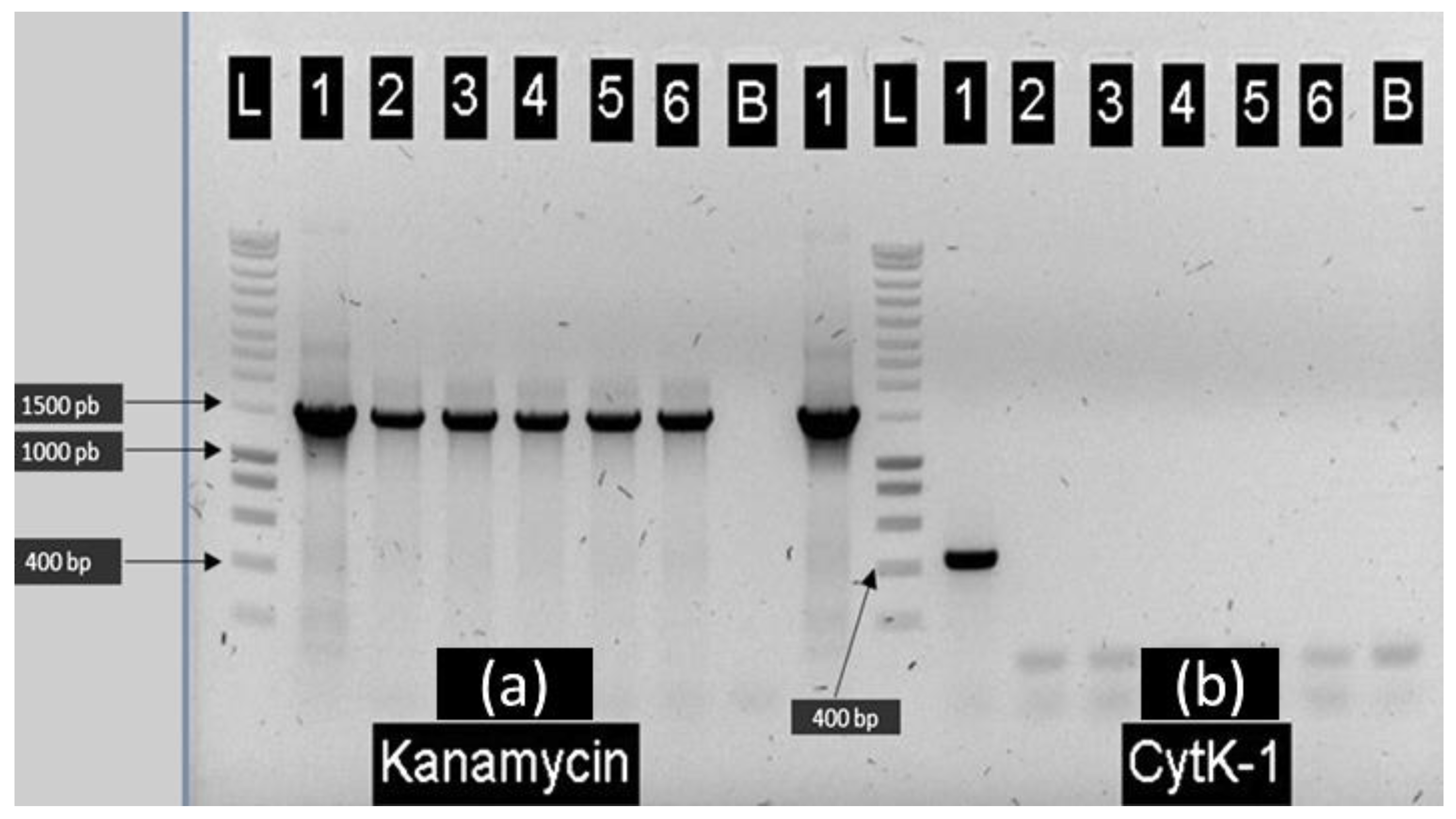
2.1. Construction of E28.3cytK-1 KO-A and E8.1cytK-1 KO B. cytotoxicus Mutants Lacking cytK-1

Since the initial isolation of B. cytotoxicus from a fatal food poisoning incident [1], its toxicity has become somewhat controversial. Indeed, it has even been suggested that the cytotoxicity of this species was overestimated in the past [20]. To help circumscribe the role of CytK-1 toxin, we knocked out the cytK-1 gene in two B. cytotoxicus strains, E28.3 and E8.1. Both strains were isolated from potato flakes. In contrast to the reference strain NVH 391-98 showing no visible plasmid, E28.3 and E8.1 display large and small plasmids. These two B. cytotoxicus strains share the same RAPD profile, which is different from that of NVH 391-98 [23]. They also display distinct plasmid profiles. While strain E28.3 is one of the rare B. cytotoxicus of the collection to be susceptible to electroporation, E8.1 is the only strain in which the cytK-1 KO mutation could be successfully transferred via conjugation (see below).
Using a double-recombination approach based on a thermo-sensitive shuttle plasmid, a KO-mutant of the cytK-1 gene was built in strain E28.3 by replacing this locus by a kanamycin-resistance (kanR) gene. Five recombinant strains (A–E) were screened for cytK-1 and kanamycin resistance genes. As shown in Figure 1, all the potential recombinant clones carried the resistance gene (Lanes a2 to a6), while they all lost the cytk-1 gene (Lanes b2 to b6). Additional PCR experiments confirmed the validity of the gene swap (data not shown). Based on these results, the E28.3cytK-1-KO-A recombinant clone was retained for further experiments.
It was recently shown that the mega-plasmid pXO16 could mobilize chromosomal loci between members of the B. cereus group during its conjugation [24]. Using this approach, the KO-mutation of E28.3cytK−1-KO-A was transferred to strain E8.1 via filter mating conjugation. The kanamycin-resistant (KanR) candidate transconjugants were then verified by RAPD profiling [23] to confirm that they displayed the same pattern as their parental strain E8.1. As shown in Figure 2, the clone present in Lane 2 displayed the RAPD pattern identical to that of its parental strains E8.1. Additional PCR confirmed that this clone (named E8.1cytK−1-KO-B) had acquired the locus where cytK-1 was replaced by the KanR gene (data not shown). This clone was retained for the cytotoxicity experiments.
Given that viable mutants of B. cytotoxicus strains lacking the cytK-1 gene were obtained, it can be suggested that this gene is not essential to B. cytotoxicus. However, it is worth mentioning that attempts to create a similar mutant in the reference type-strain NVH 391-98 through electroporation, as well as via pXO16 conjugation, have failed (data not shown). The reasons for these unsuccessful attempts remain so far unknown. Future trials to construct mutants of other highly cytotoxic B. cytotoxicus strains, such as CH_213 [18], should certainly be considered.

2.2. Cytotoxicity of B. cytotoxicus Wild-Type Strains E28.3 and E8.1, Their Derived Mutants Lacking the cytK-1 Gene and the Complemented Mutant

Caco-2 cells were exposed to the B. cytotoxicus supernatant of the reference strain NVH 391-98, as well as that of wild-type strains E8.1 and E28.3 and that of the derived cytK-1 KO-mutants. The cytotoxicity was assayed using the tetrazolium salt method (MTT) which assesses cell viability. After two hours, the highly cytotoxic strain, NVH 391-98, was able to impair Caco-2 viability while little cytotoxicity effect was observed for the wild-type strains E8.1 and E28.3. However, after twelve hours of exposure, these two strains were almost as cytotoxic as NVH 391-98 (Figure 3).
Interestingly, deleting the cytK-1 gene had a striking effect: in both genetic backgrounds, removing the gene reduced by more than 90% the deleterious effect of the E28.3cytK−1-KO-A and E8.1cytK−1-KO-B supernatants on Caco-2 cell viability, indicating a drastic reduction of cytotoxicity and suggesting that CytK-1 toxin plays a major role in B. cytotoxicus cytotoxicity. In order to confirm that the observed effects were directly related to the cytK-1 gene deletion, a complementation experiment was conducted by cloning cytK-1 into the pHT304-18Z shuttle vector and introducing it in the E28.3cytK−1-KO-A mutant by electroporation. As shown in Figure 3, the resulting complemented mutant displayed almost the same activity as its wild-type counterpart, reinforcing the idea that CytK-1 is responsible for most of the B. cytotoxicus cytotoxicity, at least on Caco2 cells.
The toxicity of NVH 391-98, initially isolated from the fatal case in 1998, is in line with previous reports [1,20]. Recently, Stevens et al. [17] explored the relationship between genomic diversity and cytotoxicity among strains pertaining to four B. cytotoxicus genomic clades (A–D). It was then suggested that potentially highly cytotoxic strains are gathered in clades A and B, while those of clades C and D are presumably less cytotoxic. More recently, the draft genome of another highly cytotoxic B. cytotoxicus strain, CH_213, pertaining to genomic clade A has been released [18]. In addition, whole genome sequencing of wild-type strains E8.1 and E28.3 indicated that they relate to clade C [25]. With clade C strains displaying relatively high cytotoxicity toward cell lines, the current findings indicate that highly cytotoxic B. cytotoxicus strains are not necessarily restricted to clades A and B. Nevertheless, a better understanding of the link between genomic grouping and virulence would require cytotoxic studies on other B. cytotoxicus strains, from different food matrices and genomic clades.
The inactivation of cytK-1 correlates with a drastic drop in the cytotoxicity of our B. cytotoxicus strains. This is in contrast to the findings of Romarao and Lereclus, in which there was no significant cytotoxicity reduction (both on Caco-2 and HeLa cell lines) in a B. thuringiensis mutant lacking its cytK-2 gene [26]. It is noteworthy that the cytK-2 gene harbored by B. thuringiensis encodes for the less cytotoxic variant of the CytK toxin [15]. Assessing the various wild-type B. cytotoxicus and B. thuringiensis strains and their cytK-KO mutants in the same assay could be valuable to resolve this seeming discrepancy.
Despite displaying an impaired cytotoxicity, our KO-mutants potentially possessed remaining toxicity (Figure 3) that could be imputable to other to-be-investigated enterotoxins or virulence factors in B. cytotoxicus. In fact, previous reports have shown that B. cytotoxicus lacks the Hbl genes but harbors a novel variant of Nhe genes [1,16]. Nhe is a three-component toxin that was shown to be necrotic at high concentrations, while inducing apoptosis in sub-necrotic concentrations. It has also been recently reported to trigger inflammation synergistically with the Hbl toxin [13,14]. As for several B. cereus enterotoxins, Nhe and CytK-1 are under the control of the PlcR regulator [10], and it has been reported that NVH 391-98 overproduces the CytK-1 toxin [16]. We can therefore speculate that the high cytotoxicity of some B. cytotoxicus strains could be imputable to a synergistic action of large amount of produced CytK-1 and/or Nhe toxins. It remains to be seen whether the remaining toxicity displayed by the cytK-1 KO-mutants can be attributed to these other putative enterotoxins.

3. Conclusions

In conclusion, viable B. cytotoxicus mutants lacking the cytK-1 gene were successfully created. Cytotoxicity tests showed that these mutants were less cytotoxic than the parental wild-type strains. To give further credence to these observations, a complementation of the knockout mutants with the wild-type cytK-1 gene was constructed. The complemented mutant displayed cytotoxic activity comparable to that of the wild-type strains. Together, these results suggest that CytK-1 toxin is indubitably implicated in B. cytotoxicus cytotoxicity. The exact contribution of this activity to the diarrheal syndrome caused by the B. cytotoxicus strains remains, however, to be clarified. Similarly, it is plausible that high cytotoxicity observed in certain B. cytotoxicus strains could be imputable to the association of CytK-1 with one or more additional enterotoxin(s) and/or enzyme(s).

4. Materials and Methods

4.1. Bacterial Strains, Growth Media and Plasmids

The bacterial strains and plasmids used in this study are summarized in Table 1. B. cytotoxicus strain E28.3 was used to create the cytK-1 KO mutant through the homologous recombination method (see below). NEBuilder HiFi DNA Assembly Cloning Kit (New England Biolabs™, Ipswich, MA, USA) was used to insert the PCR amplicons into the thermo-sensitive plasmid pMAD [27]. Chemically competent Escherichia coli NEB 5-alpha and dam/dcm C2925 strains (New England Biolabs™, Ipswich, MA, USA) were used for the cloning steps. Plasmid pXO16 conjugation was used to transfer, via mobilization, the cytK-1-KO mutation into strain E8.1. Bacterial strains were grown at 37 °C (E. coli) or 30 °C (B. cytotoxicus) on Luria-Bertani (LB) medium (broth or supplemented with 1.5% agar) containing the appropriated antibiotics: ampicillin (Ap, 100 μg/mL), erythromycin (Ery, 10 μg/mL), kanamycin (Kan, 50 μg/mL) or tetracycline (Tet, 50 μg/mL).

4.2. Construction of the E28.3 Mutant Strain Lacking the cytK-1 Gene

A fresh overnight colony of B. cytotoxicus strain E28.3 from LB agar was mixed in 50 μL of deionized water for the DNA extraction. Using Q5 High-Fidelity polymerase (Promega, Leiden, The Netherlands), PCR amplification of upstream (987 bp) and downstream (1001 bp) regions of cytK-1 gene, as well the kanamycin resistance gene from plasmid pDG783 [28], were performed. The primers used in this study are listed in Table 2. Electrophoresis gel migration of the PCR products was performed as described elsewhere [23]. The PCR amplicons were purified using GenElute™ PCR Clean-Up Kit (Sigma-Aldrich™, Overijse, Belgium) and the DNA quality was check with the Nanodrop spectrophotometer (Isogen, De Meern, The Netherlands).
Table 1. Bacterial strains and plasmids used.
StrainsMain FeaturesReferences
B. cytotoxicus strains
E8.1Wild-type strain isolated from potato flakes; RAPD pattern A, plasmid profile PP10[23]
E8.1cytK−1-KO-BcytK-1 knockout mutant of E8.1This work
E28.3Wild-type strain isolated from potato flakes; RAPD pattern A and plasmid profile PP8[23]
E28.3cytK−1-KO-AcytK-1 knockout mutant of E28.3This work
E28.3.1Derivative of E28.3 containing pXO16::Tn5401, TetR[29]
E28.3.1cytK−1-KO-ADerivative of E28.3cytK−1-KO-A containing pXO16::Tn5401, TetRThis work
E28.3cytK−1-KO-A(pHT304-18Z::cytK-1)Complementation of the mutant lacking cytK-1 geneThis work
NVH 391-98Reference strain; highly cytotoxic[1]
E. colistrains
E. coli C2925 dam/dcmK12 derivative deficient in adenine and cytosine methyl-transferases, chemically competentNew England Biolabs
E. coli NEB 5-alphaDH5-alpha derivative, T1 phage resistant and endA1 deficient, chemically competentNew England Biolabs
Plasmids
pHT304-18ZShuttle vector used in the complementation; EryR[30]
pHT304-18Z::cytK-1CytK-1 gene of strain E28.3 cloned into the pHT304-18Z shuttle vectorThis work
pMADGram+/Gram- shuttle vector containing a thermo-sensitive Gram+ replicon; EryR, AmpR[27]
pMAD::UDpMAD derivative containing the KanR cassette with up- and down-stream regions of the cytK-1 gene, EryR, AmpR, KanRThis work
pDG783Plasmid carrying a KanR cassette; KanR[28]
pXO16::Tn5401Large conjugative plasmid from B. thuringiensis sv. israelensis, tagged with a Tetracycline-resistance gene; TetR[31]
As previously described by Gibson et al. [32], the purified PCR amplicons were mixed with the PCR-opened pMAD shuttle vector [27], deionized water, and NEBuilder HiFi DNA Assembly MasterMix. The HiFi DNA assembly reaction product was chemically transformed into E. coli NEB 5-alpha and incubated at 37 °C (1 h, 180 rpm). The bacterial culture was then spread on a LB agar supplemented with ampicillin and incubated overnight at 37 °C. Next, E. coli transformants were PCR-checked for the presence of the expected construct. The recombinant plasmid, pMAD::UD (containing the KanR cassette flanked by the upstream and downstream sequences of cytK-1) was extracted using GenElute™ Plasmid Miniprep Kit (Sigma-Aldrich™, Overijse, Belgium) and checked through DNA sequencing (Macrogen Europe, Amsterdam, The Netherlands). Thereafter, the construct was demethylated in E. coli C2925 before subsequent electroporation into B. cytotoxicus.
Table 2. Primers used in the cloning steps.
DesignationPrimersSequences (5′ to 3′)Tm (°C)Amplicon (pb)Reference
KanR geneKana_RGTTTTTTACTATCGATACAAATTCCTCGTAG601.490This study
Kana_FACATATATCGTGATAAACCCAGCGAACC
CytK-1 upstream regioncytK-1_Up_RGGGTTTATCACGATATATGTCGTATTTCACATATATC58987This study
cytK-1_Up_FAGATCTATCGATGCATGCCAGAAGTTTTAGGTTCATACATTTG
CytK-1 downstream regioncytK-1_Down_RCGGATCCATATGACGTCGACAATGCGAGAGACGTTGCG621.001This study
cytK-1_Down_FTTGTATCGATAGTAAAAAACAACACTGACAAACTC
CytK-1 genecytK-1_RCCTCGTGCATCTGTTTCATGAG61436[33]
cytK-1_FCAATTCCAGGGGCAAGTGTC
The B. cereus electroporation protocols previously described [34,35] were adapted for B. cytotoxicus. Briefly, a single fresh colony of E28.3 was sub-cultured in 25 mL of brain-heart infusion (BHI) (Bio-Rad, Richmond, CA, USA) and incubated O/N (30 °C, 120 rpm). Next, the bacterial culture was centrifuged (6000 rpm, 4 °C, 10 min), the cell pellet washed three times with chilled deionized water (4 °C) and resuspended in 400 μL PEG6000 (40% wt/v). Using the Gene Pulser and in a 2 mm cuvette (Bio-Rad, Richmond, CA, USA), the electro-competent B. cytotoxicus cells were electroporated with 1 to 2 μg of demethylated pMAD::UD plasmid. Electroporated bacterial cells were immediately suspended in 1 mL of LB broth and incubated at 30 °C, (1 h, 120 rpm). Next, 100 μL of the bacterial suspension were spread on LB agar supplemented with erythromycin and incubated O/N at 30 °C. The transformants were PCR-checked for the construct presence.
Swapping of the cytK-1 gene by the kanR gene in B. cytotoxicus strain E28.3 was performed following the plasmid homologous recombination approach described by Makart et al. [24,36]. B. cytotoxicus transformants containing the shuttle pMAD::UD underwent successive non-permissive incubation cycles as follow: two at 43 °C, one at 45 °C and a last one at 50 °C. Up to 10−7 serial dilutions were made from the last cycle culture (50°C) and spread on LB agar supplemented with kanamycin and 20 μg/μL of X-Gal (Sigma-Aldrich™, Overijse, Belgium). Since pMAD contains a β-galactosidase cassette, five white colonies (indicating pMAD absence) on X-Gal LB agar were re-streaked on LB agar supplemented with kanamycin and incubated overnight at 30 °C.

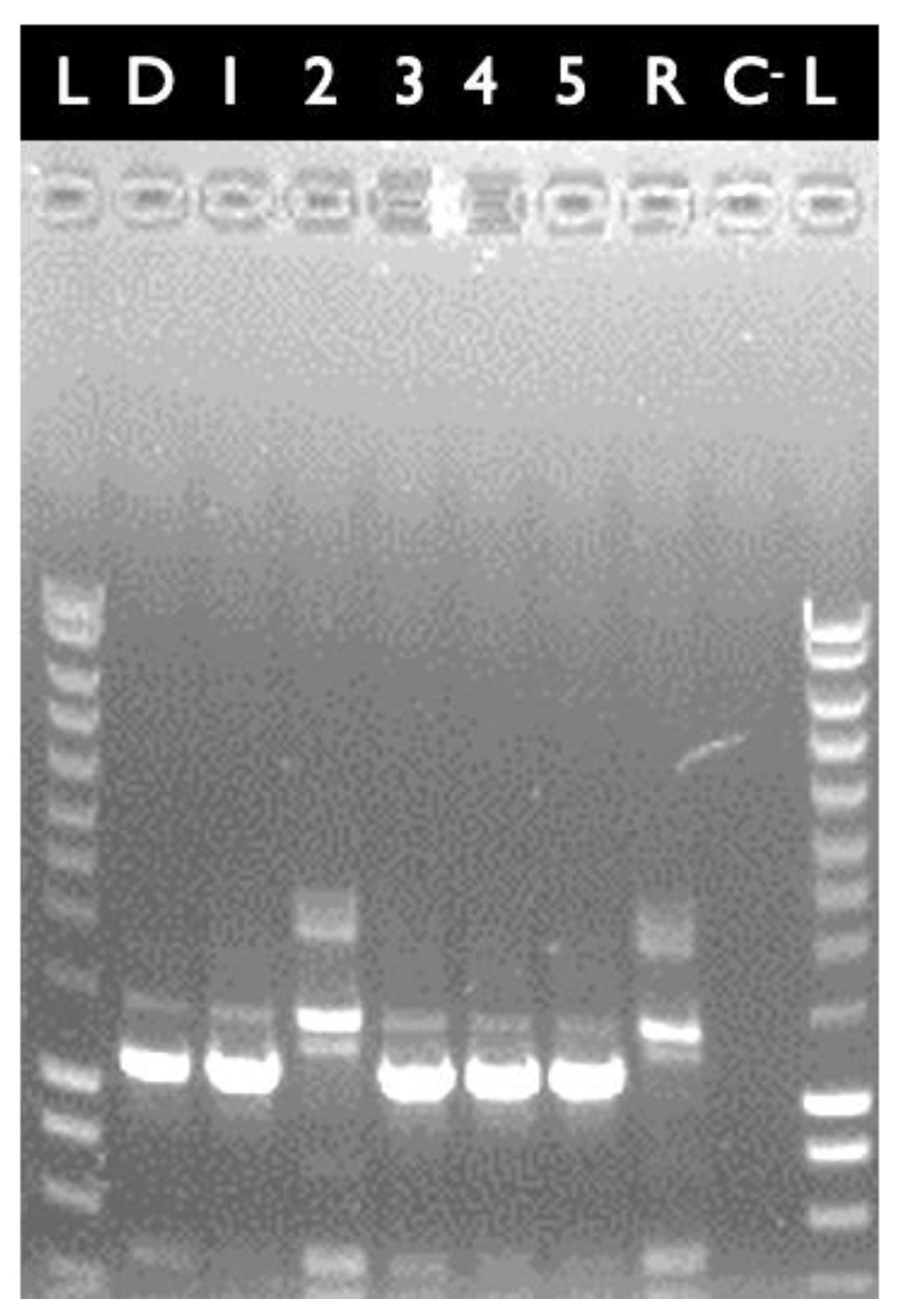
4.3. Transfer of the cytK-1 KO Locus from Strain E28.3 to Strain E8.1

Using filter mating as described by Hinnekens et al. [29], the cytK-1 KO mutation was mobilized from B. cytotoxicus strain E28.3.1cytK−1-KO-A (Derivative of E28.3cytK−1-KO-A containing pXO16::Tn5401) into E8.1 via conjugation [24]. The candidate transconjugants strains were PCR-screened to check the effective replacement of cytK-1 gene by KanR gene. RAPD patterns of the final transconjugant were also checked as previously described by Koné et al. [23].

4.4. Complementation of the cytK-1 KO Mutant

Primers flanked with overlapping restriction sites sequences of BamH1 and Pst1 enzymes (New England Biolabs™, Ipswich, MA, USA) (Table 2) were used to amplify the cytK-1 gene from B. cytotoxicus wild-type strain E28.3. As described by Makart et al. [24], the PCR product was cloned in the shuttle pHT304-18Z and transformed into chemically competent E. coli 10-beta cells. The resulting construct pHT304-18Z::cytK-1 was retrieved using the GeneElute Plasmid Miniprep Kit (Sigma-Aldrich™, Overijse, Belgium) and checked through sequencing (Macrogen Europe, Amsterdam, The Netherlands). The pHT304::18Z-cytK-1 construct was then demethylated by passing it through E. coli C2925 and subsequently electroporated into the B. cytotoxicus strain cytK-1-KO mutant to obtain E28.3cytK−1-KO-A(pHT304-18Z::cytK-1).

4.5. Cytotoxicity of E28.3, E8.1, Their Mutants Lacking cytK-1 and the Complemented Strain

4.5.1. Cell Culture

The Caco-2 cell culture described by Rajkovic et al. [37] was adapted as follows. The human colorectal carcinoma cell line Caco-2 (HTB-37™) was obtained from American Type Culture Collection (ATCC) (Manassas, VA, USA). Cells were maintained in high glucose, Glutamax Dulbecco’s Modified Eagle Medium (DMEM) (Gibco-Thermo Fisher, Geel, Belgium), supplemented with (i) 10% heat-inactivated foetal bovine serum (Greiner Bio One, Wemmel, Belgium), (ii) 1% non-essential amino acids (Gibco-Thermo Fisher, Geel, Belgium), (iii) 1% penicillin/streptomycin (Gibco-Thermo Fisher, Geel, Belgium) in the humidified 10% CO2 atmosphere at 37 °C (Memmert GmbH & Co., Nurnberg, Germany). Cell morphology was regularly checked with phase-contrast microscopy (VWR, Leuven, Belgium) and the growth medium was changed every other day. After reaching 80–90% of confluency, the cells were sub-cultured by trypsinization using 0.5% trypsin-EDTA (Gibco-Thermo Fisher, Geel, Belgium) until cells were realized from the bottom of 75 cm2 neck tissue culture flasks (SPL Life Sciences, Gyeonggi-do, Korea). The cells were counted with a Bürker counting chamber according to the Trypan blue staining method. Next, they were seeded in a 96-well plate at a concentration of 20,000 cells per well to achieve optimal distribution of cells. In all experiments, the cells were passaged less than 25 times.

4.5.2. Preparation of Bacterial Cell-Free Supernatants

To assess the effects of the wild-type B. cytotoxicus strains E28.3, E8.1, their mutant cytK-1-KO derivatives and the complemented strain E28.3cytK−1-KO-A(pHT304-18Z::cytK-1), cell-free supernatants were prepared. B. cytotoxicus reference strain NVH 391-98 cell-free supernatant and the untreated Caco-2 cells were used as positive and negative controls, respectively. Single separated colonies of selected strains were inoculated in serum and antibiotic-free DMEM and incubated for overnight at 37 °C. The cultures were centrifuged at 5000 g for 10 min, and supernatants were filter-sterilized through a 0.22 um filter (Millipore Inc., Billerica, MA, USA) and used freshly. Two-fold diluted supernatants were used for Caco-2 cell exposure, to better discriminate cytotoxicity of the strains. Exposure was performed for 2, 4, and 12 h. The effects were the most notable after 12 h of exposure.

4.5.3. MTT Assay

Cytotoxic effects were characterized through the tetrazolium salt or MTT [(3-(4,5-dimethylthiazol-2-yl)-2,5-diphenyltetrazolium bromide]. This method assesses the cell’s mitochondrial activity as an indicator of cell viability and cytotoxicity, initially described by Mosmann [38]. After exposure, 20 μL of MTT (5 mg/mL in PBS) was added to each well, and the plates were incubated at 37 °C for 2 h. Upon incubation, liquid was discarded, and the purple formazan crystals were solubilized in dimethyl-sulfoxide (DMSO). SpectraMax plate reader (Molecular Devices, Sunnyvale, CA, USA) was used to record the absorbance at 570 nm.

4.5.4. Statistical Analysis

The normality of the data was investigated using the Kolmogorov–Smirnov test. Microsoft Excel 2016 was used to compute mean values and standard deviations (N = 2, n = 6) for each test condition. To determine whether the data were significantly different (p < 0.05), a t-test (two-tailed with unequal variance) was performed with SPSS Statistics 26 (Chicago, IL, USA).

Author Contributions

Conceptualization, K.M.K. and P.H.; methodology, K.M.K., P.H. and J.J.; software, K.M.K., P.H. and J.J.; validation, K.M.K., P.H., J.J., A.R. and J.M.; formal analysis, K.M.K., P.H. and J.J.; investigation, K.M.K., P.H. and J.J.; resources, K.M.K., P.H. and J.J.; data curation, K.M.K., P.H. and J.J.; writing-original draft preparation, K.M.K.; writing-review and editing, K.M.K., P.H., J.J.,A.R. and J.M.; visualization, K.M.K., P.H. and J.J.; supervision, J.M. and A.R.; project administration, K.M.K., P.H., J.J., A.R. and J.M.; funding acquisition, J.M. and A.R. All authors have read and agreed to the published version of the manuscript.

Funding

This work was funded by the International Office for Cooperation of Université Catholique de Louvain, Belgium (UCLouvain, grant to K.M.K.), the National Foundation for Scientific Research (FNRS, grants to P.H. and J.M.) and the Special Research Fund (Ghent University) BOFSTA2017004201 (grant to A.R.). This work has been also part of European Union’s Horizon 2020 research and innovation programme project FoodEnTwin under grant agreement No 810752, providing material for selected case studies, and GlobalMinds project of Ghent University (grant to J.J.).

Institutional Review Board Statement

Not applicable.

Informed Consent Statement

Not applicable.

Conflicts of Interest

The authors declare no conflict of interest. The funders had no role in the design of the study; in the collection, analyses, or interpretation of data; in the writing of the manuscript, or in the decision to publish the results.

References

  1. Lund, T.; De Buyser, M.L.; Granum, P.E. A new cytotoxin from Bacillus cereus that may cause necrotic enteritis. Mol. Microbiol. 2000, 38, 254–261. [Google Scholar] [CrossRef]
  2. Guinebretière, M.H.; Auger, S.; Galleron, N.; Contzen, M.; De Sarrau, B.; De Buyser, M.L.; Lamberet, G.; Fagerlund, A.; Granum, P.E.; Lereclus, D.; et al. Bacillus cytotoxicus sp. nov. is a novel thermotolerant species of the Bacillus cereus Group occasionally associated with food poisoning. Int. J. Syst. Evol. Microbiol. 2013, 63, 31–40. [Google Scholar] [CrossRef]
  3. Stenfors Arnesen, L.P.; Fagerlund, A.; Granum, P.E. From soil to gut: Bacillus cereus and its food poisoning toxins. FEMS Microbiol. Rev. 2008, 32, 579–606. [Google Scholar] [CrossRef] [PubMed] [Green Version]
  4. Federici, B.A. Insecticidal bacteria: An overwhelming success for invertebrate pathology. J. Invert. Pathol. 2005, 89, 30–38. [Google Scholar] [CrossRef] [PubMed]
  5. Spencer, R.C. Bacillus anthracis. J. Clin. Pathol. 2003, 56, 182–187. [Google Scholar] [CrossRef] [PubMed] [Green Version]
  6. Bottone, E.J. Bacillus cereus, a volatile human pathogen. Clin. Microbiol. Rev. 2010, 23, 382–398. [Google Scholar] [CrossRef] [PubMed] [Green Version]
  7. Hoton, F.M.; Andrup, L.; Swiecicka, I.; Mahillon, J. The cereulide genetic determinants of emetic Bacillus cereus are plasmid-borne. Microbiology 2005, 151, 2121–2124. [Google Scholar] [CrossRef] [PubMed]
  8. Ehling-Schulz, M.; Fricker, M.; Scherer, S. Bacillus cereus, the causative agent of an emetic type of foodborne illness. Mol. Nutr. Food Res. 2004, 48, 479–487. [Google Scholar] [CrossRef] [PubMed]
  9. Senesi, S.; Ghelardi, E. Production, secretion and biological activity of Bacillus cereus enterotoxins. Toxins 2010, 2, 1690–1703. [Google Scholar] [CrossRef]
  10. Gohar, M.; Faegri, K.; Perchat, S.; Ravnum, S.; Okstad, O.A.; Gominet, M.; Kolsto, A.B.; Lereclus, D. The PlcR virulence regulon of Bacillus cereus. PLoS ONE 2008, 3, e2793. [Google Scholar] [CrossRef]
  11. Glasset, B.; Herbin, S.; Guillier, L.; Cadel-Six, S.; Vignaud, M.-L.; Grout, J.; Pairaud, S.; Michel, V.; Hennekinne, J.-A.; Ramarao, N.; et al. Bacillus cereus-induced food-borne outbreaks in France, 2007 to 2014: Epidemiology and genetic characterisation. Eur. Commun. Dis. Bull. 2016, 21, 30413. [Google Scholar] [CrossRef] [Green Version]
  12. Ceuppens, S.; Boon, N.; Uyttendaele, M. Diversity of Bacillus cereus group strains is reflected in their broad range of pathogenicity and diverse ecological lifestyles. FEMS Microbiol. Ecol. 2013, 84, 433–450. [Google Scholar] [CrossRef] [Green Version]
  13. Liu, X.; Ding, S.; Shi, P.; Dietrich, R.; Martlbauer, E.; Zhu, K. Non-hemolytic enterotoxin of Bacillus cereus induces apoptosis in Vero cells. Cell Microbiol. 2017, 19, e12684. [Google Scholar] [CrossRef]
  14. Fox, D.; Mathur, A.; Xue, Y.; Liu, Y.; Tan, W.H.; Feng, S.; Pandey, A.; Ngo, C.; Hayward, J.; Atmosukarto, I.I.; et al. Bacillus cereus non-haemolytic enterotoxin activates the NLRP3 inflammasome. Nat. Commun. 2020, 11, 760. [Google Scholar] [CrossRef] [PubMed]
  15. Fagerlund, A.; Ween, O.; Lund, T.; Hardy, S.P.; Granum, P.E. Genetic and functional analysis of the cytK family of genes in Bacillus cereus. Microbiology 2004, 150, 2689–2697. [Google Scholar] [CrossRef] [PubMed] [Green Version]
  16. Fagerlund, A.; Brillard, J.; Fürst, R.; Guinebretière, M.-H.; Granum, P.E. Toxin production in a rare and genetically remote cluster of strains of the Bacillus cereus group. BMC Microbiol. 2007, 7, 43. [Google Scholar] [CrossRef] [PubMed] [Green Version]
  17. Stevens, M.J.A.; Tasara, T.; Klumpp, J.; Stephan, R.; Ehling-Schulz, M.; Johler, S. Whole-genome-based phylogeny of Bacillus cytotoxicus reveals different clades within the species and provides clues on ecology and evolution. Sci. Rep. 2019, 9, 1984. [Google Scholar] [CrossRef] [PubMed] [Green Version]
  18. Stevens, M.J.A.; Johler, S. Draft genome sequence of CH_213, a highly cytotoxic Bacillus cytotoxicus strain isolated from mashed potatoes. Microbiol. Resour. Announc. 2020, 9, e00836-20. [Google Scholar] [CrossRef] [PubMed]
  19. Ceuppens, S.; Rajkovic, A.; Heyndrickx, M.; Tsilia, V.; Van De Wiele, T.; Boon, N.; Uyttendaele, M. Regulation of toxin production by Bacillus cereus and its food safety implications. Crit. Rev. Microbiol. 2011, 37, 188–213. [Google Scholar] [CrossRef]
  20. Heini, N.; Stephan, R.; Ehling-Schulz, M.; Johler, S. Characterization of Bacillus cereus group isolates from powdered food products. Int. J. Food Microbiol. 2018, 283, 59–64. [Google Scholar] [CrossRef] [Green Version]
  21. Jessberger, N.; Dietrich, R.; Granum, P.E.; Martlbauer, E. The Bacillus cereus food infection as multifactorial process. Toxins 2020, 12, 701. [Google Scholar] [CrossRef]
  22. Burtscher, J.; Etter, D.; Biggel, M.; Schlaepfer, J.; Johler, S. Further Insights into the Toxicity of Bacillus cytotoxicus Based on Toxin Gene Profiling and Vero Cell Cytotoxicity Assays. Toxins 2021, 13, 234. [Google Scholar] [CrossRef]
  23. Koné, K.M.; Douamba, Z.; Halleux, M.; Bougoudogo, F.; Mahillon, J. Prevalence and diversity of the thermotolerant bacterium Bacillus cytotoxicus among dried food products. J. Food Prot. 2019, 82, 1210–1216. [Google Scholar] [CrossRef] [PubMed]
  24. Makart, L.; Commans, F.; Gillis, A.; Mahillon, J. Horizontal transfer of chromosomal markers mediated by the large conjugative plasmid pXO16 from Bacillus thuringiensis serovar israelensis. Plasmid 2017, 91, 76–81. [Google Scholar] [CrossRef] [PubMed]
  25. Koné, K.M.; Fayad, N.; Gillis, A.; Mahillon, J. Bacillus Cytotoxicus Genomics: Chromosomal Diversity and Plasmidome Versatility. under revision.
  26. Ramarao, N.; Lereclus, D. Adhesion and cytotoxicity of Bacillus cereus and Bacillus thuringiensis to epithelial cells are FlhA and PlcR dependent, respectively. Microbes Infect. 2006, 8, 1483–1491. [Google Scholar] [CrossRef]
  27. Arnaud, M.; Chastanet, A.; Debarbouillé, M. New vector for efficient allelic replacement in naturally nontransformable, low-GC-content, Gram-positive bacteria. Appl. Environ. Microbiol. 2004, 70, 6887–6891. [Google Scholar] [CrossRef] [PubMed] [Green Version]
  28. Guerout-Fleury, A.M.; Shazand, K.; Frandsen, N.; Stragier, P. Antibiotic-resistance cassettes for Bacillus subtilis. Gene 1995, 167, 335–336. [Google Scholar] [CrossRef]
  29. Hinnekens, P.; Koné, K.M.; Fayad, N.; Leprince, A.; Mahillon, J. pXO16, the large conjugative plasmid from Bacillus thuringiensis serovar israelensis displays an extended host spectrum. Plasmid 2019, 102, 46–50. [Google Scholar] [CrossRef] [PubMed]
  30. Agaisse, H.; Lereclus, D. Structural and functional analysis of the promoter region involved in full expression of the cryIIIA toxin gene of Bacillus thuringiensis. Mol. Microbiol. 1994, 13, 97–107. [Google Scholar] [CrossRef]
  31. Jensen, G.B.; Andrup, L.; Wilcks, A.; Smidt, L.; Poulsen, O.M. The aggregation-mediated conjugation system of Bacillus thuringiensis subsp. israelensis: Host range and kinetics of transfer. Cur. Microbiol. 1996, 33, 228–236. [Google Scholar] [CrossRef]
  32. Gibson, D.G.; Young, L.; Chuang, R.Y.; Venter, J.C.; Hutchison, C.A., III; Smith, H.O. Enzymatic assembly of DNA molecules up to several hundred kilobases. Nat. Methods 2009, 6, 343–345. [Google Scholar] [CrossRef]
  33. Guinebretière, M.H.; Fagerlund, A.; Granum, P.E.; Nguyen-The, C. Rapid discrimination of cytK-1 and cytK-2 genes in Bacillus cereus strains by a novel duplex PCR system. FEMS Microbiol. Lett. 2006, 259, 74–80. [Google Scholar] [CrossRef] [PubMed] [Green Version]
  34. Mahillon, J.; Lereclus, D. Electroporation of Bacillus thuringiensis and Bacillus cereus. In Electrotransformation of Bacteria; Eynard, N., Teissié, J., Eds.; Springer: Berlin/Heidelberg, Germany, 2000; pp. 242–252. [Google Scholar]
  35. Turgeon, N.; Laflamme, C.; Ho, J.; Duchaine, C. Elaboration of an electroporation protocol for Bacillus cereus ATCC 14579. J. Microbiol. Methods 2006, 67, 543–548. [Google Scholar] [CrossRef]
  36. Makart, L.; Gillis, A.; Hinnekens, P.; Mahillon, J. A novel T4SS-mediated DNA transfer used by pXO16, a conjugative plasmid from Bacillus thuringiensis serovar israelensis. Environ. Microbiol. 2018, 20, 1550–1561. [Google Scholar] [CrossRef]
  37. Rajkovic, A.; Grootaert, C.; Butorac, A.; Cucu, T.; De Meulenaer, B.; van Camp, J.; Bracke, M.; Uyttendaele, M.; Bačun-Družina, V.; Cindrić, M. Sub-emetic toxicity of Bacillus cereus toxin cereulide on cultured human enterocyte-like Caco-2 cells. Toxins 2014, 6, 2270–2290. [Google Scholar] [CrossRef] [Green Version]
  38. Mosmann, T. Rapid colorimetric assay for cellular growth and survival: Application to proliferation and cytotoxicity assays. J. Immunol. Methods 1983, 65, 55–63. [Google Scholar] [CrossRef]
Figure 1. PCR-based confirmation of the five B. cytotoxicus KO-mutants selected after homologous recombination. L: DNA molecular weight markers (200 bp to 10 kb); B: negative control (buffer only). Panel (a): PCR-screening of the Kanamycin resistance gene: lanes 1 to 6: positive control (pDG173), E28.3cytK−1-KO-A, E28.3cytK−1-KO-B, E28.3cytK−1-KO-C, E28.3cytK−1-KO-D and E28.3cytK−1-KO-E. Panel (b): Detection of the cytK-1 gene: lanes 1 to 6: positive control (E28.3 wild-type), E28.3cytK−1-KO-A, E28.3cytK−1-KO-B, E28.3cytK−1-KO-C, E28.3cytK−1-KO-D and E28.3cytK−1-KO-E.
Toxins 13 00698 g001
Figure 2. RAPD pattern of B. cytotoxicus strains using the OPA9 primer. L: DNA molecular weight markers (200 bp to 10 kb). D: donor strain E28.3cytK−1-KO-A; R: recipient strain E8.1; C-: negative control (buffer only); Lanes 1 to 5: candidate transconjugants. Lane #2 contains a bona fide E8.1 transconjugant containing the desired genetic loci (i.e., kanR gene replacing the cytK-1 gene). It was named E8.1cytK−1-KO-B.
Toxins 13 00698 g002
Figure 3. Tetrazolium salt method (MTT) used to assess viability of Caco-2 cells after 12 h of exposure to B. cytotoxicus supernatants. Cells treated with supernatant of wild-type (WT) NVH 391-98 and untreated Caco-2 cells were used as positive and negative control, respectively. Mutants (E8.1cytK−1-KO and E28.3cytK−1-KO-A) lacking the cytK-1 gene are less cytotoxic than the WT strains of E8.1 and E28.3 (p < 0.05). The complemented mutant of E28.3cytK−1-KO-A is as cytotoxic as the WT E28.3.
Toxins 13 00698 g003
Publisher’s Note: MDPI stays neutral with regard to jurisdictional claims in published maps and institutional affiliations.

