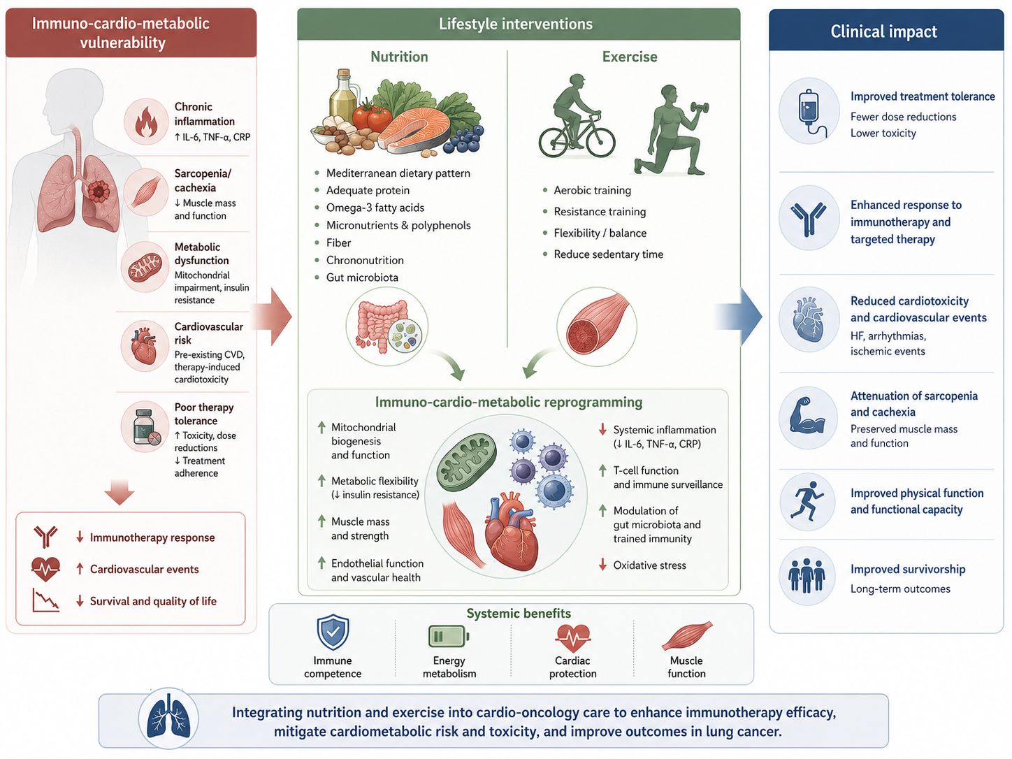
Integrating Nutrition and Exercise to Mitigate Cardiometabolic Risk and Enhance Outcomes in Lung Cancer During the Era of Immunotherapy and Targeted Therapy
 , , , , and
Abstract
1. Introduction
- Endothelial dysfunction: loss of endothelial integrity impairs the function of endothelial cells, which act as key sensors of hemodynamic forces and play a central role in vascular homeostasis, resilience, and adaptation to environmental stressors (exposome).
- Chronic inflammation: persistent inflammatory activation promotes immune dysregulation, contributing to the development of several noncommunicable diseases, including CVD and cancer, supporting the concept of a common pathogenic soil.
- Cellular proliferation: activation of mitogenic pathways can promote uncontrolled cell growth as well as cardiovascular remodeling and cardiac fibrosis.
- Resistance to cell death: dysregulation of cellular stress responses and apoptotic pathways favors cell survival under adverse conditions.
- Neurohormonal activation: increased levels of cardiovascular neurohormones play a pivotal role in acute and chronic heart failure; however, similar mediators may also be produced by malignant cells within the tumor microenvironment.
- Angiogenesis: angiogenic pathways are involved in endothelial cell survival and contribute to tumor growth, invasion, and metastatic spread.
- Genomic instability: genomic alterations, including clonal hematopoiesis of indeterminate potential (CHIP), have emerged as risk factors for CVD and may also be linked to chronic inflammatory and atherosclerotic processes through sustained stimulation of hematopoietic stem cell proliferation.
- Metabolic reprogramming: Both cardiomyocytes and cancer cells exhibit remarkable metabolic adaptability, enabling the preservation of cellular functions during stress conditions. Various stressors may disrupt the balance between ATP demand and oxidative metabolism. In the failing heart, adaptive responses include a shift from oxidative phosphorylation toward glycolytic ATP production, with specific metabolites such as ketone bodies potentially acting as epigenetic regulators. A similar metabolic phenotype is observed in cancer cells; however, whereas metabolic adaptation in the heart serves a compensatory and protective role, in tumors metabolic remodeling sustains malignant progression and phenotype maintenance.
2. Metabolic and Cardiovascular Alterations in Lung Cancer
2.1. Systemic Inflammation and Cardiometabolic Risk
2.2. Cancer Cachexia and Sarcopenia
2.3. Cancer Therapy-Induced Cardiac Metabolic Dysfunction
3. Nutritional Status and Energy Requirements
3.1. Assessment of Nutritional Risk
3.2. Energy and Protein Requirements
3.3. Macronutrient Composition and Dietary Patterns
3.4. Micronutrients
3.5. Meal Timing and Chrononutrition
3.6. The Role of Trained Immunity
4. Exercise
4.1. The Multitargeted Effects of Exercise: From Myokines to Exerkines
4.2. Role of Exercise in Cancer Treatment-Induced Metabolic Syndrome (CTMS)
4.3. Clinical Implementation and Safety
4.4. Barriers/Solutions (Training at Home and Hybrid Training)
4.5. Integration with Nutritional Support
4.6. Physical Activity and Trained Immunity
5. Integrative Approaches and Clinical Evidence
5.1. Multimodal Interventions
5.2. The Multifaceted Network of Gut Microbiota
5.3. Personalized Lifestyle Interventions
6. Future Perspectives
7. Conclusions
Author Contributions
Funding
Data Availability Statement
Conflicts of Interest
Abbreviations
| AECs | Airborne environmental contaminants |
| ALK/ROS1 | Anaplastic lymphoma kinase/c-ros oncogene 1 |
| AHA | American Heart Association |
| BAT | Brown adipose tissue |
| BC | Breast cancer |
| BIA | Bioimpedance analysis |
| BP | Blood Pressure |
| BRAF/MEK V | Raf murine sarcoma viral oncogene homolog B/mitogen-activated extracellular signal-regulated kinase |
| ChemR23 | Chemerin Receptor 23 |
| CKM | Cardiovascular–kidney–metabolic |
| CONUT | Controlling Nutritional Status |
| CRF | Cardiorespiratory fitness |
| CRP | C-reactive protein |
| CT | Computed tomography |
| CTGF | Connective tissue growth factor |
| CTMS | Cancer treatment-induced metabolic syndrome |
| CV | Cardiovascular |
| CVD | Cardiovascular disease |
| DASH | Dietary Approaches to Stop Hypertension |
| DEXA | Dual-energy X-ray absorptiometry |
| EC | Endothelial cells |
| FGF | Fibroblast growth factor |
| GM | Gut microbiota |
| HDL | High-density lipoprotein |
| HF | Heart failure |
| H2S | Hydrogen sulfide |
| hs-CRP | High-sensitivity C-reactive protein |
| ICIs | Immune checkpoint inhibitors |
| IL | Interleukin |
| IGF | Insulin-like growth factor |
| IGFBP | Insulin-like growth factor-binding protein |
| irAEs | Immune-related adverse events |
| LC | Lung cancer |
| LDL | Low-density lipoprotein |
| LRR | [leucine-rich repeat]-containing |
| MD | Mediterranean diet |
| MMP | Matrix metalloproteinase |
| mtDNA | Mitochondrial DNA |
| MUST | Malnutrition Universal Screening Tool |
| NCCN | National Comprehensive Cancer Network |
| NK | Natural killer |
| NLRP3 NOD | [Nucleotide oligomerization domain]-containing, LRR [leucine-rich repeat]-containing, and PYD [pyrin domain]-containing protein3 |
| NRS-2002 | Nutritional risk score (screening)-2002 |
| PDGF | Platelet-derived growth factor |
| PG-SGA | Patient-Generated Subjective Global Assessment |
| RCTs | Randomized controlled trials (RCTs) |
| RET | Rearranged during Transfection |
| SCFA | Short-chain fatty acids |
| SDOH | Social determinants of health |
| TGF-b | Transforming growth factor beta |
| TRE | Time-restricted eating |
| VEGF | Vascular endothelial growth factor |
| WAT | White adipose tissue |
References
- Bray, F.; Laversanne, M.; Sung, H.; Ferlay, J.; Siegel, R.L.; Soerjomataram, I.; Jemal, A. Global cancer statistics 2022: GLOBOCAN estimates of incidence and mortality worldwide for 36 cancers in 185 countries. CA Cancer J. Clin. 2024, 74, 229–263. [Google Scholar] [CrossRef] [PubMed]
- Noone, A.M.; Howlader, N.; Krapcho, M.; Miller, D.; Brest, A.; Yu, M.; Ruhl, J.; Tatalovich, Z.; Mariotto, A.; Lewis, D.R.; et al. SEER Cancer Statistics Review. National Cancer Institute. 2015. Available online: https://seer.cancer.gov/csr/1975_2015/sections.html (accessed on 20 April 2026).
- Divo, M.J.; Martinez, C.H.; Mannino, D.M. Ageing and the epidemiology of multimorbidity. Eur. Respir. J. 2014, 44, 1055–1068. [Google Scholar] [CrossRef] [PubMed]
- Chang, W.H.; Lai, A.G. Pan-cancer analyses of the associations between 109 pre-existing conditions and cancer treatment patterns across 19 adult cancers. Sci. Rep. 2024, 14, 464. [Google Scholar] [CrossRef] [PubMed] [PubMed Central]
- Moore, S.C.; Lee, I.M.; Weiderpass, E.; Campbell, P.T.; Sampson, J.N.; Kitahara, C.M.; Keadle, S.K.; Arem, H.; Berrington de Gonzalez, A.; Hartge, P.; et al. Association of Leisure-Time Physical Activity with Risk of 26 Types of Cancer in 1.44 Million Adults. JAMA Intern. Med. 2016, 176, 816–825. [Google Scholar] [CrossRef] [PubMed]
- Xue, X.J.; Gao, Q.; Qiao, J.H.; Zhang, J.; Xu, C.P.; Liu, J. Red and processed meat consumption and the risk of lung cancer: A dose-response meta-analysis of 33 published studies. Int. J. Clin. Exp. Med. 2014, 7, 1542–1553. [Google Scholar] [PubMed]
- Leiter, A.; Veluswamy, R.R.; Wisnivesky, J.P. The global burden of lung cancer: Current status and future trends. Nat. Rev. Clin. Oncol. 2023, 20, 624–639. [Google Scholar] [CrossRef] [PubMed]
- Edwards, B.K.; Noone, A.M.; Mariotto, A.B.; Simard, E.P.; Boscoe, F.P.; Henley, S.J.; Jemal, A.; Cho, H.; Anderson, R.N.; Kohler, B.A.; et al. Annual Report to the Nation on the status of cancer, 1975–2010, featuring prevalence of comorbidity and impact on survival among persons with lung, colorectal, breast, or prostate cancer. Cancer 2014, 120, 1290–1314. [Google Scholar] [CrossRef] [PubMed]
- Islam, K.M.M.; Jiang, X.; Anggondowati, T.; Lin, G.; Ganti, A.K. Comorbidity and survival in lung cancer patients. Cancer Epidemiol. Biomark. Prev. 2015, 24, 1079–1085. [Google Scholar] [CrossRef] [PubMed]
- Pluchart, H.; Chanoine, S.; Moro-Sibilot, D.; Chouaid, C.; Frey, G.; Villa, J.; Degano, B.; Giaj Levra, M.; Bedouch, P.; Toffart, A.C. Lung cancer, comorbidities, and medication: The infernal trio. Front. Pharmacol. 2024, 14, 1016976. [Google Scholar] [CrossRef] [PubMed] [PubMed Central]
- Ganatra, S.; Barac, A.; Armenian, S.; Cambareri, C.; Denlinger, C.S.; Dent, S.F.; Hayek, S.; Ky, B.; Leja, M.; Lucas, C.H.; et al. Diagnosis and Management of Cardiovascular Adverse Effects of Targeted Oncology Therapies: Bruton’s Tyrosine Kinase, Immune Checkpoint, and Vascular Endothelial Growth Factor Inhibitors: 2025 ACC Concise Clinical Guidance: A Report of the American College of Cardiology Solution Set Oversight Committee. J. Am. Coll. Cardiol. 2026, 87, 654–682. [Google Scholar] [CrossRef] [PubMed]
- Battisti, N.M.L.; Welch, C.A.; Sweeting, M.; de Belder, M.; Deanfield, J.; Weston, C.; Peake, M.D.; Adlam, D.; Ring, A. Prevalence of Cardiovascular Disease in Patients with Potentially Curable Malignancies: A National Registry Dataset Analysis. JACC CardioOncol. 2022, 4, 238–253. [Google Scholar] [CrossRef] [PubMed]
- Mitchell, J.D.; Laurie, M.; Xia, Q.; Dreyfus, B.; Jain, N.; Jain, A.; Lane, D.; Lenihan, D.J. Risk profiles and incidence of cardiovascular events across different cancer types. ESMO Open 2023, 8, 101830. [Google Scholar] [CrossRef] [PubMed]
- Ogedegbe, O.J.; Odugbemi, O.P.; Tabowei, G.; Alugba, G.; Pius, R.; Nwogwugwu, E.; Nwaezeapu, K.I. Rising Cardiovascular mortality in Lung cancer patients results from a large cancer database retrospective cohort study. J. Am. Coll. Cardiol. 2025, 85, 2874. [Google Scholar] [CrossRef]
- Sun, J.Y.; Zhang, Z.Y.; Qu, Q.; Wang, N.; Zhang, Y.M.; Miao, L.F.; Wang, J.; Wu, L.D.; Liu, Y.; Zhang, C.Y.; et al. Cardiovascular disease-specific mortality in 270,618 patients with non-small cell lung cancer. Int. J. Cardiol. 2021, 330, 186–193. [Google Scholar] [CrossRef] [PubMed]
- Batra, A.; Sheka, D.; Kong, S.; Cheung, W.Y. Impact of pre-existing cardiovascular disease on treatment patterns and survival outcomes in patients with lung cancer. BMC Cancer 2020, 20, 1004. [Google Scholar] [CrossRef] [PubMed]
- Kobo, O.; Raisi-Estabragh, Z.; Gevaert, S.; Rana, J.S.; Van Spall, H.G.C.; Roguin, A.; Petersen, S.E.; Ky, B.; Mamas, M.A. Impact of cancer diagnosis on distribution and trends of cardiovascular hospitalizations in the USA between 2004 and 2017. Eur. Heart J. Qual. Care Clin. Outcomes 2022, 8, 787–797. [Google Scholar] [CrossRef] [PubMed]
- Bell, C.F.; Lei, X.; Haas, A.; Baylis, R.A.; Gao, H.; Luo, L.; Giordano, S.H.; Wehner, M.R.; Nead, K.T.; Leeper, N.J. Risk of Cancer After Diagnosis of Cardiovascular Disease. JACC CardioOncol. 2023, 5, 431–440. [Google Scholar] [CrossRef] [PubMed]
- Epeslidou, E.; Scott, J.S.; de Klein, B.; Cudia, J.T.; Melgert, B.; Prekovic, S. Microplastics as environmental modifiers of lung disease. EMBO Mol. Med. 2026, 18, 381–395. [Google Scholar] [CrossRef] [PubMed] [PubMed Central]
- Narayan, V.; Thompson, E.W.; Demissei, B.; Ho, J.E.; Januzzi, J.L., Jr.; Ky, B. Mechanistic Biomarkers Informative of Both Cancer and Cardiovascular Disease: JACC State-of-the-Art Review. J. Am. Coll. Cardiol. 2020, 75, 2726–2737. [Google Scholar] [CrossRef] [PubMed]
- Wilcox, N.S.; Amit, U.; Reibel, J.B.; Berlin, E.; Howell, K.; Ky, B. Cardiovascular disease and cancer: Shared risk factors and mechanisms. Nat. Rev. Cardiol. 2024, 21, 617–631. [Google Scholar] [CrossRef] [PubMed] [PubMed Central]
- Karlstaedt, A.; Taegtmeyer, H. Cardio-Onco-Metabolism—Metabolic vulnerabilities in cancer and the heart. J. Mol. Cell. Cardiol. 2022, 171, 71–80. [Google Scholar] [CrossRef] [PubMed] [PubMed Central]
- Miramontes-González, J.P.; Rodrigo-Alaíz, Á.; Briongos-Figuero, L.; Olivares-Hernández, A.; Carretero-Gómez, J.; Gabella-Martín, M.; Sebastián-Rodríguez, J.; Corral-Gudino, L. Nutrition in lung cancer treatment: The forgotten pillar of care? Chin. Clin. Oncol. 2025, 14, 43. [Google Scholar] [CrossRef] [PubMed]
- Yang, M.; Shen, Y.; Tan, L.; Li, W. Prognostic Value of Sarcopenia in Lung Cancer: A Systematic Review and Meta-analysis. Chest 2019, 156, 101–111. [Google Scholar] [CrossRef] [PubMed]
- El-Rayes, M.; Nardi Agmon, I.; Yu, C.; Osataphan, N.; Yu, H.A.; Hope, A.; Sacher, A.; Yu, A.F.; Abdel-Qadir, H.; Thavendiranathan, P. Lung Cancer and Cardiovascular Disease: Common Pathophysiology and Treatment-Emergent Toxicity. JACC CardioOncol. 2025, 7, 325–344. [Google Scholar] [CrossRef] [PubMed] [PubMed Central]
- Hawryszko, M.; Sławiński, G.; Tomasik, B.; Lewicka, E. Cardiac Arrhythmias in Patients Treated for Lung Cancer: A Review. Cancers 2023, 15, 5723. [Google Scholar] [CrossRef] [PubMed] [PubMed Central]
- Howlader, N.; Forjaz, G.; Mooradian, M.J.; Meza, R.; Kong, C.Y.; Cronin, K.A.; Mariotto, A.B.; Lowy, D.R.; Feuer, E.J. The Effect of Advances in Lung-Cancer Treatment on Population Mortality. N. Engl. J. Med. 2020, 383, 640–649. [Google Scholar] [CrossRef] [PubMed]
- Wei, X.; Zhu, C.; Ji, M.; Fan, J.; Xie, J.; Huang, Y.; Jiang, X.; Xu, J.; Yin, R.; Du, L.; et al. Diet and Risk of Incident Lung Cancer: A Large Prospective Cohort Study in UK Biobank. Am. J. Clin. Nutr. 2021, 114, 2043–2051. [Google Scholar] [CrossRef] [PubMed]
- Ortiz, C.; López-Cuadrado, T.; Rodríguez-Blázquez, C.; Pastor-Barriuso, R.; Galán, I. Clustering of unhealthy lifestyle behaviors, self-rated health and disability. Prev. Med. 2022, 155, 106911. [Google Scholar] [CrossRef] [PubMed]
- Ding, D.; Rogers, K.; van der Ploeg, H.; Stamatakis, E.; Bauman, A.E. Traditional and Emerging Lifestyle Risk Behaviors and All-Cause Mortality in Middle-Aged and Older Adults: Evidence from a Large Population-Based Australian Cohort. PLoS Med. 2015, 12, e1001917. [Google Scholar] [CrossRef] [PubMed]
- Zhang, Y.; Yang, J.; Na, X.; Zhao, A. Association between β-carotene supplementation and risk of cancer: A meta-analysis of randomized controlled trials. Nutr. Rev. 2023, 81, 1118–1130. [Google Scholar] [CrossRef] [PubMed]
- McAleer, J.P.; Kolls, J.K. Contributions of the intestinal microbiome in lung immunity. Eur. J. Immunol. 2018, 48, 39–49. [Google Scholar] [PubMed]
- Koh, A.; De Vadder, F.; Kovatcheva-Datchary, P.; Bäckhed, F. From dietary fiber to host physiology: Short-chain fatty acids as key bacterial metabolites. Cell 2016, 165, 1332–1345. [Google Scholar] [CrossRef] [PubMed]
- Roeland, E.J.; Bohlke, K.; Baracos, V.E.; Bruera, E.; Del Fabbro, E.; Dixon, S.; Fallon, M.; Herrstedt, J.; Lau, H.; Platek, M.; et al. Management of Cancer Cachexia: ASCO Guideline. J. Clin. Oncol. 2020, 38, 2438–2453. [Google Scholar] [CrossRef] [PubMed]
- Baguley, B.J.; Edbrooke, L.; Denehy, L.; Prado, C.M.; Kiss, N. A rapid review of nutrition and exercise approaches to managing unintentional weight loss, muscle loss, and malnutrition in cancer. Oncologist 2025, 30, oyae261. [Google Scholar] [CrossRef] [PubMed] [PubMed Central]
- Libby, P.; Kobold, S. Inflammation: A common contributor to cancer, aging, and cardiovascular diseases-expanding the concept of cardio-oncology. Cardiovasc. Res. 2019, 115, 824–829. [Google Scholar] [CrossRef] [PubMed] [PubMed Central]
- Tilg, H.; Ianiro, G.; Gasbarrini, A.; Adolph, T.E. Adipokines: Masterminds of metabolic inflammation. Nat. Rev. Immunol. 2025, 25, 250–265. [Google Scholar] [CrossRef] [PubMed]
- Yao, J.; Sterling, K.; Wang, Z.; Zhang, Y.; Song, W. The role of inflammasomes in human diseases and their potential as therapeutic targets. Signal Transduct. Target. Ther. 2024, 9, 10. [Google Scholar] [CrossRef] [PubMed]
- Tall, A.R.; Bornfeldt, K.E. Inflammasomes and Atherosclerosis: A Mixed Picture. Circ. Res. 2023, 132, 1505–1520. [Google Scholar] [CrossRef] [PubMed]
- Gimbrone, M.A., Jr.; García-Cardeña, G. Endothelial cell dysfunction and the pathobiology of atherosclerosis. Circ. Res. 2016, 118, 620–636. [Google Scholar] [CrossRef] [PubMed]
- Eelen, G.; de Zeeuw, P.; Simons, M.; Carmeliet, P. Endothelial cell metabolism in normal and diseased vasculature. Circ. Res. 2015, 116, 1231–1244. [Google Scholar] [CrossRef] [PubMed]
- Hsu, T.; Nguyen-Tran, H.H.; Trojanowska, M. Active roles of dysfunctional vascular endothelium in fibrosis and cancer. J. Biomed. Sci. 2019, 26, 86. [Google Scholar] [CrossRef] [PubMed]
- Engelen, S.E.; Robinson, A.J.B.; Zurke, Y.X.; Monaco, C. Therapeutic strategies targeting inflammation and immunity in atherosclerosis: How to proceed? Nat. Rev. Cardiol. 2022, 19, 522–542. [Google Scholar] [CrossRef] [PubMed]
- Silva, E.M.; Mariano, V.S.; Pastrez, P.R.; Pinto, M.C.; Castro, A.G.; Syrjanen, K.J.; Longatto-Filho, A. High systemic IL-6 is associated with worse prognosis in patients with non-small cell lung cancer. PLoS ONE 2017, 12, e0181125. [Google Scholar] [CrossRef] [PubMed] [PubMed Central]
- Wen, Y.; Wang, X.; Meng, W.; Guo, W.; Duan, C.; Cao, J.; Kang, L.; Guo, N.; Lin, Q.; Lv, P.; et al. TNF-α-dependent lung inflammation upregulates PD-L1 in monocyte-derived macrophages to contribute to lung tumorigenesis. FASEB J. 2022, 36, e22595. [Google Scholar] [CrossRef] [PubMed]
- Li, R.; Ong, S.L.; Tran, L.M.; Jing, Z.; Liu, B.; Park, S.J.; Huang, Z.L.; Walser, T.C.; Heinrich, E.L.; Lee, G.; et al. Chronic IL-1β-induced inflammation regulates epithelial-to-mesenchymal transition memory phenotypes via epigenetic modifications in non-small cell lung cancer. Sci. Rep. 2020, 10, 377, Erratum in Sci. Rep. 2020, 10, 4386. https://doi.org/10.1038/s41598-020-61341-3. [Google Scholar] [CrossRef] [PubMed] [PubMed Central]
- Hu, W.; Ru, Z.; Zhou, Y.; Xiao, W.; Sun, R.; Zhang, S.; Gao, Y.; Li, X.; Zhang, X.; Yang, H. Lung cancer-derived extracellular vesicles induced myotube atrophy and adipocyte lipolysis via the extracellular IL-6-mediated STAT3 pathway. Biochim. Biophys. Acta Mol. Cell Biol. Lipids 2019, 1864, 1091–1102. [Google Scholar] [CrossRef] [PubMed]
- Zheng, L.; Chen, X.; He, X.; Wei, H.; Li, X.; Tan, Y.; Min, J.; Chen, M.; Zhang, Y.; Dong, M.; et al. METTL4-Mediated Mitochondrial DNA N6-Methyldeoxyadenosine Promoting Macrophage Inflammation and Atherosclerosis. Circulation 2025, 151, 946–965. [Google Scholar] [CrossRef] [PubMed] [PubMed Central]
- Sharma, B.; Dabur, R. Role of Pro-inflammatory Cytokines in Regulation of Skeletal Muscle Metabolism: A Systematic Review. Curr. Med. Chem. 2020, 27, 2161–2188. [Google Scholar] [CrossRef] [PubMed]
- Abid, H.; Ryan, Z.C.; Delmotte, P.; Sieck, G.C.; Lanza, I.R. Extramyocellular interleukin-6 influences skeletal muscle mitochondrial physiology through canonical JAK/STAT signaling pathways. FASEB J. 2020, 34, 14458–14472. [Google Scholar] [CrossRef] [PubMed]
- Murphy, S.P.; Kakkar, R.; McCarthy, C.P.; Januzzi, J.L., Jr. Inflammation in Heart Failure: JACC State-of-the-Art Review. J. Am. Coll. Cardiol. 2020, 75, 1324–1340. [Google Scholar] [CrossRef] [PubMed]
- Zhang, H.; Dhalla, N.S. The Role of Pro-Inflammatory Cytokines in the Pathogenesis of Cardiovascular Disease. Int. J. Mol. Sci. 2024, 25, 1082. [Google Scholar] [CrossRef] [PubMed] [PubMed Central]
- Steyers, C.M., 3rd; Miller, F.J., Jr. Endothelial dysfunction in chronic inflammatory diseases. Int. J. Mol. Sci. 2014, 15, 11324–11349. [Google Scholar] [CrossRef] [PubMed] [PubMed Central]
- Henein, M.Y.; Vancheri, S.; Longo, G.; Vancheri, F. The Role of Inflammation in Cardiovascular Disease. Int. J. Mol. Sci. 2022, 23, 12906. [Google Scholar] [CrossRef] [PubMed] [PubMed Central]
- Agca, S.; Kir, S. The role of interleukin-6 family cytokines in cancer cachexia. FEBS J. 2024, 291, 4009–4023. [Google Scholar] [CrossRef] [PubMed]
- Karlstaedt, A.; Barrett, M.; Hu, R.; Gammons, S.T.; Ky, B. Cardio-Oncology: Understanding the Intersections Between Cardiac Metabolism and Cancer Biology. JACC Basic Transl. Sci. 2021, 6, 705–718. [Google Scholar] [CrossRef] [PubMed] [PubMed Central]
- Bazoukis, G.; Loscalzo, J.; Hall, J.L.; Bollepalli, S.C.; Singh, J.P.; Armoundas, A.A. Impact of Social Determinants of Health on Cardiovascular Disease. J. Am. Heart Assoc. 2025, 14, e039031. [Google Scholar] [CrossRef] [PubMed] [PubMed Central]
- Parcha, V.; Josey, G.; Patton, M.; Verma, A.; Armstrong, N.; Irvin, M.; Sethu, P.; Payne, G.; Clarkson, S. Abstract 02: Systemic Inflammation and Traditional Cardiovascular-Kidney-Metabolic Risk Mediate the Association Between Social Determinants of Health and Incident Heart Failure: Insights from A Multi-Ethnic Cohort. Circulation 2026, 153. [Google Scholar] [CrossRef]
- Powell-Wiley, T.M.; Baumer, Y.; Baah, F.O.; Baez, A.S.; Farmer, N.; Mahlobo, C.T.; Pita, M.A.; Potharaju, K.A.; Tamura, K.; Wallen, G.R. Social determinants of cardiovascular disease. Circ. Res. 2022, 130, 782–799. [Google Scholar] [CrossRef] [PubMed]
- Gilmore, L.A.; Willmann, J.; Olaechea, S.; Gilmore, B.W.; Dee, E.C.; Rao, M.; Gannavarapu, B.S.; Venkateswaran, S.; Alvarez, C.M.; Ahn, C.; et al. Prevalence and Development of Cachexia Before and After Diagnosis of Non-small Cell Lung Cancer. Am. J. Clin. Oncol. 2025, 48, 470–476. [Google Scholar] [CrossRef] [PubMed]
- Arends, J.; Strasser, F.; Gonella, S.; Solheim, T.S.; Madeddu, C.; Ravasco, P.; Buonaccorso, L.; de van der Schueren, M.A.E.; Baldwin, C.; ESMO Guidelines Committee; et al. Cancer cachexia in adult patients: ESMO Clinical Practice Guidelines. ESMO Open 2021, 6, 100092, Erratum in ESMO Open 2022, 7, 100614. https://doi.org/10.1016/j.esmoop.2022.100614. [Google Scholar] [CrossRef] [PubMed] [PubMed Central]
- Madeddu, C.; Busquets, S.; Donisi, C.; Lai, E.; Pretta, A.; López-Soriano, F.J.; Argilés, J.M.; Scartozzi, M.; Macciò, A. Effect of Cancer-Related Cachexia and Associated Changes in Nutritional Status, Inflammatory Status, and Muscle Mass on Immunotherapy Efficacy and Survival in Patients with Advanced Non-Small Cell Lung Cancer. Cancers 2023, 15, 1076. [Google Scholar] [CrossRef] [PubMed] [PubMed Central]
- Cortellini, A.; Verna, L.; Porzio, G.; Bozzetti, F.; Palumbo, P.; Masciocchi, C.; Cannita, K.; Parisi, A.; Brocco, D.; Tinari, N.; et al. Predictive value of skeletal muscle mass for immunotherapy with nivolumab in non-small cell lung cancer patients: A “hypothesis-generator” preliminary report. Thorac. Cancer 2019, 10, 347–351. [Google Scholar] [CrossRef] [PubMed]
- Cruz-Jentoft, A.J.; Sayer, A.A. Sarcopenia. Lancet 2019, 393, 2636–2646, Erratum in Lancet 2019, 393, 2590. https://doi.org/10.1016/S0140-6736(19)31465-5. [Google Scholar] [CrossRef] [PubMed]
- Damluji, A.A.; Alfaraidhy, M.; AlHajri, N.; Rohant, N.N.; Kumar, M.; Al Malouf, C.; Bahrainy, S.; Ji Kwak, M.; Batchelor, W.B.; Forman, D.E.; et al. Sarcopenia and Cardiovascular Diseases. Circulation 2023, 147, 1534–1553. [Google Scholar] [CrossRef] [PubMed]
- Wang, J.; Cao, L.; Xu, S. Sarcopenia affects clinical efficacy of immune checkpoint inhibitors in non-small cell lung cancer patients: A systematic review and meta-analysis. Int. Immunopharmacol. 2020, 88, 106907. [Google Scholar] [CrossRef] [PubMed]
- Ren, B.; Shen, J.; Qian, Y.; Zhou, T. Sarcopenia as a Determinant of the Efficacy of Immune Checkpoint Inhibitors in Non-Small Cell Lung Cancer: A Meta-Analysis. Nutr. Cancer 2023, 75, 685–695. [Google Scholar] [PubMed]
- Guzman-Prado, Y.; Ben Shimol, J.; Samson, O. Sarcopenia and the risk of adverse events in patients treated with immune checkpoint inhibitors: A systematic review. Cancer Immunol. Immunother. 2021, 70, 2771–2780. [Google Scholar] [CrossRef] [PubMed] [PubMed Central]
- Baracos, V.E.; Martin, L.; Korc, M.; Guttridge, D.C.; Fearon, K.C.H. Cancer-associated cachexia. Nat. Rev. Dis. Prim. 2018, 4, 17105. [Google Scholar] [CrossRef] [PubMed]
- Argilés, J.M.; Busquets, S.; Stemmler, B.; López-Soriano, F.J. Cancer cachexia: Understanding the molecular basis. Nat. Rev. Cancer 2014, 14, 754–762. [Google Scholar] [CrossRef] [PubMed]
- Chan, S.H.Y.; Fitzpatrick, R.W.; Layton, D.; Webley, S.; Salek, S. Cancer Therapy-Induced Cardiotoxicity: Results of the Analysis of the UK DEFINE Database. Cancers 2025, 17, 311. [Google Scholar] [CrossRef] [PubMed]
- Demkow, U.; Stelmaszczyk-Emmel, A. Cardiotoxicity of cisplatin-based chemotherapy in advanced non-small cell lung cancer patients. Respir. Physiol. Neurobiol. 2013, 187, 64–67. [Google Scholar] [CrossRef] [PubMed]
- Bennetts, J.D.; Sverdlov, A.L.; Ngo, D.T. Cardiometabolic perturbations arising from treatment with novel anticancer therapies. Trends Cardiovasc. Med. 2025, 36, 218–228. [Google Scholar] [CrossRef] [PubMed]
- Walls, G.M.; Bergom, C.; Mitchell, J.D.; Rentschler, S.L.; Hugo, G.D.; Samson, P.P.; Robinson, C.G. Cardiotoxicity following thoracic radiotherapy for lung cancer. Br. J. Cancer 2025, 132, 311–325, Erratum in Br. J. Cancer 2025, 132, 401–407. [Google Scholar] [CrossRef] [PubMed]
- Gougis, P.; Jochum, F.; Abbar, B.; Dumas, E.; Bihan, K.; Lebrun-Vignes, B.; Moslehi, J.; Spano, J.P.; Laas, E.; Hotton, J.; et al. Clinical spectrum and evolution of immune-checkpoint inhibitors toxicities over a decade-a worldwide perspective. EClinicalMedicine 2024, 70, 102536. [Google Scholar] [CrossRef] [PubMed]
- Jain, P.; Bugarin, J.G.; Guha, A.; Jain, C.; Patil, N.; Shen, T.; Stanevich, I.; Nikore, V.; Margolin, K.; Ernstoff, M.; et al. Cardiovascular adverse events are associated with usage of immune checkpoint inhibitors in real-world clinical data across the United States. ESMO Open 2021, 6, 100252, Erratum in ESMO Open 2021, 6, 100286. [Google Scholar] [CrossRef] [PubMed]
- Cheng, X.; Lin, J.; Wang, B.; Huang, S.; Liu, M.; Yang, J. Clinical characteristics and influencing factors of anti-PD-1/PD-L1-related severe cardiac adverse event: Based on FAERS and TCGA databases. Sci. Rep. 2024, 14, 22199. [Google Scholar] [CrossRef] [PubMed]
- D’Souza, M.; Nielsen, D.; Svane, I.M.; Iversen, K.; Rasmussen, P.V.; Madelaire, C.; Fosbøl, E.; Køber, L.; Gustafsson, F.; Andersson, C.; et al. The risk of cardiac events in patients receiving immune checkpoint inhibitors: A nationwide Danish study. Eur. Heart J. 2021, 42, 1621–1631. [Google Scholar] [CrossRef] [PubMed]
- Li, H.; Zheng, Y.; Li, B.; Zhi, Y.; Chen, M.; Zeng, J.; Jiao, Q.; Tao, Y.; Liu, X.; Shen, Z.; et al. Association among major adverse cardiovascular events with immune checkpoint inhibitors: A systematic review and meta-analysis. J. Intern. Med. 2025, 297, 36–46. [Google Scholar] [PubMed]
- Delombaerde, D.; Oeste, C.L.; Geldhof, V.; Croes, L.; Bassez, I.; Verbiest, A.; Tack, L.; Hens, D.; Franssen, C.; Debruyne, P.R.; et al. Cardiovascular toxicities in cancer patients treated with immune checkpoint inhibitors: Multicenter study using natural language processing on Belgian hospital data. ESMO Real World Data Digit. Oncol. 2025, 7, 100111. [Google Scholar] [CrossRef] [PubMed]
- Zheng, Y.; Liu, Z.; Chen, D.; Zhang, J.; Yuan, M.; Zhang, Y.; Liu, S.; Zhang, G.; Yang, G. The Cardiotoxicity Risk of Immune Checkpoint Inhibitors Compared with Chemotherapy: A Systematic Review and Meta-analysis of Observational Studies. Cardiovasc. Toxicol. 2025, 25, 805–819. [Google Scholar] [CrossRef] [PubMed]
- Power, J.R.; Dolladille, C.; Ozbay, B.; Procureur, A.; Ederhy, S.; Palaskas, N.L.; Lehmann, L.H.; Cautela, J.; Courand, P.Y.; Hayek, S.S.; et al. International ICI-Myocarditis Registry. Immune checkpoint inhibitor-associated myocarditis: A novel risk score. Eur. Heart J. 2026, 47, 1050–1062, Erratum in Eur. Heart J. 2026, 47, 1717. https://doi.org/10.1093/eurheartj/ehaf529. [Google Scholar] [CrossRef] [PubMed]
- Salem, J.E.; Manouchehri, A.; Moey, M.; Lebrun-Vignes, B.; Bastarache, L.; Pariente, A.; Gobert, A.; Spano, J.P.; Balko, J.M.; Bonaca, M.P.; et al. Cardiovascular toxicities associated with immune checkpoint inhibitors: An observational, retrospective, pharmacovigilance study. Lancet Oncol. 2018, 19, 1579–1589. [Google Scholar] [CrossRef] [PubMed]
- Nielsen, D.L.; Juhl, C.B.; Nielsen, O.H.; Chen, I.M.; Herrmann, J. Immune checkpoint inhibitor- induced cardiotoxicity. JAMA Oncol. 2024, 10, 1390–1399. [Google Scholar] [CrossRef] [PubMed]
- Nguyen, L.S.; Cooper, L.T.; Kerneis, M.; Funck-Brentano, C.; Silvain, J.; Brechot, N.; Hekimian, G.; Ammirati, E.; Ben M’Barek, B.; Redheuil, A.; et al. Systematic analysis of drug-associated myocarditis reported in the World Health Organization pharmacovigilance database. Nat. Commun. 2022, 13, 25. [Google Scholar] [CrossRef] [PubMed]
- Waliany, S.; Zhu, H.; Wakelee, H.; Padda, S.K.; Das, M.; Ramchandran, K.; Myall, N.J.; Chen, T.; Witteles, R.M.; Neal, J.W. Pharmacovigilance Analysis of Cardiac Toxicities Associated with Targeted Therapies for Metastatic NSCLC. J. Thorac. Oncol. 2021, 16, 2029–2039. [Google Scholar] [CrossRef] [PubMed]
- Ma, Z.; Cao, F.; Liao, M.; Min, R.; Zheng, R.; Sun, X.; Chen, X.; Gong, Y.; Ai, S.; Kang, X. Cardiovascular adverse events associated with epidermal growth factor receptor tyrosine kinase inhibitors in EGFR-mutated non-small cell lung cancer: Systematic review and network meta-analysis. BMJ 2025, 390, e082834. [Google Scholar] [CrossRef] [PubMed] [PubMed Central]
- Shaw, A.T.; Bauer, T.M.; de Marinis, F.; Felip, E.; Goto, Y.; Liu, G.; Mazieres, J.; Kim, D.W.; Mok, T.; Polli, A.; et al. CROWN Trial Investigators. First-Line Lorlatinib or Crizotinib in Advanced ALK-Positive Lung Cancer. N. Engl. J. Med. 2020, 383, 2018–2029. [Google Scholar] [CrossRef] [PubMed]
- Sikkema, B.; de Leeuw, S.; Pruis, M.; Mohseni, M.; Veerman, G.D.M.; Paats, M.; Dumolin, D.W.; Smit, E.F.; Schols, A.M.W.M.; Mathissen, R.H.I.; et al. Analysis of significant weight gain in patients using alectinib for ALK-positive lung cancer. J. Clin. Oncol. 2023, 41, e21138. [Google Scholar] [CrossRef]
- Campia, U.; Moslehi, J.J.; Amiri-Kordestani, L.; Barac, A.; Beckman, J.A.; Chism, D.D.; Cohen, P.; Groarke, J.D.; Herrmann, J.; Reilly, C.M.; et al. Cardio-Oncology: Vascular and Metabolic Perspectives: A Scientific Statement from the American Heart Association. Circulation 2019, 139, e579–e602, Erratum in Circulation 2019, 139, e838–e839. https://doi.org/10.1161/CIR.0000000000000687. [Google Scholar] [CrossRef] [PubMed] [PubMed Central]
- Zhou, H.-J.; Deng, L.-J.; Wang, T.; Chen, J.-X.; Jiang, S.-Z.; Yang, L.; Liu, F.; Weng, M.-H.; Hu, J.-W.; Tan, J.-Y. Clinical practice guidelines for the nutritional risk screening and assessment of cancer patients: A systematic quality appraisal using the AGREE II instrument. Support. Care Cancer 2021, 29, 2885–2893. [Google Scholar] [CrossRef] [PubMed]
- Trestini, I.; Gkountakos, A.; Carbognin, L.; Avancini, A.; Lanza, M.; Molfino, A.; Friso, S.; Corbo, V.; Tortora, G.; Scarpa, A.; et al. Muscle derangement and alteration of the nutritional machinery in NSCLC. Crit. Rev. Oncol. Hematol. 2019, 141, 43–53. [Google Scholar] [CrossRef] [PubMed]
- Voulgaridou, G.; Tyrovolas, S.; Detopoulou, P.; Tsoumana, D.; Drakaki, M.; Apostolou, T.; Chatziprodromidou, I.P.; Papandreou, D.; Giaginis, C.; Papadopoulou, S.K. Diagnostic Criteria and Measurement Techniques of Sarcopenia: A Critical Evaluation of the Up-to-Date Evidence. Nutrients 2024, 16, 436. [Google Scholar] [CrossRef] [PubMed] [PubMed Central]
- Kondrup, J.; Rasmussen, H.H.; Hamberg, O.; Stanga, Z. An ad hoc ESPEN Working Group. Nutritional risk screening (NRS 2002): A new method based on an analysis of controlled clinical trials. Clin. Nutr. 2003, 22, 321–336. [Google Scholar] [CrossRef] [PubMed]
- Ottery, F.D. Definition of standardized nutritional assessment and interventional pathways in oncology. Nutrition 1996, 12, S15–S19. [Google Scholar] [CrossRef] [PubMed]
- Sandhu, A.; Mosli, M.; Yan, B.; Gregor, J.; Chande, N.; Ponich, T.; Beaton, M.; Wu, T.; Rahman, A. Self-Screening for malnutrition risk in outpatient inflammatory bowel disease patients using the malnutrition universal screening tool (MUST). J. Parenter. Enter. Nutr. 2016, 40, 507–510. [Google Scholar]
- Pagliaro, R.; Scalfi, L.; Di Fiore, I.; Leoni, A.; Masi, U.; D’Agnano, V.; Picone, C.; Scial, F.; Perrotta, F.; Bianco, A. Controlling Nutritional Status (CONUT) Score as a Predictor of Prognosis in Non-Small Cell Lung Cancer. Nutrients 2025, 17, 3416. [Google Scholar] [CrossRef] [PubMed]
- Cornier, M.A.; Després, J.P.; Davis, N.; Grossniklaus, D.A.; Klein, S.; Lamarche, B.; Lopez-Jimenez, F.; Rao, G.; St-Onge, M.P.; Towfighi, A.; et al. Assessing adiposity: A scientific statement from the American Heart Association. Circulation 2011, 124, 1996–2019. [Google Scholar] [CrossRef] [PubMed]
- Lee, S.Y.; Gallagher, D. Assessment methods in human body composition. Curr. Opin. Clin. Nutr. Metab. Care 2008, 11, 566–572. [Google Scholar] [CrossRef] [PubMed] [PubMed Central]
- Jung, S.H.; Hao, J.; Shivakumar, M.; Nam, Y.; Kim, J.; Kim, M.J.; Ryoo, S.B.; Choe, E.K.; Jeong, S.Y.; Park, K.J.; et al. Development and validation of a novel strong prognostic index for colon cancer through a robust combination of laboratory features for systemic inflammation: A prognostic immune nutritional index. Br. J. Cancer 2022, 126, 1539–1547. [Google Scholar] [CrossRef] [PubMed] [PubMed Central]
- An, S.; Kim, S.; Eo, W.; Lee, S. The Prognostic Immune and Nutritional Index as a Predictor of Survival in Resected Non-Small Cell Lung Cancer. Medicina 2025, 61, 1763. [Google Scholar] [CrossRef] [PubMed] [PubMed Central]
- Cederholm, T.; Bosaeus, I. Malnutrition in Adults. N. Engl. J. Med. 2024, 391, 155–165. [Google Scholar] [CrossRef] [PubMed]
- Cruz-Jentoft, A.J.; Volkert, D. Malnutrition in Older Adults. N. Engl. J. Med. 2025, 392, 2244–2255. [Google Scholar] [CrossRef] [PubMed]
- Tobberup, R.; Rasmussen, H.H.; Holst, M.; Jensen, N.A.; Falkmer, U.G.; Bøgsted, M.; Delekta, A.M.; Carus, A. Exploring the dietary protein intake and skeletal muscle during first-line anti-neoplastic treatment in patients with non-small cell lung cancer. Clin. Nutr. ESPEN 2019, 34, 94–100. [Google Scholar] [CrossRef] [PubMed]
- Montalvo, R.N.; Hardee, J.P.; VanderVeen, B.N.; Carson, J.A. Resistance Exercise’s Ability to Reverse Cancer-Induced Anabolic Resistance. Exerc. Sport. Sci. Rev. 2018, 46, 247–253. [Google Scholar] [CrossRef] [PubMed] [PubMed Central]
- Engelen, M.P.K.J.; Safar, A.M.; Bartter, T.; Koeman, F.; Deutz, N.E.P. High anabolic potential of essential amino acid mixtures in advanced nonsmall cell lung cancer. Ann. Oncol. 2015, 26, 1960–1966. [Google Scholar] [CrossRef] [PubMed] [PubMed Central]
- Ligibel, J.A.; Bohlke, K.; May, A.M.; Clinton, S.K.; Demark-Wahnefried, W.; Gilchrist, S.C.; Irwin, M.L.; Late, M.; Mansfield, S.; Marshall, T.F.; et al. Exercise, Diet, and Weight Management During Cancer Treatment: ASCO Guideline. J. Clin. Oncol. 2022, 40, 2491–2507. [Google Scholar] [CrossRef] [PubMed]
- Yi, S.Y.; Steffen, L.M.; Guan, W.; Duprez, D.; Lakshminarayan, K.; Jacobs, D.R., Jr. Dietary carbohydrate quality, fibre-rich food intake, and left ventricular structure and function: The CARDIA study. Eur. Heart J. 2025, 46, 4329–4337. [Google Scholar] [CrossRef] [PubMed] [PubMed Central]
- Mente, A.; Miller, V.; Yusuf, S. Diet and heart failure: Evidence is limited to make recommendations. Eur. Heart J. 2025, 46, 4338–4340. [Google Scholar] [CrossRef] [PubMed]
- Vest, A.R.; DiDomenico, R.J.; Lichtenstein, L.; Slater, T.; Ekpo, E.; Damluji, A.A.; Bohula, E.; Alviar, C.L. American Heart Association Acute Cardiac Care and General Cardiology Committee of the Council on Clinical Cardiology; Council on Cardiovascular and Stroke Nursing. Malnutrition and Cachexia in Inpatients with Acute Cardiac Conditions: A Scientific Statement from the American Heart Association. Circulation 2026, 153, e1078–e1105. [Google Scholar] [CrossRef] [PubMed]
- Driggin, E.; Cohen, L.P.; Gallagher, D.; Karmally, W.; Maddox, T.; Hummel, S.L.; Carbone, S.; Maurer, M.S. Nutrition Assessment and Dietary Interventions in Heart Failure: JACC Review Topic of the Week. J. Am. Coll. Cardiol. 2022, 79, 1623–1635. [Google Scholar] [CrossRef] [PubMed] [PubMed Central]
- Torres-Peña, J.D.; Rangel-Zuñiga, O.A.; Alcala-Diaz, J.F.; Lopez-Miranda, J.; Delgado-Lista, J. Mediterranean Diet and Endothelial Function: A Review of its Effects at Different Vascular Bed Levels. Nutrients 2020, 12, 2212. [Google Scholar] [CrossRef] [PubMed] [PubMed Central]
- Martínez-González, M.A.; Gea, A.; Ruiz-Canela, M. The Mediterranean Diet and Cardiovascular Health. Circ. Res. 2019, 124, 779–798. [Google Scholar] [CrossRef] [PubMed]
- Zou, T.; Zhu, M.; Ma, Y.C.; Xiao, F.; Yu, X.; Xu, L.; Ma, L.Q.; Yang, J.; Dong, J.Z. MicroRNA-410-5p exacerbates high-fat diet-induced cardiac remodeling in mice in an endocrine fashion. Sci. Rep. 2018, 8, 8780. [Google Scholar] [CrossRef] [PubMed] [PubMed Central]
- Freitas, R.D.S.; Campos, M.M. Protective effects of omega-3 fatty acids in cancer-related complications. Nutrients 2019, 11, 945. [Google Scholar] [CrossRef] [PubMed]
- Sanchez-Lara, K.; Turcott, J.G.; Juarez-Hernandez, E.; Nunez-Valencia, C.; Villanueva, G.; Guevara, P.; De la Torre-Vallejo, M.; Mohar, A.; Arrieta, O. Effects of an oral nutritional supplement containing eicosapentaenoic acid on nutritional and clinical outcomes in patients with advanced non-small cell lung cancer: Randomised trial. Clin. Nutr. 2014, 33, 1017–1023. [Google Scholar] [CrossRef] [PubMed]
- Razquin, C.; Ruiz-Canela, M.; Toledo, E.; Hernández-Alonso, P.; Clish, C.B.; Guasch-Ferré, M.; Li, J.; Wittenbecher, C.; Dennis, C.; Alonso-Gómez, A.; et al. Metabolomics of the tryptophan-kynurenine degradation pathway and risk of atrial fibrillation and heart failure: Potential modification effect of Mediterranean diet. Am. J. Clin. Nutr. 2021, 114, 1646–1654, Erratum in Am. J. Clin. Nutr. 2022, 115, 310. https://doi.org/10.1093/ajcn/nqab393. [Google Scholar] [CrossRef] [PubMed] [PubMed Central]
- Lichtenstein, A.H.; Khera, A.; Anderson, C.A.M.; Appel, L.J.; DeSilva, D.M.; Gardner, C.; Hu, F.B.; Jones, D.W.; Petersen, K.S.; American Heart Association. 2026 Dietary Guidance to Improve Cardiovascular Health: A Scientific Statement from the American Heart Association. Circulation 2026, 153, e1285–e1295. [Google Scholar] [CrossRef] [PubMed]
- Cortellino, S.; Quagliariello, V.; Delfanti, G.; Blaževitš, O.; Chiodoni, C.; Maurea, N.; Di Mauro, A.; Tatangelo, F.; Pisati, F.; Shmahala, A.; et al. Fasting mimicking diet in mice delays cancer growth and reduces immunotherapy-associated cardiovascular and systemic side effects. Nat. Commun. 2023, 14, 5529. [Google Scholar] [CrossRef] [PubMed]
- Huang, R.; Shen, Z.Y.; Huang, D.; Zhao, S.-H.; Dan, L.-X.; Wu, P.; Tang, Q.-Z.; Ma, Z.-G. Microbiota-indole-3-propionic acid-heart axis mediates the protection of leflunomide against αPD1-induced cardiotoxicity in mice. Nat. Commun. 2025, 16, 2651. [Google Scholar] [CrossRef] [PubMed]
- Sliwa, K.; Viljoen, C.A.; Hasan, B.; Ntusi, N.A.B. Nutritional Heart Disease and Cardiomyopathies: JACC Focus Seminar 4/4. J. Am. Coll. Cardiol. 2022, 81, 187–202. [Google Scholar] [CrossRef] [PubMed]
- Bikle, D.D. Vitamin D and cancer: The promise not yet fulfilled. Endocrine 2014, 46, 29–38. [Google Scholar] [CrossRef] [PubMed]
- Lewis, P.; Oster, H.; Korf, H.W.; Foster, R.G.; Erren, T.C. Food as a circadian time cue: Evidence from human studies. Nat. Rev. Endocrinol. 2020, 16, 213–223. [Google Scholar] [CrossRef] [PubMed]
- Knutson, K.L.; Dixon, D.D.; Grandner, M.A.; Jackson, C.L.; Kline, C.E.; Maher, L.; Makarem, N.; Martino, T.A.; St-Onge, M.P.; Johnson, D.A.; et al. Role of Circadian Health in Cardiometabolic Health and Disease Risk: A Scientific Statement from the American Heart Association. Circulation 2025, 152, e408–e419. [Google Scholar] [CrossRef] [PubMed]
- Stringer, E.J.; Cloke, R.W.G.; Van der Meer, L.; Murphy, R.A.; Macpherson, N.A.; Lum, J.J. The Clinical Impact of Time-restricted Eating on Cancer: A Systematic Review. Nutr. Rev. 2025, 83, e1660–e1676. [Google Scholar] [CrossRef] [PubMed] [PubMed Central]
- Verde, L.; Galasso, M.; Savastano, S.; Colao, A.; Barrea, L.; Muscogiuri, G. “Time” for obesity-related cancers: The role of chrononutrition in cancer prevention and treatment. Semin. Cancer Biol. 2025, 114, 15–28. [Google Scholar] [CrossRef] [PubMed]
- Bekkering, S.; Quintin, J.; Joosten, L.A.; van der Meer, J.W.; Netea, M.G.; Riksen, M.P. Oxidized low-density lipoprotein induce long-term proinflammatory cytokine production and foam cell formation via epigenetic reprogramming of monocytes. Arterioscler. Thromb. Vasc. Biol. 2014, 34, 1731–1738. [Google Scholar] [CrossRef] [PubMed]
- Riksen, N.P.; de Mast, Q. Diet and Trained Immunity in Cardiovascular Diseases. Arterioscler. Thromb. Vasc. Biol. 2026, 46, 51–58. [Google Scholar] [CrossRef] [PubMed]
- Riksen, N.P.; Netea, M.G.; Ait-Oufella, H.; Chavakis, T.; Hajishengallis, G. Trained immunity in cardiovascular disease. Eur. Heart J. 2026, 47, 1159–1170. [Google Scholar] [CrossRef] [PubMed]
- Christ, A.; Gunther, P.; Lautebach, M.A.R.; Duewell, P.; Biswas, D.; Pelka, K.; Scholz, C.J.; Oosting, M.; Haendler, K.; BaBier, G.K.; et al. Western diet triggers NLRP3-dependent innate immune reprogramming. Cell 2018, 172, 162–175.e14. [Google Scholar] [CrossRef] [PubMed] [PubMed Central]
- Lavillegrand, J.R.; Al-Rifai, R.; Thietart, S.; Guyon, T.; Vandestienne, M.; Cohen, R.; Duval, V.; Zhong, X.; Yen, D.; Ozturk, M.; et al. Alternating high-fat diet enhances atherosclerosis by neutrophil reprogramming. Nature 2024, 634, 447–456. [Google Scholar] [CrossRef] [PubMed] [PubMed Central]
- Mazzocchi, A.; Leone, L.; Agostoni, C.; Pali-Schöll, I. The Secrets of the Mediterranean Diet. Does [Only] Olive Oil Matter? Nutrients 2019, 11, 2941. [Google Scholar] [CrossRef] [PubMed] [PubMed Central]
- Barrea, L.; Muscogiuri, G.; Frias-Toral, E.; Laudisio, D.; Pugliese, G.; Castellucci, B.; Garcia-Velasquez, E.; Savastano, S.; Colao, A. Nutrition and immune system: From the Mediterranean diet to dietary supplementary through the microbiota. Crit. Rev. Food Sci. Nutr. 2021, 61, 3066–3090. [Google Scholar] [CrossRef] [PubMed]
- Di Tolla, M.F.; Libutti, M.; D’Onofrio, G.; Riccio, A.; Cabaro, S.; Longo, M.; Parascandolo, A.; Ferraro, G.; Formisano, E.; D’Esposito, V.; et al. Unraveling the anti-inflammatory effects of Mediterranean diet in patients with cancer remission. Front. Immunol. 2025, 16, 1666611. [Google Scholar] [CrossRef] [PubMed] [PubMed Central]
- Piercy, K.L.; Troiano, R.P.; Ballard, R.M.; Carlson, S.A.; Fulton, J.E.; Galuska, D.A.; George, S.M.; Olson, R.D. The Physical Activity Guidelines for Americans. JAMA 2018, 320, 2020–2028. [Google Scholar] [CrossRef] [PubMed] [PubMed Central]
- Poorhabibi, H.; Weiss, K.; Rosemann, T.; Knechtle, B.; Eslami, R.; Tartibian, B.; Tayebi, S.M.; Sheikhhoseini, R. Short-Lived Exercise-Induced Exerkines Modulate Inflammation for Chronic Disease Prevention: A Systematic Review and Meta-Analysis. Biomolecules 2025, 15, 1590. [Google Scholar] [CrossRef] [PubMed] [PubMed Central]
- Ruegsegger, G.N.; Pataky, M.W.; Simha, S.; Robinson, M.M.; Klaus, K.A.; Nair, K.S. High-intensity aerobic, but not resistance or combined, exercise training improves both cardiometabolic health and skeletal muscle mitochondrial dynamics. J. Appl. Physiol. (1985) 2023, 135, 763–774. [Google Scholar] [CrossRef] [PubMed] [PubMed Central]
- Zhao, Y.C.; Gao, B.H. Integrative effects of resistance training and endurance training on mitochondrial remodeling in skeletal muscle. Eur. J. Appl. Physiol. 2024, 124, 2851–2865. [Google Scholar] [CrossRef] [PubMed]
- Tucker, W.; Fegers-Wustrow, I.; Halle, M.; Haykowsky, M.J.; Chung, E.H.; Kovacic, J.C. Exercise for Primary and Secondary Prevention of Cardiovascular Disease: JACC Focus Seminar 1/4. J. Am. Coll. Cardiol. 2022, 80, 1091–1106. [Google Scholar] [CrossRef] [PubMed]
- Vega, R.B.; Konhilas, J.P.; Kelly, D.P.; Leinwand, L.A. Molecular Mechanisms Underlying Cardiac Adaptation to Exercise. Cell Metab. 2017, 25, 1012–1026. [Google Scholar] [CrossRef] [PubMed] [PubMed Central]
- Smith, J.A.B.; Murach, K.A.; Dyar, K.A.; Zierath, J.R. Exercise metabolism and adaptation in skeletal muscle. Nat. Rev. Mol. Cell Biol. 2023, 24, 607–632. [Google Scholar] [CrossRef] [PubMed] [PubMed Central]
- Penna, C.; Alloatti, G.; Crisafulli, A. Mechanisms Involved in Cardioprotection Induced by Physical Exercise. Antioxid. Redox Signal. 2020, 32, 1115–1134. [Google Scholar] [CrossRef] [PubMed]
- Wang, Q.; Huang, Z.; Chair, S.Y. Exercise-based interventions for preventing and treating cancer therapy-related cardiovascular toxicity: A systematic review and meta-analysis. BMC Cardiovasc. Disord. 2025, 25, 433. [Google Scholar] [CrossRef] [PubMed] [PubMed Central]
- Safdar, A.; Saleem, A.; Tarnopolsky, M.A. The potential of endurance exercise-derived exosomes to treat metabolic diseases. Nat. Rev. Endocrinol. 2016, 12, 504–517. [Google Scholar] [CrossRef] [PubMed]
- Chow, L.S.; Gerszten, R.E.; Taylor, J.M.; Pedersen, B.K.; van Praag, H.; Trappe, S.; Febbraio, M.A.; Galis, Z.S.; Gao, Y.; Haus, J.M.; et al. Exerkines in health, resilience and disease. Nat. Rev. Endocrinol. 2022, 18, 273–289. [Google Scholar] [CrossRef] [PubMed] [PubMed Central]
- Raschke, S.; Eckel, J. Adipo-myokines: Two sides of the same coin--mediators of inflammation and mediators of exercise. Mediat. Inflamm. 2013, 2013, 320724. [Google Scholar] [CrossRef] [PubMed] [PubMed Central]
- Tichy, L.; Parry, T.L. Low-Intensity Exercise Attenuates Immune Checkpoint Inhibitor-Induced Cardiotoxicity via Regulation of Metabolism and Autophagy. Cancers 2025, 18, 138. [Google Scholar] [CrossRef] [PubMed] [PubMed Central]
- Liu, L.; Deng, Z.; Fang, R.; Zou, M.; Ren, J.; Gao, Y.; Peng, J.; Hao, L. Exercise and CD8+ T cells: Mechanisms of immune modulation in antitumor responses. J. Mol. Med. 2026, 104, 54. [Google Scholar] [CrossRef] [PubMed]
- Hapuarachi, B.; Danson, S.; Wadsley, J.; Muthana, M. Exercise to transform tumours from cold to hot and improve immunotherapy responsiveness. Front. Immunol. 2023, 14, 1335256. [Google Scholar] [CrossRef] [PubMed] [PubMed Central]
- Koelwyn, G.J.; Quail, D.F.; Zhang, X.; White, R.M.; Jones, L.W. Exercise-dependent regulation of the tumour microenvironment. Nat. Rev. Cancer 2017, 17, 620–632. [Google Scholar] [CrossRef] [PubMed]
- Gundakaram, S.; Sirigireddy, S.; McDonald, A.; Emily King, E.; Goparaju, P.; Edwards, J.C. Evaluating the role of exercise in modulating immunity and immunotherapy outcomes in cancer: A systematic review. J. Clin. Oncol. 2025, 43, 2646. [Google Scholar] [CrossRef]
- Ochi, E.; Fukushima, T.; Katsushima, U.; Yamashita, T.; Nakano, J. Effects of exercise on people living with advanced lung cancer: A systematic review and meta-analysis. Support. Care Cancer 2026, 34, 179. [Google Scholar] [CrossRef] [PubMed]
- Ficarra, S.; Kang, D.W.; Wilson, R.L.; Gonzalo-Encabo, P.; Christopher, C.N.; Normann, A.J.; Lopez, P.; Lakićević, N.; Dieli-Conwright, C.M. Exercise medicine for individuals diagnosed with Lung Cancer: A systematic review and meta-analysis of health outcomes. Lung Cancer 2025, 201, 108413. [Google Scholar] [CrossRef] [PubMed]
- Berger, A.M.; Amy Pickar Abernethy, A.P.; Atkinson, A.; Barsevick, A.M.; Breitbart, W.S.; Cella, D.; Cimprich, B.; Cleeland, C.; Eisenberger, M.A.; Escalante, C.P.; et al. Cancer-Related Fatigue. J. Natl. Compr. Canc. Netw. 2010, 8, 904–931. [Google Scholar] [CrossRef]
- Peternos, L.L.; McDonough, A.L.; Sanft, T.; Ansbaugh, S.M.; Ariza-Heredia, E.J.; Armenian, S.; Scott Baker, K.; Ballinger, T.J.; Cathcart-Rake, E.J.; Cohen, S.H.; et al. NCCN Guidelines Version 2.2026 Survivorship. J. Natl. Compr. Cancer Netw. 2026. [Google Scholar]
- Riely, G.J.; Wood, D.E.; Aisner, D.L.; Axtell, A.L.; Bauman, J.R.; Bharat, A.; Chang, J.Y.; Desai, A.; Dilling, T.J.; Dowell, J.; et al. Non-Small Cell Lung Cancer, Version 4.2026, NCCN Clinical Practice Guidelines in Oncology. J. Natl. Compr. Cancer Netw. 2026, 24, e260017. [Google Scholar] [CrossRef] [PubMed]
- Emery, J.; Butow, P.; Lai-Kwon, J.; Nekhlyudov, L.; Rynderman, M.; Jefford, M. Management of common clinical problems experienced by survivors of cancer. Lancet 2022, 399, 1537–1550. [Google Scholar] [CrossRef] [PubMed]
- Bai, X.L.; Li, Y.; Feng, Z.F.; Cao, F.; Wang, D.D.; Ma, J.; Yang, D.; Li, D.R.; Fang, Q.; Wang, Y.; et al. Impact of exercise on health outcomes in people with cancer: An umbrella review of systematic reviews and meta-analyses of randomized controlled trials. Br. J. Sports Med. 2025, 59, 1010–1020. [Google Scholar] [CrossRef] [PubMed]
- Thomas, G.A.; Alvarez-Reeves, M.; Lu, L.; Yu, H.; Irwin, M.L. Effect of exercise on metabolic syndrome variables in breast cancer survivors. Int. J. Endocrinol. 2013, 2013, 168797. [Google Scholar] [CrossRef] [PubMed] [PubMed Central]
- Dieli-Conwright, C.M.; Courneya, K.S.; Demark-Wahnefried, W.; Sami, N.; Lee, K.; Buchanan, T.A.; Spicer, D.V.; Tripathy, D.; Bernstein, L.; Mortimer, J.E. Effects of Aerobic and Resistance Exercise on Metabolic Syndrome, Sarcopenic Obesity, and Circulating Biomarkers in Overweight or Obese Survivors of Breast Cancer: A Randomized Controlled Trial. J. Clin. Oncol. 2018, 36, 875–883, Erratum in J. Clin. Oncol. 2020, 38, 1370. https://doi.org/10.1200/JCO.20.00521. Erratum in J. Clin. Oncol. 2020, 38, 2115. https://doi.org/10.1200/JCO.20.01277. [Google Scholar] [CrossRef] [PubMed] [PubMed Central]
- Gonzalo-Encabo, P.; Christopher, C.N.; Lee, K.; Normann, A.J.; Yunker, A.G.; Norris, M.K.; Wang, E.; Dieli-Conwright, C.M. High-intensity interval training improves metabolic syndrome in women with breast cancer receiving Anthracyclines. Scand. J. Med. Sci. Sports 2023, 33, 475–484. [Google Scholar] [CrossRef] [PubMed]
- Han, X.; Robinson, L.A.; Jensen, R.E.; Smith, T.G.; Yabroff, K.R. Factors associated with health-related quality of life among cancer survivors in the United States. JNCI Cancer Spectr. 2021, 5, pkaa123. [Google Scholar] [CrossRef] [PubMed]
- Huang, I.-C.; Hudson, M.M.; Robison, L.L.; Krull, K.R. Differential impact of symptom prevalence and chronic conditions on quality of life in cancer survivors and non-cancer individuals: A population study. Cancer Epidemiol. Biomark. Prev. 2017, 26, 1124–1132. [Google Scholar] [CrossRef]
- Rigney, M.; Rapsomaniki, E.; Carter-Harris, L.; King, J.C. A 10-year cross-sectional analysis of public, oncologist, and patient attitudes about lung cancer and associated stigma. J. Thorac. Oncol. 2021, 16, 151–155. [Google Scholar] [CrossRef] [PubMed]
- Sommer, M.S.; Staerkind, M.E.B.; Christensen, J.; Vibe-Petersen, J.; Larsen, K.R.; Holst Pedersen, J.; Langberg, H. Effect of postsurgical rehabilitation programmes in patients operated for lung cancer: A systematic review and meta-analysis. J. Rehabil. Med. 2018, 50, 236–245. [Google Scholar] [CrossRef] [PubMed]
- Sun, J.; Chen, D.; Qin, C.; Liu, R. The effect of mind-body exercise in lung cancer patients: A meta-analysis of RCTs. Support. Care Cancer 2023, 31, 650. [Google Scholar] [CrossRef] [PubMed]
- Molassiotis, A.; Smith, J.A.; Mazzone, P.; Blackhall, F.; Irwin, R.S.; Panel, C.E.C. Symptomatic Treatment of Cough Among Adult Patients with Lung Cancer: CHEST Guideline and Expert Panel Report. Chest 2017, 151, 861–874. [Google Scholar] [CrossRef] [PubMed]
- Batchelor, T.J.P.; Rasburn, N.J.; Abdelnour-Berchtold, E.; Brunelli, A.; Cerfolio, R.J.; Gonzalez, M.; Ljungqvist, O.; Petersen, R.H.; Popescu, W.M.; Slinger, P.D.; et al. Guidelines for enhanced recovery after lung surgery: Recommendations of the Enhanced Recovery After Surgery (ERAS®) Society and the European Society of Thoracic Surgeons (ESTS). Eur. J. Cardiothorac. Surg. 2019, 55, 91–115. [Google Scholar] [CrossRef] [PubMed]
- Mina, D.S.; Langelier, D.; Adams, S.C.; Alibhai, S.M.H.; Chasen, M.; Campbell, K.L.; Oh, P.; Jones, J.M.; Chang, E. Exercise as part of routine cancer care. Lancet Oncol. 2018, 19, e433–e436. [Google Scholar] [CrossRef] [PubMed]
- Stout, N.L.; Santa Mina, D.; Lyons, K.D.; Robb, K.; Silver, J.K. A systematic review of rehabilitation and exercise recommendations in oncology guidelines. CA Cancer J. Clin. 2021, 71, 149–175. [Google Scholar] [CrossRef] [PubMed] [PubMed Central]
- World Health Organization (WHO). Rehabilitation 2030ߞA Call for Action Meeting Report; WHO Organization: Geneva, Switzerland, 2017. [Google Scholar]
- Gimigliano, F.; Negrini, S. The World Health Organization “Rehabilitation 2030—A call for action”. Eur. J. Phys. Rehabil. Med. 2017, 53, 155–168. [Google Scholar] [CrossRef] [PubMed]
- Avancini, A.; Sartori, G.; Gkountakos, A.; Casali, M.; Trestini, I.; Tregnago, D.; Bria, E.; Jones, L.W.; Milella, M.; Lanza, M.; et al. Physical Activity and Exercise in Lung Cancer Care: Will Promises Be Fulfilled? Oncologist 2020, 25, e555–e569. [Google Scholar] [CrossRef] [PubMed] [PubMed Central]
- Kline-Quiroz, C.; Andrews, C.; Martone, P.; Pastrnak, J.T.; Power, K.; Smith, S.R.; Wisotzky, E. Rehabilitation in Oncology Care Guidelines: A Gap Analysis. J. Natl. Compr. Cancer Netw. 2024, 22, 543–548. [Google Scholar] [CrossRef] [PubMed]
- Erlik, M.; Timm, H.; Larsen, A.T.S.; Quist, M. Reasons for non-participation in cancer rehabilitation: A scoping literature review. Support. Care Cancer 2024, 32, 346. [Google Scholar] [CrossRef] [PubMed] [PubMed Central]
- Voorn, M.J.J.; Franssen, R.F.W.; Hoogeboom, T.J.; van Kampen-van den Boogaart, V.E.M.; Bootsma, G.P.; Bongers, B.C.; Janssen-Heijnen, M.L.G. Evidence base for exercise prehabilitation suggests favourable outcomes for patients undergoing surgery for non-small cell lung cancer despite being of low therapeutic quality: A systematic review and meta-analysis. Eur. J. Surg. Oncol. 2023, 49, 879–894. [Google Scholar] [CrossRef] [PubMed]
- Cho, A.R.; Najafi, T.; Ramanakumar, A.V.; Ferri, L.; Spicer, J.; Najmeh, S.; Cools-Lartigue, J.; Sirois, C.; Soh, S.; Kim, D.J.; et al. The effect of multimodal prehabilitation on postoperative outcomes in lung cancer surgery. J. Thorac. Cardiovasc. Surg. 2025, 169, 1631–1644.e2. [Google Scholar] [CrossRef] [PubMed]
- Ricketts, W.; Sandsund, C.; Merchant, Z.; Franks, K.; Pompili, C.; Petrova, A.; Fernando, A.; Dalrymple, P.; Naidu, B.; Gossage, L.; et al. Delivering equitable access to prehabilitation services to optimise outcomes for patients with lung cancer—Best practice recommendations from a UK roundtable event. Lung Cancer 2025, 210, 108805. [Google Scholar] [CrossRef] [PubMed]
- Holliday, A.M.; Hashmi, A.Z.; Okoli-Umeweni, A.O.; Khan, A.; Jindal, S.K.; Gaur, S.; Rivera, V.; Patel, N.K. American Geriatrics Society Position Statement: Telehealth Policy for Older Adults. J. Am. Geriatr. Soc. 2025, 73, 3646–3654. [Google Scholar] [CrossRef] [PubMed]
- Mollica, M.A.; Zaleta, A.K.; Gallicchio, L.; Brick, R.; Jacobsen, P.B.; Tonorezos, E.; Castro, K.M.; Miller, M.F. Financial toxicity among people with metastatic cancer: Findings from the Cancer Experience Registry. Support. Care Cancer 2024, 32, 137. [Google Scholar] [CrossRef] [PubMed]
- Ngan, T.T.; Tien, T.H.; Donnelly, M.; O’Neill, C. Financial toxicity among cancer patients, survivors and their families in the United Kingdom: A scoping review. J. Public Health 2023, 45, e702–e713. [Google Scholar] [CrossRef]
- Valero-Elizondo, J.; Chouairi, F.; Khera, R.; Grandhi, G.R.; Saxena, A.; Warraich, H.J.; Virani, S.S.; Desai, N.R.; Sasangohar, F.; Krumholz, H.M.; et al. Atherosclerotic cardiovascular disease, cancer, and financial toxicity among adults in the United States. Cardio Oncol. 2021, 3, 236–246. [Google Scholar] [CrossRef]
- Sukumar, S.; Wasfy, J.H.; Januzzi, J.L.; Peppercorn, J.; Chino, F.; Warraich, H.J. Financial toxicity of medical management of heart failure: JACC review topic of the week. J. Am. Coll. Cardiol. 2023, 81, 2043–2055. [Google Scholar] [CrossRef] [PubMed]
- Carrera, P.M.; Curigliano, G.; Santini, D.; Sharp, L.; Chan, R.J.; Pisu, M.; Perrone, F.; Karjalainen, S.; Numico, G.; Cherny, N.; et al. ESMO expert consensus statements on the screening and management of financial toxicity in patients with cancer. ESMO Open 2024, 9, 102992. [Google Scholar] [CrossRef] [PubMed]
- Menezes, H.J.; D’Souza, S.R.B.; Padmakumar, R.; Babu, A.S.; Rao, R.R.; Garg, M.; Kotebagilu, N.P.; Kamath, V.G.; Kamath, A.; Satyamurthy, A.; et al. Technology-bAsed cardiac rehabilitation therapy (TaCT) for women: Intervention implementability, usability, engagement and acceptability in a middle-income setting. J. Educ. Health Promot. 2025, 14, 287. [Google Scholar] [CrossRef] [PubMed] [PubMed Central]
- Avancini, A.; Borsati, A.; Adamoli, G.; Toniolo, L.; Ciurnelli, C.; Trevisan, A.; Belluomini, L.; Trestini, I.; Tregnago, D.; Sposito, M.; et al. Real-world feasibility and effectiveness of a personalized exercise program during cancer treatment: Results from the CHOiCE prospective study. Oncologist 2026, 31, oyag153. [Google Scholar] [CrossRef] [PubMed]
- Avancini, A.; Belluomini, L.; Giannarelli, D.; Insolda, J.; Borsati, A.; Sposito, M.; Menis, J.; Lavagnolo, P.; Tregnago, D.; Trestini, I.; et al. Impact of the Oncologist’s Recommendation on Exercise Levels and Quality of Life in Patients with Lung Cancer: The ORE Randomized Controlled Trial. Cancer Med. 2026, 15, e71857. [Google Scholar] [CrossRef] [PubMed] [PubMed Central]
- Ghezeljeh, T.N.; Cho, A.R.; Guigui, A.; Douglas, L.; Schwartzman, K.; Ramanakumar, A.V.; Tsang, J.; Ferri, L.; Cools-Lartigue, J.; Spicer, J.D.; et al. Cost Evaluation of a Multimodal Prehabilitation Program for High-risk Lung Cancer Surgery. J. Thorac. Cardiovasc. Surg. 2026; in press. [CrossRef] [PubMed]
- Damanti, S.; Azzolino, D.; Roncaglione, C.; Arosio, B.; Rossi, P.; Cesari, M. Efficacy of Nutritional Interventions as Stand-Alone or Synergistic Treatments with Exercise for the Management of Sarcopenia. Nutrients 2019, 11, 1991. [Google Scholar] [CrossRef] [PubMed] [PubMed Central]
- Tang, E.J.; Phillips, S.M. Maximizing muscle protein anabolism: The role of protein quality. Curr. Opin. Clin. Nutr. Metab. Care 2009, 12, 66–71. [Google Scholar] [CrossRef] [PubMed]
- Gilchrist, S.C.; Barac, A.; Ades, P.A.; Alfano, C.M.; Franklin, B.A.; Jones, L.W.; La Gerche, A.; Ligibel, J.A.; Lopez, G.; Madan, K.; et al. Cardio-Oncology Rehabilitation to Manage Cardiovascular Outcomes in Cancer Patients and Survivors: A Scientific Statement from the American Heart Association. Circulation 2019, 139, e997–e1012. [Google Scholar] [CrossRef] [PubMed]
- Cavalheri, V.; Burtin, C.; Formico, V.R.; Nonoyama, M.L.; Jenkins, S.; Spruit, M.A.; Hill, K. Exercise training undertaken by people within 12 months of lung resection for non-small cell lung cancer. Cochrane Database Syst. Rev. 2019, 6, CD009955. [Google Scholar] [CrossRef] [PubMed]
- Chastin, S.F.M.; Abaraogu, U.; Bourgois, J.G.; Dall, P.M.; Darnborough, J.; Duncan, E.; Dumortier, J.; Pavón, D.J.; McParland, J.; Roberts, N.J.; et al. Effects of Regular Physical Activity on the Immune System, Vaccination and Risk of Community-Acquired Infectious Disease in the General Population: Systematic Review and Meta-Analysis. Sports Med. 2021, 51, 1673–1686. [Google Scholar] [CrossRef] [PubMed] [PubMed Central]
- Krüger, K.; Mooren, F.C.; Pilat, C. The Immunomodulatory Effects of Physical Activity. Curr. Pharm. Des. 2016, 22, 3730–3748. [Google Scholar] [CrossRef] [PubMed]
- Simpson, R.J.; Kunz, H.; Agha, N.; Graff, R. Exercise and the Regulation of Immune Functions. Prog. Mol. Biol. Transl. Sci. 2015, 135, 355–380. [Google Scholar] [CrossRef] [PubMed]
- Duggal, N.A.; Niemiro, G.; Harridge, S.D.R.; Simpson, R.J.; Lord, J.M. Can Physical Activity Ameliorate Immunosenescence and Thereby Reduce Age-Related Multi-Morbidity? Nat. Rev. Immunol. 2019, 19, 563–572. [Google Scholar] [CrossRef] [PubMed]
- Al-Mhanna, S.B.; Wan Ghazali, W.S.; Mohamed, M.; Rabaan, A.A.; Santali, E.Y.H.; Alestad, J.; Santali, E.Y.; Arshad, S.; Ahmed, N.; Afolabi, H.A. Effectiveness of physical activity on immunity markers and quality of life in cancer patient: A systematic review. PeerJ 2022, 10, e13664. [Google Scholar] [CrossRef] [PubMed] [PubMed Central]
- Westerink, N.L.; Nuver, J.; Lefrandt, J.D.; Vrieling, A.H.; Gietema, J.A.; Walenkamp, A.M. Cancer treatment induced metabolic syndrome: Improving outcome with lifestyle. Crit. Rev. Oncol. Hematol. 2016, 108, 128–136. [Google Scholar] [CrossRef] [PubMed]
- Silver, J.K.; Baima, J. Cancer prehabilitation: An opportunity to decrease treatment-related morbidity, increase cancer treatment options, and improve physical and psychological health outcomes. Am. J. Phys. Med. Rehabil. 2013, 92, 715–727. [Google Scholar] [CrossRef] [PubMed]
- Squires, R.W.; Shultz, A.M.; Herrmann, J. Exercise Training and Cardiovascular Health in Cancer Patients. Curr. Oncol. Rep. 2018, 20, 27. [Google Scholar] [CrossRef] [PubMed]
- Pouwels, S.; Fiddelaers, J.; Teijink, J.A.W.; Woorst, J.F.; Siebenga, J.; Smeenk, F.W.J.M. Preoperative exercise therapy in lung surgery patients: A systematic review. Respir. Med. 2015, 109, 1495–1504. [Google Scholar] [CrossRef] [PubMed]
- Leviatan, S.; Shoer, S.; Rothschild, D.; Gorodetski, M.; Segal, E. An expanded reference map of the human gut microbiome reveals hundreds of previously unknown species. Nat. Commun. 2022, 13, 3863. [Google Scholar] [CrossRef] [PubMed]
- Chen, H.C.; Tang, T.W.H.; Pasaribu, S.N.N.; Wu, D.C.; Rey, F.E.; Hsieh, P.C.H. Gut-Heart Axis in Myocardial Repair: Mechanisms, Cross-Organ Networks, and Therapeutic Opportunities. Circ. Res. 2026, 138, e326978. [Google Scholar] [CrossRef] [PubMed] [PubMed Central]
- Snelson, M.R.; Muralitharan, R.; Liu, C.F.; Markó, L.; Forslund, S.K.; Marques, F.Z.; Tang, W.H.W. Gut-Heart Axis: The Role of Gut Microbiota and Metabolites in Heart Failure. Circ. Res. 2025, 136, 1382–1406. [Google Scholar] [CrossRef] [PubMed] [PubMed Central]
- Saponaro, F.; Bertolini, A.; Baragatti, R.; Galfo, L.; Chiellini, G.; Saba, A.; D’Urso, G. Myokines and Microbiota: New Perspectives in the Endocrine Muscle-Gut Axis. Nutrients 2024, 16, 4032. [Google Scholar] [CrossRef] [PubMed] [PubMed Central]
- Saeed, R.F.; Shaheed, S.u. Nutrition and Dietary Intervention in Cancer; Springer: Berlin/Heidelberg, Germany, 2024; Chapter 7; pp. 164–169. [Google Scholar]
- Avery, E.G.; Bartolomaeus, H.; Ruch, A.; Chen, C.Y.; N’Diaye, G.; Löber, U.; Bartolomaeus, T.U.P.; Fritsche-Guenther, R.; Rodrigues, A.F.; Yarritu, A.; et al. Quantifying the impact of gut microbiota on inflammation and hypertensive organ damage. Cardiovasc. Res. 2023, 119, 1441–1452. [Google Scholar] [PubMed]
- Gabriel, C.L.; Ferguson, J.F. Gut Microbiota and Microbial Metabolism in Early Risk of Cardiometabolic Disease. Circ. Res. 2023, 132, 1674–1691. [Google Scholar] [CrossRef] [PubMed] [PubMed Central]
- García-Montero, C.; Fraile-Martínez, O.; Gómez-Lahoz, A.M.; Pekarek, L.; Castellanos, A.J.; Noguerales-Fraguas, F.; Coca, S.; Guijarro, L.G.; García-Honduvilla, N.; Asúnsolo, A.; et al. Nutritional Components in Western Diet Versus Mediterranean Diet at the Gut Microbiota-Immune System Interplay. Implications for Health and Disease. Nutrients 2021, 13, 699. [Google Scholar] [CrossRef] [PubMed] [PubMed Central]
- Gopalakrishnan, V.; Helmink, B.A.; Spencer, C.N.; Reuben, A.; Wargo, J.A. The influence of the gut microbiome on cancer, immunity, and cancer immunotherapy. Cancer Cell 2018, 33, 570–580. [Google Scholar] [CrossRef] [PubMed]
- Gao, Y.Q.; Tan, Y.J.; Fang, J.Y. Roles of the gut microbiota in immune-related adverse events: Mechanisms and therapeutic intervention. Nat. Rev. Clin. Oncol. 2025, 22, 499–516. [Google Scholar] [CrossRef] [PubMed]
- Komatsu, H.; Sugimoto, T.; Ogata, Y.; Miura, T.; Aida, M.; Nishiyama, H.; Kawai, M.; Yano, Y.; Mori, M.; Shishido, Y. Characteristics of the gut microbiota in patients with advanced non-small cell lung cancer who responded to immune checkpoint inhibitors. Sci. Rep. 2025, 15, 23398. [Google Scholar] [CrossRef] [PubMed] [PubMed Central]
- Routy, B.; Le Chatelier, E.; Derosa, L.; Duong, C.P.M.; Alou, M.T.; Daillère, R.; Fluckiger, A.; Messaoudene, M.; Rauber, C.; Roberti, M.P.; et al. Gut microbiome influences efficacy of PD-1-based immunotherapy against epithelial tumors. Science 2018, 359, 91–97. [Google Scholar] [CrossRef] [PubMed]
- Matson, V.; Fessler, J.; Bao, R.; Chongsuwat, T.; Zha, Y.; Alegre, M.L.; Luke, J.J.; Gajewski, T.F. The commensal microbiome is associated with anti-PD-1 efficacy in metastatic melanoma patients. Science 2018, 359, 104–108. [Google Scholar] [CrossRef] [PubMed] [PubMed Central]
- Hakozaki, T.; Richard, C.; Elkrief, A.; Hosomi, Y.; Benlaïfaoui, M.; Mimpen, I.; Terrisse, S.; Derosa, L.; Zitvogel, L.; Routy, B.; et al. The Gut Microbiome Associates with Immune Checkpoint Inhibition Outcomes in Patients with Advanced Non-Small Cell Lung Cancer. Cancer Immunol. Res. 2020, 8, 1243–1250. [Google Scholar] [CrossRef] [PubMed]
- Lu, H.; Gao, N.L.; Tong, F.; Wang, J.; Li, H.; Zhang, R.; Ma, H.; Yang, N.; Zhang, Y.; Wang, Y.; et al. Alterations of the Human Lung and Gut Microbiomes in Non-Small Cell Lung Carcinomas and Distant Metastasis. Microbiol. Spectr. 2021, 9, e0080221. [Google Scholar] [CrossRef] [PubMed] [PubMed Central]
- Shah, H.; Ng, T.L. A narrative review from gut to lungs: Non-small cell lung cancer and the gastrointestinal microbiome. Transl. Lung Cancer Res. 2023, 12, 909–926. [Google Scholar] [CrossRef] [PubMed] [PubMed Central]
- Kozdrowicki, M.; Szczepaniak, P.; Kyslyi, V.; Carnevale, L.; Carnevale, D.; Lembo, G.; Guzik, T.J.; Mikołajczyk, T.P. The impact of inflammation, neuromodulation, and gut microbiota on developing cardiac fibrosis and hypertension. Cardiovasc. Res. 2026, 122, 681–706. [Google Scholar] [CrossRef] [PubMed] [PubMed Central]
- Allayee, H.; Hazen, S.L. Contribution of gut bacteria to lipid levels: Another metabolic role for microbes? Circ. Res. 2015, 117, 750–754. [Google Scholar] [CrossRef] [PubMed]
- Kaye, D.M.; Shihata, W.A.; Jama, H.A.; Tsyganov, K.; Ziemann, M.; Kiriazis, H.; Horlock, D.; Vijay, A.; Giam, B.; Vinh, A.; et al. Deficiency of prebiotic fiber and insufficient signaling through gut metabolite-sensing receptors leads to cardiovascular disease. Circulation 2020, 141, 1393–1403. [Google Scholar] [CrossRef] [PubMed]
- Gill, P.A.; van Zelm, M.C.; Muir, J.G.; Gibson, P.R. Review article: Short chain fatty acids as potential therapeutic agents in human gastrointestinal and inflammatory disorders. Aliment. Pharmacol. Ther. 2018, 48, 15–34. [Google Scholar] [CrossRef] [PubMed]
- Weber, G.J.; Foster, J.; Pushpakumar, S.B.; Sen, U. Altered microRNA regulation of short chain fatty acid receptors in the hypertensive kidney is normalized with hydrogen sulfide supplementation. Pharmacol. Res. 2018, 134, 157–165. [Google Scholar] [CrossRef] [PubMed]
- Barber, T.M.; Kabisch, S.; Pfeiffer, A.F.H.; Weickert, M.O. The Effects of the Mediterranean Diet on Health and Gut Microbiota. Nutrients 2023, 15, 2150. [Google Scholar] [CrossRef] [PubMed]
- Perrone, P.; D’Angelo, S. Gut Microbiota Modulation Through Mediterranean Diet Foods: Implications for Human Health. Nutrients 2025, 17, 948. [Google Scholar] [CrossRef] [PubMed]
- Wan, Y.; Yuan, J.; Li, J.; Li, H.; Zhang, J.; Tang, J.; Ni, Y.; Huang, T.; Wang, F.; Zhao, F.; et al. Unconjugated and secondary bile acid profiles in response to higher-fat, lower-carbohydrate diet and associated with related gut microbiota: A 6-month randomized controlled-feeding trial. Clin. Nutr. 2020, 39, 395–404. [Google Scholar] [CrossRef] [PubMed]
- Hu, T.; Wu, Q.; Yao, Q.; Jiang, K.; Yu, J.; Tang, Q. Short-chain fatty acid metabolism and multiple effects on cardiovascular diseases. Ageing Res. Rev. 2022, 81, 101706. [Google Scholar] [CrossRef] [PubMed]
- Liu, Y.; Wang, Y.; Ni, Y.; Cheung, C.K.Y.; Lam, K.S.L.; Wang, Y.; Xia, Z.; Ye, D.; Guo, J.; Tse, M.A.; et al. Gut Microbiome Fermentation Determines the Efficacy of Exercise for Diabetes Prevention. Cell Metab. 2020, 31, 77–91.e5. [Google Scholar] [CrossRef] [PubMed]
- Scheiman, J.; Luber, J.M.; Chavkin, T.A.; MacDonald, T.; Tung, A.; Pham, L.D.; Wibowo, M.C.; Wurth, R.C.; Punthambaker, S.; Tierney, B.T.; et al. Meta-omics analysis of elite athletes identifies a performance-enhancing microbe that functions via lactate metabolism. Nat. Med. 2019, 25, 1104–1109. [Google Scholar] [CrossRef] [PubMed] [PubMed Central]
- McEwen, B.S.; Stellar, E. Stress and the individual. Mechanisms leading to disease. Arch. Intern. Med. 1993, 153, 2093–2101. [Google Scholar] [CrossRef] [PubMed]
- McEwen, B.S. Stress, adaptation, and disease. Allostasis and allostatic load. Ann. N. Y. Acad. Sci. 1998, 840, 33–44. [Google Scholar] [CrossRef] [PubMed]
- Stabellini, N.; Cullen, J.; Bittencourt, M.S.; Moore, J.X.; Sutton, A.; Nain, P.; Hamerschlak, N.; Weintraub, N.L.; Dent, S.; Tsai, M.H.; et al. Allostatic Load/Chronic Stress and Cardiovascular Outcomes in Patients Diagnosed with Breast, Lung, or Colorectal Cancer. J. Am. Heart Assoc. 2024, 13, e033295. [Google Scholar] [CrossRef] [PubMed]
- Fernandes, M.R.; Aggarwal, P.; Costa, R.G.F.; Cole, A.M.; Trinchieri, G. Targeting the gut microbiota for cancer therapy. Nat. Rev. Cancer 2022, 22, 703–722. [Google Scholar] [CrossRef] [PubMed]
- Evans, B.R.; Schulz, J.; Triantafyllidou, V.; Yerly, A.; Thakur, M.; Angliker, N.; Siegrist, M.; Jansen, Y.; Yan, Y.; Maas, S.L.; et al. Chemr23 prevents phenotypic switching of vascular smooth muscle cells into macrophage-like foam cells in atherosclerosis. Cardiovasc. Res. 2026, 122, 195–213. [Google Scholar] [CrossRef] [PubMed]
- Krol, K.; Szczepaniak, P.; Mikolajczyk, T.P. ChemR23 signalling as a potential therapeutic target in atherosclerosis and cardiovascular diseases. Cardiovasc. Res. 2026, 122, 166–168. [Google Scholar] [CrossRef] [PubMed]
- Zheng, D.; Liwinski, T.; Elinav, E. Interaction between microbiota and immunity in health and disease. Cell Res. 2020, 30, 492–506. [Google Scholar] [CrossRef] [PubMed]
Disclaimer/Publisher’s Note: The statements, opinions and data contained in all publications are solely those of the individual author(s) and contributor(s) and not of MDPI and/or the editor(s). MDPI and/or the editor(s) disclaim responsibility for any injury to people or property resulting from any ideas, methods, instructions or products referred to in the content. |
© 2026 by the authors. Licensee MDPI, Basel, Switzerland. This article is an open access article distributed under the terms and conditions of the Creative Commons Attribution (CC BY) license.
Share and Cite
Gallucci, G.; Inno, A.; Fugazzaro, S.; Costi, S.; Leo, S.D.; Pezzuolo, D.; Zanelli, F.; Ciammella, P.; Navazio, A.; Pinto, C.; et al. Integrating Nutrition and Exercise to Mitigate Cardiometabolic Risk and Enhance Outcomes in Lung Cancer During the Era of Immunotherapy and Targeted Therapy. Nutrients 2026, 18, 2290. https://doi.org/10.3390/nu18142290
Gallucci G, Inno A, Fugazzaro S, Costi S, Leo SD, Pezzuolo D, Zanelli F, Ciammella P, Navazio A, Pinto C, et al. Integrating Nutrition and Exercise to Mitigate Cardiometabolic Risk and Enhance Outcomes in Lung Cancer During the Era of Immunotherapy and Targeted Therapy. Nutrients. 2026; 18(14):2290. https://doi.org/10.3390/nu18142290
Chicago/Turabian StyleGallucci, Giuseppina, Alessandro Inno, Stefania Fugazzaro, Stefania Costi, Silvia Di Leo, Debora Pezzuolo, Francesca Zanelli, Patrizia Ciammella, Alessandro Navazio, Carmine Pinto, and et al. 2026. "Integrating Nutrition and Exercise to Mitigate Cardiometabolic Risk and Enhance Outcomes in Lung Cancer During the Era of Immunotherapy and Targeted Therapy" Nutrients 18, no. 14: 2290. https://doi.org/10.3390/nu18142290
APA StyleGallucci, G., Inno, A., Fugazzaro, S., Costi, S., Leo, S. D., Pezzuolo, D., Zanelli, F., Ciammella, P., Navazio, A., Pinto, C., & Tarantini, L. (2026). Integrating Nutrition and Exercise to Mitigate Cardiometabolic Risk and Enhance Outcomes in Lung Cancer During the Era of Immunotherapy and Targeted Therapy. Nutrients, 18(14), 2290. https://doi.org/10.3390/nu18142290