Article

Maternal palmitoleic acid supplementation attenuates neurodegenerative programming in aged offspring exposed to gestational diabetes


1
Department of Food and Nutrition, Takasaki University Graduate School of Health and Welfare, 37-1, Nakaorui-machi, Takasaki 370-0033, Gunma, Japan
2
Department of General Surgical Science, Graduate School of Medicine, Gunma University, Maebashi 371-8511, Gunma, Japan
3
Department of Nutrition, Takasaki University of Health and Welfare, 37-1, Nakaorui-machi, Takasaki 370-0033, Gunma, Japan
4
Department of Public Health, Graduate School of Medicine, Gunma University, Maebashi 371-8511, Gunma, Japan
5
Department of Molecular Nutrition, Graduate School of Human Life Sciences, Jissen Women’s University, 4-1-1, Osakaue, Hino 191-8510, Tokyo, Japan
*
Author to whom correspondence should be addressed.
Nutrients 2026, 18(11), 1748; https://doi.org/10.3390/nu18111748
Submission received: 27 April 2026 / Revised: 23 May 2026 / Accepted: 26 May 2026 / Published: 29 May 2026
(This article belongs to the Special Issue Maternal Nutrition: Developmental Origins of Health and Disease)

Abstract

Background/Objectives: Maternal hyperglycemia is associated with adverse neurodevelopmental outcomes in offspring; however, its long-term effects on brain aging remain unclear. This study investigated whether maternal hyperglycemia induces persistent molecular and behavioral alterations in aged male offspring and whether maternal palmitoleic acid supplementation exerts protective effects. Methods: The pregnant rats were divided into four groups: PCM, PDM, PDM/CPA, and PDM/TPA. Male offspring were analyzed at 48 weeks of age. Results: Maternal hyperglycemia significantly increased cerebral RAGE expression (~1.6-fold) and tau phosphorylation (~1.8-fold), accompanied by reduced Akt phosphorylation, impaired Nrf2-related antioxidant responses, and increased inflammatory gene expression. These molecular alterations are associated with impaired recognition memory, as reflected by a marked reduction in the discrimination index in the novel object recognition test. Maternal CPA/TPA supplementation partially attenuated these abnormalities. Conclusions: These findings suggest that maternal hyperglycemia may induce long-lasting molecular alterations associated with neuroinflammation, impaired insulin-related signaling, and cognitive dysfunction in aged offspring. Maternal palmitoleic acid supplementation may partially mitigate the adverse developmental alterations associated with intrauterine hyperglycemic exposure.

1. Introduction

Gestational diabetes mellitus (GDM) is a form of glucose intolerance that is first recognized or diagnosed during pregnancy, and its prevalence has been increasing worldwide in recent years. GDM affects maternal metabolism and the long-term health of the offspring. Exposure to a hyperglycemic intrauterine environment has been suggested to influence postnatal metabolic abnormalities and neurodevelopment. Epidemiological studies have reported that children born to mothers with diabetes are at an increased risk of obesity and type 2 diabetes and neurodevelopmental disorders such as attention-deficit/hyperactivity disorder and autism spectrum disorder [1,2,3]. These findings strongly support the concept of Developmental Origins of Health and Disease, suggesting that the intrauterine metabolic environment influences postnatal health outcomes [4].
Therefore, several studies have been actively conducted using animal models of gestational diabetes. In offspring of streptozotocin-induced gestational diabetic rats, reduced neuronal density, impaired synapse formation, and decreased neural plasticity have been reported in the hippocampus and cerebral cortex [5,6,7]. These structural and molecular alterations may impair neural circuit formation and synaptic connectivity, thereby contributing to cognitive dysfunction [8,9].
Furthermore, abnormalities in cognitive and emotional behaviors have been reported in offspring of gestational diabetes models. For example, impaired recognition memory in the novel object recognition test and deficits in spatial learning in the water maze test have been observed [9,10]. Additionally, anxiety-like behavior and altered exploratory activity have been reported in the elevated plus maze and open field tests [9,11]. These findings suggest that fetal exposure to hyperglycemia exerts long-term effects on neural circuit formation and brain function [8,9].
The involvement of glycation stress and neuroinflammation has recently attracted attention as a potential mechanism underlying the effects of gestational diabetes on the fetal brain. Under hyperglycemic conditions, the formation of advanced glycation end products (AGEs) is accelerated, and AGEs activate inflammatory signaling by binding to their receptor, receptor of AGE (RAGE) [12]. The AGE–RAGE signaling pathway induces inflammatory cytokine production and oxidative stress via the nuclear factor kappa B (NF-κB) pathway [13]. Increased expression of glial fibrillary acidic protein (GFAP) and ionized calcium-binding adapter molecule 1 (Iba1) has been reported in the brains of offspring in animal models of gestational diabetes, suggesting enhanced neuroinflammation [10,14]. Furthermore, abnormalities in the insulin signaling pathways and alterations in synapse-related proteins have been reported, indicating that the fetal metabolic environment may influence neural function [15,16]. Accumulating evidence suggests that chronic neuroinflammation, oxidative stress, and insulin signaling dysfunction are closely associated with brain aging and neurodegenerative disorders, including Alzheimer’s disease [17,18,19]. Persistent AGE–RAGE signaling has been implicated in age-related cognitive decline by promoting oxidative stress, microglial activation, and synaptic dysfunction [20,21]. These findings raise the possibility that fetal exposure to hyperglycemia may increase the susceptibility to age-associated neurological dysfunction later in life.
We have previously reported enhanced AGE–RAGE signaling and impaired insulin-related signaling in the fetal brains of gestational diabetes model rats. Although previous studies have suggested neurodevelopmental abnormalities in offspring exposed to gestational diabetes, most investigations have focused on the fetal, neonatal, or young adult stages. Consequently, it remains unclear whether intrauterine hyperglycemia induces persistent molecular alterations that contribute to brain aging and late-life neurodegenerative vulnerabilities. In particular, the long-term involvement of AGE–RAGE signaling, impaired insulin signaling, oxidative stress responses, and tau-related neurodegenerative pathways in aged offspring has not been sufficiently investigated. Additionally, administration of palmitoleic acids, including cis-palmitoleic acid (CPA) and trans-palmitoleic acid (TPA), ameliorated these molecular abnormalities in pregnant rats [22]. Cis- and trans-palmitoleic acids were examined separately, because they differ in their endogenous origins and dietary sources. Cis-palmitoleic acid is synthesized endogenously from palmitic acid via stearoyl-CoA desaturase in the human body and is abundant in foods such as macadamia nut oil and avocado oil. In contrast, trans-palmitoleic acid is primarily derived from dairy and ruminant fats. Therefore, we analyzed cis- and trans-palmitoleic acids separately to evaluate whether different dietary sources and structural isomers exert distinct biological effects as functional lipids.
We hypothesized that fetal exposure to maternal hyperglycemia induces persistent activation of AGE–RAGE signaling and impairs insulin signaling and Nrf2-dependent antioxidant defense pathways, thereby promoting neuroinflammation, tau phosphorylation, and age-related cognitive dysfunction in the offspring. To test this hypothesis, we evaluated the aged offspring of rats with gestational diabetes to determine whether fetal hyperglycemic exposure promotes molecular and behavioral features associated with brain aging and neurodegenerative vulnerability. Furthermore, we investigated whether maternal administration of palmitoleic acids (CPA and TPA) could attenuate these long-term alterations.

2. Materials and Methods

2.1. Animals

All animal experiments were approved by the Animal Experiment Committee of Takasaki University of Health and Welfare (approval number: 2508; 37-1 Nakaimachi, Takasaki, Gunma 370-0033, Japan). Pregnant Wistar rats on gestational day 1 were purchased from CLEA Japan Inc. (from Tokyo, Japan). and randomly assigned to either the control group (n = 5) or the GDM group (n = 15). Experimental GDM was induced by intravenous injection of streptozotocin (streptozotocin; 40 mg/kg; FUJIFILM Wako Pure Chemical Corporation, Osaka, Japan) dissolved in 50 mM citrate buffer (pH 4.5) on gestational day two. Pregnant rats with blood glucose levels exceeding 350 mg/dL at 24 h after streptozotocin injection were considered diabetic and included in the GDM group.
The GDM rats were further divided into three treatment groups receiving oral administration of cis-palmitoleic acid (CPA; n = 5, 150 mg/kg/day; Cayman Chemical, Ann Arbor, MI, USA), transpalmitoleic acid (TPA; n = 5, 150 mg/kg/day; ABITEC, Larodan Research Grade Lipids, Solna, Sweden), or water (n = 5). CPA and TPA doses were determined based on a previous study [22].
Four groups of offspring were established: offspring of control dams (PCM group, n = 5), offspring of CPA-treated hyperglycemic dams (PDM/CPA group, n = 5), offspring of TPA-treated hyperglycemic dams (PDM/TPA group, n = 5), and offspring of hyperglycemic dams (PDM group, n = 5). All animals were fed a standard laboratory diet (CE-2; CLEA Japan, Inc., Tokyo, Japan) throughout the experimental period. The composition of the CE-2 diet was 4.8% fat, 49.8% carbohydrates, 25.1% protein, 4.4% fiber, and 8.9% moisture, with a caloric value of approximately 3424 kcal/kg.
Animals were individually housed in plastic cages with wood chip bedding under controlled environmental conditions (60 ± 5% humidity) and maintained on a 12 h light/dark cycle. Only male offspring were used for subsequent analyses to minimize potential confounding effects of sex differences. All offspring were reared under identical conditions and maintained until 48 weeks of age, representing an aged stage in rats. At this point, behavioral assessments and analyses of cerebral tissue were performed. At the conclusion of the study, all rats were euthanized by isoflurane inhalation in accordance with the institutional guidelines.

2.2. Western Blot Analysis

Brain tissues from 48-week-old offspring were processed using the NucleoSpin® TriPrep Total DNA, RNA, and Protein Isolation Kit (MACHEREY-NAGEL GmbH & Co. KG, Düren, Germany). Protein pellets were dissolved in the Laemmli sample buffer. Equal amounts of total protein were separated using sodium dodecyl sulfate–polyacrylamide gel electrophoresis and transferred onto polyvinylidene difluoride membranes (Merck Millipore, Tokyo, Japan). The membranes were blocked with EveryBlot blocking buffer (Bio-Rad, Hercules, CA, USA) and incubated overnight at 4 °C with primary antibodies (1:1000). After washing, the membranes were incubated with horseradish peroxidase-conjugated secondary antibodies (1:5000) for 1 h at room temperature.
Protein bands were visualized using the Immobilon Western Chemiluminescent HRP Substrate (Merck Millipore, Tokyo, Japan) and quantified using a Fusion S Imaging Analyzer (Vilber Lourmat, Collégien, France).
The following primary antibodies were used: anti-protein kinase B (Akt) (#9272), anti-phospho-Akt (p-Akt473) (Ser473; #9267), β-tubulin (#2128), anti-Tau (Pan-Tau) (Tau46; #4019), anti-phospho-Tau (p-Tau181) (Thr181; #12885), anti-phospho-Tau (p-Tau231) (Thr231; #71429), anti-phospho-Tau (p-Tau217) (Thr217; #51625), anti-Iba1/AIF-1 (Iba1) (#17198), anti-Glial Fibrillary Acidic Protein (GFAP) (#12389), anti-postsynaptic density protein 95 (PSD-95) (#3450), anti-Synaptophysin (#36406), and anti-NeuN (#24307) (all from Cell Signaling Technology, Danvers, MA, USA); anti-AGEs antibody (KH001) from TransGenic Inc. (from Fukuoka, Japan); and anti-RAGE antibody (ab3611) from Abcam plc (from Cambridge, UK). Horseradish peroxidase-conjugated anti-rabbit IgG (#7074) and anti-mouse IgG (#7076) antibodies were purchased from Cell Signaling Technology (from MA, USA).

2.3. RNA Extraction and RT-qPCR

Total RNA was extracted from brain tissues using the NucleoSpin® TriPrep Total DNA, RNA, and Protein Isolation Kit (MACHEREY-NAGEL GmbH & Co. KG, Düren, Germany). Complementary DNA was synthesized using ReverTra Ace® qPCR RT Master Mix with gDNA Remover (TOYOBO Co., Ltd., Osaka, Japan). RT-qPCR was performed using the QuantStudio™ 3 Real-Time PCR System (Thermo Fisher Scientific, Waltham, MA, USA) and KOD SYBR qPCR Mix (TOYOBO Co., Ltd., Osaka, Japan).
PCR conditions were as follows: initial denaturation at 98 °C for 2 min, followed by 40 cycles of 98 °C for 10 s, 60 °C for 10 s, and 68 °C for 30 s. Primer sequences used in this study are listed in Supplementary Table S1. The mRNA expression levels of Slc6a3 (Solute Carrier Family 6 Member 3), Drd2 (Dopamine Receptor D2), Comt (Catechol-O-Methyltransferase), Snap25 (Synaptosome Associated Protein 25), Ntrk2 (Neurotrophic Receptor Tyrosine Kinase 2), Mecp2 (Methyl-CpG Binding Protein 2), Nrf2 (NFE2 Like BZIP Transcription Factor 2), Hmox1 (Heme Oxygenase 1), Gclm (Glutamate-Cysteine Ligase Modifier Subunit), Txnrd1 (Thioredoxin Reductase 1), Cat (Catalase), Sod1 (Superoxide Dismutase 1), BDNF (Brain Derived Neurotrophic Factor), IL-6 (Interleukin 6), TNF-α (Tumor Necrosis Factor), RAGE (Advanced Glycosylation End-Product Specific Receptor), Map2 (Microtubule Associated Protein 2), Rbfox3 (RNA Binding Fox-1 Homolog 3) and Tubb3 (Tubulin Beta 3 Class III) were normalized to the endogenous reference genes Rplp0 (Ribosomal Protein Lateral Stalk Subunit P0) and HPRT1 (Hypoxanthine Phosphoribosyltransferase 1). Relative expression levels were calculated using the comparative Ct method (2−ΔΔCt).

2.4. Behavioral Assessment

Behavioral tests were conducted on 48-week-old male offspring rats to evaluate their cognitive and emotional functions. The following tests were performed according to standard protocols.
  • Y-maze test (short-term spatial memory)
  • Elevated plus maze test (anxiety-like behavior)
  • Novel object recognition test (recognition memory)
All behavioral analyses were performed by investigators who were blinded to the treatment groups. Behavioral parameters were recorded by direct observation without an automated video tracking system and manually scored according to predefined behavioral criteria.

2.4.1. Y-Maze Test

The Y-maze test was used to evaluate spatial working memory and exploratory behavior. Each rat was placed at the end of one arm and allowed to explore freely for 8 min. The exploratory activity was defined as the total number of arm entries. Spatial memory performance was evaluated based on spontaneous alternation behavior. Spontaneous alternation was defined as consecutive entries into all three arms. The percentage of spontaneous alternations was calculated as follows:
Spontaneous alternation (%) = [A/(E − 2)] × 100
where A represents the number of alternations and E represents the total number of arm entries.

2.4.2. Elevated Plus Maze Test

The elevated plus maze test was used to assess anxiety-like behavior. The apparatus consisted of two open arms (50 × 10 cm) and two closed arms (50 × 10 cm, with 40 cm-high walls), both elevated 50 cm above the floor. Before testing, rats were habituated to the test room for 30 min. Each animal was placed in the center of the maze and allowed to explore freely for 5 min. The time spent in the open arms and the percentage of open-arm entries were recorded.

2.4.3. Novel Object Recognition Test

A novel object recognition test was conducted using an open-field apparatus (60 cm × 60 cm × 40 cm) to evaluate recognition memory. During the habituation phase, rats were allowed to explore the empty apparatus for 10 min. During the training phase, two identical objects were placed diagonally in the apparatus, and the rats were allowed to explore for 10 min. After 24 h, one familiar object was replaced with a novel object, and the rats were allowed to explore for 5 min.
Exploratory behavior was defined as directing the nose toward, sniffing, or touching an object within 2 cm. Recognition memory was quantified using the discrimination index (DI).
DI = (time spent exploring the novel object − time spent exploring the familiar object)/total exploration time.

2.5. Blood Glucose Levels and Body Weight

Blood glucose levels and body weights were measured weekly. Blood glucose levels were measured using the Glutest Neo sensor (SANWA KAGAKU KENKYUSHO CO., LTD., Nagoya, Japan) based on the glucose dehydrogenase-flavin adenine dinucleotide electrode method. Subsequently, 50% glucose solution (3 g/kg) was administered, and blood glucose levels were measured at 0, 30, 60, 90 and 120 min. The area under the curve (AUC) was calculated.

2.6. Statistical Analysis

Normality of data distribution was assessed using the Shapiro–Wilk test, and homogeneity of variance was evaluated using Levene’s test before group comparisons. When the assumption of equal variance was satisfied, a one-way ANOVA followed by Tukey’s HSD post hoc test was applied. When the homogeneity of variance was violated, Welch’s ANOVA followed by Games–Howell post hoc testing was used. Statistical significance was defined as p < 0.05. Post hoc power analyses for the behavioral experiments were performed using G*Power software (version 3.1, Heinrich Heine University, Düsseldorf, Germany) based on one-way ANOVA models. Statistical power (1 − β) was calculated using the observed effect sizes, sample sizes, and significance level (α = 0.05) obtained from the behavioral data analyses.

3. Results

3.1. Effects of CPA and TPA on Glucose Tolerance and Body Weight in GDM Rats (Table 1 and Figure 1)

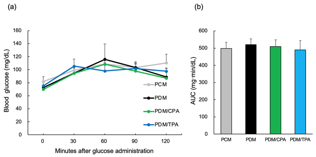
An oral glucose tolerance test (OGTT) was performed to evaluate the effects of maternal hyperglycemia and CPA or TPA intake during pregnancy on offspring glucose tolerance. Analysis of blood glucose levels at 0 min (before glucose loading) and at 30, 60, 90, and 120 min after loading, as well as the area under the OGTT curve, revealed no significant differences between the groups. No differences in body weight were observed between the groups.
Table 1. Body weights and blood glucose levels of the rat offspring groups.
PCMPDMPDM/CPAPDM/TPA
Blood glucose (mg/dL)90.1 ± 15.292.3 ± 13.797.7 ± 18.795.3 ± 11.1
Body weight (g)503.7 ± 15.9503.2 ± 14.8512.5 ± 18.1508.5 ± 16.3
Each value represents the mean ± SEM. PDM, offspring of hyperglycemic dams; PCM, offspring of control dams. The rat offspring of hyperglycemic dams fed CPA or TPA were designated as PDM/CPA or PDM/TPA, respectively.
Figure 1. Effects of maternal diabetes and palmitoleic acid supplementation on glucose tolerance in aged offspring rats. An oral glucose tolerance test (OGTT) was performed on 48-week-old male offspring of control dams (PCM), hyperglycemic dams (PDM), and hyperglycemic dams treated with cis- (PDM/CPA) or trans-palmitoleic acid (PDM/TPA) during pregnancy. Following oral glucose administration (3 g/kg), blood glucose levels were measured at the indicated time points. (a) Time-course changes in blood glucose levels during the OGTT. (b) Area under the curve (AUC) of glucose levels during OGTT. Data are presented as mean ± SEM (n = 5 per group).
Nutrients 18 01748 g001

3.2. AGEs–RAGE Signaling and Insulin Signaling in the Brain (Figure 2)

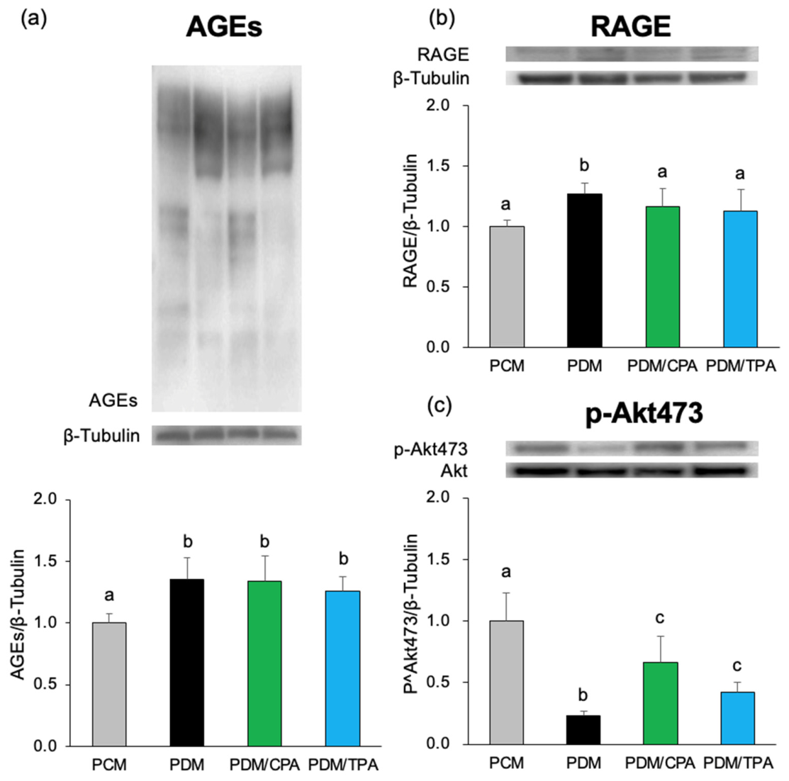
The effects of maternal hyperglycemia on glycation stress and insulin signaling in the cerebrum of adult offspring were evaluated. AGE expression was significantly higher in the PDM group than in the PCM group. Expression of RAGE, an AGE receptor, was also significantly higher in the PDM group than in the PCM group. However, the administration of CPA or TPA decreased RAGE expression compared with the PDM group.
Figure 2. Increased AGE–RAGE signaling and impaired Akt phosphorylation in the brains of aged offspring exposed to maternal hyperglycemia. Western blot analysis was performed using cerebral tissue obtained from 48-week-old male offspring of control dams (PCM), hyperglycemic dams (PDM), and hyperglycemic dams treated with cis-palmitoleic acid (PDM/CPA) or trans-palmitoleic acid (PDM/TPA) during pregnancy. Representative immunoblots of AGEs, RAGE, phosphorylated Akt at Ser473 (p-Akt473), total Akt, and β-tubulin are shown. (a) Quantification of AGEs normalized to β-tubulin. (b) Quantification of RAGE normalized to β-tubulin. (c) Quantification of p-Akt473 normalized to total Akt levels. Data are presented as mean ± SEM (n = 5 per group). Statistical analysis was performed using one-way ANOVA, followed by Tukey’s HSD post hoc test. Different letters (a–c) indicate statistically significant differences between groups (p < 0.05).
Nutrients 18 01748 g002
Furthermore, the expression of p-Akt473, an indicator of insulin signaling, was lower in the PDM group than in the PCM group. Phosphorylation of Akt increased in the PDM/CPA and PDM/TPA groups compared with the PDM group, suggesting a tendency toward recovery of insulin signaling.

3.3. Neuroinflammation and Synaptic Protein Expression (Figure 3)

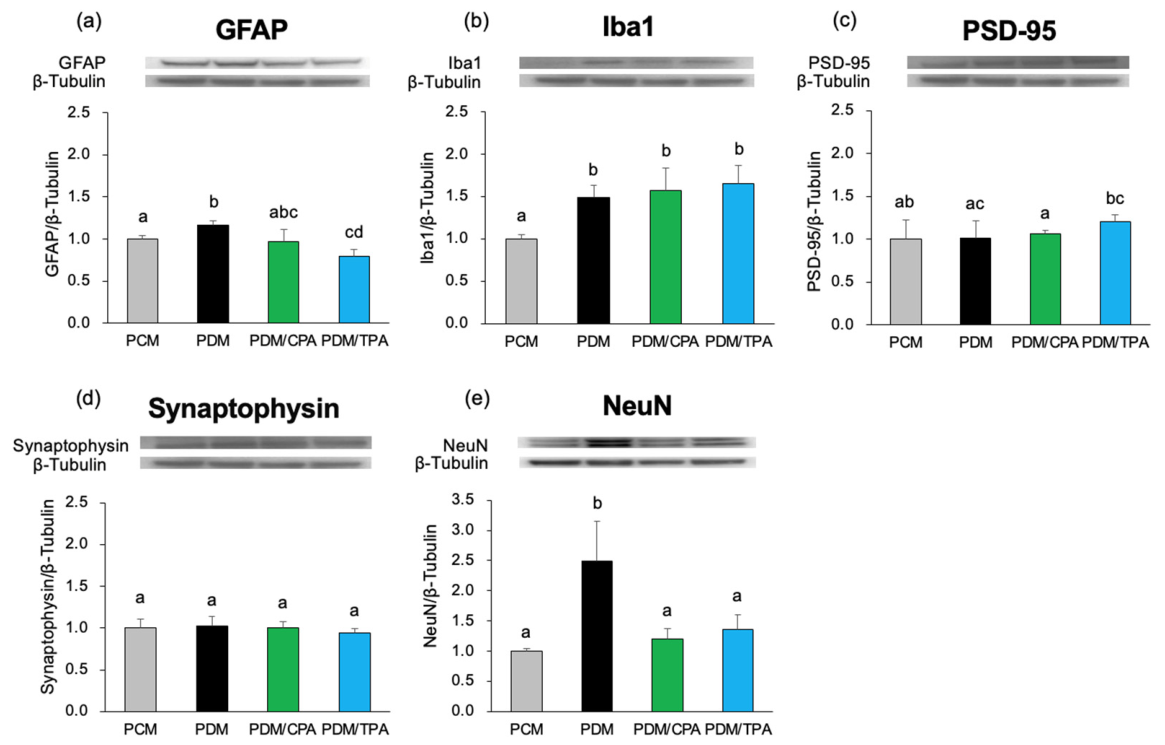
The effects of maternal hyperglycemia on neuroinflammation and synapse-related proteins in the brains of offspring were evaluated. The expression of GFAP, an astrocyte marker, was higher in the PDM group than in the PCM group. In contrast, expression appeared to be lower in the PDM/CPA and PDM/TPA groups compared with the PDM group. Expression of Iba1, a microglial marker, was higher in the PDM group than in the PCM group.
Figure 3. Alterations in neuroinflammation and synaptic proteins in offspring brains. Western blot analysis of neuroinflammatory markers and synaptic proteins in the brain tissue. (ae) Representative immunoblots for GFAP, Iba1, NeuN, PSD-95, and synaptophysin. Protein expression was normalized to β-tubulin. Data are presented as mean ± SEM. Statistical analyses were performed using one-way analysis of variance (ANOVA), followed by Tukey’s honest significant difference post hoc test. Different letters (a–d) indicate statistically significant differences between groups (p < 0.05).
Nutrients 18 01748 g003
There were no differences in the expression of PSD-95 and synaptophysin between the PCM and PDM groups. The expression of NeuN, a neuronal marker, was higher in the PDM group than in the PCM group, but lower in the PDM/CPA and PDM/TPA groups than in the PDM group.

3.4. Tau Phosphorylation in the Brain (Figure 4)

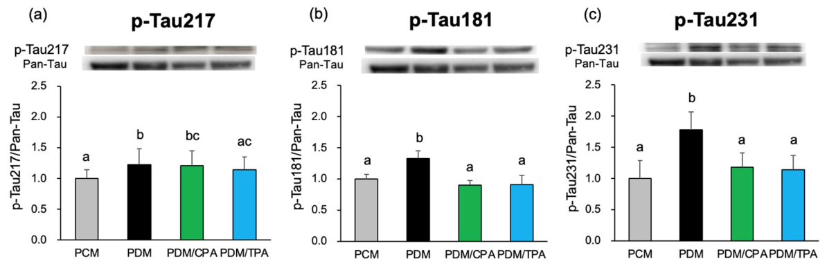
Tau phosphorylation was analyzed to evaluate neurodegenerative signaling. In the PDM group, p-Tau217 expression was increased compared with the PCM group, but in the PDM/TPA group, it was reduced compared with the PDM group.
Figure 4. Alterations in tau phosphorylation in offspring brains. Western blot analysis of tau phosphorylation in brain tissue. Representative immunoblots of phosphorylated Tau (p-Tau217, p-Tau181, p-Tau231) and total Tau (Pan-Tau). (ac) Quantification of phosphorylated and total tau levels. Data are presented as mean ± SEM. Statistical analyses were performed using one-way analysis of variance (ANOVA), followed by Tukey’s honest significant difference post hoc test. Different letters (a–c) indicate statistically significant differences between groups (p < 0.05).
Nutrients 18 01748 g004
The expression of p-Tau181 was higher in the PDM group than in the PCM group and lower in the PDM/CPA and PDM/TPA groups than in the PDM group. Similarly, p-Tau231 expression was increased in the PDM group and decreased in the PDM/CPA and PDM/TPA groups. Pan-tau expression was also higher in the PDM group than in the PCM group, and lower in the PDM/CPA and PDM/TPA groups than in the PDM group.

3.5. Gene Expression Changes in the Brain

To evaluate the effects of maternal hyperglycemia on the offspring’s cerebrum, mRNA expression levels of genes involved in neurotransmission, inflammation, and oxidative stress were analyzed.

3.5.1. Inflammation-Related Genes (Figure 5)

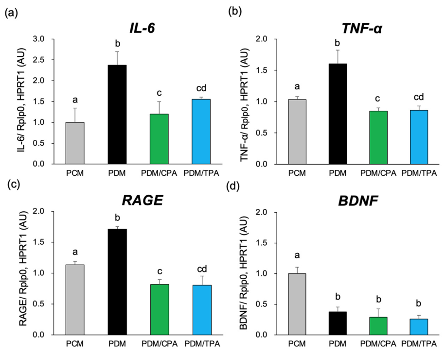
In the analysis of inflammation-related genes, mRNA expression levels of IL-6 and TNF-α were higher in the PDM group than in the PCM group. Similarly, RAGE expression, which encodes the receptor for AGEs, increased in the PDM group.
Figure 5. Increased inflammatory gene expression and decreased BDNF in PDM offspring. The mRNA expression levels of inflammatory cytokines and neurotrophic factors in the brain tissues were measured using RT-qPCR. (ac) Relative mRNA expression of IL-6, TNF-α, and RAGE. (d) Relative mRNA expression of BDNF. Gene expression levels were normalized to those of housekeeping genes and expressed relative to PCM. Data are presented as mean ± SEM. Statistical analyses were performed using one-way analysis of variance (ANOVA), followed by Tukey’s honest significant difference post hoc test. Different letters (a–d) indicate statistically significant differences between groups (p < 0.05).
Nutrients 18 01748 g005
In contrast, the expression of these inflammation-related genes was lower in the PDM/CPA and PDM/TPA groups than in the PDM group. The expression of BDNF, a neurotrophic factor, was lower in the PDM group than in the PCM group. BDNF expression was lower in the PDM/CPA and PDM/TPA groups than in the PCM group.

3.5.2. Neurotransmission-Related Genes (Figure 6)

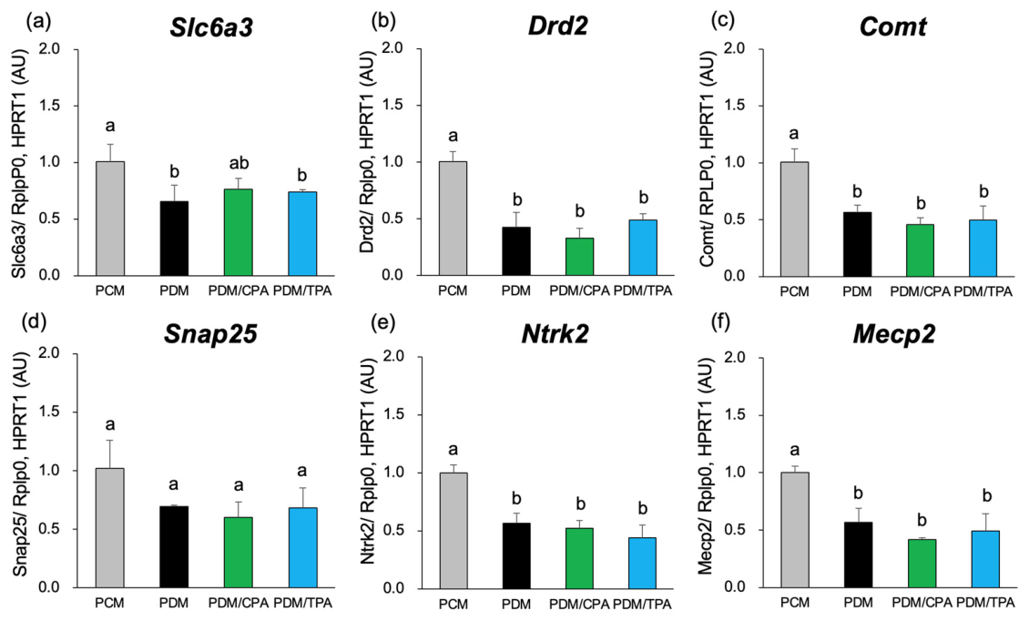
Analysis of neurotransmission-related genes revealed that the expression levels of Slc6a3, Drd2, and Comt were lower in the PDM group than in the PCM group. No significant changes in gene expression were observed in the PDM/CPA and PDM/TPA groups compared with the PDM group. The expression of Snap25, a synapse-related gene, was also tended to decrease in the PDM group compared with that in the PCM group, with no significant changes observed in the PDM/CPA and PDM/TPA groups (Figure 6).
Figure 6. Altered expression of neuronal function-related genes in PDM offspring. mRNA expression levels of genes associated with neuronal function in the brain tissue. (af) Relative mRNA expression of Slc6a3, Drd2, Comt, Snap25, Ntrk2, and Mecp2. Gene expression levels were normalized to those of housekeeping genes and expressed relative to PCM. Data are presented as mean ± SEM. Statistical analyses were performed using one-way analysis of variance (ANOVA), followed by Tukey’s honest significant difference post hoc test. Different letters (a,b) indicate statistically significant differences between groups (p < 0.05).
Nutrients 18 01748 g006

3.5.3. Neurodevelopment-Related Genes (Figure 6)

Analysis of neurodevelopment-related genes revealed that Ntrk2 and Mecp2 expression levels were lower in the PDM group than in the PCM group. No significant differences in gene expression were observed between the PDM/CPA and PDM/TPA groups (Figure 6).

3.5.4. Neuronal Markers Genes (Figure 7)

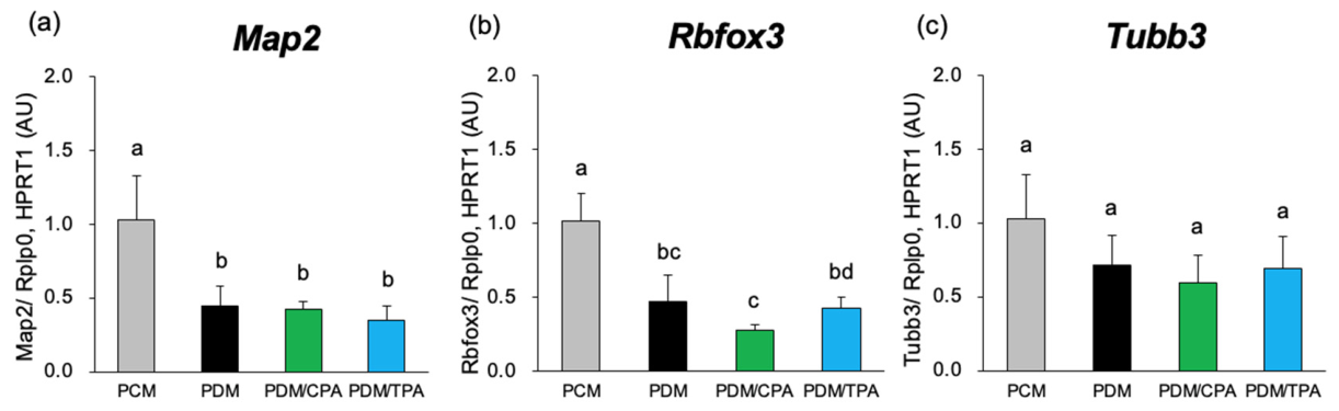
The expression levels of Map2 and Rbfox3 (NeuN) in the PDM group were lower than those in the PCM group. No difference in Tubb3 expression levels was observed between the groups.
Figure 7. Expression of neuronal marker genes in the brains of aged offspring rats. Relative mRNA expression levels of neuronal marker genes in brain tissues from aged offspring rats were analyzed by RT-qPCR. (ac) Relative mRNA expression levels of Map2, Rbfox3, and Tubb3 were normalized to housekeeping genes (Rplp0 and HPRT1) and expressed relative to the PCM group. Data are presented as mean ± SEM (n = 5 per group). Statistical analyses were performed using one-way ANOVA followed by Tukey’s HSD post hoc test. Different letters (a–d) indicate statistically significant differences between groups (p < 0.05).
Nutrients 18 01748 g007

3.5.5. Oxidative Stress-Related Genes (Figure 8)

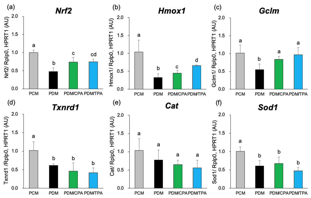
Analysis of antioxidant-related genes revealed that Nrf2 expression was lower in the PDM group than in the PCM group. In the PDM/CPA and PDM/TPA groups, Nrf2 expression was higher than that in the PDM group (Figure 8). The expression levels of Hmox1 and Gclm decreased in the PDM group and increased in the PDM/CPA and PDM/TPA groups compared to those in the PDM group. In contrast, the expression levels of Txnrd1 and Sod1 decreased in the PDM group, but no significant changes were observed in the PDM/CPA and PDM/TPA groups compared with the PDM group. No marked differences in the expression of Cat were observed among the groups.
Figure 8. Downregulation of antioxidant-related genes in PDM offspring. The relative mRNA expression levels of antioxidant-related genes associated with the Nrf2 antioxidant pathway were analyzed in the brain tissue of aged offspring. (af) Relative mRNA expression levels of Nrf2, Hmox1, Gclm, Txnrd1, Cat, and Sod1 were determined by RT-qPCR. Gene expression levels were normalized to housekeeping genes and expressed relative to the PCM group. Data are presented as mean ± SEM (n = 5 per group). Statistical analyses were performed using one-way analysis of variance (ANOVA), followed by Tukey’s honest significant difference post hoc test. Different letters (a–d) indicate statistically significant differences between groups (p < 0.05).
Nutrients 18 01748 g008

3.6. Behavioral Alterations in Offspring Exposed to Maternal Hyperglycemia

Behavioral analyses were performed on 48-week-old male offspring to evaluate the long-term effects of fetal hyperglycemic exposure on cognitive and emotional functions.

3.6.1. Y-Maze Test (Figure 9)

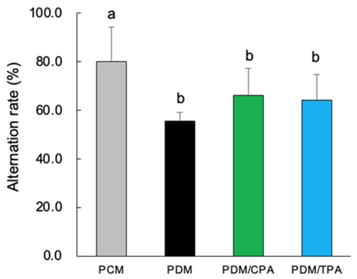
The Y-maze test was used to evaluate the spatial working memory. The percentage of spontaneous alteration was significantly lower in the PDM group than in the PCM group (Figure 9). In contrast, the PDM/CPA and PDM/TPA groups showed a tendency toward increased spontaneous alternation compared to the PDM group, although the differences were not statistically significant. In the Y-maze test, the PDM group showed a tendency toward reduced spontaneous alternation compared to the PCM group, suggesting impaired spatial working memory; however, post hoc analysis did not reach statistical significance after correction for unequal variances.
Figure 9. Impaired spatial working memory in PDM offspring assessed using the Y-maze test. Spontaneous alternation behavior in the Y-maze was used to evaluate spatial working memory in the aged offspring of normoglycemic mothers (PCM), diabetic mothers (PDM), and diabetic mothers treated with CPA or TPA (PDM/CPA, PDM/TPA). Spontaneous alternation rates (%) were calculated as an index of working memory performance. Data are presented as mean ± SEM (n = 5). Statistical analyses were performed using Welch’s ANOVA, followed by Games–Howell post hoc testing owing to unequal variances among groups. Different letters (a,b) indicate statistically significant differences between groups (p < 0.05). A tendency toward reduced spontaneous alternation behavior was observed in the PDM group compared to the PCM group.
Nutrients 18 01748 g009

3.6.2. Elevated Plus Maze Test (Figure 10)

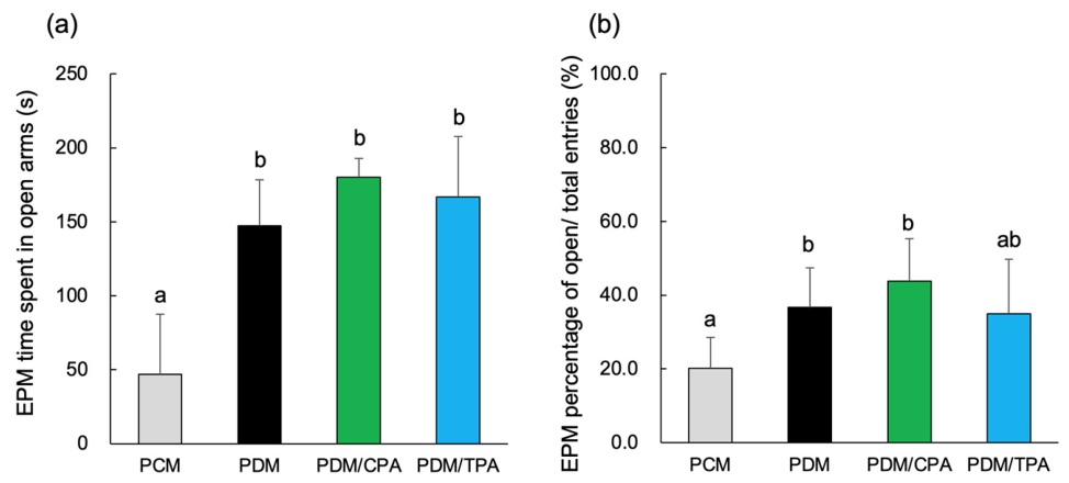
In the elevated plus maze test, the time spent in the open arms was higher in the PDM group than in the PCM group (Figure 10a). In addition, the percentage of open-arm entries relative to the total number of entries increased in the PDM group (Figure 10b). No significant differences in these parameters were observed between the PDM/CPA and PDM/TPA groups and the PDM group.
Figure 10. Altered anxiety-like behavior in PDM offspring assessed using the elevated plus maze. Behavioral performance in the elevated plus maze (EPM) test. (a) Time spent in the open arms during a 300 s test session. (b) Percentage of open-arm entries relative to the total arm entries. Data are presented as mean ± SEM (n = 5). Statistical analyses were performed using one-way ANOVA, followed by Tukey’s HSD post hoc test after confirmation of the normality and homogeneity of variance. Different letters (a,b) indicate statistically significant differences between groups (p < 0.05).
Nutrients 18 01748 g010

3.6.3. Novel Object Recognition Test (Figure 11)

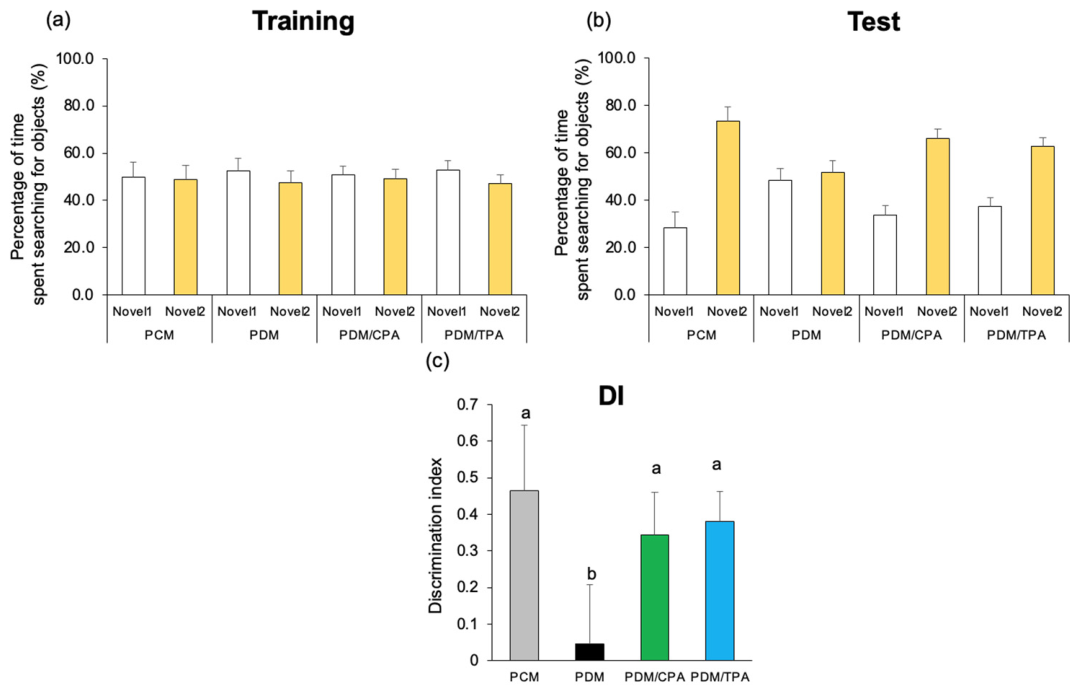
Recognition memory was evaluated using the novel object recognition test. During the training session, no differences in exploration time for the two objects were observed between the groups (Figure 11a). During the test session, the PCM, PDM/CPA, and PDM/TPA groups spent more time exploring the novel than the familiar object, whereas no clear difference was observed between the two objects in the PDM group (Figure 11b).
Figure 11. Impaired recognition memory in PDM offspring assessed using the novel object recognition test. A novel object recognition (NOR) test was performed to evaluate recognition memory. (a) Training session: exploration time for two identical objects (Novels 1 and 2). (b) Test session: exploration time for familiar and novel objects. (c) The discrimination index (DI) is calculated as a measure of recognition memory. Data are presented as mean ± SEM (n = 5). Statistical analyses were performed using one-way ANOVA, followed by Tukey’s HSD post hoc test after confirmation of the normality and homogeneity of variance. Different letters (a,b) indicate statistically significant differences between groups (p < 0.05).
Nutrients 18 01748 g011
The discrimination index (DI) was significantly lower in the PDM group than in the PCM group (Figure 11c). In the PDM/CPA and PDM/TPA groups, the DI was higher than that in the PDM group.

4. Discussion

The present study suggests that maternal hyperglycemia may induce persistent activation of the AGE–RAGE signaling pathway and disruption of Nrf2-dependent antioxidant defense mechanisms in the offspring’s brain, resulting in neuroinflammation, impaired insulin signaling, tau phosphorylation, neural dysfunction, and cognitive decline. Specifically, we evaluated aged (48-week-old) male offspring to provide insights into the long-term effects of fetal hyperglycemic exposure on brain aging. This is particularly important in the context of the Developmental Origins of Health and Disease, as many previous studies have focused on early-life outcomes rather than late-life neurological functions.
The OGTT results showed no apparent impairment of glucose tolerance in the adult offspring. This finding suggests that the effects of gestational diabetes do not necessarily persist as systemic metabolic abnormalities but may instead exert selective and long-lasting effects on the central nervous system. Such “latent central abnormalities” may reflect developmental programming induced by the fetal environment [23,24].
Furthermore, the present study confirmed increased expression of AGEs and RAGE in the PDM group, which is consistent with previous reports showing increased RAGE expression in the fetal brain in gestational diabetes models [25]. RAGE activation has been suggested to induce inflammation and oxidative stress through NF-κB signaling and function upstream of neural dysfunction [25]. Additionally, increased RAGE expression is associated with enhanced hippocampal excitability and behavioral abnormalities [9,25], which is consistent with the present findings.
Importantly, p-Akt473, an indicator of insulin signaling, was markedly decreased in the PDM group. The PI3K/Akt pathway is a major insulin signaling pathway in the brain, and its impairment contributes to cognitive decline and neurodegeneration-related molecular changes such as “brain insulin resistance” [26]. Indeed, changes in insulin receptors and IGF-1 signaling in fetal and neonatal hippocampi have been reported in gestational diabetes models [27], suggesting that impaired insulin signaling may be programmed during the fetal period and contribute to increased susceptibility to age-related neural dysfunction later in life [10,17].
Moreover, it has been reported that diabetes and hyperglycemia promote Tau phosphorylation through impairment of the PI3K/Akt pathway, leading to neurodegeneration-related molecular changes and cognitive decline [28,29]. Recent evidence supports an association between diabetes-related metabolic dysfunction, tau phosphorylation, and accelerated cognitive decline [29]. Therefore, the reduction in p-Akt levels observed in the present study may serve as a molecular basis for subsequent tau phosphorylation and neural dysfunction.
Although reduced Akt phosphorylation and increased Tau phosphorylation were observed, direct assessments of insulin receptor signaling, GSK-3β activity, or neuronal degeneration were not performed in the present study. Therefore, these molecular alterations should be interpreted as indirect evidence of impaired insulin-related signaling and neurodegeneration-related pathways, rather than definitive proof of brain insulin resistance or neurodegeneration.
A particularly important finding of this study was that despite enhanced oxidative and glycation stress, the expression of Nrf2 and its downstream antioxidant genes was reduced. This “failure of antioxidant responses in the presence of stress” suggests not merely insufficient compensation but also dysfunction of the Nrf2 pathway itself, driven by chronic AGE–RAGE signaling and aging [18,30]. Recent studies have highlighted the importance of Nrf2 dysfunction and mitochondrial oxidative stress in age-related molecular changes underlying neurodegeneration [30].
Several mechanisms may underlie this impaired Nrf2 response: (1) age-related decline in the Nrf2/ARE system, (2) chronic inflammation mediated by AGE–RAGE signaling, which may favor NF-κB signaling and suppress Nrf2-dependent antioxidant responses, and (3) abnormalities in intracellular signaling, represented by decreased p-Akt, which may inhibit the upstream activation of Nrf2 [21,31,32].
Additionally, analysis of molecules related to neural function revealed increased tau phosphorylation and decreased expression of BDNF, Slc6a3, Drd2, Ntrk2, and Mecp2 in the PDM group. These changes suggest that glycation stress and inflammation extend to neural dysfunction [18,19,33], and that reduced Akt signaling and disruption of Nrf2-dependent defense mechanisms may contribute to neurodegeneration-related molecular changes. These findings suggest that impairment of Nrf2-dependent antioxidant defenses and decreased Akt signaling may disrupt neural plasticity and synaptic function, ultimately leading to cognitive decline and neurodegeneration-related molecular changes [18].
Furthermore, the increased expression of GFAP and Iba1 increases neuroinflammation. Previous studies using animal models of gestational diabetes have also reported glial activation and increased levels of inflammatory cytokines in the hippocampus [10,14,34], which are consistent with the present findings. Excessive microglial activation contributes to abnormal neural circuit formation and cognitive dysfunction [35], suggesting that the neuroinflammation observed in this study may represent an important pathological basis for neural circuit dysfunction and cognitive impairment in offspring exposed to maternal hyperglycemia [10,35]. Recent reviews have also emphasized persistent microglial activation and neuroinflammation as key mechanisms linking gestational diabetes exposure to long-term neuropsychiatric abnormalities in the offspring [26].
Although no marked changes were observed in the synapse-related proteins PSD-95 and synaptophysin [36], decreases in these proteins have been reported in models of diabetes and Alzheimer’s disease [37], suggesting that there may be differences depending on the brain region or disease stage. The increase in NeuN protein expression does not necessarily directly reflect an increase in neuronal number but may instead indicate altered neuronal status or dysregulated neuronal expression [38]. Supporting this interpretation, additional RT-qPCR analyses revealed decreased expression of neuronal markers, including Map2 and Rbfox3 (NeuN), in the PDM group, whereas Tubb3 expression was relatively preserved (Figure 7). These observations indicate that maternal hyperglycemia may induce alterations in neuronal phenotype or integrity rather than simple neuronal proliferation [38].
Interestingly, despite substantial alterations in inflammatory signaling, Tau phosphorylation, and antioxidant-related pathways, synaptic markers such as PSD-95 and synaptophysin showed relatively limited changes in the present study. These findings may suggest that the observed molecular abnormalities reflect early or partial neurodegeneration-related processes rather than severe synaptic degeneration. Alternatively, synaptic alterations may occur in a brain region-specific or temporally restricted manner that was not fully captured in the present analyses.
Moreover, increased tau phosphorylation (p-Tau217, p-Tau181, and p-Tau231) indicated enhanced signaling in neurodegeneration-related molecular changes, consistent with previous reports of tau abnormalities associated with diabetes and insulin resistance [17,39,40]. In particular, decreased p-Akt is known to promote Tau phosphorylation through activation of GSK-3β [41], while AGE–RAGE signaling can also enhance Tau phosphorylation via inflammation and oxidative stress [20]. Therefore, the present findings support a molecular mechanism by which AGE–RAGE signaling induces inflammation and insulin resistance, thereby promoting tau phosphorylation. Emerging evidence suggests a bidirectional relationship between insulin resistance and tau pathology, forming a vicious cycle that promotes neurodegenerative molecular changes [42].
In contrast, although the effects of CPA and TPA on AGE–RAGE signaling were limited, recovery of p-Akt, reduction in inflammatory cytokines, and partial restoration of Nrf2-related genes were observed. These findings suggest that palmitoleic acid primarily acts on insulin signaling and anti-inflammatory/antioxidant pathways, thereby attenuating the negative effects of “metabolic memory.” Palmitoleic acid is recognized as a lipokine with insulin-sensitizing and anti-inflammatory properties [43], which is associated with our results.
In the Y-maze test, the PDM group showed a reduced spontaneous alternation rate, suggesting an impaired spatial working memory. Furthermore, a decreased discrimination index was observed in a novel object recognition test, indicating impaired recognition memory. These observations suggest that fetal exposure to hyperglycemia may have long-lasting effects on memory. Previous studies on the offspring of gestational diabetes model animals have reported hippocampus-dependent memory impairment, particularly reduced learning and memory performance in the Y-maze and Morris Water Maze tests [9,11]. The present findings are consistent with these reports and suggest that fetal hyperglycemic exposure may affect hippocampal function.
Furthermore, the reduction in BDNF levels and the suppression of the Nrf2 pathway observed in this study may contribute to memory impairment by decreasing synaptic plasticity and impairing neuroprotective mechanisms [8,44]. Increased Tau phosphorylation is closely associated with synaptic dysfunction and may represent the molecular basis of impaired recognition memory [45]. The novel object recognition test reflects the function of the hippocampus and entorhinal cortex. Impairments in this test are associated with neurodegeneration-related molecular changes and synaptic dysfunction [46]. Similar memory deficits have been reported in gestational diabetes models [11,47,48], which are consistent with the present findings.
In contrast, in the elevated plus maze test, the PDM group spent more time on the open arms and had a higher percentage of open-arm entries. These changes may reflect altered emotional or exploratory behavior. However, increased open-arm exploration in the elevated plus maze can be influenced by multiple behavioral dimensions, including reduced anxiety-like behavior, disinhibition, exploratory drive, and locomotor activity. Because locomotor activity was not independently quantified, these findings should be interpreted cautiously. Previous studies using gestational diabetes models have reported altered anxiety-related behavior and increased activity [9,10,11,48,49], which may be associated with Attention-Deficit/Hyperactivity Disorder-like behavior. Additionally, epidemiological studies have reported an increased risk of ADHD in children born to mothers with diabetes [1,3]. Therefore, the behavioral findings of the present study are consistent with those of previous epidemiological and developmental studies in humans [50]. A recent large-scale synthesis study reported increased risks of ADHD and autism spectrum disorders in children with gestational diabetes in utero.
These behavioral changes are likely closely related to the molecular abnormalities observed in this study. In the PDM group, enhanced AGE–RAGE signaling and increased inflammatory cytokine levels were accompanied by decreased Akt signaling, reduced BDNF expression, and downregulation of Nrf2-related genes. These changes may contribute to cognitive impairment by disrupting neural plasticity, synaptic integrity, and neuronal signaling, and enhanced tau phosphorylation is considered an important factor in neural dysfunction and cognitive decline associated with neurodegenerative processes [40,45,51,52].
Abnormalities in the dopaminergic system may also contribute to the observed behavioral changes. In the present study, reduced expression of Slc6a3 and Drd2 was observed, suggesting impaired dopaminergic neurotransmission. The dopamine system plays a critical role in motor control and attention, and its dysfunction is associated with hyperactivity and impulsivity [53]. Therefore, the hyperactivity-like behavior observed in this study may be closely related to impaired dopaminergic neurotransmission and reward-related signaling, particularly the dysfunction of dopamine transporters and dopamine receptor pathways associated with attention and motor regulation [53,54].
Although the observed molecular alterations are biologically consistent with previous reports on neuroinflammatory and neurodegeneration-related pathways, the present study did not provide direct mechanistic evidence linking AGE–RAGE signaling, Akt suppression, Nrf2 dysfunction, tau phosphorylation, and behavioral abnormalities. Therefore, these relationships should be interpreted as hypothetical or associative mechanisms requiring further experimental validation.
In contrast, the CPA- and TPA-treated groups showed a tendency toward partial attenuation of molecular and behavioral alterations in both the Y-maze and novel object recognition tests. These changes may be associated with reduced RAGE expression, reduced inflammation, and the partial recovery of Akt signaling and antioxidant responses. Palmitoleic acid is known to improve insulin sensitivity and exert anti-inflammatory effects [55], which are associated with p-Akt and the reduction in inflammatory gene expression observed in this study. In particular, restoration of brain insulin signaling has been reported to improve synaptic plasticity and memory function [17].
However, the behavioral improvements induced by CPA and TPA were only partial, and no significant recovery was observed in any parameter. This finding suggests that neurodevelopmental abnormalities induced by fetal hyperglycemia, such as reduced BDNF and Mecp2 expression, may persist irreversibly.
Although CPA and TPA supplementation partially attenuated several molecular and behavioral abnormalities, some behavioral improvements were not statistically significant. Therefore, the protective effects of palmitoleic acid should be interpreted with caution. Additional studies using larger cohorts and more comprehensive behavioral analyses are necessary to confirm these effects.
The CPA and TPA doses used in the present study were selected based on previous experimental studies that demonstrated their biological activity in maternal hyperglycemia models. However, the translational relevance of these supplementation protocols for human nutritional intake remains unclear. Although palmitoleic acids are naturally occurring dietary fatty acids present in dairy products and certain plant oils, the doses administered in the present animal study may have exceeded typical human dietary exposure. Therefore, the present findings should be interpreted as proof-of-concept evidence, and future studies are required to determine physiologically relevant intake ranges, long-term safety, and clinical applicability in humans.
Although these molecular changes appear to be associated with the observed behavioral abnormalities, further studies are needed to address the limitations of this study.
Several limitations of the present study should be acknowledged. First, the STZ-induced maternal hyperglycemia model used in this study does not fully replicate the pathophysiology of classical human gestational diabetes mellitus, particularly insulin resistance-driven metabolic abnormalities characteristic of human GDM. Because STZ administration at gestational day 2 induces hyperglycemia from early pregnancy, this model may reflect a relatively severe maternal hyperglycemic condition rather than late-onset human GDM. Second, only male offspring were analyzed in order to reduce variability associated with estrous cycle-related hormonal fluctuations and to facilitate stable long-term behavioral analyses; therefore, potential sex-specific responses could not be evaluated. In addition, although molecular alterations associated with AGE–RAGE signaling, impaired Akt phosphorylation, Nrf2-related pathways, and Tau phosphorylation were observed, direct mechanistic relationships among these pathways and behavioral abnormalities were not established. Oxidative stress and inflammatory responses were evaluated primarily using gene expression and signaling-related analyses without direct biochemical measurements of ROS production, cytokine protein levels, or antioxidant enzyme activities. Furthermore, behavioral analyses were performed using manual scoring rather than automated tracking systems, and locomotor activity was not independently evaluated using an open field test. Therefore, the elevated plus maze findings should be interpreted cautiously because altered open-arm behavior may reflect multiple behavioral dimensions, including anxiety-related behavior, exploratory activity, disinhibition, or locomotor alterations. Collectively, these limitations indicate that the present findings should be interpreted as exploratory and hypothesis-generating rather than definitive mechanistic evidence.
Taken together, maternal hyperglycemia appears to induce persistent activation of the AGE–RAGE axis and disruption of Nrf2-dependent antioxidant defense mechanisms by altering the fetal metabolic environment. This state of “inflammation dominance and impaired defense” may subsequently progress to neural dysfunction through impaired insulin signaling and enhanced Tau phosphorylation. The present study demonstrates that the fetal metabolic environment can exert long-term effects on brain function later in life and suggests that interventions targeting Nrf2 may represent a promising therapeutic strategy. Therefore, the present findings should be interpreted as exploratory and hypothesis-generating rather than as definitive mechanistic evidence.

5. Conclusions

The present study suggests that maternal hyperglycemia is associated with long-lasting alterations in molecular signaling and cognitive function in aged male offspring, potentially involving persistent activation of AGE–RAGE signaling, impaired insulin signaling, disruption of Nrf2-dependent antioxidant defense pathways, neuroinflammation, and enhanced tau phosphorylation. Maternal palmitoleic acid supplementation partially ameliorated several of these molecular and behavioral abnormalities, including inflammatory signaling and antioxidant responses.
These findings further suggest that an intrauterine hyperglycemic environment may contribute to increased vulnerability to age-related neurological dysfunction later in life, supporting the concept of developmental programming associated with maternal metabolic status. However, because the present study used an experimental animal model and did not establish direct causal relationships between the observed molecular alterations and behavioral phenotypes, the findings should be interpreted cautiously.
This study had several limitations. First, the STZ-induced maternal hyperglycemia model does not fully reproduce the pathophysiology of human gestational diabetes mellitus, particularly the insulin resistance-driven mechanisms. Second, only male offspring were analyzed; therefore, potential sex-specific differences could not be evaluated. Third, behavioral analyses were performed manually, which may have introduced observer bias despite efforts to standardize procedures. In addition, although multiple molecular pathways were investigated, mechanistic interventions targeting AGE–RAGE, Nrf2, or insulin signaling were not performed, limiting conclusions regarding causality. Furthermore, detailed brain region-specific analyses and histopathological evaluations were not performed.
Future studies using larger cohorts and experimental models that more closely replicate human gestational diabetes, particularly diet-induced insulin resistance models, will be important for validating and extending the present findings. Inclusion of both male and female offspring may further clarify sex-specific differences in neurodevelopmental and neurodegenerative outcomes. In addition, mechanistic studies targeting AGE–RAGE, Nrf2, and insulin-related pathways, together with region-specific brain analyses, histopathological evaluation, and advanced neuroimaging approaches, may help clarify the causal mechanisms linking maternal hyperglycemia to late-life neurological dysfunction. Longitudinal investigations tracking the progression from early metabolic alterations to age-related cognitive decline may further improve understanding of developmental programming mechanisms and potential preventive nutritional interventions.

Supplementary Materials

The following supporting information can be downloaded at: https://www.mdpi.com/article/10.3390/nu18111748/s1, Table S1: Primer sequences used in this study.

Author Contributions

Conceptualization, R.K. (Ritsuko Kawaharada) and A.N.; methodology, R.K. (Ritsuko Kawaharada) and A.N.; software: R.K. (Ritsuko Kawaharada) and R.K. (Reiko Kimura); validation: R.K. (Ritsuko Kawaharada) and R.K. (Reiko Kimura); formal analysis, R.K. (Ritsuko Kawaharada); investigation, R.K. (Ritsuko Kawaharada), R.K. (Reiko Kimura), E.M., and R.N.; resources, A.T., R.N., and E.M.; data curation, R.K. (Ritsuko Kawaharada); writing—original draft preparation, R.K. (Ritsuko Kawaharada) and A.N.: writing, review, and editing, R.K. (Ritsuko Kawaharada) and A.N.; visualization, R.K. (Ritsuko Kawaharada); supervision, A.N. and R.K. (Ritsuko Kawaharada); project administration, A.N.; funding acquisition, A.N. and R.K. (Ritsuko Kawaharada). All authors have read and agreed to the published version of the manuscript.

Funding

This research was funded by JSPS KAKENHI [grant numbers 25K14940, 24K14683, and 22K11882 to Ri. K. and A.N.], the Joint Research Project at Takasaki University of Health and Welfare [to Ri. K.], the Smoking Research Foundation [2023G015], and the Research Program of Jissen Women’s University [to A.N.].

Institutional Review Board Statement

The animal study protocol was approved by the Animal Ethics Committee of Takasaki University of Health and Welfare (protocol codes 2508 and 1 April 2025).

Informed Consent Statement

Not applicable.

Data Availability Statement

All data generated or analyzed during this study are included in this published article.

Acknowledgments

We are grateful to the members of our laboratory for their assistance.

Conflicts of Interest

The authors declare no conflicts of interest.

Abbreviations

The following abbreviations are used in this manuscript:
GDMGestational diabetes mellitus
CPACis-palmitoleic acid
TPATrans-palmitoleic acid
PCMOffspring of control dams
PDMOffspring of hyperglycemic dams
AGEsAdvanced glycation end products
RAGEReceptor for advanced glycation end products
NF-κBNuclear factor kappa B
GFAPGlial fibrillary acidic protein
Iba1Ionized calcium-binding adapter molecule 1
RT-qPCRReverse transcription quantitative polymerase chain reaction
DIDiscrimination index
SEMStandard error of the mean
ANOVAAnalysis of variance
OGTTOral glucose tolerance test
AUCArea under the curve
p-Akt473Phosphorylated Akt at Ser473
AktProtein kinase B
PSD-95Postsynaptic density protein 95
NeuNNeuronal nuclei
IL-6Interleukin-6
TNF-αTumor necrosis factor-alpha
BDNFBrain-derived neurotrophic factor
Slc6a3Solute carrier family 6 member 3
Drd2Dopamine receptor D2
ComtCatechol-O-methyltransferase
Snap25Synaptosomal-associated protein 25
Ntrk2Neurotrophic receptor tyrosine kinase 2
Mecp2Methyl-CpG-binding protein 2
Nrf2Nuclear factor erythroid 2-related factor 2
Hmox1Heme oxygenase 1
GclmGlutamate-cysteine ligase modifier subunit
Txnrd1Thioredoxin reductase 1
CatCatalase
Sod1Superoxide dismutase 1
EPMElevated plus maze
NORNovel object recognition
IGF-1Insulin-like growth factor-1
PI3KPhosphoinositide 3-kinase
GSK-3βGlycogen synthase kinase-3 beta

References

  1. Xu, G.; Jing, J.; Bowers, K.; Liu, B.; Bao, W. Maternal Diabetes and the Risk of Autism Spectrum Disorders in the Offspring: A Systematic Review and Meta-Analysis. J. Autism Dev. Disord. 2014, 44, 766–775. [Google Scholar] [CrossRef]
  2. Xiang, A.H.; Wang, X.; Martinez, M.P.; Page, K.; Buchanan, T.A.; Feldman, R.K. Maternal Type 1 Diabetes and Risk of Autism in Offspring. JAMA 2018, 320, 89–91. [Google Scholar] [CrossRef]
  3. Xiang, A.H.; Wang, X.; Martinez, M.P.; Getahun, D.; Page, K.A.; Buchanan, T.A.; Feldman, K. Maternal Gestational Diabetes Mellitus, Type 1 Diabetes, and Type 2 Diabetes During Pregnancy and Risk of ADHD in Offspring. Diabetes Care 2018, 41, 2502–2508. [Google Scholar] [CrossRef] [PubMed]
  4. Barker, D.J.P. The Developmental Origins of Adult Disease. J. Am. Coll. Nutr. 2004, 23, 588S–595S. [Google Scholar] [CrossRef]
  5. Ornoy, A.; Ratzon, N.; Greenbaum, C.; Wolf, A.; Dulitzky, M. School-Age Children Born to Diabetic Mothers and to Mothers with Gestational Diabetes Exhibit a High Rate of Inattention and Fine and Gross Motor Impairment. J. Pediatr. Endocrinol. Metab. 2001, 14, 681–689. [Google Scholar] [CrossRef]
  6. Vafaei-Nezhad, S.; Hami, J.; Sadeghi, A.; Ghaemi, K.; Hosseini, M.; Abedini, M.R.; Haghir, H. The Impacts of Diabetes in Pregnancy on Hippocampal Synaptogenesis in Rat Neonates. Neuroscience 2016, 318, 122–133. [Google Scholar] [CrossRef]
  7. Lotfi, N.; Hami, J.; Hosseini, M.; Haghir, D.; Haghir, H. Diabetes during Pregnancy Enhanced Neuronal Death in the Hippocampus of Rat Offspring. Int. J. Dev. Neurosci. 2016, 51, 28–35. [Google Scholar] [CrossRef] [PubMed]
  8. Park, H.; Poo, M.M. Neurotrophin Regulation of Neural Circuit Development and Function. Nat. Rev. Neurosci. 2013, 14, 7–23. [Google Scholar] [CrossRef]
  9. Chandna, A.R.; Kuhlmann, N.; Bryce, C.A.; Greba, Q.; Campanucci, V.A.; Howland, J.G. Chronic Maternal Hyperglycemia Induced during Mid-Pregnancy in Rats Increases RAGE Expression, Augments Hippocampal Excitability, and Alters Behavior of the Offspring. Neuroscience 2015, 303, 241–260. [Google Scholar] [CrossRef]
  10. Vuong, B.; Odero, G.; Rozbacher, S.; Stevenson, M.; Kereliuk, S.M.; Pereira, T.J.; Dolinsky, V.W.; Kauppinen, T.M. Exposure to Gestational Diabetes Mellitus Induces Neuroinflammation, Derangement of Hippocampal Neurons, and Cognitive Changes in Rat Offspring. J. Neuroinflammation 2017, 14, 80. [Google Scholar] [CrossRef]
  11. Huerta-Cervantes, M.; Peña-Montes, D.J.; López-Vázquez, M.Á.; Montoya-Pérez, R.; Cortés-Rojo, C.; Olvera-Cortés, M.E.; Saavedra-Molina, A. Effects of Gestational Diabetes in Cognitive Behavior, Oxidative Stress and Metabolism on the Second-Generation Off-Spring of Rats. Nutrients 2021, 13, 1575. [Google Scholar] [CrossRef]
  12. Singh, V.P.; Bali, A.; Singh, N.; Jaggi, A.S. Advanced Glycation End Products and Diabetic Complications. Korean J. Physiol. Pharmacol. 2014, 18, 1–14. [Google Scholar] [CrossRef]
  13. Schmidt, A.M.; Yan, S.D.; Yan, S.F.; Stern, D.M. The Multiligand Receptor RAGE as a Progression Factor Amplifying Immune and Inflammatory Responses. J. Clin. Investig. 2001, 108, 949–955. [Google Scholar] [CrossRef]
  14. Piazza, F.V.; Segabinazi, E.; de Meireles, A.L.F.; Mega, F.; Spindler, C.F.; Augustin, O.A.; Salvalaggio, G.D.S.; Achaval, M.; Kruse, M.S.; Coirini, H.; et al. Severe Uncontrolled Maternal Hyperglycemia Induces Microsomia and Neurodevelopment Delay Accompanied by Apoptosis, Cellular Survival, and Neuroinflammatory Deregulation in Rat Offspring Hippocampus. Cell. Mol. Neurobiol. 2019, 39, 401–414. [Google Scholar] [CrossRef] [PubMed]
  15. Duarte, J.M.N.; Agostinho, P.M.; Carvalho, R.A.; Cunha, R.A. Caffeine Consumption Prevents Diabetes-Induced Memory Impairment and Synaptotoxicity in the Hippocampus of NONcZNO10/LTJ Mice. PLoS ONE 2012, 7, e21899. [Google Scholar] [CrossRef]
  16. Hami, J.; Kerachian, M.A.; Karimi, R.; Haghir, H.; Sadr-Nabavi, A. Effects of Streptozotocin-Induced Type 1 Maternal Diabetes on PI3K/AKT Signaling Pathway in the Hippocampus of Rat Neonates. J. Recept. Signal Transduct. Res. 2016, 36, 254–260. [Google Scholar] [CrossRef] [PubMed]
  17. Arnold, S.E.; Arvanitakis, Z.; Macauley-Rambach, S.L.; Koenig, A.M.; Wang, H.Y.; Ahima, R.S.; Craft, S.; Gandy, S.; Buettner, C.; Stoeckel, L.E.; et al. Brain Insulin Resistance in Type 2 Diabetes and Alzheimer Disease: Concepts and Conundrums. Nat. Rev. Neurol. 2018, 14, 168–181. [Google Scholar] [CrossRef]
  18. George, M.; Tharakan, M.; Culberson, J.; Reddy, A.P.; Reddy, P.H. Role of Nrf2 in Aging, Alzheimer’s and Other Neurodegenerative Diseases. Ageing Res. Rev. 2022, 82, 101756. [Google Scholar] [CrossRef]
  19. Brackhan, M.; Arribas-Blazquez, M.; Lastres-Becker, I. Aging, NRF2, and TAU: A Perfect Match for Neurodegeneration? Antioxidants 2023, 12, 1564. [Google Scholar] [CrossRef] [PubMed]
  20. Li, X.-H.; Du, L.-L.; Cheng, X.-S.; Jiang, X.; Zhang, Y.; Lv, B.-L.; Liu, R.; Wang, J.-Z.; Zhou, X.-W. Glycation Exacerbates the Neuronal Toxicity of β-Amyloid. Cell Death Dis. 2013, 4, e673. [Google Scholar] [CrossRef]
  21. Sivandzade, F.; Prasad, S.; Bhalerao, A.; Cucullo, L. NRF2 and NF-κB Interplay in Cerebrovascular and Neurodegenerative Disorders: Molecular Mechanisms and Possible Therapeutic Approaches. Redox Biol. 2019, 21, 101059. [Google Scholar] [CrossRef] [PubMed]
  22. Okami, H.; Kawaharada, R.; Yoshizaki, H.; Toriumi, A.; Tsutsumi, S.; Nakamura, A. Maternal n-7 Unsaturated Fatty Acids Protect the Fetal Brain from Neuronal Degeneration in an Intrauterine Hyperglycemic Animal Model. Nutrients 2023, 15, 3434. [Google Scholar] [CrossRef]
  23. Sousa, R.A.L.; Torres, Y.S.; Figueiredo, C.P.; Passos, G.F.; Clarke, J.R. Consequences of Gestational Diabetes to the Brain and Behavior of the Offspring. An. Acad. Bras. Cienc. 2018, 90, 2279–2291. [Google Scholar] [CrossRef] [PubMed]
  24. Huang, H.; Apaijai, N.; Thonusin, C.; Suntornsaratoon, P.; Chattipakorn, N.; Charoenphandhu, N.; Chattipakorn, S.C. Mothers with Obesity and Gestational Diabetes Did Not Induce Brain Pathologies or Premature Brain Aging in Their Adolescent and Early Adult Offspring in Rats. Neuroscience 2025, 568, 454–464. [Google Scholar] [CrossRef]
  25. Tang, X.; Qin, Q.; Xie, X.; He, P. Protective Effect of sRAGE on Fetal Development in Pregnant Rats with Gestational Diabetes Mellitus. Cell Biochem. Biophys. 2015, 71, 549–556. [Google Scholar] [CrossRef]
  26. Yan, Z.; Pu, J.; Li, D.; Liu, M.; Xu, Z.; Tang, J. GDM-Related Neurodevelopmental and Neuropsychiatric Disorders in the Mothers and Their Progeny, and the Underlying Mechanisms. J. Pers. Med. 2026, 16, 19. [Google Scholar] [CrossRef]
  27. Hami, J.; Sadr-Nabavi, A.; Sankian, M.; Balali-Mood, M.; Haghir, H. The Effects of Maternal Diabetes on Expression of Insulin-Like Growth Factor-1 and Insulin Receptors in Male Developing Rat Hippocampus. Brain Struct. Funct. 2013, 218, 73–84. [Google Scholar] [CrossRef]
  28. Zhao, L.; Yuan, J.; Yang, Q.; Ma, J.; Yang, F.; Zou, Y.; Liu, K.; Liu, F. Diabetes and Its Complications: Molecular Mechanisms, Prevention and Treatment. Signal Transduct. Target. Ther. 2026, 11, 22. [Google Scholar] [CrossRef]
  29. Vargas-Soria, M.; Corraliza-Gomez, M.; Infante-Garcia, C.; Stitt, A.W.; Simó, R.; Garcia-Alloza, M. Diabetes-Linked Metabolic Dysfunction Relates with Distinct Tau Phosphorylation Patterns, Neuroinflammation and Cognitive Impairment in Mouse Models of Alzheimer’s Disease. Brain Behav. Immun. 2026, 132, 106204. [Google Scholar] [CrossRef] [PubMed]
  30. Saso, L.; Ates, I.; Tunc, R.; Yilmaz, B.; Gallorini, M.; Carradori, S.; Suzen, S. Modulation of Nrf2 and Mitochondrial Function: Pharmacological Implications. Pharmaceuticals 2025, 18, 1698. [Google Scholar] [CrossRef] [PubMed]
  31. Li, L.; Meng, F.; Li, D. Downregulation of Nrf2 in the Hippocampus Contributes to Postoperative Cognitive Dysfunction in Aged Rats by Sensitizing Oxidative Stress and Neuroinflammation. Oxid. Med. Cell. Longev. 2023, 2023, 7272456. [Google Scholar] [CrossRef] [PubMed]
  32. Díaz, M.; Valdés-Baizabal, C.; de Pablo, D.P.; Marin, R. Age-Dependent Changes in Nrf2/Keap1 and Target Antioxidant Protein Expression Correlate to Lipoxidative Adducts, and Are Modulated by Dietary n-3 LCPUFA in the Hippocampus of Mice. Antioxidants 2024, 13, 206. [Google Scholar] [CrossRef] [PubMed]
  33. Chu, C.-T.; Uruno, A.; Katsuoka, F.; Yamamoto, M. Role of NRF2 in Pathogenesis of Alzheimer’s Disease. Antioxidants 2024, 13, 1529. [Google Scholar] [CrossRef]
  34. Głombik, K.; Trojan, E.; Kurek, A.; Budziszewska, B.; Basta-Kaim, A. Inflammatory Consequences of Maternal Diabetes on the Offspring Brain: A Hippocampal Organotypic Culture Study. Neurotox. Res. 2019, 36, 357–375. [Google Scholar] [CrossRef] [PubMed]
  35. Hong, S.; Dissing-Olesen, L.; Stevens, B. New Insights on the Role of Microglia in Synaptic Pruning in Health and Disease. Curr. Opin. Neurobiol. 2016, 36, 128–134. [Google Scholar] [CrossRef]
  36. Sun, X.; Guo, X.; Wu, T.; Tao, W.; Lin, R.; Song, Y.; Zheng, Y.; Ma, C.; Shen, F.; Zhou, Y.; et al. Identification of Hub Genes and Pathways Associated with Sevoflurane-Induced Synaptic Loss in Diabetic Offspring via Comprehensive Transcriptome Analysis. Metab. Brain Dis. 2025, 40, 313. [Google Scholar] [CrossRef]
  37. Carvalho, C.; Santos, M.S.; Oliveira, C.R.; Moreira, P.I. Alzheimer’s Disease and Type 2 Diabetes-Related Alterations in Brain Mitochondria, Autophagy and Synaptic Markers. Biochim. Biophys. Acta Mol. Basis Dis. 2015, 1852, 1665–1675. [Google Scholar] [CrossRef] [PubMed]
  38. Gusel’nikova, V.V.; Korzhevskiy, D.E. NeuN as a Neuronal Nuclear Antigen and Neuron Differentiation Marker. Acta Naturae 2015, 7, 42–47. [Google Scholar] [CrossRef]
  39. Sun, H.; Gao, X.; Niu, J.; Chen, P.; He, S.; Xu, S.; Ge, J. AD-Like Neuropsychiatric Dysfunction in a Mice Model Induced by a Combination of High-Fat Diet and Intraperitoneal Injection of Streptozotocin. eNeuro 2024, 11. [Google Scholar] [CrossRef]
  40. Planel, E.; Tatebayashi, Y.; Miyasaka, T.; Liu, L.; Wang, L.; Herman, M.; Yu, W.H.; Luchsinger, J.A.; Wadzinski, B.; Duff, K.E.; et al. Insulin Dysfunction Induces In Vivo Tau Hyperphosphorylation through Distinct Mechanisms. J. Neurosci. 2007, 27, 13635–13648. [Google Scholar] [CrossRef]
  41. Kim, B.; Feldman, E.L. Insulin Resistance in the Nervous System. Trends Endocrinol. Metab. 2012, 23, 133–141. [Google Scholar] [CrossRef]
  42. Mu, J.; Zhang, Z.; Jiang, C.; Geng, H.; Duan, J. Role of Tau Protein Hyperphosphorylation in Diabetic Retinal Neurodegeneration. J. Ophthalmol. 2025, 2025, 3278794. [Google Scholar] [CrossRef]
  43. Guillocheau, E.; Legrand, P.; Rioux, V. Trans-Palmitoleic Acid (trans-9-C16:1, or trans-C16:1 n-7): Nutritional Impacts, Metabolism, Origin, Compositional Data, Analytical Methods and Chemical Synthesis: A Review. Biochimie 2020, 169, 144–160. [Google Scholar] [CrossRef]
  44. Ma, Q. Role of Nrf2 in Oxidative Stress and Toxicity. Annu. Rev. Pharmacol. Toxicol. 2013, 53, 401–426. [Google Scholar] [CrossRef] [PubMed]
  45. Wang, Y.; Mandelkow, E. Tau in Physiology and Pathology. Nat. Rev. Neurosci. 2016, 17, 5–21. [Google Scholar] [CrossRef]
  46. Antunes, M.; Biala, G. The Novel Object Recognition Memory: Neurobiology, Test Procedure, and Its Modifications. Cogn. Process. 2012, 13, 93–110. [Google Scholar] [CrossRef]
  47. Zou, K.; Ren, J.; Luo, S.; Zhang, J.; Zhou, C.; Tan, C.; Lv, P.; Sun, X.; Sheng, J.; Liu, X.; et al. Intrauterine Hyperglycemia Impairs Memory across Two Generations. Transl. Psychiatry 2021, 11, 434. [Google Scholar] [CrossRef]
  48. Huerta-Cervantes, M.; Peña-Montes, D.J.; Montoya-Pérez, R.; Trujillo, X.; Huerta, M.; López-Vázquez, M.Á.; Olvera-Cortés, M.E.; Saavedra-Molina, A. Gestational Diabetes Triggers Oxidative Stress in Hippocampus and Cerebral Cortex and Cognitive Behavior Modifications in Rat Offspring: Age- and Sex-Dependent Effects. Nutrients 2020, 12, 376. [Google Scholar] [CrossRef] [PubMed]
  49. Ramanathan, M.; Jaiswal, A.K.; Bhattacharya, S.K. Hyperglycaemia in Pregnancy: Effects on the Offspring Behaviour with Special Reference to Anxiety Paradigms. Indian J. Exp. Biol. 2000, 38, 231–236. [Google Scholar] [PubMed]
  50. Ornoy, A.; Reece, E.A.; Pavlinkova, G.; Kappen, C.; Miller, R.K. Effect of Maternal Diabetes on the Embryo, Fetus, and Children: Congenital Anomalies, Genetic and Epigenetic Changes and Developmental Outcomes. Birth Defects Res. C Embryo Today 2015, 105, 53–72. [Google Scholar] [CrossRef]
  51. Safwan, Z.; Ijaz, E.; Shamim, H.; Fatima, A.; Ahmad, A.; Khalid, A.M. The Impact of Gestational Diabetes Mellitus on Fetal Neural Development and Long-Term Neurodevelopmental Outcomes: A Review. Brain Dev. 2026, 48, 104521. [Google Scholar] [CrossRef]
  52. Oikonomou, E.; Chatzakis, C.; Stavros, S.; Potiris, A.; Nikolettos, K.; Sotiriou, S.; Domali, E.; Nikolettos, N.; Sotiriadis, A.; Gerede, A. A Review of the Impact of Gestational Diabetes on Fetal Brain Development: An Update on Neurosonographic Markers During the Last Decade. Life 2025, 15, 210. [Google Scholar] [CrossRef] [PubMed]
  53. Volkow, N.D.; Wang, G.J.; Kollins, S.H.; Wigal, T.L.; Newcorn, J.H.; Telang, F.; Fowler, J.S.; Zhu, W.; Logan, J.; Ma, Y.; et al. Evaluating Dopamine Reward Pathway in ADHD: Clinical Implications. JAMA 2009, 302, 1084–1091. [Google Scholar] [CrossRef]
  54. Dum, R.; Ghahramani, A.; Baweja, R.; Bellon, A. Dopamine Receptor Expression and the Pathogenesis of Attention-Deficit Hyperactivity Disorder: A Scoping Review of the Literature. Curr. Dev. Disord. Rep. 2022, 9, 127–136. [Google Scholar] [CrossRef]
  55. Cao, H.; Gerhold, K.; Mayers, J.R.; Wiest, M.M.; Watkins, S.M.; Hotamisligil, G.S. Identification of a Lipokine, a Lipid Hormone Linking Adipose Tissue to Systemic Metabolism. Cell 2008, 134, 933–944. [Google Scholar] [CrossRef] [PubMed]
Disclaimer/Publisher’s Note: The statements, opinions and data contained in all publications are solely those of the individual author(s) and contributor(s) and not of MDPI and/or the editor(s). MDPI and/or the editor(s) disclaim responsibility for any injury to people or property resulting from any ideas, methods, instructions or products referred to in the content.