Article

The Neuroprotective Role of Quinoa (Chenopodium quinoa, Wild) Supplementation in Hippocampal Morphology and Memory of Adolescent Stressed Rats

by
Gonzalo Terreros
1,2,
Miguel Ángel Pérez
3,
Pablo Muñoz-LLancao
4,
Amanda D’Espessailles
1,
Enrique A. Martínez
5 and
Alexies Dagnino-Subiabre
2,6,*
1
Instituto de Ciencias de la Salud, Universidad de O’Higgins, Rancagua 8370993, Chile
2
Auditory and Cognition Center (AUCO), Santiago 8320000, Chile
3
Health Sciences School, Universidad Viña del Mar, Viña del Mar 2580022, Chile
4
Department of Cell Biology, School of Medicine, Yale University, New Haven, CT 06510, USA
5
Foyer de Charité de Provence, 13410 Lambesc, France
6
Laboratory of Stress Neurobiology, Faculty of Sciences, Institute of Physiology, Universidad de Valparaíso, Valparaíso 2360102, Chile
*
Author to whom correspondence should be addressed.
Nutrients 2024, 16(3), 381; https://doi.org/10.3390/nu16030381
Submission received: 10 November 2023 / Revised: 26 December 2023 / Accepted: 29 December 2023 / Published: 27 January 2024
(This article belongs to the Section Micronutrients and Human Health)

Abstract

:
Brain physiology and morphology are vulnerable to chronic stress, impacting cognitive performance and behavior. However, functional compounds found in food may alleviate these alterations. White quinoa (Chenopodium quinoa, Wild) seeds contain a high content of n-3 fatty acids, including alpha-linolenic acid. This study aimed to evaluate the potential neuroprotective role of a quinoa-based functional food (QFF) in rats. Prepubertal male Sprague-Dawley rats were fed with rat chow or QFF (50% rat chow + 50% dehydrated quinoa seeds) and exposed or not to restraint stress protocol (2 h/day; 15 days). Four experimental groups were used: Non-stressed (rat chow), Non-stressed + QFF, Stressed (rat chow) and Stressed + QFF. Weight gain, locomotor activity (open field), anxiety (elevated plus maze, light-dark box), spatial memory (Y-maze), and dendritic length in the hippocampus were measured in all animals. QFF intake did not influence anxiety-like behaviors, while the memory of stressed rats fed with QFF improved compared to those fed with rat chow. Additionally, QFF intake mitigated the stress-induced dendritic atrophy in pyramidal neurons located in the CA3 area of the hippocampus. The results suggest that a quinoa-supplemented diet could play a protective role in the memory of chronically stressed rats.

1. Introduction

Stress refers to the physiological response induced in living organisms when they are exposed to adversity and try to adapt to it, a process known as allostasis [1]. Non-adaptation generates allostatic overload in the brain, increasing vulnerability to mental disorders [2]. In animal models, allostatic overload triggers morphological changes in neurons, such as dendritic atrophy and neurogenesis reduction in the hippocampus, dendritic arborization in the amygdala, and dendritic remodeling in the medial prefrontal cortex [3,4]. These morphological changes impact synaptic function, leading to cognitive and behavioral impairments [5,6]. The hippocampus, a brain area associated with episodic memory, is highly vulnerable to allostatic overload. Chronic stress induces dendritic atrophy in the hippocampal CA3 pyramidal neurons [5,7] and dendritic hypertrophy in the basolateral amygdala, both leading to spatial memory impairments [4,8] and enhancing anxiety [9], respectively.
The adolescent brain is highly responsive and susceptible to environmental demands, such as stressors [10]. For instance, adolescent rats exposed to restraint stress exhibited higher plasma corticosterone levels than adult rats [11]. The serum corticosterone levels of acutely stressed adolescent rats remained elevated for twice as long as those of adult rats subjected to the same conditions [12]. In humans, both male and female adolescents also demonstrate an increased cortisol response to stress exposure compared to male and female children and adults [13].
External factors, such as nutrition and exercise, can modulate vulnerability to stress [14]. In line with this, polyunsaturated fatty acids (PUFAs) play an essential role in brain development [15] and promote anti-stress effects in rats [14]. n-3 PUFA supplementation improves memory and GABAergic activity in the hippocampus, modulated by the cannabinoid receptor type 1 [14]. An optimal n-6:n-3 ratio (1:5) of PUFA promotes anti-inflammatory and antioxidant activities, as has been shown in animal models [16] and in humans [17]. Moreover, a high n-6:n-3 ratio is associated with depression symptoms, a stress-related disorder [18]. As n-3 PUFA consumption has decreased in recent years, triggering an imbalance in the n-6:n-3 ratio, it is crucial to promote and study foods high in these fatty acids [19].
Quinoa (Chenopodium quinoa, Wild) is an ancient Andean grain with exceptional nutritional value. Quinoa has a high content of unsaturated fatty acids, constituting 88% of the total fatty acids, primarily oleic (OA, 18:1 n-9, 19.7–29.5%), linoleic (LA: 18:2 n-6, 49.0–56.4%) and alpha-linolenic (ALA: 18:3 n-3, 8.7–11.7%) acids [20]. ALA and LA are essential fatty acids, and dietary intake serves as a source of PUFAs [21]. Quinoa consumption has been associated with improved cardiovascular health and reduction of risk factors [22]. Recently, a red-quinoa seed extract prevented memory deficit induced by scopolamine in mice, suggesting that quinoa may have a neuroprotective effect [23]. Considering that hippocampal neurogenesis and maturation in the adolescent brain are particularly susceptible to stress, we aimed to evaluate the effects of a quinoa-based functional food intake in a repeated restraint stress protocol in adolescent rats. Anxiety, spatial memory, and dendritic length in long-shaft pyramidal neurons of the CA3 hippocampal region were measured in rats supplemented (or not) with quinoa-based functional food and exposed (or not) to a restraint stress.

2. Materials and Methods

2.1. Animals and Restraint Protocols

Prepubertal male Sprague-Dawley rats, 21 postnatal days (PND) old, were housed in groups of three on a 12 h light/dark cycle (light: 350 lux at 8:00 a.m.). All rats had ad libitum access to food and water at 21 ± 1 °C and 55% humidity.
Table 1 shows the experimental groups and the number of rats used in each group. Non-stressed animals, littermates of the Stressed group animals, were housed separately and not subjected to any experimental stress. Stressed groups were restrained during the light phase of the cycle at 10 a.m. Rats were placed in a custom-made plastic restrainer (12 cm long and 6 cm diameter ×20 cm, with length adjusted for growth) in their home cages for 2 h daily, from 36 to 51 PND (15 days). This protocol adapts the repeated restraint paradigm used by McLaughlin et. al. [24]. Rats were fed with commercial rat chow (Champion®, Rancagua, Chile) or quinoa functional food (QFF) for 31 or 32 days starting on the weaning day (day 21). The stress protocol was initiated on PND 36 (15 days after weaning) (Figure 1). To monitor the general effects of the stress protocol, we measured the weight gain in all of the animals throughout the experiments.

2.2. Quinoa Functional Food: Raw Material and Formulation

The quinoa-supplemented rat food for QFF groups was prepared by mixing commercial rat chow (Champion S.A.®, Rancagua, Chile) and quinoa in a 1:1 ratio, following previously described methods [25]. Quinoa seeds were obtained from a crop grown in coastal central Chile at Cahuil, Region of O’Higgins. Quinoa seeds were washed under stirring in water for 1 h to extract saponins. Subsequently, 125 g of quinoa was blanched in 250 mL of water at 80 °C for 15 min. Finally, quinoa was combined with an equal amount of rat chow (125 g each) and 300 mL of water. The mixture was homogenized using a Meat Mincer (TJ12 model, Australia) and molded into a pellet, similar to commercial rat chow. The pellets were dried for 36 h at 50 °C and 60% humidity in an industrial furnace.

2.3. Behavioral Procedures

Tests were performed between 8:00 am and 12:00 pm on naive rats recorded by cameras connected to a computer in an adjacent room. Videos were acquired by NUUO SCB-IP+ 16 software (Version 3.3.0) (NUUO, Miaoli City, Miaoli Country, Taiwan, China) and analyzed offline using EthoVision® XT 18 version (Noldus, Wageningen, The Netherlands). Mazes were cleaned with 5% ethanol solution between trials. Animals from all experimental groups were evaluated simultaneously in a sound-proof and temperature-controlled (21 ± 1 °C) room. The background noise level in the room was 40 dB (Precision sound level meter, Quest Technologies, Medley, FL, USA).
(a)
Open Field (OF)
Locomotor activity was analyzed 24 h after the last stress session. The open field arena (70 × 70 × 40 cm) was digitally divided into 16 grids using the EthoVision® XT 18 version (Noldus, Wageningen, The Netherlands), and it was illuminated to 300 lux. The open field test was performed by placing the animal in the center of the cage for 5 min. Total distance traveled and time spent in the center were measured as locomotor and anxiety-like behaviors, respectively. Immediately afterward, anxiety levels were measured using elevated plus maze and light-dark box tests.
(b)
Elevated plus maze (EPM)
The elevated plus maze consisted of two open (60 × 15 cm) and two closed (60 × 15 × 20 cm) arms arranged opposite each other extending from a central platform (15 × 15 cm). It was elevated 100 cm and illuminated by a ceiling bulb giving 300 ± 10 lux in open arms and 210 ± 10 lux in closed arms. Each rat was placed at the center of the maze, always facing the same open arm, and the frequency of entries into the open and closed arms during a 5 min test period was recorded. Entries into an arm were defined as occurring when the animal placed 70% of its body onto the arm. Open-arm entries were used as a measure of anxiety level.
(c)
Light Dark Box (LDB) paradigm
Since the LDB paradigm has a different sensibility than EPM in evaluating anxiety levels [26], a new group of rats was used to perform this test. The LDB consisted of a two-compartment Plexiglass box, each measuring 50 cm × 50 cm × 40 cm each. The lighted chamber was illuminated from above by a white light bulb (500 lux at floor level), while the dark chamber was made with black Plexiglas with a light intensity of 5 lux on the floor. Both chambers were separated by a black partition with a small opening (8 cm × 8 cm) at the bottom. Each rat was placed in the center of the lighted box, facing away from the door, and released during the 5 min test. The frequency of full-body entries into the light side was measured by using EthoVision® XT 18 version (Noldus, Wageningen, The Netherlands).
(d)
Y-maze
Y-maze is a robust and well-documented test to measure spatial and working memory for chronically stressed rats [4]. A new group of rats underwent the Y-maze task 24 h after completing the evaluations for locomotor activity and anxiety. Each rat was placed in the center of a black Plexiglass cage (70 × 70 × 40 cm) for 5 min with arena illuminated at 300 ± 20 lux. One arm of the maze was designated as the “novel arm”, and the start and alternate arms were labeled as the “other arm” with assignments counterbalanced among rats. Stressed and non-stressed rats were simultaneously tested in different rooms and mazes.
The test comprised two phases. Initially, a training session was conducted where the novel arm was blocked, and the animals were placed on the start arm to explore the two free arms for 15 min. After training, rats were returned to their home cages, and the novel arm was unblocked. Four hours later, rats places back on the same start arm used during training and allowed to explore all three arms for 5 min. Entry into the arm was defined as when the animal placed all limbs or 70 percent of the body onto the arm. The number of entries in the novel and other arms was recorded.

2.4. Morphological Data Analysis

Hippocampal morphology was assessed in a new group of rats. Animals were euthanized under deep anesthesia 1 day after the end of the stress paradigm. The brain was extracted and processed using the FD Rapid Golgi Stain kit (FD Neuro Technologies, Inc., Columbia, MD, USA). Each rat was anaesthetized with 5% isoflurane (Novafarma Service S.A, Quilicura, Chile) in O2 using a gas anesthesia system (Ugo Basile, Gemonio, Italy) and then decapitated. The brain tissue was cut in the coronal plane, preserving the middle part containing the hippocampus.
Coronal sections, 150 µm thick, were obtained using a cryostat (Microm International, Walldorf, Germany) and collected onto super-frost plus slides. Subsequently, the sections were serially hydrated in a descending alcohol battery, cleared in xylene, and cover-slipped. The drawing of stained cells (long-shaft pyramidal neurons of CA3 hippocampal neurons) was generated using Camera Lucida tracking (BX31-U-DAL 10X, Olympus Co., Tokyo, Japan). Neurons were randomly selected based on the following criteria: (1) presence of untruncated dendrites, (2) consistent and dark impregnation along the entire dendritic field, and (3) relative isolation from neighboring impregnated neurons. The drawn cells were scanned (eight-bit grayscale TIFF images with 1200 d.p.i. resolution; EPSON ES-1000C, Torrance, CA, USA) along with a calibrated scale for subsequent computerized image analysis. A researcher conducted data analysis (dendritic arbor length per neuron) in a double-blind fashion. ImageJ 1.47 software (Wayne Rasband, National Institute of Health, Bethesda, MA, USA) was used for the morphometric analysis of digitized images.

2.5. Statistical Analysis

First, all variables were analyzed for normal distribution using the Shapiro–Wilk test, whereas the homoscedasticity was assessed using the Levene test. Weight gain differences were analyzed by two-way ANOVA [groups (stress, diet) × days (1, 5, 10, 15)] followed by Bonferroni post-test. Results from the OF (total distance traveled, time spent in the center), EPM (percentage of open-arm entries), LDB (light side entries), and Y-maze tests (total entries into the novel arm) and dendritic morphology were analyzed by two-way ANOVA for stress (non-stressed or stressed) and diet (rat chow or QFF), followed by a Bonferroni post-test. Statistical analyses were performed using Prism 7 software (GraphPad Software Inc., La Jolla, CA, USA). Results are presented as mean ± SEM, and a probability level of 0.05 or less was accepted as significant.

3. Results

3.1. QFF Intake Did Not Affect Body Weight Gain in Rats Subjected to a Restraint Stress

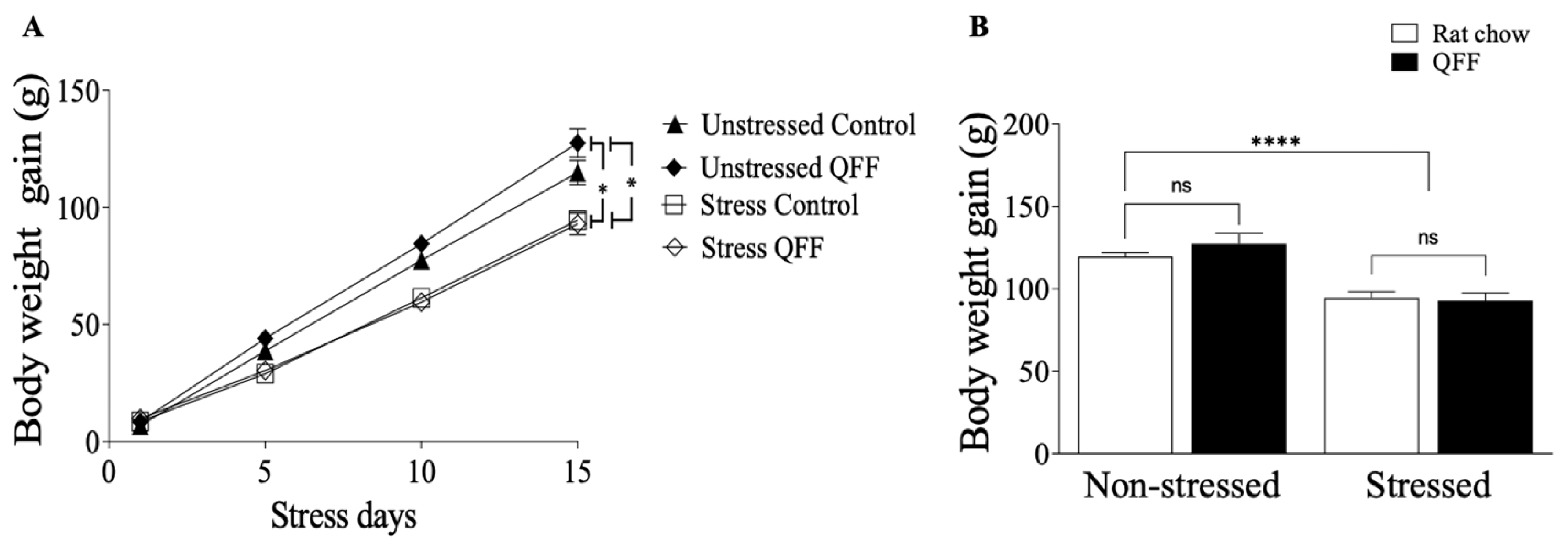
To assess the effect of the stress protocol and QFF intake, we measured the body weight gain during the stress protocol. Figure 2A displays the body weight gain over the 15 days of the stress protocol. Rats under the stress protocol exhibited significantly lower weight gain compared to the non-stressed rats (p < 0.0001). Conversely, the QFF diet did not affect body weight gain compared to chow diet (p > 0.05). Figure 2B displays the reduction in total weight gain in the stressed group at the end of the 15-day protocol (F(1.31) = 42.94, p < 0.0001), with no significant differences in body weight gain observed for diet. This suggests that QFF intake did not alter body weight gain and did not prevent the stress-induced effects on this parameter (Figure 2B).

3.2. QFF Consumption Did Not Affect Locomotor Activity and Anxiety-like Behavior

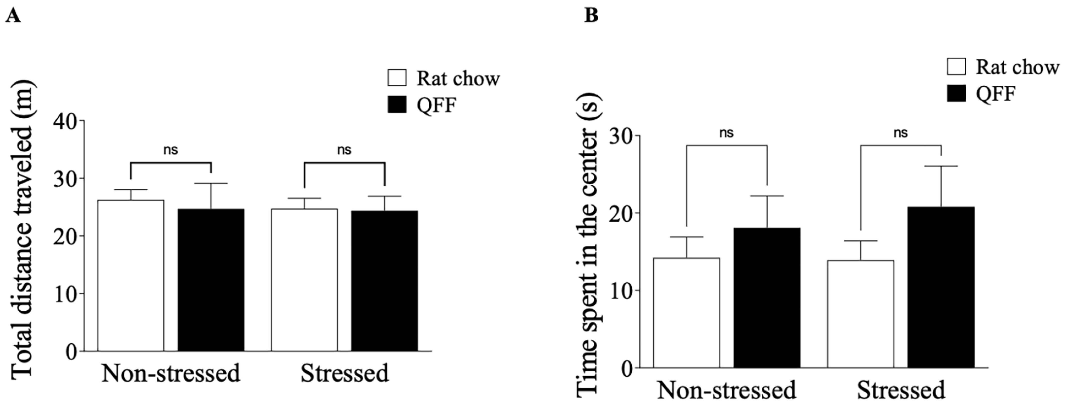
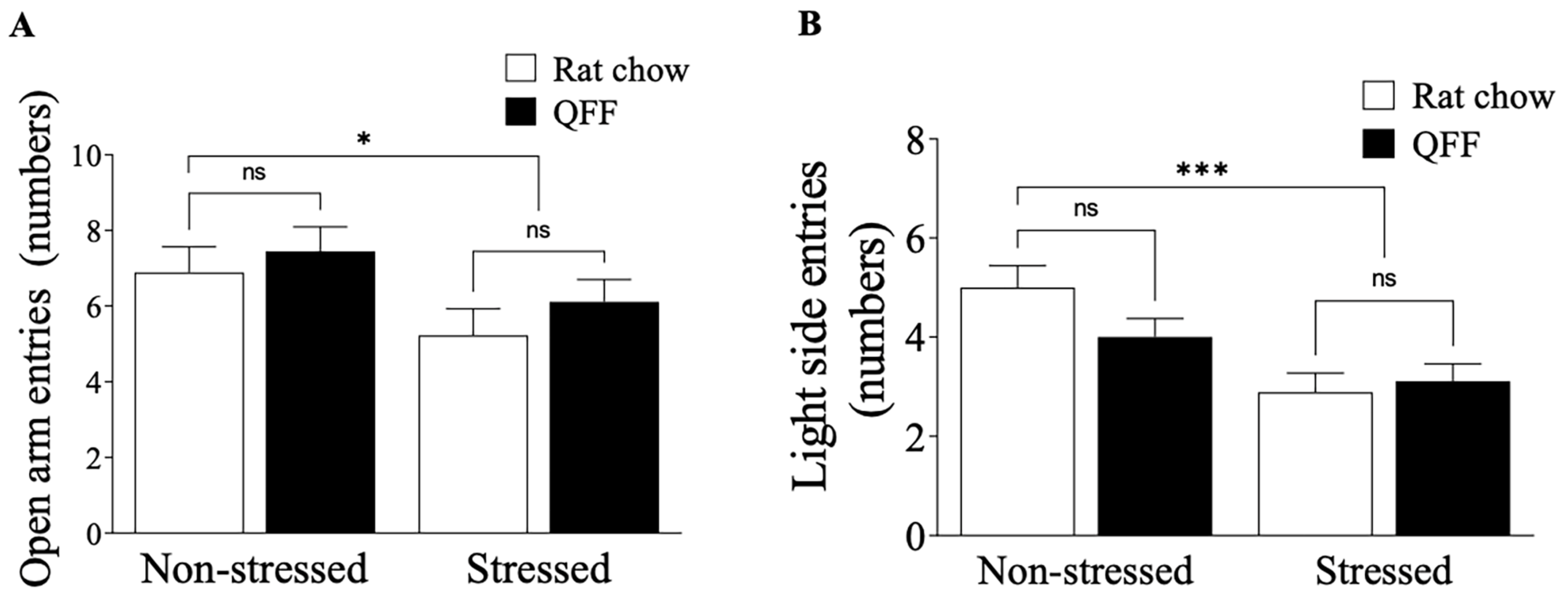
We measured the locomotor activity in the OF test and anxiety-like behavior in the OF, EPM, and LDB tests 24 h after the last stress session. Figure 3A shows that the locomotor activity was not affected by stress protocol or diet (p > 0.05). Similarly, the analysis of the anxiety-like behavior indicator in the OF test, measured as the time spent in the center of the field (Figure 3B), was not affected by stress protocol or diet (F(1.32) = 0.1028, p = 0.7506). To complement the OF test results, we analyzed the anxiety levels in the EPM and LDB tests at PND 52. Figure 4A shows that the restraint stress reduced the number of open-arm entries in the EPM, confirming that our chronic stress protocol induced an anxiety-like behavior (Figure 4A). There was no main effect of diet (F(1.32) = 1.2, p = 0.2784) or diet–treatment interaction (F(1.32) = 0.065, p = 0.8008) on anxiety levels measured in the EPM (Figure 4A). We next assessed anxiety in the LDB (Figure 4B), where rats subjected to the stress protocol showed a significant decrease in the number of entries to the light side compared to the non-stressed groups (F(1.32)= 14.80, p < 0.001). There were no main effects of diet or interaction (diet–stress) on the numbers of entries to the light side (Figure 4B). These results showed that quinoa did not affect either locomotor activities or anxiety behavior in our model.

3.3. QFF Consumption Prevented Spatial Memory Impairment Induced by Restraint Stress

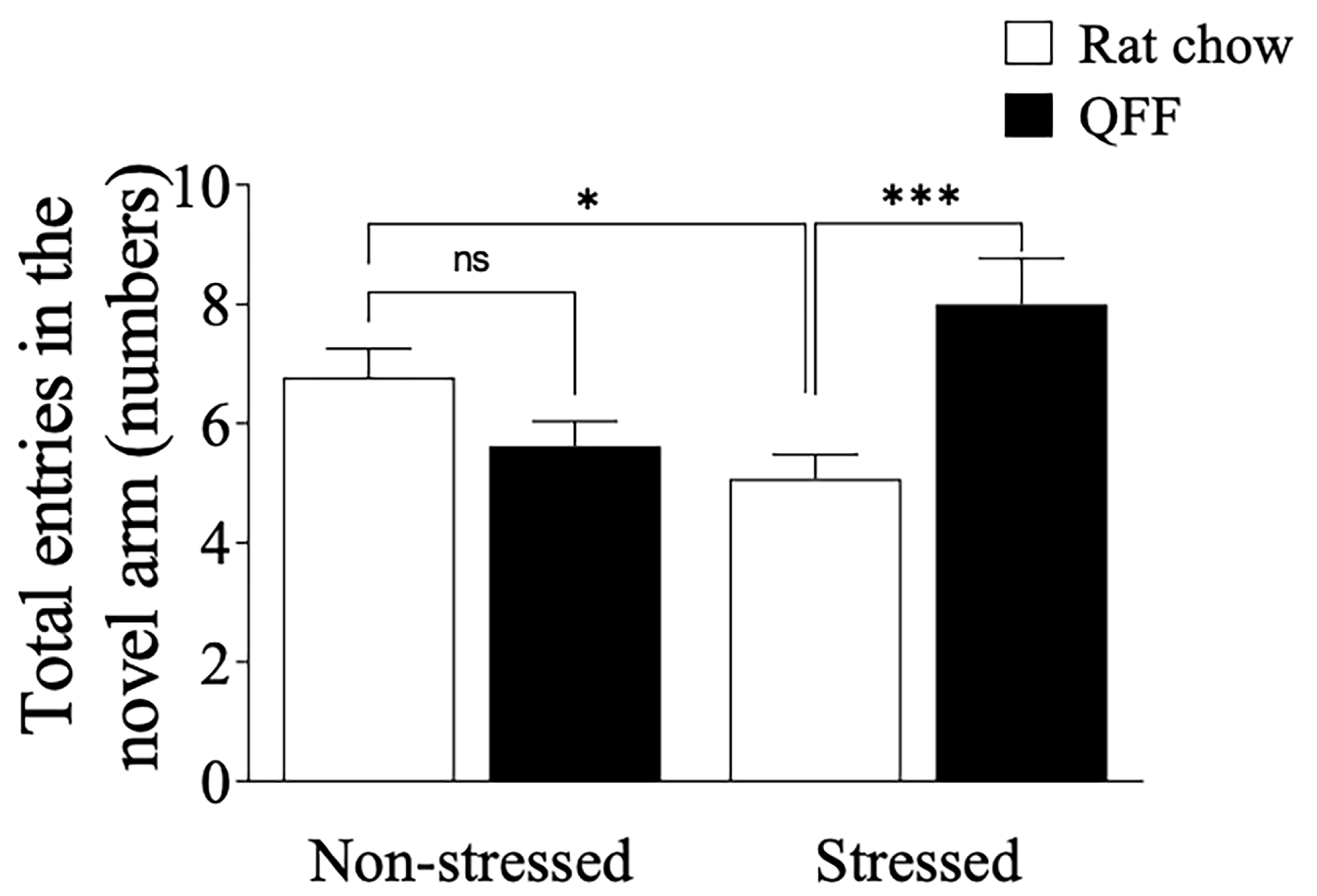
To determine whether QFF consumption could modulate cognitive parameters in our stress protocol, we evaluated spatial memory impairments in the animals using the Y-maze task (Figure 5) 48 h after the last stress session. Restraint stress significantly decreased the number of entries made into the novel arms of the Y-maze (p < 0.05). There was not a main effect of diet on the number of entries made into the novel arms of the Y-maze. However, there was a main interaction effect (diet × tress) on the number of entries made into the novel arms of the Y-maze (F(1.49) = 15, p = 0.0004). Further analysis showed that QFF consumption prevented the memory deficit induced by stress (p < 0.05; Stressed (rat chow) vs. Stressed + QFF entries to the novel arm), allowing us to conclude that quinoa consumption was effective in modulating spatial memory in our model of stress.

3.4. QFF Consumption Reduced the Stress-Induced Dendritic Atrophy in the Hippocampus

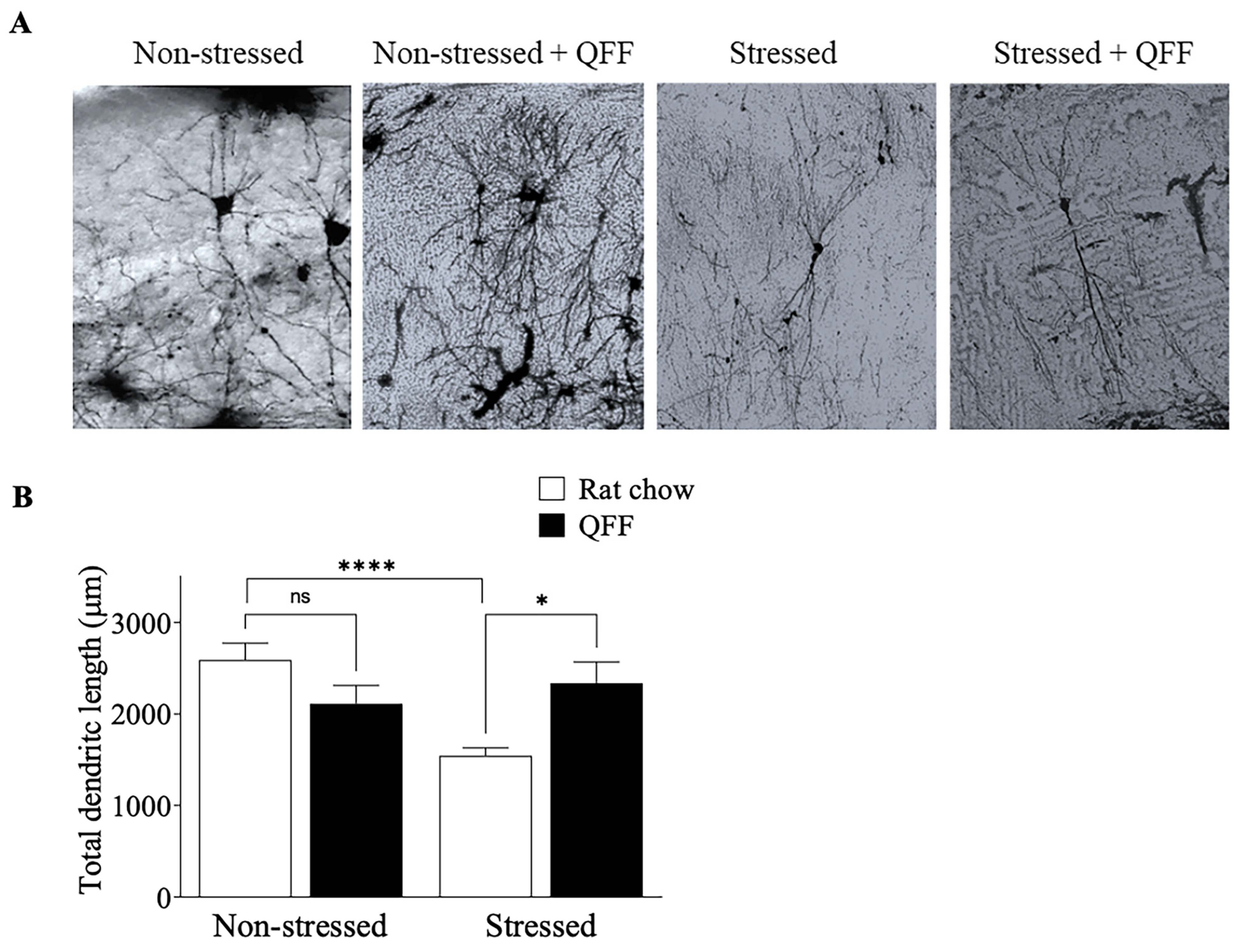
To determine whether the effect of QFF consumption on spatial memory was associated with morphological changes in the hippocampus, we compared the total dendritic length of CA3 pyramidal neurons of rats from non-stressed and stressed groups. First, the Bonferroni–Dunn post-test analysis revealed that the stress protocol decreased the total dendritic length compared to that of non-stressed animals (p < 0.05) (Figure 6). Second, QFF consumption effectively increased the total dendritic length to the values observed in non-stressed animals (stressed 1544 ± 486.3 µm, stressed + QFF 2338 ± 1107 µm) (Figure 6). Moreover, QFF supplementation in the non-stressed group did not have a significant effect (p > 0.05) on the dendritic length of the CA3 pyramidal neurons. These results suggest that quinoa supplementation prevented the dendritic atrophy in hippocampal neurons in our stress model.

4. Discussion

In this study, we demonstrate that the consumption of quinoa-based food during adolescence enhances the memory of stressed rats and mitigates stress-induced dendritic atrophy in the hippocampus. Initially, we examined whether our stress protocol effectively elicited stress responses. Stressed rats from all experimental groups (rat chow and QFF) exhibited less body weight gain than non-stressed rats. This indicates the effectiveness of the stress protocol, and the intake of QFF did not prevent the reduction in weight gain induced by restraint stress. However, other studies have reported that the dietary supplementation of hydrolyzed quinoa (2000 mg/kg, 30 days) in sedentary Wistar rats resulted in decreased body weight gain, food intake, fat deposition, and triglycerides [27]. Additionally, supplementation with fermented or sprouted quinoa (47 days) reduced food intake, blood glucose, and lipid levels in rats fed with a high-carbohydrate diet [28]. Both studies reported a decrease in weight gain after quinoa supplementation, which may be associated with reduced food intake [28] linked to lower levels of ghrelin, leptin, and cholecystokinin [29].
On the other hand, we did not observe changes in the body weight gain in the non-stressed group fed with QFF compared to rat chow, which could be explained by differences in food intake, forms of administration, and nutritional properties of the food. We suggest that the administration of anxiogenic protocols (oral administration or orogastric gavage) and reduction in food intake result in less body weight gain. In support of this idea, previous studies have shown that oral administration of n-3 PUFAs is considered a stressor for rats because it increases corticosterone levels in non-stressed rats [30], thereby reducing body weight gain. Moreover, feeding the animals with a solution of hydrolyzed quinoa through orogastric gavage reduces food intake and decreases body weight gain [27].
Food intake is strongly influenced by stress and the composition of the diet. Rats subjected to stress consumed less commercial chow than the non-stressed rats [31]. Interestingly, rats given the option to choose between two diets prefer high-caloric food [31]. In our study, we found no difference in food intake. This may suggest that our method of administration of food is less stressful or that, due to nutritional characteristics, quinoa is more palatable for rats.
Chronic stress-induced anxiety arises from alterations in neuronal morphology in the basolateral amygdala [32]. In the brain, the medial prefrontal cortex and the amygdala are extensively interconnected, working together to regulate the expression of emotions such as fear and anxiety [33]. It is possible that the chronic stress protocol used in our study induced hyperactivation of the basolateral amygdala and/or the bed nucleus of the stria terminalis [33,34], leading to increased anxiety. On the contrary, it has been demonstrated that supplementation with n-3 PUFAs can reduce chronic stress-induced anxiety [30,35]. The anxiolytic effect of n-3 PUFAs was notably different from controls only in subgroups with a higher dosage (at least 2000 mg/d) and not in subgroups with a lower dosage (<2000 mg/d) [36]. This suggests that the anxiolytic effect depends on the concentration of PUFA, specifically docosahexaenoic acid (DHA: 22:6 n-3) that is synthesized from ALA. In our experiments, QFF consumption did not affect anxiety in the EPM and LDB tests. Quinoa seeds are rich in ALA [37], but while humans can convert ALA into eicosapentaenoic acid (EPA: 20:5 n-3), which is the main substrate for synthesizing DHA, the efficiency of this conversion is limited, and thus EPA and DHA must also be obtained from the diet to maintain adequate levels [38]. Therefore, we believe that due to the low concentration of DHA achieved from the conversion of ALA, it is not possible to obtain an anxiolytic effect.
The chronic restraint protocol induces atrophy in dendritic arborization of pyramidal neurons from the CA3 region of the hippocampus, characterized by a reduction in dendritic length and number of branch points [39]. Moreover, dendrite atrophy has been associated with deficits in hippocampal-dependent spatial memory [8,39]. This evidence aligns with our findings, where the restraint protocol led to a decrease in dendritic length and spatial memory deficits assessed in the Y-maze. However, QFF intake in our study mitigated these effects, alleviating the stress-induced damage in both dendritic length and spatial memory. Notably, prior studies have demonstrated that an extract from red quinoa seeds prevented memory deficits induced by scopolamine treatment in mice [23]. This supports our findings and establishes the protective role of a quinoa-enriched diet in cognitive performance and/or behavioral alterations in rodents.
Diet significantly influences anxiety behavior [40], learning, and memory in rats [41]. When combined with stress, a diet rich in saturated fatty acids induces the retraction of dendrites in neurons in the CA3 region of the hippocampus, leading to arterial hypertension and an increase in corticosterone levels [14,42]. In contrast, n-6 and n-3 PUFA consumption improves the cognitive and emotional state in both rodents [43] and humans [44]. Quinoa seeds have a high content of ALA and LA, two essential fatty acids, which represent 55–63% of its lipid fraction [45]. ALA is associated with health, reproduction, and mammalian development [46]. Importantly, ALA can be converted to long-chain PUFAs (LCPUFAs) in the liver, enabling the synthesis of EPA and DHA. DHA, the predominant fatty acid in the brain, constituting about 15% of the total fatty acids in that tissue [47], plays a crucial role. Inadequate DHA intake has been linked to poor performance in attention and learning tests [48], increased aggression, and elevated anxiety levels [49] in rodents. Additionally, rats deficient in n-3 LCPUFAs exhibit excessive stress, anxiety, and fear responses, which are reversed after DHA supplementation [50]. Moreover, EPA and DHA supplementation improved memory and prevented hippocampal dendritic atrophy in chronically stressed rats [51]. DHA may prevent stress-induced behavior through modulating GABA receptor activity [50] and restoring GABA release probability in the CA1 region of the hippocampus [51]. Conversely, the consumption of a fish oil-enriched diet (high in EPA and DHA) alters the phospholipid composition of the rat brain, increasing levels of phosphatidylserine and influencing cortical and striatal dopaminergic function. This dietary change enhances cognitive function, spatial memory, and locomotor activity in aged rats [52]. Furthermore, n-3 PUFA induces the expression of genes involved in learning, memory, and neuronal metabolism [53]. Interestingly, high ALA supplementation in rats did not alter anxiety levels [52,54], aligning with our findings that QFF did not affect anxiety in the EPM and LDB tests.
In summary, the collective evidence suggests that QFF’s effects in preventing spatial memory decline and dendritic impairment induced by the restraint protocol may be attributed to the PUFA content in quinoa. However, the observed differences between anxiety and memory effects could be attributed to varying requirements in the concentration of PUFAs, emphasizing the need for further studies.
In addition to the lipid profile of quinoa, it has been suggested that its high antioxidant content may play a role in the neuroprotective effects of these seeds. Indeed, dietary supplementation with red quinoa extract, rich in phenolic and flavonoid compounds, demonstrated a neuroprotective effect in mice treated with scopolamine [23].

5. Conclusions

We presented evidence supporting the neuroprotective effects of a quinoa-supplemented diet on neuronal morphology and memory. Additionally, we found that quinoa did not alleviate anxiogenic behavior or changes in weight gain induced by stress. This study enhances our understanding of the positive impact of quinoa on brain physiology. Future research should investigate whether quinoa’s effects on behavior are primarily due to the ALA content or other nutrients found in various quinoa seeds. Ultimately, considering its potential, this amaranthaceous plant could be considered a healthy dietary option to mitigate the impact of toxic stress (allostatic overload) in humans. For example, in stress-related disorders where memory deteriorates, such as in Alzheimer’s disease, dietary interventions using quinoa could be an alternative to enhance the therapeutic effect of drugs currently available for these neurodegenerative diseases.

Author Contributions

Conceptualization, G.T., P.M.-L. and E.A.M.; Methodology, G.T., M.Á.P., P.M.-L., A.D. and A.D.-S.; Formal Analysis, G.T., M.Á.P., A.D. and A.D.-S.; Resources, P.M.-L., E.A.M. and A.D.-S.; Writing—Original Draft Preparation, G.T., M.Á.P. and P.M.-L.; Writing—Review and Editing, G.T., A.D., E.A.M. and A.D.-S.; Funding Acquisition, A.D.-S. All authors have read and agreed to the published version of the manuscript.

Funding

This work has been funded by FONDECYT Regular Nº 1100413, Nº 1141276, and Anillo de Ciencia y Tecnología Nº ADI-09 grants (AD-S). Health Sciences School, Universidad Viña del Mar grant (MAP).

Institutional Review Board Statement

The study was conducted according to the guidelines of the Declaration of Helsinki, and approved by the Institutional Animal Ethics Committee of the Universidad de Valparaíso (Ref. No: AEB-022-2013, approval date: 10 September 2013).

Informed Consent Statement

Not applicable.

Data Availability Statement

The data that support the findings of this study are available from the corresponding author, A.D.-S., upon reasonable request.

Acknowledgments

This article is a testimony of the effort, friendship and love for science of Gonzalo Terreros, Miguel Ángel Pérez and Pablo Muñoz-Llancao. We acknowledge the contribution of laboratory facilities and preparation of the experiments by Antonio Vega Galvez and Ximena Moncada (respectively, ULS and CEAZA labs).

Conflicts of Interest

The authors declare no conflicts of interest.

References

  1. Selye, H. A Syndrome Produced by Diverse Nocuous Agents. Nature 1936, 138, 32. [Google Scholar] [CrossRef]
  2. McEwen, B.S.; Akil, H. Revisiting the Stress Concept: Implications for Affective Disorders. J. Neurosci. 2020, 40, 12–21. [Google Scholar] [CrossRef] [PubMed]
  3. Cook, S.C.; Wellman, C.L. Chronic Stress Alters Dendritic Morphology in Rat Medial Prefrontal Cortex. J. Neurobiol. 2004, 60, 236–248. [Google Scholar] [CrossRef] [PubMed]
  4. Conrad, C.D.; Galea, L.A.M.; Kuroda, Y.; McEwen, B.S. Chronic Stress Impairs Rat Spatial Memory on the Y Maze, and This Effect Is Blocked by Tianeptine Pretreatment. Behav. Neurosci. 1996, 110, 1321–1334. [Google Scholar] [CrossRef] [PubMed]
  5. McEwen, B.S. Neurobiological and Systemic Effects of Chronic Stress. Chronic Stress 2017, 1, 2470547017692328. [Google Scholar] [CrossRef] [PubMed]
  6. Vyas, A.; Bernal, S.; Chattarji, S. Effects of Chronic Stress on Dendritic Arborization in the Central and Extended Amygdala. Brain Res. 2003, 965, 290–294. [Google Scholar] [CrossRef] [PubMed]
  7. Magariños, A.M.; Deslandes, A.; McEwen, B.S. Effects of Antidepressants and Benzodiazepine Treatments on the Dendritic Structure of CA3 Pyramidal Neurons after Chronic Stress. Eur. J. Pharmacol. 1999, 371, 113–122. [Google Scholar] [CrossRef] [PubMed]
  8. Kleen, J.K.; Sitomer, M.T.; Killeen, P.R.; Conrad, C.D. Chronic Stress Impairs Spatial Memory and Motivation for Reward without Disrupting Motor Ability and Motivation to Explore. Behav. Neurosci. 2006, 120, 842–851. [Google Scholar] [CrossRef]
  9. Vyas, A.; Mitra, R.; Shankaranarayana Rao, B.S.; Chattarji, S. Chronic Stress Induces Contrasting Patterns of Dendritic Remodeling in Hippocampal and Amygdaloid Neurons. J. Neurosci. 2002, 22, 6810–6818. [Google Scholar] [CrossRef]
  10. Hueston, C.M.; Cryan, J.F.; Nolan, Y.M. Stress and Adolescent Hippocampal Neurogenesis: Diet and Exercise as Cognitive Modulators. Transl. Psychiatry 2017, 7, e1081. [Google Scholar] [CrossRef]
  11. Foilb, A.R.; Lui, P.; Romeo, R.D. The Transformation of Hormonal Stress Responses throughout Puberty and Adolescence. J. Endocrinol. 2011, 210, 391–398. [Google Scholar] [CrossRef] [PubMed]
  12. Romeo, R.D.; Karatsoreos, I.N.; McEwen, B.S. Pubertal Maturation and Time of Day Differentially Affect Behavioral and Neuroendocrine Responses Following an Acute Stressor. Horm. Behav. 2006, 50, 463–468. [Google Scholar] [CrossRef] [PubMed]
  13. Gunnar, M.R.; Wewerka, S.; Frenn, K.; Long, J.D.; Griggs, C. Developmental Changes in Hypothalamus-Pituitary-Adrenal Activity over the Transition to Adolescence: Normative Changes and Associations with Puberty. Dev. Psychopathol. 2009, 21, 69–85. [Google Scholar] [CrossRef] [PubMed]
  14. Baran, S.E.; Campbell, A.M.; Kleen, J.K.; Foltz, C.H.; Wright, R.L.; Diamond, D.M.; Conrad, C.D. Combination of High Fat Diet and Chronic Stress Retracts Hippocampal Dendrites. Neuroreport 2005, 16, 39–43. [Google Scholar] [CrossRef] [PubMed]
  15. Bazinet, R.P.; Layé, S. Polyunsaturated Fatty Acids and Their Metabolites in Brain Function and Disease. Nat. Rev. Neurosci. 2014, 15, 771–785. [Google Scholar] [CrossRef] [PubMed]
  16. Simopoulos, A.P. An Increase in the Omega-6/Omega-3 Fatty Acid Ratio Increases the Risk for Obesity. Nutrients 2016, 8, 128. [Google Scholar] [CrossRef] [PubMed]
  17. Michelle Bosquet, K.J.B. Effects of Prenatal Social Stress and Maternal Dietary Fatty Acid Ratio on Infant Temperament: Does Race Matter? Epidemiol. Open Access 2014, 4, 1000167. [Google Scholar] [CrossRef]
  18. ter Horst, D.M.; Schene, A.H.; Figueroa, C.A.; Assies, J.; Lok, A.; Bockting, C.L.H.; Ruhé, H.G.; Mocking, R.J.T. Cortisol, Dehydroepiandrosterone Sulfate, Fatty Acids, and Their Relation in Recurrent Depression. Psychoneuroendocrinology 2019, 100, 203–212. [Google Scholar] [CrossRef]
  19. Abedi, E.; Sahari, M.A. Long-Chain Polyunsaturated Fatty Acid Sources and Evaluation of Their Nutritional and Functional Properties. Food Sci. Nutr. 2014, 2, 443–463. [Google Scholar] [CrossRef]
  20. Vilcacundo, R.; Hernández-Ledesma, B. Nutritional and Biological Value of Quinoa (Chenopodium quinoa Willd.). Curr. Opin. Food Sci. 2017, 14, 1–6. [Google Scholar] [CrossRef]
  21. Vega-Gálvez, A.; Miranda, M.; Vergara, J.; Uribe, E.; Puente, L.; Martínez, E.A. Nutrition Facts and Functional Potential of Quinoa (Chenopodium quinoa Willd.), an Ancient Andean Grain: A Review. J. Sci. Food Agric. 2010, 90, 2541–2547. [Google Scholar] [CrossRef] [PubMed]
  22. Farinazzi-Machado, F.M.V.; Barbalho, S.M.; Oshiiwa, M.; Goulart, R.; Pessan Junior, O. Use of Cereal Bars with Quinoa (Chenopodium quinoa W.) to Reduce Risk Factors Related to Cardiovascular Diseases. Food Sci. Technol. 2012, 32, 239–244. [Google Scholar] [CrossRef]
  23. Souza, S.P.; Roos, A.A.; Gindri, A.L.; Domingues, V.O.; Ascari, J.; Guerra, G.P. Neuroprotective Effect of Red Quinoa Seeds Extract on Scopolamine-Induced Declarative Memory Deficits in Mice: The Role of Acetylcholinesterase and Oxidative Stress. J. Funct. Foods 2020, 69, 103958. [Google Scholar] [CrossRef]
  24. McLaughlin, K.J.; Gomez, J.L.; Baran, S.E.; Conrad, C.D. The Effects of Chronic Stress on Hippocampal Morphology and Function: An Evaluation of Chronic Restraint Paradigms. Brain Res. 2007, 1161, 56–64. [Google Scholar] [CrossRef] [PubMed]
  25. Vega-Gálvez, A.; Dagnino-Subiabre, A.; Terreros, G.; López, J.; Miranda, M.; Di Scala, K. Mathematical Modeling of Convective Air Drying of Quinoa-Supplemented Feed for Laboratory Rats. Brazilian Arch. Biol. Technol. 2011, 54, 161–171. [Google Scholar] [CrossRef]
  26. Biała, G.; Kruk, M. Amphetamine-Induced Anxiety-Related Behavior in Animal Models. Pharmacol. Rep. 2007, 59, 636–644. [Google Scholar]
  27. de Oliveira Lopes, C.; de Fátima Píccolo Barcelos, M.; de Goes Vieira, C.N.; de Abreu, W.C.; Ferreira, E.B.; Pereira, R.C.; de Angelis-Pereira, M.C. Effects of Sprouted and Fermented Quinoa (Chenopodium quinoa) on Glycemic Index of Diet and Biochemical Parameters of Blood of Wistar Rats Fed High Carbohydrate Diet. J. Food Sci. Technol. 2019, 56, 40–48. [Google Scholar] [CrossRef]
  28. Meneguetti, Q.A.; Brenzan, M.A.; Batista, M.R.; Bazotte, R.B.; Silva, D.R.; Garcia Cortez, D.A. Biological Effects of Hydrolyzed Quinoa Extract from Seeds of Chenopodium quinoa Willd. J. Med. Food 2011, 14, 653–657. [Google Scholar] [CrossRef]
  29. Mithila, M.V.; Khanum, F. Effectual Comparison of Quinoa and Amaranth Supplemented Diets in Controlling Appetite; a Biochemical Study in Rats. J. Food Sci. Technol. 2015, 52, 6735–6741. [Google Scholar] [CrossRef]
  30. Pérez, M.Á.; Terreros, G.; Dagnino-Subiabre, A. Long-Term ω-3 Fatty Acid Supplementation Induces Anti-Stress Effects and Improves Learning in Rats. Behav. Brain Funct. 2013, 9, 12–25. [Google Scholar] [CrossRef]
  31. Ortolani, D.; Oyama, L.M.; Ferrari, E.M.; Melo, L.L.; Spadari-Bratfisch, R.C. Effects of Comfort Food on Food Intake, Anxiety-like Behavior and the Stress Response in Rats. Physiol. Behav. 2011, 103, 487–492. [Google Scholar] [CrossRef]
  32. Mitra, R.; Jadhav, S.; McEwen, B.S.; Vyas, A.; Chattarji, S. Stress Duration Modulates the Spatiotemporal Patterns of Spine Formation in the Basolateral Amygdala. Proc. Natl. Acad. Sci. USA 2005, 102, 9371–9376. [Google Scholar] [CrossRef] [PubMed]
  33. Liu, W.Z.; Zhang, W.H.; Zheng, Z.H.; Zou, J.X.; Liu, X.X.; Huang, S.H.; You, W.J.; He, Y.; Zhang, J.Y.; Wang, X.D.; et al. Identification of a Prefrontal Cortex-to-Amygdala Pathway for Chronic Stress-Induced Anxiety. Nat. Commun. 2020, 11, 1–15. [Google Scholar] [CrossRef] [PubMed]
  34. Daviu, N.; Bruchas, M.R.; Moghaddam, B.; Sandi, C.; Beyeler, A. Neurobiological Links between Stress and Anxiety. Neurobiol. Stress 2019, 11, 100191. [Google Scholar] [CrossRef] [PubMed]
  35. Ferraz, A.C.; Delattre, A.M.; Almendra, R.G.; Sonagli, M.; Borges, C.; Araujo, P.; Andersen, M.L.; Tufik, S.; Lima, M.M.S. Chronic ω-3 Fatty Acids Supplementation Promotes Beneficial Effects on Anxiety, Cognitive and Depressive-like Behaviors in Rats Subjected to a Restraint Stress Protocol. Behav. Brain Res. 2011, 219, 116–122. [Google Scholar] [CrossRef] [PubMed]
  36. Su, K.P.; Tseng, P.T.; Lin, P.Y.; Okubo, R.; Chen, T.Y.; Chen, Y.W.; Matsuoka, Y.J. Association of Use of Omega-3 Polyunsaturated Fatty Acids With Changes in Severity of Anxiety Symptoms: A Systematic Review and Meta-Analysis. JAMA Netw. Open 2018, 1, e182327. [Google Scholar] [CrossRef]
  37. Repo-Carrasco Valencia, R. Chapter 2.1: Quinoa. In Andean Indigenous Food Crops: Nutritional Value and Bioactive Compounds; University of Turku: Turku, Finland, 2011; ISBN 9789512946044. [Google Scholar]
  38. Burdge, G. α-Linolenic Acid Metabolism in Men and Women: Nutritional and Biological Implications. Curr. Opin. Clin. Nutr. Metab. Care 2004, 7, 137–144. [Google Scholar] [CrossRef]
  39. Conrad, C.D.; McLaughlin, K.J.; Harman, J.S.; Foltz, C.; Wieczorek, L.; Lightner, E.; Wright, R.L. Chronic Glucocorticoids Increase Hippocampal Vulnerability to Neurotoxicity under Conditions That Produce CA3 Dendritic Retraction but Fail to Impair Spatial Recognition Memory. J. Neurosci. 2007, 27, 8278–8285. [Google Scholar] [CrossRef]
  40. Aslani, S.; Vieira, N.; Marques, F.; Costa, P.S.; Sousa, N.; Palha, J.A. The Effect of High-Fat Diet on Rat’s Mood, Feeding Behavior and Response to Stress. Transl. Psychiatry 2015, 5, e684. [Google Scholar] [CrossRef]
  41. Khazen, T.; Hatoum, O.A.; Ferreira, G.; Maroun, M. Acute Exposure to a High-Fat Diet in Juvenile Male Rats Disrupts Hippocampal-Dependent Memory and Plasticity through Glucocorticoids. Sci. Rep. 2019, 9, 12270. [Google Scholar] [CrossRef]
  42. Bruder-Nascimento, T.; Campos, D.H.S.; Alves, C.; Thomaz, S.; Cicogna, A.C.; Cordellini, S. Effects of Chronic Stress and High-Fat Diet on Metabolic and Nutritional Parameters in Wistar Rats. Arq. Bras. Endocrinol. Metabol. 2013, 57, 642–649. [Google Scholar] [CrossRef] [PubMed]
  43. Cardoso, C.; Afonso, C.; Bandarra, N.M. Dietary DHA and Health: Cognitive Function Ageing. Nutr. Res. Rev. 2016, 29, 281–294. [Google Scholar] [CrossRef] [PubMed]
  44. Bauer, I.; Hughes, M.; Rowsell, R.; Cockerell, R.; Pipingas, A.; Crewther, S.; Crewther, D. Omega-3 Supplementation Improves Cognition and Modifies Brain Activation in Young Adults. Hum. Psychopharmacol. 2014, 29, 133–144. [Google Scholar] [CrossRef] [PubMed]
  45. Repo-Carrasco, R.; Espinoza, C.; Jacobsen, S.E. Nutritional Value and Use of the Andean Crops Quinoa (Chenopodium quinoa) and Kañiwa (Chenopodium pallidicaule). Food Rev. Int. 2003, 19, 179–189. [Google Scholar] [CrossRef]
  46. Simopoulos, A.P. Evolutionary Aspects of Diet: The Omega-6/Omega-3 Ratio and the Brain. Mol. Neurobiol. 2011, 44, 203–215. [Google Scholar] [CrossRef] [PubMed]
  47. Chang, C.Y.; Ke, D.S.; Chen, J.Y. Essential Fatty Acids and Human Brain. Acta Neurol. Taiwan 2009, 18, 231–241. [Google Scholar]
  48. Fedorova, I.; Salem, N. Omega-3 Fatty Acids and Rodent Behavior. Prostagland. Leukot. Essent. Fat. Acids 2006, 75, 271–289. [Google Scholar] [CrossRef]
  49. Mathieu, G.; Oualian, C.; Denis, I.; Lavialle, M.; Gisquet-Verrier, P.; Vancassel, S. Dietary N-3 Polyunsaturated Fatty Acid Deprivation Together with Early Maternal Separation Increases Anxiety and Vulnerability to Stress in Adult Rats. Prostagland. Leukot. Essent. Fat. Acids 2011, 85, 129–136. [Google Scholar] [CrossRef]
  50. Takeuchi, T.; Iwanaga, M.; Harada, E. Possible Regulatory Mechanism of DHA-Induced Anti-Stress Reaction in Rats. Brain Res. 2003, 964, 136–143. [Google Scholar] [CrossRef]
  51. Pérez, M.Á.; Peñaloza-Sancho, V.; Ahumada, J.; Fuenzalida, M.; Dagnino-Subiabre, A. N-3 Polyunsaturated Fatty Acid Supplementation Restored Impaired Memory and GABAergic Synaptic Efficacy in the Hippocampus of Stressed Rats. Nutr. Neurosci. 2018, 21, 556–569. [Google Scholar] [CrossRef]
  52. Chalon, S.; Delion-Vancassel, S.; Belzung, C.; Guilloteau, D.; Leguisquet, A.M.; Besnard, J.C.; Durand, G. Dietary Fish Oil Affects Monoaminergic Neurotransmission and Behavior in Rats. J. Nutr. 1998, 128, 2512–2519. [Google Scholar] [CrossRef] [PubMed]
  53. Kitajka, K.; Puskás, L.G.; Zvara, Á.; Hackler, L.; Barceló-Coblijn, G.; Yeo, Y.K.; Farkas, T. The Role of N-3 Polyunsaturated Fatty Acids in Brain: Modulation of Rat Brain Gene Expression by Dietary n-3 Fatty Acids. Proc. Natl. Acad. Sci. USA 2002, 99, 2619–2624. [Google Scholar] [CrossRef] [PubMed]
  54. Nakashima, Y.; Yuasa, S.; Hukamizu, Y.; Okuyama, H.; Ohhara, T.; Kameyama, T.; Nabeshima, T. Effect of a High Linoleate and a High α-Linolenate Diet on General Behavior and Drug Sensitivity in Mice. J. Lipid Res. 1993, 34, 239–247. [Google Scholar] [CrossRef] [PubMed]
Figure 1. Schematic drawing of the experimental design. Male Sprague-Dawley rats of 21 postnatal days (PND) were fed either with rat chow or quinoa functional food (QFF, (50% rat chow + 50% dehydrated quinoa seeds)). At PND 36, animals were submitted to a restraint protocol for 2 h per day for 15 days, establishing four groups (n = 27 per experimental group): non-stressed (rat chow), non-stressed + QFF, stressed, and stressed + QFF. At PND 52, motor and anxiety behavior were evaluated on the open field (OF), elevated plus maze (EPM), and light-dark box (LDB). Finally, at PND 53, the memory performance was evaluated on the Y-maze test, and brain tissue was used for Golgi staining. Created with BioRender.com.
Nutrients 16 00381 g001
Figure 2. Effect of diet type on body weight gain during the stress protocol: (A) Daily measure of body weight during the 15 days of stress protocol, and (B) total body weight gain at the end of day 15 of the stress protocol. There were no significant differences in body weight gain induced by diet intake. However, exposure to the restraint stress protocol was associated with a significant reduction in body weight in both rat chow and QFF groups (n = 9, in each). Values are expressed as mean ± SEM (n = 9). * p < 0.05, **** p < 0.0001. Non-significant, n.s; quinoa functional food, QFF.
Nutrients 16 00381 g002
Figure 3. Effect of diet type on locomotor activity measured in the open field test. (A) Total distance traveled (m) and (B) time spent in the center in an open-field test (s). Locomotor activity at PND 52 was not affected by the restraint stress protocol in any experimental group. The anxiety-like behavior measured by the time spent in the center of the field did not show a significant difference in any of the groups (n = 9, in each). Values are expressed as mean ± SEM (n = 9). Non-significant, n.s; quinoa functional food, QFF.
Nutrients 16 00381 g003
Figure 4. Effect of diet type on anxiety-like behavior measured in the elevated plus-maze and light-dark box test. (A) Total number of open arm entries made in the elevated plus-maze. Stress protocol significantly decreased the number of open-arm entries in both the rat chow and QFF groups. Anxiety levels measured in the EPM were not affected by diet. (B) Total number of light side entries made in the light-dark box test. Stressed animals had a smaller number of entries to the light side compared to the non-stressed animals in both the rat chow and QFF groups. Anxiety levels measured in LDB were not affected by diet between groups (n = 9, in each). Values are expressed as mean ± SEM (n = 9) * p < 0.05, *** p < 0.001. Non-significant, n.s; quinoa functional food, QFF.
Nutrients 16 00381 g004
Figure 5. Influence of quinoa functional food diet on Y-maze performance. Total number of entries made in the novel arm in the Y-maze. Chronic stress protocol induced spatial memory impairment in the rat chow but not in the QFF group (n = 9, in each). Values are expressed as mean ± SEM (n = 9), * p < 0.05, *** p < 0.001. Non-significant, n.s; quinoa functional food, QFF.
Nutrients 16 00381 g005
Figure 6. Effect of quinoa functional food on CA3 dendritic morphology. (A) Representative photographs and (B) quantification of the total dendritic length of CA3 pyramidal neurons in the hippocampus. QFF diet reduced the dendritic impairment induced by stress compared to the rat chow group (n = 9, in each). Values are expressed as mean ± SEM (n = 9), * p < 0.05, **** p < 0.0001. Non-significant, n.s; quinoa functional food, QFF.
Nutrients 16 00381 g006
Table 1. Number of rats used in each experiment.
DescriptionExperimental GroupsTotal
Non-StressedNon-Stressed + QFFStressedStressed + QFF
Locomotor activity and anxietyn = 9n = 9n = 9n = 9
Golgi stainingn = 9n = 9n = 9n = 9
Behavioral tasksn = 9n = 9n = 9n = 9
Totaln = 27n = 27n = 27n = 27n = 108
Disclaimer/Publisher’s Note: The statements, opinions and data contained in all publications are solely those of the individual author(s) and contributor(s) and not of MDPI and/or the editor(s). MDPI and/or the editor(s) disclaim responsibility for any injury to people or property resulting from any ideas, methods, instructions or products referred to in the content.