Article

Factors Influencing Breast Milk Antibody Titers during the Coronavirus Disease 2019 Pandemic: An Observational Study

1
Department of Pediatrics II (Neonatology), Medical University of Innsbruck, Anichstraße 35, 6020 Innsbruck, Austria
2
Division for Nutrition and Dietetics, University Hospital Innsbruck, 6020 Innsbruck, Austria
3
Institute of Public Health, Medical Decision Making and Health Technology Assessment, Department of Public Health, Health Services Research and Health Technology Assessment, UMIT TIROL—University for Health Sciences and Technology, 6060 Hall in Tirol, Austria
*
Author to whom correspondence should be addressed.
Nutrients 2024, 16(14), 2320; https://doi.org/10.3390/nu16142320
Submission received: 25 June 2024 / Revised: 9 July 2024 / Accepted: 17 July 2024 / Published: 18 July 2024

Abstract

:
The COVID-19 pandemic has highlighted the role of breastfeeding in providing passive immunity to infants via specific anti-SARS-CoV-2 antibodies in breast milk. We aimed to quantify these antibodies across different lactation stages and identify influencing factors. This prospective study involved mother–child dyads from Innsbruck University Hospital, Austria, with a positive maternal SARS-CoV-2 test during pregnancy or peripartum between 2020 and 2023. We collected breast milk samples at various lactation stages and analyzed anti-Spike S1 receptor-binding domain (S1RBD) immunoglobulins (Ig). Maternal and neonatal data were obtained from interviews and medical records. This study included 140 mothers and 144 neonates. Anti-S1RBD-IgA (72.0%), -IgG (86.0%), and -IgM (41.7%) were highly present in colostrum and decreased as milk matured. Mothers with natural infection and vaccination exhibited higher anti-S1RBD-IgA and -IgG titers in all milk stages. Mothers with moderate to severe infections had higher concentrations of anti-S1RBD-IgA and -IgG in transitional milk and higher anti-S1RBD-IgA and -IgM in mature milk compared to those with mild or asymptomatic infections. Variations in antibody responses were also observed with preterm birth and across different virus waves. This study demonstrates the dynamic nature of breast milk Ig and underscores the importance of breastfeeding during a pandemic.

Graphical Abstract

1. Introduction

In the wake of the unprecedented global health crisis triggered by the COVID-19 pandemic, the significance of breastfeeding and its potential implications on infant health have garnered increased attention. In the very beginning of the pandemic, newborns were separated from SARS-CoV-2-positive mothers, and breastfeeding was discouraged due to fear of infection [1,2]. However, soon, studies reported significant levels of specific anti-SARS-CoV-2 antibodies in breast milk samples from COVID-19-recovered donors [1,3,4,5,6] and vaccinated breastfeeding mothers [5,7,8], indicating the transfer of passive immunity. Additional in vitro studies demonstrated the neutralizing capacity of SARS-CoV-2-specific antibodies in human breast milk [4,9,10].
Ever since, the World Health Organization (WHO), the U.S. Centers for Disease Control and Prevention (CDC), and other national healthcare systems have advocated for breastfeeding with active SARS-CoV-2 infection, as the benefits of human milk seem to far exceed the minimal risk of potential SARS-CoV-2 transmission [11,12].
Studies show that the mechanism for acquiring immunity influences breast milk responses in terms of titer development and predominant immunoglobulin (Ig) class; natural infection with SARS-CoV-2 induces a rapid rise in Spike-reactive IgA [3,13], whereas vaccination mainly induces an IgG response [5,14]. However, little is known about the effect of milk maturation and other factors on anti-SARS-CoV-2 Ig levels and composition in breast milk [5].
The present study aimed to quantify breast milk Ig specifically directed against the Spike S1 subunit of the SARS-CoV-2 receptor-binding domain (S1RBD) in all stages of lactation throughout the course of the pandemic and to assess factors that influence concentrations of anti-S1RBD Ig in human milk.

2. Materials and Methods

2.1. Study Design and Population

Mother–child dyads included in the present analysis were participants of the prospective study “Evaluation of potential effects of a maternal SARS-CoV-2 infection on the newborn” conducted at the Department of Pediatrics II (Neonatology) at Innsbruck University Hospital, Austria. We invited all women who gave birth at Innsbruck University Hospital between July 2020 and January 2023 and had a positive nasopharyngeal swab test result for SARS-CoV-2 during pregnancy or peripartum to participate in this study.
We employed convenience sampling and determined our sample size based on resource constraints.
This study was conducted according to the guidelines of the Declaration of Helsinki and was approved by the Institutional Review Board of the Medical University of Innsbruck, Austria (approval number 1084/2020, 11 May 2020), and all participants gave written informed consent for participation.

2.2. Maternal and Neonatal Data Collection

Data on maternal past medical history, first positive test results for SARS-CoV-2, and disease course and symptom severity, as well as vaccination status, were collected by means of personal interviews and medical records. Maternal SARS-CoV-2 infection was categorized as asymptomatic or pre-symptomatic disease if the mother had a positive SARS-CoV-2 test without symptoms. Mild disease was defined as flu-like symptoms, such as fever, cough, myalgia, and anosmia. Moderate disease was defined as lower respiratory tract disease with dyspnea, pneumonia, refractory fever, and abnormal blood gas results while maintaining an oxygen saturation above 93%. Severe disease was defined as a respiratory rate greater than 30 breaths per minute and the need for oxygen supplementation. Critical disease was defined as multi-organ failure, respiratory failure requiring high-flow nasal cannula therapy, or mechanical ventilation [15].
Virus waves were categorized according to the timing of infection. Data on the prevailing SARS-CoV-2 variant at any given time point were obtained from the website of the Global Initiative on Sharing all Influenza Data (GISAID) [16] and the regular local GISAID reports, which are published on the homepage of the Austrian Agency for Health and Food Safety GmBH, Vienna, Austria (AGES) [17].
All relevant perinatal and neonatal data were collected from medical records.

2.3. Sample Collection and Processing

2.3.1. Neonatal Samples

After obtaining informed consent, every newborn was swab-tested for SARS-CoV-2. Specimens were analyzed at the ISO-certified Central Institute of Clinical and Chemical Laboratory Diagnostics at Innsbruck University Hospital, Austria.
Whole-blood anti-S1RBD-IgG and -IgM were quantified in dried blood spot (DBS) samples obtained from umbilical cord blood and/or neonatal venous blood collected at 48 h of life. DBS samples from patients without SARS-CoV-2 infection or vaccination were used as negative controls. Samples exhibiting anti-S1RBD-IgG concentrations greater than 30 U/mL and anti-S1RBD-IgM concentrations greater than 100 U/mL were considered positive. Methodological details are provided in the Supplementary Materials.

2.3.2. Breast Milk Samples

Breast milk samples were self-collected by participants into sterile 1.8 mL cryotubes (CryoPure, Sarstedt, Nümbrecht, Germany) at different time points, as follows: (i) colostrum (in the first 3 days after delivery), (ii) transitional milk (4 to 14 days after delivery), and (iii) mature milk (>14 days after delivery). Samples were centrifuged three times at 4100 rpm/4 °C for 15 min to obtain fat-free, acellular whey milk and stored at −20 °C until further analysis.
Pre-diluted samples were plated in duplicate on a 96-well plate coated with S1RBD protein and incubated for 1 h (IEQ-CoVS1RBD-IgA, -IgM and -IgG, Raybiotech, GA, USA). For background subtraction, samples were further added to a human albumin-coated plate (IgA, IgM). After washing, plates were incubated with biotinylated anti-human IgA, IgM, and IgG and, subsequently, with HRP-Streptavidin concentrate. 3,3′,5,5′-tetramethylbenzidine (TMB) substrate solution was added, and the reaction was stopped with sulphuric acid. Plates were read at 450 nm (Hidex Sense, HVD Lifesciences, Vienna, Austria). An inactivated serum sample was used to create a standard curve to calculate Ig concentrations.
To determine the detection thresholds (DT), five breast milk samples from healthy donors without prior SARS-CoV-2 infection or vaccination were included as negative controls in every run. DT was calculated as the mean of negative controls + 3× SD (DT IgA: 7.52 U/mL; DT IgG: 0.72 U/mL; DT IgM: 1.32 U/mL).

2.4. Statistical Analysis

A statistical analysis was performed using SPSS version 29.0 for Windows (IBM Corporation, Armonk, NY, USA) and GraphPad Prism version 10.1.2 for Windows (GraphPad Software, Boston, MA, USA). For all Ig titers, values below the respective lower limits of detection (LOD) were censored as LOD/√(2). Data distribution was evaluated by means of a histogram analysis as well as the Shapiro–Wilk test. For the analysis of non-normally distributed data, the Mann–Whitney U test was applied. If more than two groups were compared at a time, overall differences between groups were detected with the Kruskal–Wallis test. A post hoc analysis was conducted by means of the Mann–Whitney U test with Bonferroni correction for multiple comparisons. Associations between two variables were assessed by means of Spearman’s rank correlation coefficient. The results were regarded as statistically significant when p < 0.05 or at the respective adjusted level of significance if the Bonferroni correction was applied.

3. Results

A total number of 140 mothers with 144 neonates (four twin births) participated in this study. Most of the mothers were infected during pregnancy (70.0%), while 30.0% had peripartum active COVID-19. Two-thirds (67.1%) acquired SARS-CoV-2 immunity by natural infection only; the rest were additionally vaccinated. In all participating mothers, natural infection was the last antigen contact. Of the included newborns, 34 (23.6%) were admitted to the neonatal intensive care unit (NICU) due to prematurity or common neonatal disease entities. Three neonates (2.1%) tested positive in the nasal swab. None of them developed COVID-19-like symptoms or had to be admitted to the NICU. The median DBS anti-S1RBD-IgG concentrations in all participating neonates were 90.8 U/mL, while anti-S1RBD-IgM was not detectable in any of the analyzed samples. Table 1 provides detailed information on participants’ characteristics.
In breast milk, anti-S1RBD-IgA was detectable in 72.0%, -IgG was detectable in 86.0%, and -IgM was detectable in 41.7% of colostrum samples. In transitional milk samples, anti-S1RBD-IgA was detectable in 46.3%, -IgG was detectable in 53.7%, and -IgM was detectable in 33.7%. Mature milk samples were collected at a median of 16 days after delivery (25th percentile: 15 days, 75th percentile: 22 days). In mature milk samples, anti-S1RBD-IgA was detectable in 45.7%, -IgG was detectable in 50.7%, and -IgM was detectable in 23.0%. Detailed information on sample availability is provided in Supplementary Table S1. The most abundant Ig class in colostrum was anti-S1RBD-IgA, with a median concentration of 38.5 U/mL, followed by anti-S1RBD-IgG, with a median concentration of 22.2 U/mL. With milk maturation, the concentrations of anti-S1RBD Ig decreased, with median IgA concentrations falling below the detection threshold, while median IgG concentrations remained above the detection threshold. The median concentrations of anti-S1RBD-IgM were below the detection threshold in all types of breast milk samples. Details are provided in Figure 1.
Anti-S1RBD-IgA (r = 0.366, p = 0.002) and -IgG (r = 0.494, p < 0.001) concentrations in colostrum were significantly correlated with DBS anti-S1RBD-IgG concentrations in neonatal blood samples, while anti-S1RBD-IgM in colostrum and all Ig classes in other breast milk sample types did not correlate (all p > 0.05). We also did not observe positive or negative correlations between breast milk anti-S1RBD Ig concentrations and the interval from the last maternal antigen contact to the time of sample collection (all p > 0.05). However, we found significantly higher anti-S1RBD-IgA and -IgG concentrations in mature milk samples of mothers with active peripartum SARS-CoV-2 infection in comparison to those with infection during pregnancy. The concentrations of anti-S1RBD-IgA and -IgG were significantly higher in colostrum, transitional milk, and mature milk samples of mothers who had received at least one dose of mRNA vaccination in addition to natural infection, whereas anti-S1RBD-IgM concentrations were significantly lower. Other types of vaccination were not recorded in our study cohort. Mothers with moderate to severe infection had significantly higher anti-S1RBD-IgA and -IgG concentrations in transitional milk and significantly higher anti-S1RBD-IgA and -IgM concentrations in mature milk than mothers with no to mild COVID-19 symptoms, while anti-S1RBD Ig concentrations in colostrum did not significantly differ between the two groups. We also observed significantly higher anti-S1RBD-IgM concentrations in mature breast milk samples of mother–child dyads with preterm birth in comparison to term birth. No significant differences in breast milk anti-S1RBD-Ig concentrations were detected in relation to advanced maternal age (≥35 years). Detailed information is provided in Table 2.
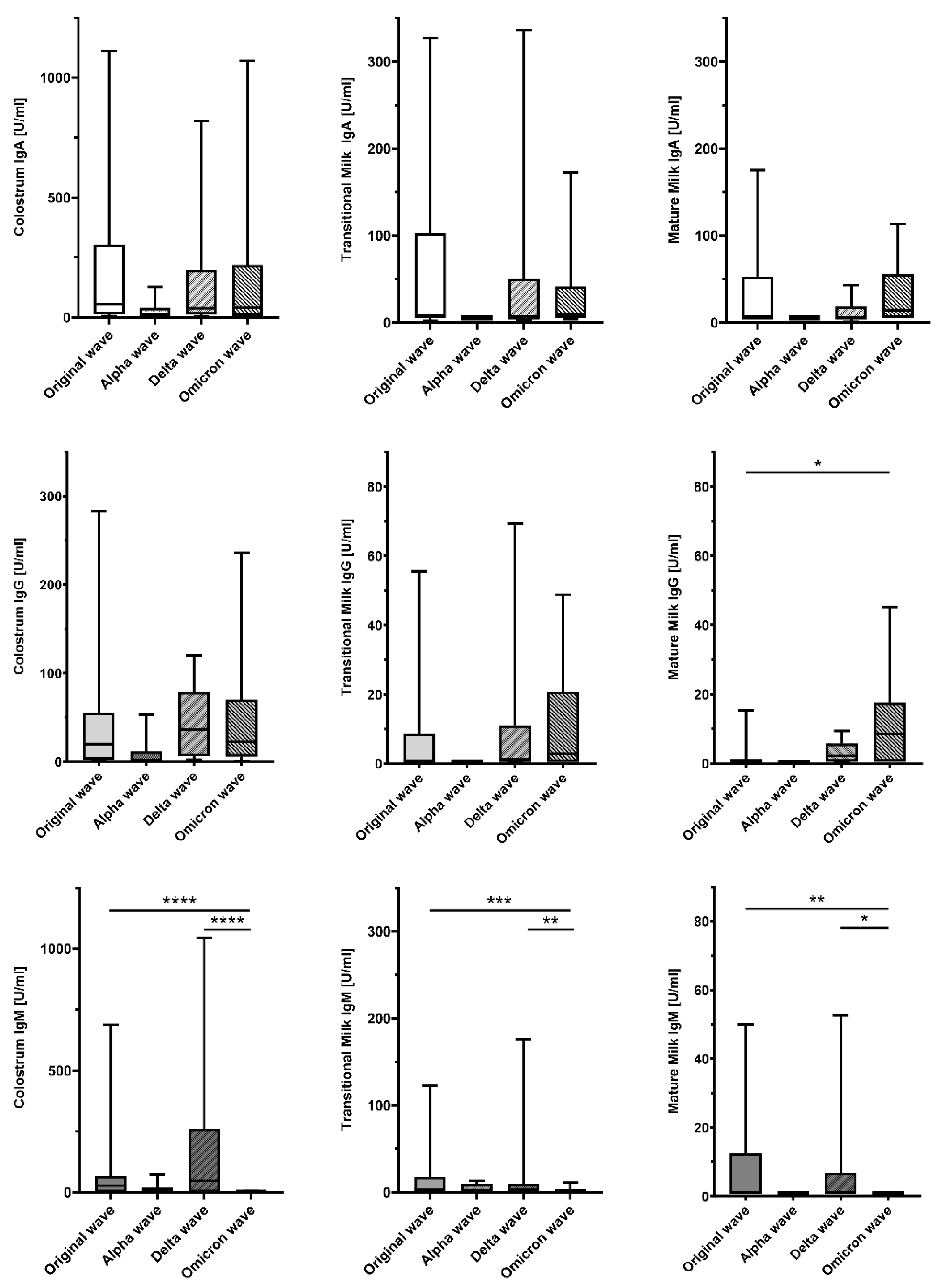
With regard to virus waves, we detected significant overall differences in anti-S1RBD-IgG concentrations in mature milk and in anti-S1RBD-IgM concentrations in colostrum, transitional milk, and mature milk. A post hoc analysis with pairwise comparisons revealed significant Ig- and sample type-specific differences between the original wave and the Omicron wave, as well as the Delta wave and the Omicron wave. Detailed results are presented in Table 3 and Figure 2.

4. Discussion

Breast milk is the physiological form of infant nutrition, widely recognized for its numerous benefits, including its role as a source of immunological factors that protect infants from infectious agents [18,19]. During the COVID-19 pandemic, numerous studies have demonstrated the potential protective effect of passive immunity conveyed through Ig in breast milk [4,9,10]. However, the factors that influence immunoglobulin titers and the role of milk maturation remain incompletely understood [19].
Our study uniquely elucidates the dynamics of anti-SARS-CoV-2 antibodies in breast milk across different lactation stages. By analyzing breast milk Ig in a large cohort of mothers with diverse COVID-19 infection timelines, disease severities, and vaccination statuses throughout the pandemic, we provide comprehensive insights into breast milk immunity.
Specifically, we measured anti-S1RBD-Ig of the classes IgA, IgG, and IgM in colostrum, transitional, and mature milk, elucidating the influence of various factors on their concentrations. The most abundant Ig in the analyzed samples was anti-S1RBD-IgA, which is consistent with previous research [1,6,20]. Notably, anti-S1RBD-IgA levels were higher in colostrum than in mature milk, which also accords with prior findings in the field [19]. A high detection rate of IgG in colostrum (86.0%) corroborates earlier studies on SARS-CoV-2 antibodies in breast milk [4]. In transitional and mature milk, median anti-S1RBD-IgG concentrations remained above the detection threshold, while anti-S1RBD-IgA concentrations fell below the detection threshold. This may be attributed to the proportion of vaccinated mothers in our cohort, as vaccination is known to enhance the IgG response in breast milk [21]. For anti-S1RBD-IgM, a decline in detection rates was observed with milk maturation (41.7% vs. 33.7% vs. 23.0%), corroborating previous findings that IgM diminishes in significance during lactation [22,23]. Unlike IgG, IgM secretion into breast milk is unaffected by vaccination [10,24].
Our finding that the time since the last antigen exposure does not affect breast milk anti-S1RBD-Ig levels aligns with Szczygiol et al., who found no correlation between serum anti-SARS-CoV-2 IgA and IgG levels and the time since a positive antigen test [6]. This may also apply to breast milk, given the established correlation between maternal serum and breast milk antibodies [24,25]. Although maternal serum samples were unavailable, we found significant correlations between colostrum anti-S1RBD-IgA and -IgG and DBS anti-S1RBD-IgG in neonatal blood. The undetectable DBS anti-S1RBD-IgM suggests that these IgG antibodies are maternally derived via transplacental transfer.
In alignment with previous studies, we found higher concentrations of anti-S1RBD-IgA and -IgG in vaccinated mothers’ milk, indicating that vaccination amplifies the immune response initially triggered by natural infection [21,26]. Ramirez et al. observed robust anti-S1RBD-IgG and -IgA levels in breast milk post-mRNA vaccination, highlighting enhanced antibody titers compared to natural infection alone [26]. Our study’s results are further supported by Selma-Royo et al., who showed that breast milk anti-S1RBD-IgA and -IgG induced by mRNA-based vaccines lead to higher levels of anti-SARS-CoV-2-IgA and -IgG in breast milk compared to natural infection [21]. The lower concentrations of anti-S1RBD-IgM following combined vaccination and natural infection, compared to natural infection alone, are in accordance with previous studies showing that IgM levels in human milk do not increase with additional vaccination [10,24].
Our study indicates that the severity of maternal SARS-CoV-2 infection influences the concentration of specific anti-S1RBD-Ig in breast milk. Specifically, we observed that mothers with moderate to severe infections had significantly higher concentrations of anti-S1RBD-IgA and -IgG in transitional milk and significantly higher concentrations of anti-S1RBD-IgA and -IgM in mature milk than mothers with no to mild COVID-19 symptoms. This finding underscores the dynamic nature of the maternal immune response and its capacity to modulate breast milk composition in response to infection. It aligns with the previous literature suggesting that the severity of COVID-19 disease influences antibody titers [5]. However, the literature on this topic is conflicting, and while some studies report higher antibody levels in symptomatic individuals, other studies could not demonstrate an association between disease severity and breast milk antibody titers [6,13,27]. Comprehensive studies are required to draw definitive conclusions.
Interestingly, we detected higher mature milk anti-S1RBD-IgM titers after preterm birth. This finding accords with data indicating that preterm milk is a superior source of anti-inflammatory and anti-infective factors [28,29]. However, most research on the influence of gestational age on breast milk Ig has focused on IgA, and data on the impact of preterm birth on titers of other Ig classes are sparse. Of note, higher concentrations of IgA and IgG have been observed in preterm milk [19,30]. For IgM, earlier studies report similar concentrations in preterm and term breast milk [30,31]. However, these studies examined total Ig concentrations in breast milk in general and were not virus-specific. The impact of gestational age on breast milk responses to specific pathogens, including SARS-CoV-2, necessitates further targeted investigation.
To date, few studies have explored the impact of different virus waves on Ig concentrations in breast milk, mainly focusing on the neutralizing capacity against different virus variants following mRNA vaccination [7,25]. We are the first to report virus wave-specific anti-S1RBD differential responses depending on the type of breast milk sample and Ig class.
While our study provides valuable insights, several limitations should be acknowledged to guide future research.
First, although we did not explore potential confounding factors, such as maternal diet, stress levels, or supplementary infant feedings, our findings lay a strong foundation for subsequent studies to investigate these important aspects of infant nutrition and health.
Second, we inferred virus variants based on the timing of infection, which limits our ability to distinguish between the influences of the virus variants themselves and the public health responses during different phases of the pandemic. Including viral genome sequencing in future studies could provide more precise identification of virus variants.
Additionally, although we did not collect maternal serum samples to correlate breast milk antibody levels with maternal systemic immune activation, our findings underscore the substantial presence of antibodies in breast milk, indicating a robust adaptive maternal immune response.
Furthermore, our study was conducted at a single hospital in Austria, which might limit the generalizability of the findings. However, this controlled setting allows for a detailed and focused analysis, paving the way for multicenter studies in diverse populations. Although we did not collect data on infant outcomes or protection from COVID-19, the high levels of antibodies detected in breast milk are promising indicators of potential infant protection.
Lastly, while our focus was on breast milk antibodies, future research should explore other components of breast milk that contribute to infant immunity and assess the functional capacity of these antibodies, such as their neutralizing ability.
The major strengths of our study include its large sample size, the systematic assessment of breast milk samples at all stages of early lactation, and the comprehensive observation period spanning the entirety of the pandemic.

5. Conclusions

In conclusion, our study offers relevant insights into the dynamics of anti-SARS-CoV-2 Ig in breast milk, emphasizing the impact of maternal vaccination status and infection severity, as well as preterm birth on antibody titers. It underscores the crucial role of breastfeeding in providing tailored immunological support to infants, particularly during a pandemic. However, further research is necessary to understand the extent of protection breast milk offers against COVID-19 and the potential effects of different lactation phases. Future studies should investigate the specific influences of virus variants and maternal factors to further elucidate the complex immunological landscape of breast milk and explore the multifaceted benefits of breastfeeding in the context of maternal and infant health.

Supplementary Materials

The following supporting information can be downloaded at https://www.mdpi.com/article/10.3390/nu16142320/s1: Table S1: Data availability according to breast milk sample type and anti-SARS-CoV-2-S1RBD-immunoglobulin (Ig) class; Supplementary Materials and Methods.

Author Contributions

C.H. takes responsibility for all aspects of the reliability of the presented data and drafted the initial manuscript. He was fundamentally involved in patient recruitment and collection of biological samples. I.W. and M.H. were involved in patient recruitment and the collection of biological samples and contributed significantly to the statistical analysis. A.H. was involved in conceptualization and made technical and scientific contributions to the interpretation of data. E.H. and M.U. were responsible for Ig detection in DBS and breast milk samples at the Neonatal Research Laboratory. U.K.-K. was involved in study design and logistics, as well as funding acquisition. E.G. substantially contributed to the discussion of results and critically reviewed the manuscript for important intellectual content. A.P. is the corresponding author, acquired funding, takes responsibility for all aspects of the reliability of the presented study, and critically reviewed the manuscript for important intellectual content. All authors critically reviewed the manuscript draft for important intellectual content, approved the final manuscript as submitted, and agree to be accountable for all aspects of the work. All authors have read and agreed to the published version of the manuscript.

Funding

This study was supported by “Land Tirol—Amt der Tiroler Landesregierung”. The funder had no role in study design, data collection and analysis, decision to publish, or preparation of the manuscript.

Institutional Review Board Statement

The mother–child dyads presented in this article participated in the study “Evaluation of potential effects of a maternal SARS-CoV-2 infection on the newborn” funded by “Land Tirol—Amt der Tiroler Landesregierung”. The study protocol was reviewed and approved by the local institutional review board (Ethics Committee of the Medical University of Innsbruck; approval number 1084/2020, 11 May 2020).

Informed Consent Statement

All participating mothers gave written informed consent for the collection of perinatal data and biological samples, as well as the analysis of biological specimens. Written informed consent was obtained from all participants for the publication of information about the data collected.

Data Availability Statement

All data generated or analyzed during this study are included in this article. Further inquiries can be directed to the corresponding author.

Acknowledgments

We thank all staff members of the Department of Pediatrics II (Neonatology) and the whole Obstetrics team at Innsbruck University Hospital for their support throughout this study. Furthermore, we thank Daniela Karall, Guido Wollmann, and Alexander Egger for kindly sharing their expertise.

Conflicts of Interest

The authors have no conflicts of interest to declare.

Abbreviations

AGESAustrian Agency for Health and Food Safety GmBH
DBSdried blood spot
DTdetection threshold(s)
GISAIDGlobal Initiative on Sharing all Influenza Data
Igimmunoglobulin(s)
LODlimit(s) of detection
NICUneonatal intensive care unit
S1RBDSpike S1 receptor-binding domain
TMB3,3′,5,5′-tetramethylbenzidine

References

  1. Fernández-Buhigas, I.; Rayo, N.; Silos, J.C.; Serrano, B.; Ocón-Hernández, O.; Leung, B.W.; Delgado, J.L.; Fernández, D.S.; Valle, S.; De Miguel, L.; et al. Anti-SARS-CoV-2-specific antibodies in human breast milk following SARS-CoV-2 infection during pregnancy: A prospective cohort study. Int. Breastfeed. J. 2024, 19, 5. [Google Scholar] [CrossRef] [PubMed]
  2. Kimberlin, D.W.; Puopolo, K.M. Breast Milk and COVID-19: What Do We Know? Clin. Infect Dis. 2021, 72, 131–132. [Google Scholar] [CrossRef] [PubMed]
  3. Fox, A.; Marino, J.; Amanat, F.; Krammer, F.; Hahn-Holbrook, J.; Zolla-Pazner, S.; Powell, R.L. Robust and Specific Secretory IgA Against SARS-CoV-2 Detected in Human Milk. Iscience 2020, 23, 101735. [Google Scholar] [CrossRef] [PubMed]
  4. Pace, R.M.; Williams, J.E.; Järvinen, K.M.; Belfort, M.B.; Pace, C.D.W.; Lackey, K.A.; Gogel, A.C.; Nguyen-Contant, P.; Kanagaiah, P.; Fitzgerald, T.; et al. Characterization of SARS-CoV-2 RNA, Antibodies, and Neutralizing Capacity in Milk Produced by Women with COVID-19. mBio 2021, 12, e03192-20. [Google Scholar] [CrossRef] [PubMed]
  5. Dimitroglou, M.; Sokou, R.; Iacovidou, N.; Pouliakis, A.; Kafalidis, G.; Boutsikou, T.; Iliodromiti, Z. Anti-SARS-CoV-2 Immunoglobulins in Human Milk after Coronavirus Disease or Vaccination-Time Frame and Duration of Detection in Human Milk and Factors That Affect Their Titers: A Systematic Review. Nutrients 2023, 15, 1905. [Google Scholar] [CrossRef]
  6. Szczygioł, P.; Łukianowski, B.; Kościelska-Kasprzak, K.; Jakuszko, K.; Bartoszek, D.; Krajewska, M.; Królak-Olejnik, B. Antibodies in the breastmilk of COVID-19 recovered women. BMC Pregnancy Childbirth 2022, 22, 635. [Google Scholar] [CrossRef] [PubMed]
  7. Olearo, F.; Radmanesh, L.S.; Felber, N.; von Possel, R.; Emmerich, P.; Pekarek, N.; Pfefferle, S.; Nörz, D.; Hansen, G.; Diemert, A.; et al. Anti-SARS-CoV-2 antibodies in breast milk during lactation after infection or vaccination: A cohort study. J. Reprod. Immunol. 2022, 153, 103685. [Google Scholar] [CrossRef] [PubMed]
  8. Temocin, F.; Çaycı, Y.T.; Seren, C.; Kuruoglu, T.; Atilla, A.; Birinci, A.; Tanyel, E. Investigation of Anti-SARS-CoV-2-specific IgG Levels in Breast Milk after Vaccination or COVID-19 Infection. Am. J. Perinatol. 2024, 41, e3085–e3090. [Google Scholar] [CrossRef] [PubMed]
  9. Pace, R.M.; Williams, J.E.; Järvinen, K.M.; Belfort, M.B.; Pace, C.D.W.; Lackey, K.A.; Gogel, A.C.; Nguyen-Contant, P.; Kanagaiah, P.; Fitzgerald, T.; et al. COVID-19 and human milk: SARS-CoV-2, antibodies, and neutralizing capacity. medRxiv 2020. [Google Scholar] [CrossRef]
  10. Nicolaidou, V.; Georgiou, R.; Christofidou, M.; Felekkis, K.; Pieri, M.; Papaneophytou, C. Detection of SARS-CoV-2-Specific Antibodies in Human Breast Milk and Their Neutralizing Capacity after COVID-19 Vaccination: A Systematic Review. Int. J. Mol. Sci. 2023, 24, 2957. [Google Scholar] [CrossRef]
  11. World Health Organization. Coronavirus Disease (COVID-19): Pregnancy, Childbirth and the Postnatal Period [Place Unknown]. 2022. Available online: https://www.who.int/emergencies/diseases/novel-coronavirus-2019/question-and-answers-hub/q-a-detail/coronavirus-disease-covid-19-pregnancy-and-childbirth (accessed on 20 February 2024).
  12. Centers for Disease Control and Prevention. COVID-19 and Breastfeeding USA 2021. Available online: https://www.cdc.gov/breastfeeding-special-circumstances/hcp/illnesses-conditions/covid-19.html?CDC_AAref_Val=https://www.cdc.gov/breastfeeding/breastfeeding-special-circumstances/maternal-or-infant-illnesses/covid-19-and-breastfeeding.html (accessed on 20 February 2024).
  13. Pace, R.M.; Williams, J.E.; Järvinen, K.M.; Meehan, C.L.; Martin, M.A.; Ley, S.H.; Barbosa-Leiker, C.; Andres, A.; Yeruva, L.; Belfort, M.B.; et al. Milk From Women Diagnosed With COVID-19 Does Not Contain SARS-CoV-2 RNA but Has Persistent Levels of SARS-CoV-2-Specific IgA Antibodies. Front. Immunol. 2021, 12, 801797. [Google Scholar] [CrossRef]
  14. Conti, M.G.; Terreri, S.; Terrin, G.; Natale, F.; Pietrasanta, C.; Salvatori, G.; Brunelli, R.; Midulla, F.; Papaevangelou, V.; Carsetti, R.; et al. Severe Acute Respiratory Syndrome Coronavirus 2 Infection Versus Vaccination in Pregnancy: Implications for Maternal and Infant Immunity. Clin. Infect. Dis. 2022, 75 (Suppl. S1), S37–S45. [Google Scholar] [CrossRef]
  15. Society for Maternal-Fetal Medicine. Management Considerations for Pregnant Patients with COVID-19. 2020. Available online: https://s3.amazonaws.com/cdn.smfm.org/media/2415/SMFM_COVID_Management_of_COVID_pos_preg_patients_7-2-20.PDF_.pdf (accessed on 20 February 2024).
  16. e.V. FvG. hCoV-19 Variants Dahboard Munich 2024. Available online: https://gisaid.org/hcov-19-variants-dashboard/ (accessed on 20 February 2024).
  17. GmBh ÖAfGuE. GISIAD Österreich-Report. 2024. Available online: https://www.ages.at/forschung/wissen-aktuell/detail/gisaid-oesterreich-report (accessed on 20 February 2024).
  18. Sriraman, N.K. The Nuts and Bolts of Breastfeeding: Anatomy and Physiology of Lactation. Curr. Probl. Pediatr. Adolesc. Health Care. 2017, 47, 305–310. [Google Scholar] [CrossRef]
  19. Rio-Aige, K.; Azagra-Boronat, I.; Castell, M.; Selma-Royo, M.; Collado, M.C.; Rodríguez-Lagunas, M.J.; Pérez-Cano, F.J. The Breast Milk Immunoglobulinome. Nutrients 2021, 13, 1810. [Google Scholar] [CrossRef] [PubMed]
  20. Fox, A.; Marino, J.; Amanat, F.; Oguntuyo, K.Y.; Hahn-Holbrook, J.; Lee, B.; Zolla-Pazner, S.; Powell, R.L. The IgA in milk induced by SARS-CoV-2 infection is comprised of mainly secretory antibody that is neutralizing and highly durable over time. PLoS ONE 2022, 17, e0249723. [Google Scholar] [CrossRef] [PubMed]
  21. Selma-Royo, M.; Bäuerl, C.; Mena-Tudela, D.; Aguilar-Camprubí, L.; Pérez-Cano, F.J.; Parra-Llorca, A.; Lerin, C.; Martínez-Costa, C.; Collado, M.C. Anti-SARS-CoV-2 IgA and IgG in human milk after vaccination is dependent on vaccine type and previous SARS-CoV-2 exposure: A longitudinal study. Genome Med. 2022, 14, 42. [Google Scholar] [CrossRef]
  22. Goldsmith, S.J.; Dickson, J.S.; Barnhart, H.M.; Toledo, R.T.; Eiten-Miller, R.R. IgA, IgG, IgM and Lactoferrin Contents of Human Milk During Early Lactation and the Effect of Processing and Storage. J. Food Prot. 1983, 46, 4–7. [Google Scholar] [CrossRef]
  23. Abuidhail, J.; Al-Shudiefat, A.A.; Darwish, M. Alterations of immunoglobulin G and immunoglobulin M levels in the breast milk of mothers with exclusive breastfeeding compared to mothers with non-exclusive breastfeeding during 6 months postpartum: The Jordanian cohort study. Am. J. Hum. Biol. 2019, 31, e23197. [Google Scholar] [CrossRef] [PubMed]
  24. Jakuszko, K.; Kościelska-Kasprzak, K.; Żabińska, M.; Bartoszek, D.; Poznański, P.; Rukasz, D.; Kłak, R.; Królak-Olejnik, B.; Krajewska, M. Immune Response to Vaccination against COVID-19 in Breastfeeding Health Workers. Vaccines 2021, 9, 663. [Google Scholar] [CrossRef]
  25. Hsiao, H.M.; DiMaggio, L.S.; Perez, M.A.; Chen, X.; Stephens, K.; Gibson, T.; Anderson, E.J.; Rostad, C.A. SARS-CoV-2 Antibody Profiles in Maternal Serum and Breast Milk Following mRNA COVID-19 Vaccination: A Longitudinal Prospective Observational Cohort Study. Vaccines 2023, 11, 1643. [Google Scholar] [CrossRef]
  26. Romero Ramírez, D.S.; Lara Pérez, M.M.; Carretero Pérez, M.; Suárez Hernández, M.I.; Martín Pulido, S.; Pera Villacampa, L.; Fernández Vilar, A.M.; Rivero Falero, M.; González Carretero, P.; Reyes Millán, B.; et al. SARS-CoV-2 Antibodies in Breast Milk After Vaccination. Pediatrics 2021, 148, e2021052286. [Google Scholar] [CrossRef] [PubMed]
  27. Dutra, L.V.; Silva, M.H.; Lebrão, C.W.; Affonso-Fonseca, F.L.; Suano-Souza, F.I. Impact of the Presence Anti-SARS-CoV-2 IgA in the Colostrum of Women Infected by COVID-19 During the Pregnancy in Neonatal Clinical Outcomes: A Cross-Sectional Study. Matern. Child. Health J. 2023, 27, 737–746. [Google Scholar] [CrossRef] [PubMed]
  28. Mehta, R.; Petrova, A. Biologically active breast milk proteins in association with very preterm delivery and stage of lactation. J. Perinatol. 2011, 31, 58–62. [Google Scholar] [CrossRef] [PubMed]
  29. Trend, S.; Strunk, T.; Lloyd, M.L.; Kok, C.H.; Metcalfe, J.; Geddes, D.T.; Lai, C.T.; Richmond, P.; Doherty, D.A.; Simmer, K.; et al. Levels of innate immune factors in preterm and term mothers’ breast milk during the 1st month postpartum. Br. J. Nutr. 2016, 115, 1178–1193. [Google Scholar] [CrossRef] [PubMed]
  30. Koenig, A.; de Albuquerque Diniz, E.M.; Barbosa, S.F.; Vaz, F.A. Immunologic factors in human milk: The effects of gestational age and pasteurization. J. Hum. Lact. 2005, 21, 439–443. [Google Scholar] [CrossRef]
  31. Gross, S.J.; Buckley, R.H.; Wakil, S.S.; McAllister, D.C.; David, R.J.; Faix, R.G. Elevated IgA concentration in milk produced by mothers delivered of preterm infants. J. Pediatr. 1981, 99, 389–393. [Google Scholar] [CrossRef]
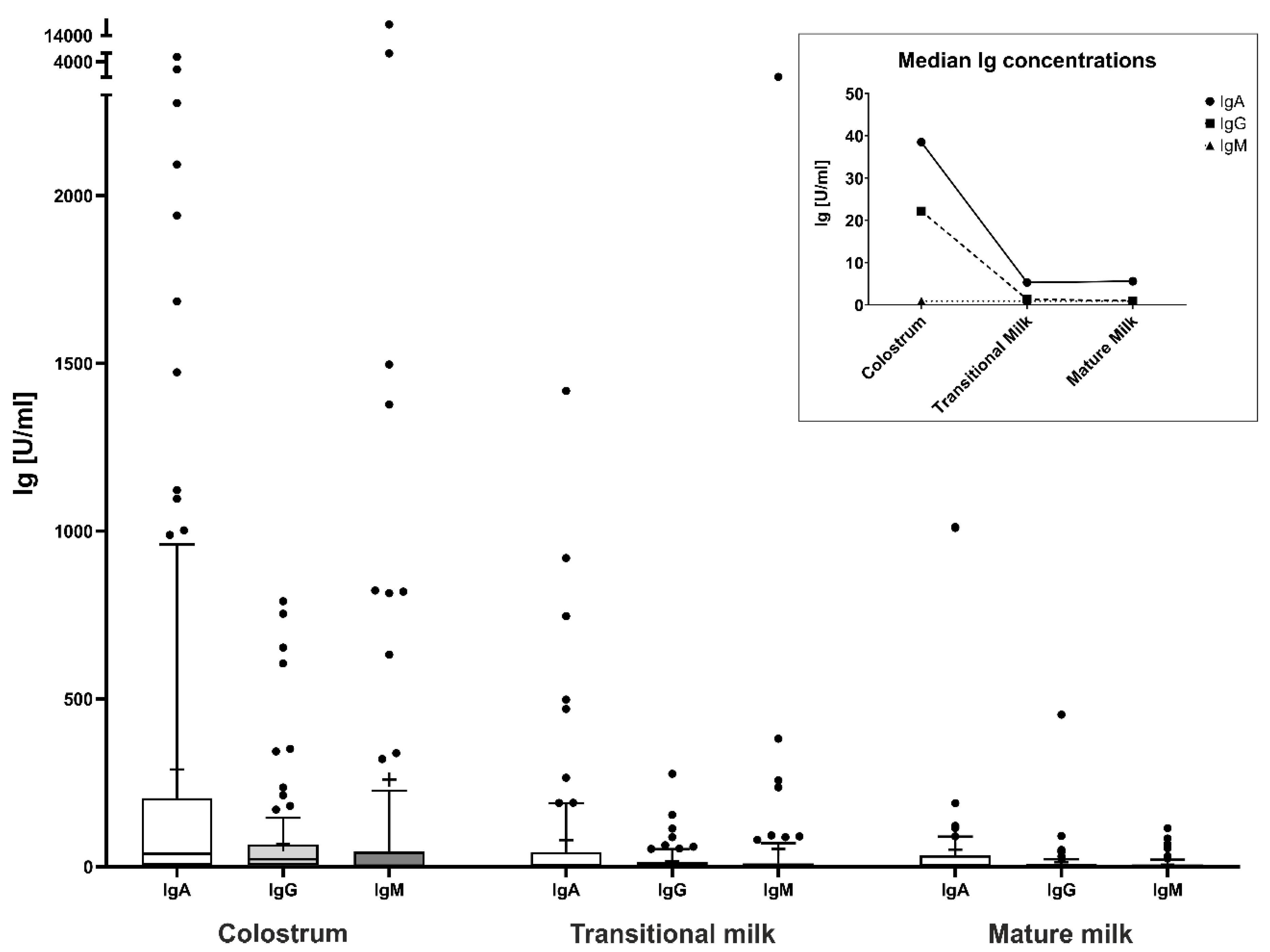
Figure 1. Concentrations of anti-SARS-CoV-2-S1RBD immunoglobulins (Ig) in different types of breast milk. This figure shows boxplots of anti-S1RBD-IgA, -IgG, and -IgM concentrations in various types of breast milk, including colostrum, transitional milk, and mature milk. Ig concentrations are plotted on the y-axis in U/mL. Center lines in boxes represent medians, box edges mark 1st and 3rd quartiles, and whiskers indicate 10th and 90th percentiles. Individual points indicate outliers. Crosses indicate mean Ig concentrations. The data in the box highlight the dynamic changes in breast milk Ig levels over the lactation period, with the highest median concentrations observed in colostrum and gradually decreasing median concentrations in transitional and mature milk. Ig, immunoglobulin; SARS-CoV-2, severe acute respiratory syndrome coronavirus 2; S1RBD, SARS-CoV-2 Spike protein S1 receptor-binding domain.
Nutrients 16 02320 g001
Figure 2. Concentrations of anti-SARS-CoV-2-S1RBD immunoglobulins (Ig) in breast milk across different SARS-CoV-2 waves. This panel displays boxplots of anti-S1RBD-IgA, -IgG, and -IgM concentrations in different types of breast milk (colostrum, transitional milk, and mature milk) across several waves of the SARS-CoV-2 pandemic. Ig concentrations are plotted on the y-axis in U/mL. Center lines in boxes represent medians, box edges mark 1st and 3rd quartiles, and whiskers indicate 10th and 90th percentiles. The influence of different SARS-CoV-2 waves on breast milk Ig concentrations is depicted. Significant differences across different pandemic waves are indicated by asterisks (* p < 0.05, ** p < 0.01, *** p < 0.001, **** p < 0.0001). Ig, immunoglobulin; SARS-CoV-2, severe acute respiratory syndrome coronavirus 2; S1RBD, SARS-CoV-2 Spike protein S1 receptor-binding domain.
Nutrients 16 02320 g002
Table 1. Characteristics of study population.
Total
n = 140 (100)
Term Birth
n = 110 (78.6)
Preterm Birth
n = 30 (21.4)
Maternal characteristics
Maternal age [years]32.0 ± 4.832.0 ± 4.631.8 ± 5.6
Gravidity, median (range)2 (1–3)2 (1–3)2 (1–3)
Parity, median (range)2 (1–2)2 (1–2)2 (1–2)
Time of infection, n (%)
   during pregnancy98 (70.0)82 (74.5)16 (53.3)
   peripartum42 (30.0)28 (25.5)14 (46.7)
Interval infection—delivery [days]44 (7.5; 102.8)50 (11; 112.3)22.5 (1.5; 86.5)
Maternal symptoms, n (%)
   a-/pre-symptomatic22 (15.7)19 (17.3)3 (10.0)
   mild95 (67.9)74 (67.3)21 (70.0)
   moderate7 (5.0)5 (4.5)2 (6.7)
   severe3 (2.1)0 (0.0)3 (10.0)
   missing information13 (9.3)12 (10.9)1 (3.3)
Vaccination, n (%)
   Non-vaccinated94 (67.1)72 (65.5)22 (73.3)
   1× vaccinated5 (3.6)3 (2.7)2 (6.7)
   2× vaccinated12 (8.6)10 (9.1)2 (6.7)
   3× vaccinated29 (20.7)25 (22.7)4 (13.3)
Virus wave, n (%)
   Original wave38 (27.2)27 (24.5)11 (36.7)
   Alpha wave9 (6.4)8 (7.3)1 (3.3)
   Delta wave29 (20.7)19 (17.3)10 (33.3)
   Omicron wave64 (45.7)56 (50.9)9 (26.7)
Neonatal characteristics
Number of participants, n (%)144 (100)111 (77.1)33 (22.9)
Multiple birth (twins), n (%)4 (2.8)1 (0.9)3 (9.1)
Infant sex, n (%)
   male85 (59.0)65 (58.6)20 (60.6)
   female59 (41.0)46 (41.4)13 (39.4)
Delivery mode
   vaginal70 (48.6)62 (55.9)8 (24.2)
   vacuum extraction6 (4.2)6 (5.4)0 (0.0)
   C-section68 (47.2)43 (38.7)25 (75.8)
Gestational age [weeks]38.7 (37.0; 39.8)39.1 (38.1; 40.1)34.4 (34.1; 35.3)
Birth weight [g]3047.3 ± 624.473277.4 ± 455.082273.2 ± 478.61
z-score birth weight−0.32 ± 1.00−0.32 ± 1.00−0.30 ± 0.83
Birth length [cm]48.6 ± 3.2949.7 ± 2.2844.7 ± 3.30
z-score birth length−0.72 ± 1.05−0.70 ± 0.99−0.81 ± 1.26
Birth head circumference [cm]34.1 ± 2.2134.8 ± 1.6331.8 ± 2.27
z-score birth head circumference−0.18 ± 1.18−0.15 ± 1.24−0.28 ± 0.98
Apgar scores
   1 min9 (8; 9)9 (9; 9)8 (6; 9)
   5 min10 (9; 10)10 (9.8; 10)9 (8; 10)
   10 min10 (10; 10)10 (10; 10)9 (9; 10)
Umbilical cord arterial pH7.259 ± 0.0737.251 ± 0.0857.293 ± 0.064
Umbilical cord arterial base excess [mmol/L]−2.7 ± 4.4−3.0 ± 4.3−0.9 ± 4.6
Neonate tested positive for SARS-CoV-2, n (%)3 (2.1)3 (2.7)0 (0.0)
Neonate admitted to NICU, n (%)34 (23.6)12 (10.8)22 (66.7)
DBS umbilical cord blood available, n (%)85 (59.0)67 (60.3)18 (54.5)
DBS venous blood neonate 48 h available, n (%)13 (9.0)10 (9.0)3 (9.1)
DBS anti-S1RBD-IgG concentrations §90.8 (23.4; 314.4)104.3 (27.1; 338.1)54.6 (15.8; 141.4)
DBS anti-S1RBD-IgM concentrations $n.d.n.d.n.d.
Categorical data are presented as counts (n) and percentages (%); continuous data are presented as median (quartile 1; quartile 3) for non-normally distributed variables or mean ± standard deviation (SD) for variables following a normal distribution, unless otherwise specified. § Limit of detection (LOD) for anti-S1RBD-IgG: 30 U/mL. Data below LOD were censored as follows: LOD/√(2). $ Limit of detection (LOD) for anti-S1RBD-IgM: 100 U/mL. NICU, neonatal intensive care unit; DBS, dried blood spot(s); n.d., not detectable.
Table 2. Potential factors influencing breast milk immunoglobulin (Ig) concentrations in various sample types.
Type of Breast MilkMaternal CharacteristicIgA Median (Q1; Q3)p-Value *IgG Median (Q1; Q3)p-Value *IgM Median (Q1; Q3)p-Value *
Colostrum
Infection during pregnancy51.3 (14.0; 221.7) 23.0 (3.7; 60.2) 0.9 (0.9; 51.4)
Peripartum infection23.2 (5.3; 187.9)0.13119.3 (4.8; 71.3)0.8960.9 (0.9; 3.2)0.063
Infection only24.0 (5.3; 128.5) 11.3 (2.2; 47.2) 7.5 (0.9; 78.7)
Infection + vaccination98.0 (12.0; 457.0)0.01342.2 (4.0; 112.5)0.0010.9 (0.9; 0.9)<0.001
Asymptomatic/mild disease48.9 (5.7; 214.2) 23.0 (4.8; 66.2) 0.9 (0.9; 47.5)
Moderate/severe disease29.9 (5.3; 1176.0)0.64236.3 (9.8; 131.4)0.20617.6 (0.9; 475.9)0.196
Term birth38.5 (5.3; 204.8) 20.7 (3.3; 63.9) 0.9 (0.9; 31.2)
Preterm birth36.0 (5.3; 217.7)0.93032.9 (10.0; 87.7)0.23618.3 (0.9; 183.1)0.065
Age < 35 years40.0 (5.3; 190.2) 22.5 (4.8; 67.7) 0.9 (0.9; 51.4)
Age ≥ 35 years26.0 (5.3; 502.1)0.61920.2 (3.1; 59.8)0.9160.9 (0.9; 16.2)0.186
Transitional milk
Infection during pregnancy5.3 (5.3; 42.5) 0.7 (0.5; 12.2) 0.9 (0.9; 6.6)
Peripartum infection12.5 (5.3; 46.7)0.3082.7 (0.5; 21.0)0.4390.9 (0.9; 21.2)0.296
Infection only5.3 (5.3; 36.0) 0.5 (0.5; 7.7) 0.9 (0.9; 12.6)
Infection + vaccination20.5 (0.0; 58.6)0.0867.3 (0.5; 33.3)0.0080.9 (0.9; 0.9)0.013
Asymptomatic/mild disease5.3 (5.3; 39.2) 0.8 (0.5; 11.4) 0.9 (0.9; 10.6)
Moderate/severe disease46.7 (33.7; 468.5)0.01220.7 (3.6; 82.4)0.02612.2 (0.9; 89.5)0.151
Term birth5.3 (5.3; 39.2) 1.4 (0.5; 12.2) 0.9 (0.9; 6.6)
Preterm birth10.6 (5.3; 121.5)0.3251.2 (0.5; 44.0)0.7950.9 (0.9; 11.9)0.671
Age < 35 years5.3 (5.3; 41.0) 1.2 (0.5; 8.9) 0.9 (0.9; 6.6)
Age ≥ 35 years17.5 (5.3; 102.8)0.4403.4 (0.5; 32.3)0.3110.9 (0.9; 55.6)0.333
Mature milk
Infection during pregnancy5.3 (5.3; 18.4) 0.5 (0.5; 4.6) 0.9 (0.9; 0.9)
Peripartum infection22.5 (8.5; 69.9)0.0048.5 (2.1; 15.7)0.0010.9 (0.9; 14.4)0.248
Infection only5.3 (5.3; 18.8) 0.5 (0.5; 2.3) 0.9 (0.9; 5.1)
Infection + vaccination20.4 (4.0; 59.7)0.01510.0 (5.6; 22.8)<0.0010.9 (0.9; 0.9)0.016
Asymptomatic/mild disease5.3 (5.3; 32.9) 0.6 (0.5; 9.0) 0.9 (0.9; 0.9)
Moderate/severe disease62.0 (21.2; 545.7)0.0128.2 (3.3; 51.6)0.13630.8 (0.9; 69.5)0.026
Term birth5.3 (5.3; 32.9) 0.6 (0.5; 9.3) 0.9 (0.9; 0.9)
Preterm birth15.6 (5.3; 59.7)0.6492.3 (0.5; 8.5)0.6000.9 (0.9; 6.9)0.018
Age < 35 years5.3 (5.3; 20.4) 0.5 (0.5; 7.7) 0.9 (0.9; 0.9)
Age ≥ 35 years22.5 (5.3; 69.9)0.1685.6 (0.5; 21.6)0.0500.9 (0.9; 3.2)0.722
Data are presented as median (quartile 1 (Q1); quartile 3 (Q3)). * Mann–Whitney-U Test.
Table 3. Breast milk immunoglobulin (Ig) concentrations in various sample types according to virus waves.
Type of Breast MilkVirus WaveIgA Median (Q1; Q3)p-Value *IgG Median (Q1; Q3)p-Value *IgM Median (Q1; Q3)p-Value *
Colostrum
Original wave54.6 (13.2; 304.4) 21.5 (2.4; 46.2) 27.2 (0.9; 67.4)
Alpha wave10.7 (1.0; 40.0) 0.9 (0.5; 11.7) 0.9 (0.9; 19.2)
Delta wave38.3 (11.7; 198.7) 36.3 (5.9; 78.8) 47.8 (0.9; 260.8)
Omicron wave41.2 (5.3; 218.7)0.27622.2 (5.7; 70.3)0.0750.9 (0.9; 0.9)<0.001
Transitional milk
Original wave8.0 (5.3; 102.8) 0.5 (0.5; 8.8) 3.2 (0.9; 18.1)
Alpha wave5.3 (5.3; 5.3) 0.5 (0.5; 0.5) 0.9 (0.9; 10.0)
Delta wave5.3 (5.3; 50.7) 1.2 (0.5; 11.1) 6.2 (0.9; 45.1)
Omicron wave9.6 (5.3; 41.3)0.6972.9 (0.5; 20.8)0.1410.9 (0.9; 0.9)0.005
Mature milk
Original wave5.3 (5.3; 52.5) 0.5 (0.5; 0.9) 0.9 (0.9; 12.4)
Alpha wave5.3 (5.3; 5.3) 0.5 (0.5; 0.6) 0.9 (0.9; 0.9)
Delta wave5.3 (4.3; 18.8) 2.3 (0.5; 5.8) 0.9 (0.9; 6.8)
Omicron wave14.4 (5.3; 55.6)0.1898.5 (0.5; 17.6)0.0040.9 (0.9; 0.9)0.004
Virus waves were categorized according to the timing of infection. Data on the prevailing SARS-CoV-2 variant at any given time point were obtained from the website of the Global Initiative on Sharing all Influenza Data (GISAID). Data are presented as median (quartile 1 (Q1); quartile 3 (Q3)). * Kruskall–Wallis Test.
Disclaimer/Publisher’s Note: The statements, opinions and data contained in all publications are solely those of the individual author(s) and contributor(s) and not of MDPI and/or the editor(s). MDPI and/or the editor(s) disclaim responsibility for any injury to people or property resulting from any ideas, methods, instructions or products referred to in the content.