Article

Circulating Omega-3 Polyunsaturated Fatty Acids Levels in Coronary Heart Disease: Pooled Analysis of 36 Observational Studies

1
Department of Medicine, Pingxiang People’s Hospital, Gannan Medical University, Pingxiang 337000, China
2
Division of Medicine, Department of Gastroenterology, Metabolism and Oncology, University Hospital Ruppin-Brandenburg, Brandenburg Medical School, 16816 Neuruppin, Germany
3
Faculty of Health Sciences, Joint Faculty of the Brandenburg University of Technology, Brandenburg Medical School and University of Potsdam, 14467 Potsdam, Germany
4
Medical Department, Division of Psychosomatic Medicine, Campus Benjamin Franklin, Charité-Universitätsmedizin Berlin, Corporate Member of Freie Universität Berlin and Humboldt-Universität zu Berlin, 10117 Berlin, Germany
5
Department of Cardiology, Beijing Anzhen Hospital, Beijing Institute of Heart, Lung and Blood Vessel Diseases, Capital Medical University, Beijing 100029, China
*
Author to whom correspondence should be addressed.
Nutrients 2024, 16(11), 1610; https://doi.org/10.3390/nu16111610
Submission received: 15 April 2024 / Revised: 19 May 2024 / Accepted: 21 May 2024 / Published: 24 May 2024
(This article belongs to the Section Lipids)

Abstract

:
Long-chain n-3 polyunsaturated fatty acid (PUFA) supplementation has shown potential benefits in the prevention of coronary heart disease (CHD); however, the impact of omega-3 fatty acid levels on CHD risk remains a subject of debate. Here, we aimed to investigate the association between n-3 PUFA levels and the risk of CHD, with particular reference to the subtypes of n-3 PUFA. Methods: Prospective studies and retrospective case-control studies analyzing n-3 PUFA levels in CHD, published up to 30 July 2022, were selected. A random effects meta-analysis was used for pooled assessment, with relative risks (RRs) expressed as 95% confidence intervals (CIs) and standardized mean differences expressed as weight mean differences (WMDs). Subgroup and meta-regression analyses were conducted to assess the impact of n-3 PUFA exposure interval on the CHD subtype variables of the study. Results: We included 20 prospective studies (cohort and nested case-control) and 16 retrospective case-control studies, in which n-3 PUFAs were measured. Higher levels of n-3 PUFAs (ALA, EPA, DPA, DHA, EPA + DHA, total n-3 PUFAs) were associated with a reduced risk of CHD, with RRs (95% CI) of 0.89 (0.81, 0.98), 0.83 (0.72, 0.96); 0.80 (0.67,0.95), 0.75 (0.64, 0.87), 0.83 (0.73, 0.95), and 0.80 (0.70, 0.93), respectively, p < 0.05. CHD patients had significantly lower n-3 PUFA levels compared to healthy controls (p < 0.05). In the subgroup analysis, a significant inverse trend was found for both fatal CHD and non-fatal CHD with n-3 PUFA (EPA + DHA) levels. Also, the link between n-3 PUFA levels in erythrocytes with total CHD was generally stronger than other lipid pools. Conclusions: n-3 PUFAs are significantly related to CHD risk, and these findings support the beneficial effects of n-3 PUFAs on CHD.

1. Introduction

The role of n-3 polyunsaturated fatty acids (PUFAs) in cardiovascular disease remains highly controversial. n-3 PUFAs, mainly α-linolenic acid (ALA), eicosapentaenoic acid (EPA), docosapentaenoic acid (DHA), and docosapentaenoic acid (DPA), were reported in the 1970s on a small group of Eskimos showing cardioprotective effects, which has ignited extensive interest and research into n-3 PUFAs within the scientific community [1]. In the following decades, scientists have provided a comprehensive explanation of the cardiovascular protective mechanisms of n-3 PUFA, and it is generally accepted to be effective in improving blood lipids and combating hypertension [2,3]. In addition, the specialized pro-resolving lipid mediators (SPMs) of EPA and DHA are also thought to mediate vascular anti-inflammation and oxidation [4,5]. As recommended by the AHA Dietary Guidelines since 2003, the health benefits of n-3 PUFAs for both primary and secondary prevention of cardiovascular disease can be achieved by eating at least two servings of fish per week [6].
Several large prospective cohort studies and randomized controlled trials have supported an inverse association between n-3 PUFA and the risk of cardiovascular disease [7,8,9,10]. However, many subsequent studies have not been entirely consistent [11,12,13,14]. A meta-analysis by Aung et al. of 10 trials with 77,917 participants revealed that supplementation with marine-derived n-3 PUFAs for a mean period of 4.4 years did not significantly lower major vascular events [15]. n-3 PUFA could decrease the risk of myocardial infarction (MI), total cardiovascular disease (CVD), and CVD death, according to Hu’s research utilizing a fixed-effects model [16]. A more recent and comprehensive meta-analysis conducted by Shen and colleagues revealed that supplementing with n-3 PUFA, regardless of dose, effectively prevented major adverse cardiovascular events (MACE), cardiovascular death, and MI, but not with all-cause mortality, stroke, or revascularization [17]. In contrast, in a pooled analysis of 79 randomized controlled trials (RCTs), Abdelhamid et al. found that n-3 PUFAs had only a minimal impact on MACE, whereas ALA could reduce coronary heart disease (CHD) mortality [18].
These conflicting results could be caused by several factors. Levels of n-3 PUFA can be influenced by genetics and dietary habits [19]. Additionally, certain medications can also impact the metabolism and overall concentration of these fatty acids (FAs) [20]. Another aspect to consider is the reliance of most studies on self-reported dietary intake of n-3 PUFA or fish oil. These types of reports are prone to inaccuracies in reflecting an individual’s n-3 PUFA levels and intake, which may weaken the reliability of findings [21]. In light of these challenges, utilizing objective measurements, such as biomarkers, can help to overcome the limitations of self-reported data.
To our knowledge, only a few pooled analyses have been conducted to report the association of individual levels of n-3 PUFA with cardiovascular risk [22,23,24]. Of these, research concerning CHD primarily dated back to before 2016 and has not been updated in several years. Meanwhile, no pooled studies have yet specifically focused on comparing blood n-3 PUFA levels between patients with CHD and healthy controls. Given this, we have updated the latest study and strictly distinguished retrospective and prospective studies assessing different types of n-3 PUFAs (ALA, EPA, DPA, DHA) on CHD and CHD subgroup (total CHD, fatal and non-fatal CHD) risk factors. Finally, we assessed the differences in n-3 PUFA levels between patients with and without CHD.

2. Materials and Methods

The study adheres to PRISMA guidelines [25] and was registered at PROSPERO (CRD42022340248).

2.1. Search Strategy

PubMed, Embase, Web of Science, and Cochrane databases were comprehensively reviewed for English-language literature from their inception to July 2022 in humans of the association between n-3 PUFAs and CHD. The primary terms used were “n-3 PUFAs” and “CHD”. To enhance the breadth of the search, we included synonyms and related terms for omega-3 fatty acids, such as “n-3 fatty acids”, “marine oils”, “EPA”, “DHA”, and “ALA”. Similarly, for CHD, we incorporated related terms such as “CHD”, “ischemic heart disease”, “cardiovascular disease”, and specific conditions under the CHD umbrella, like “myocardial infarction” and “angina pectoris”. The detailed strategy is shown in Supplementary Table S1.

2.2. Quality Assessment

We assessed the quality of cohort studies using the Newcastle–Ottawa Scale (NOS). The NOS evaluates three aspects of non-randomized studies: (1) selection, (2) comparability, and (3) assessment of either exposure or outcomes of interest. Studies can receive 2 to 4 stars within each NOS domain, respectively (Supplementary Table S2).

2.3. Exposures and Outcomes

Assessment of FA levels: We extracted different types of n-3 PUFAs (ALA, EPA, DPA, DHA) separately and the studies included in our analysis measured FAs in at least one compartment, including whole blood, red blood cells, serum, plasma, or adipose tissue. Assessment of CHD: Total CHD events were defined as non-fatal CHD (angina, non-fatal myocardial infarction (MI)), and fatal CHD (fatal MI and sudden cardiac death). For the studies reporting biomarkers of n-3 PUFA and CHD risk, inclusion criteria were: (1) prospective cohort research (nested case-control); (2) at least one type of n-3 PUFA concentration was measured; (3) the primary outcomes were risk estimates (relative risks (RRs), odds ratios (ORs), and hazard ratios (HRs)) with 95% CI for CHD events. For studies comparing n-3 PUFA levels in patients with and without CHD, the inclusion criteria were: (1) adult CHD patients and matched controls; (2) retrospective case-control study design; (3) at least one type of n-3 PUFA levels measured between two groups. The included trials were approved by their respective institutional review boards, and we assessed the risk of bias in these trials.

2.4. Data Extraction

Two authors (Yanan Xiao and Yifang Chen) independently conducted study selection based on the criteria and extracted data as follows: (1) basic information (author, year, geographical area, study type/design, sample size, etc.); (2) participant characteristics; (3) different types of n-3 PUFAs (ALA, EPA, DPA, DHA, EPA + DHA); (4) lipid pools (whole blood, erythrocytes, serum, plasma); (5) mean of n-3 PUFA levels; (6) total CHD or CHD subgroup endpoint events; (7) multivariate-adjusted risk estimates with 95% confidence intervals (CI).

2.5. Data Processing and Statistical Analysis

We recorded RRs or HR estimates and their 95% CIs for prospective studies (cohort and nested case control). Adjusted HRs were utilized and treated as equivalent risk measures to RRs. When the odds ratio (OR) varies between 0.5 and 2.5, we consider the OR and RR to be approximately equal [26]. The pooled analyses were conducted on the log scale of the RRs. For data with different categories of tertile, quartile, quintile, or quintile, we recorded the relative highest and lowest category valuations. For the retrospective case-control studies, we calculated the weight mean difference (WMD) in tissue and circulation by combining the mean and standard deviation (SD) of data from different types of n-3 PUFAs (ALA, EPA, DPA, DHA) in the CHD patients and controls. In cases where studies reported EPA and DHA levels separately, we calculated the weighted mean difference (WMD) to combine the EPA and DHA levels to illustrate the difference in blood levels of n-3 PUFAs in those patients with and without CHD. Finally, the degree of heterogeneity between these trials was calculated using the Q test and the I2 statistic. A random-effects model was applied [27,28].

2.6. Subgroup Analysis

We conducted subgroup analyses to assess the impact of different subtypes on the outcomes, focusing on (1) the lipid pool of n-3 PUFA exposure (e.g., erythrocytes, serum, plasma, whole blood, adipose tissue); (2) specific CHD outcomes (e.g., total CHD, fatal and non-fatal CHD). If studies reported data for EPA and DHA separately, we pooled the RR values for EPA + DHA using a random effects model.
Meta-regression and subgroup stratification were employed to evaluate potential sources of heterogeneity between studies. Funnel plots and Egger’s test were used to explore the potential publication bias [29]. Data analyses were carried out with Stata (version 16.0), and p < 0.05 was deemed statistically significant.

3. Results

We reviewed 4761 papers identified through database searches and other sources. Thirty-seven studies ultimately met the inclusion criteria and were included in the review after screening titles and full-text contents. The detailed flow chart is shown in Supplementary Figure S1.
The characteristics of the 20 prospective cohorts and 16 retrospective case-control studies are presented (Table 1 and Table 2). The quality of the included observational studies was assessed using the NOS. The NOS evaluation criteria and results are given in Supplementary Table S2. In Table 1, a total of 28,952 participants representing 15 national areas, with a mean age ranging from 46.4 to 79.1 years at baseline, and the proportion of enrolled women ranged from 0% to 100%. Each study quantified the levels of n-3 PUFAs in the circulation system or tissues, with 2 studies focusing on erythrocytes [10,30], 10 studies on plasma [31,32,33,34,35,36,37,38,39,40], 4 studies on serum [41,42,43,44], and 4 studies on whole blood [45,46,47,48]. In Table 2, 1148 CHD patients and 1156 healthy controls were enrolled in the study of compared n-3 PUFA levels. These participants were from 11 countries and ranged in age from 18 to 62.7 years, with five studies analyzing PUFA in erythrocytes [49,50,51,52,53], two studies in plasma [54,55], four studies in serum [56,57,58,59], one study in whole blood [60], and four studies for tissue or adipose tissue, respectively [61,62,63,64].
We examined the relationships between various types of n-3 PUFAs and the risk of CHD. Elevated levels of n-3 PUFAs (ALA, EPA, DPA, DHA, EPA + DHA) were linked with reduced CHD risk (Figure 1). Specifically, 13 studies reported the association between ALA and CHD events, with a pooled RR of 0.89 (0.81–0.98), p = 0.02, and heterogeneity: p = 0.12, I2 = 32.6%; 14 trials for EPA, RR: 0.83 (0.72–0.96), p < 0.01, heterogeneity: p < 0.01, I2 = 65.6%; 9 trials for DPA, RR: 0.80 (0.67–0.95), p < 0.01, heterogeneity: p < 0.01, I2= 62.0%; 14 trials reported DHA, RR: 0.75 (0.64–0.87), p < 0.01, heterogeneity: p < 0.01, I2 = 66.6%. In addition, 8 and 11 reported CHD events for EPA + DHA and the total n-3 PUFAs, resulting in a summary RR of 0.83 (0.73–0.95) and RR: 0.80 (0.70–0.93), p < 0.05, with heterogeneity: I2 = 67.4% and I2 = 66.6%, p < 0.01, respectively. The overall result between n-3 PUFA and CHD was 0.83 (95% CI: 0.79–0.87). In the CHD model of ALA, the heterogeneity between studies was low, and the other models were highly heterogeneous.
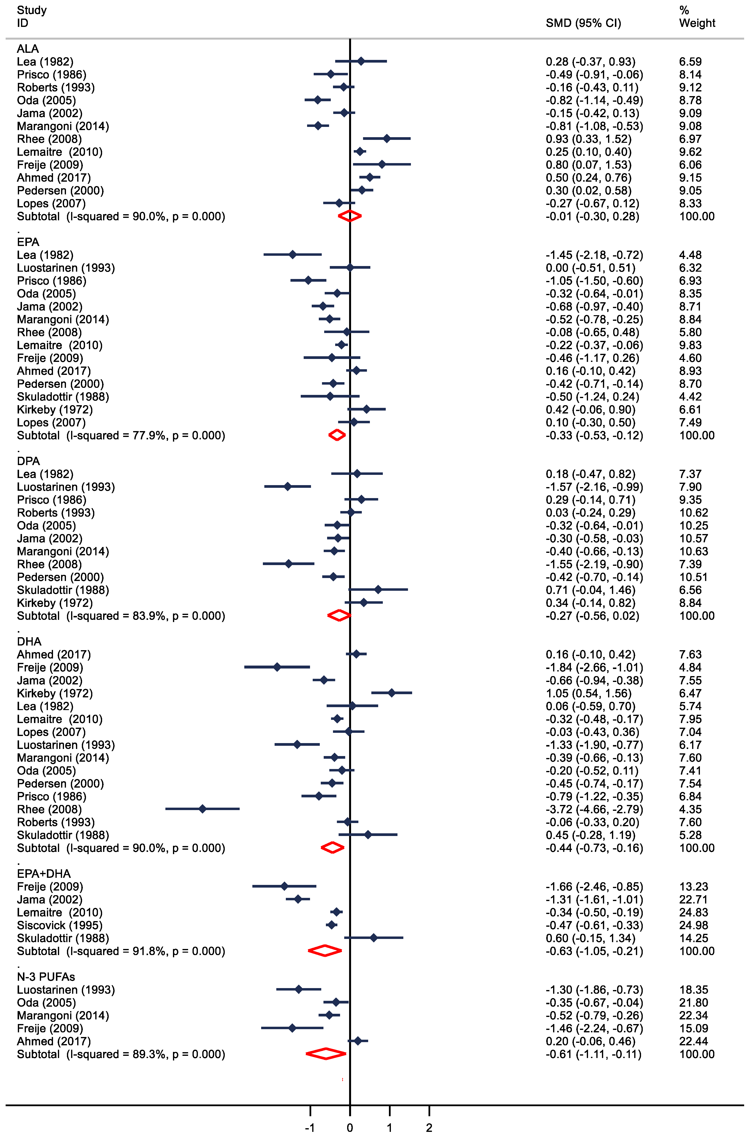
A total of 16 studies compared blood levels of n-3 PUFAs in patients with and without CHD (Figure 2), and overall, the levels of different types of n-3 PUFA (EPA, DHA, DPA, EPA + DHA, and total n-3 PUFAs) were inversely related to CHD, but not with ALA. Also, 13, 10, 14, and 5 studies provided complete data for EPA, DHA, DPA, EPA + DHA, and total n-3 PUFAs, the levels of each of these FAs were significantly lower in the CHD group (WMD = −0.33; 95% CI: −0.53 to −0.12; WMD = −0.27; 95% CI: −0.56 to 0.02; WMD = −0.44; 95% CI: −0.73 to −0.16, WMD = −0.63; 95% CI: −1.05 to −0.21; WMD = −0.61; 95% CI: −1.11 to −0.11; p < 0.01, respectively). In the 12 case-control studies, ALA levels did not change significantly in patients with and without CHD (WMD = −0.01, 95% CI: −0.30 to 0.28). Heterogeneity among studies comparing n-3 PUFA levels in patients with CHD versus those without CHD was substantial (I2: 77.9%–91.8%, p < 0.01).
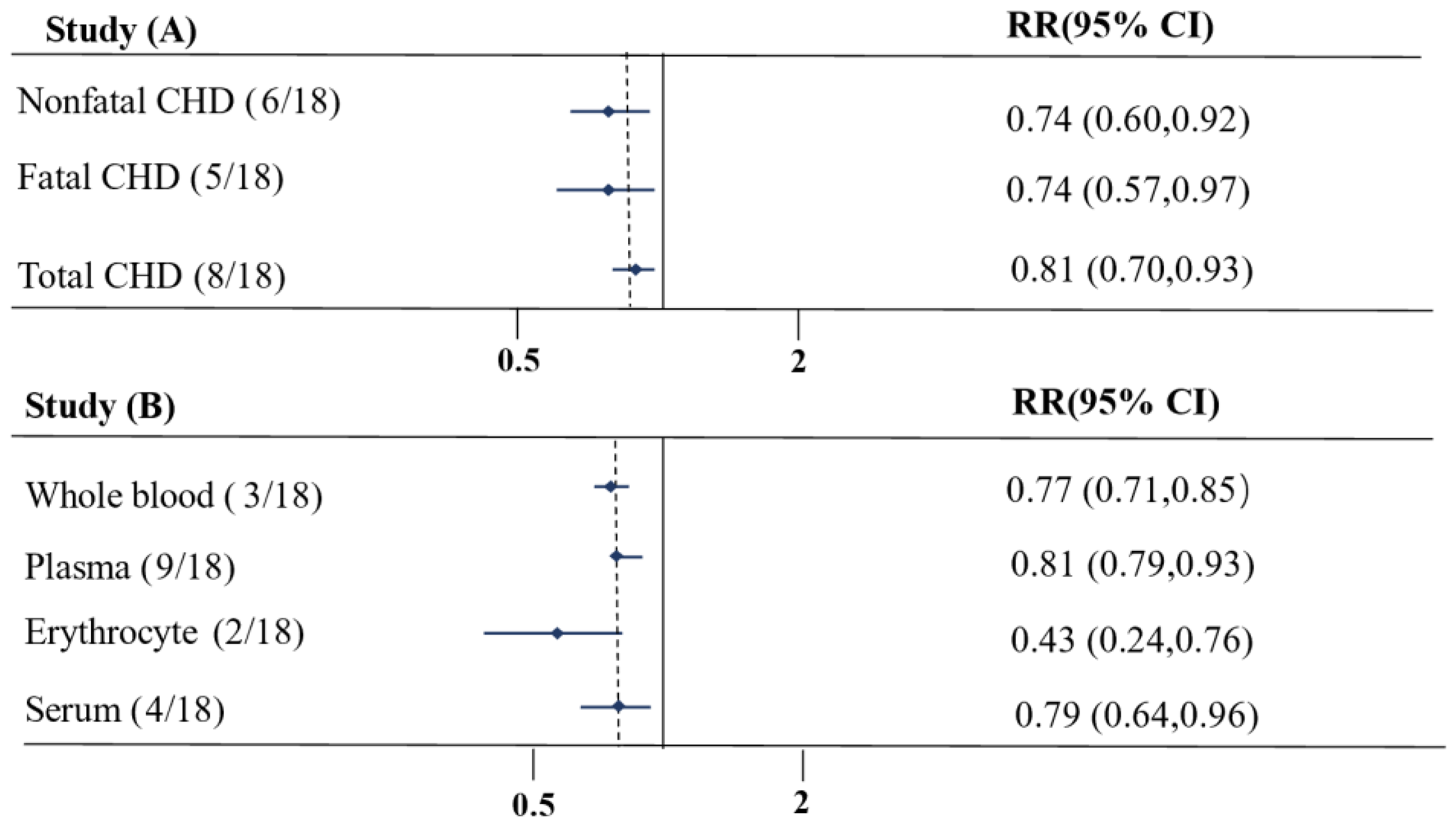
We conducted a subgroup analysis by combining the RR estimates of EPA and DHA, which included a total of 20 studies (Figure 3, Supplementary Figures S4 and S5). For the different subgroups of clinical endpoint, the observations were consistent for total CHD, fatal and non-fatal CHD (RR 0.81 (0.70–0.93 for total CHD; RR 0.74 (0.57–0.97) for fatal CHD; RR 0.74 (0.60–0.92) for non-fatal CHD, p < 0.05 for both) (Figure 3A). Regarding the different subgroups of EPA + DHA exposure compartment, whole blood, plasma, and serum had stronger correlations with CHD (RR 0.77 (0.71–0.85) for whole blood; RR 0.81 (0.70–0.93) for plasma; RR 0.79 (0.64–0.76) for serum, RR 0.43 (0.24–0.76) for erythrocytes, p <0.05 for both) (Figure 3B).
Funnel plot visualization and assessment via Egger’s test found no identified evidence of significant publication bias. Sensitivity analyses also confirmed the stability and reliability of the results produced by our statistical models. Additionally, meta-regression analysis indicated that the heterogeneity among the subgroups was not significant (Supplementary Figures S2, S3 and S6; Supplementary Table S3).

4. Discussion

The present study was a comprehensive analysis of individual-level data from 20 prospective cohort studies (cohort and nested case-control) and 16 independent case-control studies. Overall, our findings indicated that higher circulating levels of n-3 PUFAs (ALA, EPA, DPA, DHA, EPA + DHA) were related to a lower CHD risk. In subgroup analyses, EPA + DHA values exhibited a strong correlation with both fatal and non-fatal CHD, as well as total CHD. The association between n-3 PUFAs and CHD risk was generally more robust when assessed in erythrocyte lipid pools. In addition, the level of n-3 PUFAs (EPA, DPA, DHA) was significantly lower in patients with CHD compared to control subjects without CHD but with substantial heterogeneity. The ALA concentrations were similar in the two groups.
Our findings are generally in line with earlier evidence [22,23,24], but there were exceptions [15]. Gobbo et al.’s [23] pooled analysis of 19 prospective cohort studies reported that higher circulating levels of the omega-3 fatty acids ALA, DPA, and DHA were correlated with lower RRs of fatal CHD events, with RRs ranging from 0.90 to 0.91. Particularly, DPA was also associated with a reduced risk of total CHD; however, the results were not significant for EPA and CHD. Additionally, this study found generally stronger correlations for n-3 PUFA levels measured in phospholipids and total plasma versus other lipid fractions. A more recent meta-analysis by Harris et al. [22] has provided a broader perspective by evaluating the collective evidence, and their research indicated that DHA and EPA + DHA were associated with a 15% to 18% reduction in total mortality, especially CVD, whereas ALA was not.
In recent meta-analyses of RCTs, Bernasconi et al. [65] updated their dataset to include the two latest studies, further confirming that n-3 PUFAs still have favorable effects on CVD events. However, Rizos’s research [66] emphasized that the benefits were apparent only with high-dose administration. Additionally, Chao et al.’s [67] analysis revealed that protective effects were only significant in subgroups of intervention with EPA and baseline triglyceride (TG) ≥ 1.7 mmol/L, but they did not compare the blood n-3 PUFA levels between patients with CHD and controls.
The initial two large-scale randomized trials, GISSI-Prevenzione and JELIS [7,8], showed a 14–20% reduction in major coronary events in individuals with CHD using n-3 PUFA supplements of 1–1.8 g. Similar results were later observed in the REDUCE-IT trial [9], which administered a higher dose of 4 g of EPA to statin-treated patients with elevated TGs. In this study, the risk of total cardiovascular death, non-fatal myocardial infarction, and non-fatal stroke was reduced by over 20% compared to the control group. However, the outcomes of other RCTs using only 840 mg of EPA + DHA were less favorable. ORIGIN and ASCEND found that n-3 PUFAs decreased TGs while having no impact on cardiovascular events in patients with type 2 diabetes [12,68]. The VITAL trial provides further evidence for assessing the effects of vitamin D and n-3 PUFAs on the primary prevention of CVD and cancer. Although there was no direct link between n-3 PUFAs and major composite cardiovascular events, significant findings were observed across subgroups for both total and fatal MI [69]. In the STRENGTH trial, there was no cardiovascular benefit in patients treated with n-3 PUFAs in the free acid form. However, this trial only evaluated patients with a high risk of developing cardiovascular disease, and the effects of n-3 PUFAs may differ in lower-risk primary prevention populations who have yet to develop significant atherosclerosis or clinical conditions [14].
The effectiveness of n-3 PUFAs in treating dyslipidemia, particularly hypertriglyceridemia, is widely accepted [12,70,71,72], and this effect may be attributed to n-3 PUFA’s ability to accelerate the breakdown of chylomicron particles and reduce the production of very low-density lipoprotein (VLDL) cholesterol [73]. In addition to their effects on lipids, animal experiments and clinical intervention studies have also shown that n-3 PUFA possesses anti-inflammatory and antithrombotic effects [74,75,76]. Hence, the multiple physiologic advantages of n-3 PUFA may assist patients with atherosclerotic arteries [77].
ALA and DPA, unlike EPA and DHA, have received less attention. Our results are similar to those reported in previously published meta-analyses, indicating that higher ALA exposure is associated with a lower CHD risk [24]. The Singapore Chinese Health Study enrolled 63,257 adults and reported an HR of 0.81 (0.72–0.90) for the association between higher ALA intake and the risk of cardiovascular mortality [78]. However, in another large prospective cohort study from the Netherlands, ALA was only negatively linked with stroke, not the clinical endpoint of CHD [79]. However, ALA is susceptible to beta-oxidation, and a small portion is converted to EPA. Thus, ALA concentrations might not accurately reflect the ALA consumption [80].
In earlier investigations, the outcomes related to n-3 DPA have been inconsistent [30,38,81]. Mozaffarian and colleagues [38] conducted a sizeable cohort and found that higher plasma DPA levels were associated with lower all-cause mortality. However, Sun et al.’s prospective analysis demonstrated that lower levels of DPA were connected with a higher risk of non-fatal MI [30].
The concentration of n-3 PUFAs in blood and tissues seems to be essential for the pathophysiological effects in humans, yet few meta-analyses have compared the levels of n-3 PUFAs in patients with and without CHD. One concern about retrospective studies is that individuals with the disease of interest may have altered their diets in response to the diagnosis, or the disease process itself may have altered biomarker levels. This appears not to have been a concern in the present study since levels of n-3 PUFAs (EPA, DPA, DHA) were lower, not higher, in the CHD patients than in the controls. A case-control study by Block et al. suggested that there may be an inverse relationship between the levels of n-3 PUFAs and the risk of acute coronary syndrome (ACS), indicating that blood EPA + DHA levels may contribute to risk stratification [46]. Since the cardioprotective effects of omega-3 have been touted for well over 30 years now, patients with CHD might have been expected to have increased their intake of EPA and DHA, in which case the CHD patients might have had higher levels than controls. The finding that levels are lower in the cases argues against this concern and suggests that reverse causation is likely not playing a role in these studies.
In our subgroup analysis, EPA + DHA concentrations were negatively associated with total CHD, fatal and non-fatal CHD. For the different n-3 PUFA exposure compartments, the n-3 PUFA levels in erythrocytes were more closely correlated with CHD than for whole blood, plasma, and serum. n-3 PUFAs are typically found in cell membranes (to a small extent in adipose tissue), and RBCs have many qualities that make them a good sample type for monitoring n-3 PUFA status [82]. Compared to erythrocytes, serum, plasma, and whole blood may be affected by more factors. When comparing the utility of different biological matrices as biomarkers for CHD risk, erythrocytes may offer certain advantages over serum, plasma, and whole blood. Erythrocytes, by contrast, may provide a more stable environment for the measurement of certain biomarkers due to their relatively consistent lifecycle and composition [83].
The current study has several advantages: (1) We assessed different types of n-3 PUFA biomarkers separately. Furthermore, we performed subgroup analyses of different clinical endpoints (total CHD, fatal and non-fatal CHD) as well as different exposure compartments. (2) In contrast to the self-report questionnaires used in many studies, we limited our study selection to those that show measurements of n-3 PUFA in blood or tissue, which reduced the recall bias and improved the accuracy of the results. (3) By including studies from 13 countries, we minimized publication bias and increased generalizability. Sensitivity analyses showed our results were robust. This study also has some important limitations. More trials and detailed subgroup analysis information are needed to fully evaluate true subgroup effects. Furthermore, considerable heterogeneity exists between studies, which is hard to avoid due to differing methods for measuring PUFAs and pooling data from multiple sample types that can influence results. Dietary conditions in various geographic regions may also contribute to this study’s heterogeneity. Lastly, for the prospective studies, blood samples were analyzed only once at baseline. Theoretically, levels could have changed during the follow-up period, particularly if participants who were not taking supplements at baseline began taking them later.

5. Conclusions

Higher levels of circulating n-3 PUFAs (ALA, EPA, DPA, DHA, EPA + DHA) are associated with a reduced risk of CHD. Furthermore, CHD patients had significantly lower levels of n-3 PUFAs (except ALA). Our findings support a protective role of n-3 PUFAs on CHD events.

Supplementary Materials

The following supporting information can be downloaded at: https://www.mdpi.com/article/10.3390/nu16111610/s1, Figure S1: Flow chart of included studies; Figure S2: Funnel plot and Begger’s test for publication bias of n-3 PUFAs and the risk of coronary heart disease; Figure S3: Funnel plot and Begger’s test for publication bias of n-3 PUFAs in patients with and without coronary heart disease; Figure S4: Subgroup for the association between n-3 PUFA with fatal and non-fatal coronary heart disease; Figure S5: Subgroup for the association between n-3 PUFA compartments and coronary heart disease; Figure S6: Forest plot (A), begger’s test (B), and (C) sensitivity analysis of EPA + DHA and coronary heart disease; Table S1. Search strategy; Table S2. Newcastle-Ottawa Scale (NOS) for quality assessment of the studies; Table T3: Meta-regression analysis on CHD and EPA + DHA levels.

Author Contributions

Y.X.: designed the study, independently reviewed the literature and selected the studies to be included in the meta-analysis, performed the statistical analyses, and drafted the manuscript. Y.C.: independently reviewed the literature and selected the studies to be included in the meta-analysis and extracted available data. A.P.: interpreted the data and critically revised the paper. U.E.: interpreted the data and critically revised the paper. Z.F.: critically revised the paper. K.H.W.: conceived and designed the study and critically revised the paper. All authors have read and agreed to the published version of the manuscript.

Funding

This research received no external funding.

Institutional Review Board Statement

Not applicable.

Informed Consent Statement

Not applicable.

Data Availability Statement

The raw data supporting the conclusions of this article will be made available by the authors on request.

Acknowledgments

Special thanks to Zuobing Liang, for his assistance with manuscript preparation.

Conflicts of Interest

The authors declare no conflicts of interest.

References

  1. Dyerberg, J.; Bang, H.O. Hæmostatic Function and Platelet Polyunsaturated Fatty Acids in Eskimos. Lancet 1979, 314, 433–435. [Google Scholar] [CrossRef] [PubMed]
  2. Mensink, R.P.; Zock, P.L.; Kester, A.D. and Katan, M.B. Effects of dietary fatty acids and carbohydrates on the ratio of serum total to HDL cholesterol and on serum lipids and apolipoproteins: A meta-analysis of 60 controlled trials. Am. J. Clin. Nutr. 2003, 77, 1146–1155. [Google Scholar] [CrossRef] [PubMed]
  3. Russo, G.L. Dietary n−6 and n−3 polyunsaturated fatty acids: From biochemistry to clinical implications in cardiovascular prevention. Biochem. Pharmacol. 2009, 77, 937–946. [Google Scholar] [CrossRef] [PubMed]
  4. Serhan, C.N. Pro-resolving lipid mediators are leads for resolution physiology. Nature 2014, 510, 92–101. [Google Scholar] [CrossRef] [PubMed]
  5. Weylandt, K.H.; Chiu, C.Y.; Gomolka, B.; Waechter, S.F. and Wiedenmann, B. l. Omega-3 fatty acids and their lipid mediators: Towards an understanding of resolvin and protectin formation. Prostaglandins Other Lipid Mediat. 2012, 97, 73–82. [Google Scholar] [CrossRef] [PubMed]
  6. Kris-Etherton, P.M.; Harris, W.S.; Appel, L.J. Fish Consumption, Fish Oil, Omega-3 Fatty Acids, and Cardiovascular Disease. Circulation 2002, 106, 2747–2757. [Google Scholar] [CrossRef] [PubMed]
  7. Investigators, G.P. Dietary supplementation with n-3 polyunsaturated fatty acids and vitamin E after myocardial infarction: Results of the GISSI-Prevenzione trial. Lancet 1999, 354, 447–455. [Google Scholar] [CrossRef]
  8. Yokoyama, M.; Origasa, H.; Matsuzaki, M.; Matsuzawa, Y.; Saito, Y.; Ishikawa, Y.; Oikawa, S.; Sasaki, J.; Hishida, H.; Itakura, H. and Kita, T. Effects of eicosapentaenoic acid on major coronary events in hypercholesterolaemic patients (JELIS): A randomised open-label, blinded endpoint analysis. Lancet 2007, 369, 1090–1098. [Google Scholar] [CrossRef]
  9. Bhatt, D.L.; Steg, P.G.; Miller, M.; Brinton, E.A.; Jacobson, T.A.; Ketchum, S.B.; Doyle Jr, R.T.; Juliano, R.A.; Jiao, L.; Granowitz, C.; et al. Cardiovascular Risk Reduction with Icosapent Ethyl for Hypertriglyceridemia. N. Engl. J. Med. 2019, 380, 11–22. [Google Scholar] [CrossRef]
  10. Harris, W.S.; Tintle, N.L.; Etherton, M.R.; Vasan, R.S. Erythrocyte long-chain omega-3 fatty acid levels are inversely associated with mortality and with incident cardiovascular disease: The Framingham Heart Study. J. Clin. Lipidol. 2018, 12, 718–727.e6. [Google Scholar] [CrossRef]
  11. Kromhout, D.; Giltay, E.J.; Geleijnse, J.M. n–3 Fatty Acids and Cardiovascular Events after Myocardial Infarction. N. Engl. J. Med. 2010, 363, 2015–2026. [Google Scholar] [CrossRef] [PubMed]
  12. Investigators, O.T. n–3 Fatty Acids and Cardiovascular Outcomes in Patients with Dysglycemia. N. Engl. J. Med. 2012, 367, 309–318. [Google Scholar] [CrossRef] [PubMed]
  13. Manson, J.E.; Cook, N.R.; Lee, I.M.; Christen, W.; Bassuk, S.S.; Mora, S.; Gibson, H.; Albert, C.M.; Gordon, D.; Copeland, T.; et al. Marine n−3 Fatty Acids and Prevention of Cardiovascular Disease and Cancer. N. Engl. J. Med. 2019, 380, 23–32. [Google Scholar] [CrossRef] [PubMed]
  14. Nicholls, S.J.; Lincoff, A.M.; Garcia, M.; Bash, D.; Ballantyne, C.M.; Barter, P.J.; Davidson, M.H.; Kastelein, J.J.; Koenig, W.; McGuire, D.K.; et al. Effect of High-Dose Omega-3 Fatty Acids vs Corn Oil on Major Adverse Cardiovascular Events in Patients at High Cardiovascular Risk: The STRENGTH Randomized Clinical Trial. JAMA 2020, 324, 2268–2280. [Google Scholar] [CrossRef] [PubMed]
  15. Aung, T.; Halsey, J.; Kromhout, D.; Gerstein, H.C.; Marchioli, R.; Tavazzi, L.; Geleijnse, J.M.; Rauch, B.; Ness, A.; Galan, P.; et al. Associations of Omega-3 Fatty Acid Supplement Use with Cardiovascular Disease Risks: Meta-analysis of 10 Trials Involving 77 917 Individuals. JAMA Cardiol. 2018, 3, 225–233. [Google Scholar] [CrossRef] [PubMed]
  16. Hu, Y.; Hu, F.B.; Manson, J.E. Marine Omega-3 Supplementation and Cardiovascular Disease: An Updated Meta-Analysis of 13 Randomized Controlled Trials Involving 127 477 Participants. J. Am. Heart Assoc. 2019, 8, e013543. [Google Scholar] [CrossRef] [PubMed]
  17. Shen, S.; Gong, C.; Jin, K.; Zhou, L.; Xiao, Y.; Ma, L. Omega-3 Fatty Acid Supplementation and Coronary Heart Disease Risks: A Meta-Analysis of Randomized Controlled Clinical Trials. Front. Nutr. 2022, 9, 809311. [Google Scholar] [CrossRef] [PubMed]
  18. Abdelhamid, A.S.; Brown, T.J.; Brainard, J.S.; Biswas, P.; Thorpe, G.C.; Moore, H.J.; Deane, K.H.; AlAbdulghafoor, F.K.; Summerbell, C.D.; Worthington, H.V. Omega-3 fatty acids for the primary and secondary prevention of cardiovascular disease. Cochrane Database Syst. Rev. 2018, 11, CD003177. [Google Scholar] [CrossRef] [PubMed]
  19. Lattka, E.; Illig, T.; Heinrich, J.; Koletzko, B. Do FADS genotypes enhance our knowledge about fatty acid related phenotypes? Clin. Nutr. 2010, 29, 277–287. [Google Scholar] [CrossRef]
  20. de Lorgeril, M.; Salen, P.; Defaye, P.; Rabaeus, M. Recent findings on the health effects of omega-3 fatty acids and statins, and their interactions: Do statins inhibit omega-3? BMC Med. 2013, 11, 5. [Google Scholar] [CrossRef]
  21. Reigada, L.C.; Storch, B.; Alku, D.; Hazeltine, D.B.; Heppelmann, P.G.; Polokowski, A.R. Assessment of polyunsaturated fatty acids: A self-report and biomarker assessment with a racially and ethnically diverse sample of women. Prostaglandins Leukot. Essent. Fat. Acids 2021, 164, 102214. [Google Scholar]
  22. Harris, W.S.; Tintle, N.L.; Imamura, F.; Qian, F.; Korat, A.V.A.; Marklund, M.; Djoussé, L.; Bassett, J.K.; Carmichael, P.H.; Chen, Y.Y. Blood n-3 fatty acid levels and total and cause-specific mortality from 17 prospective studies. Nat. Commun. 2021, 12, 2329. [Google Scholar] [CrossRef] [PubMed]
  23. Del Gobbo, L.C.; Imamura, F.; Aslibekyan, S.; Marklund, M.; Virtanen, J.K.; Wennberg, M.; Yakoob, M.Y.; Chiuve, S.E.; Dela Cruz, L.; Frazier-Wood, A.C. ω-3 Polyunsaturated Fatty Acid Biomarkers and Coronary Heart Disease: Pooling Project of 19 Cohort Studies. JAMA Intern. Med. 2016, 176, 1155–1166. [Google Scholar] [CrossRef]
  24. Pan, A.; Chen, M.; Chowdhury, R.; Wu, J.H.; Sun, Q.; Campos, H.; Mozaffarian, D.; Hu, F.B. α-Linolenic acid and risk of cardiovascular disease: A systematic review and meta-analysis. Am. J. Clin. Nutr. 2012, 96, 1262–1273. [Google Scholar] [CrossRef]
  25. Moher, D.; Liberati, A.; Tetzlaff, J.; Altman, D.G. Preferred reporting items for systematic reviews and meta-analyses: The PRISMA statement. Int. J. Surg. 2010, 8, 336–341. [Google Scholar] [CrossRef]
  26. Zhang, J.; Yu, K.F. What’s the Relative Risk? A Method of Correcting the Odds Ratio in Cohort Studies of Common Outcomes. JAMA 1998, 280, 1690–1691. [Google Scholar] [CrossRef]
  27. Higgins, J.P.; Thompson, S.G. Quantifying heterogeneity in a meta-analysis. Stat. Med. 2002, 21, 1539–1558. [Google Scholar] [CrossRef] [PubMed]
  28. Higgins, J.P.; Thompson, S.G.; Deeks, J.J.; Altman, D.G. Measuring inconsistency in meta-analyses. BMJ 2003, 327, 557. [Google Scholar] [CrossRef] [PubMed]
  29. Egger, M.; Smith, G.D.; Schneider, M.; Minder, C. Bias in meta-analysis detected by a simple, graphical test. BMJ 1997, 315, 629. [Google Scholar] [CrossRef]
  30. Sun, Q.; Ma, J.; Campos, H.; Rexrode, K.M.; Albert, C.M.; Mozaffarian, D.; Hu, F.B. Blood concentrations of individual long-chain n–3 fatty acids and risk of nonfatal myocardial infarction1. Am. J. Clin. Nutr. 2008, 88, 216–223. [Google Scholar] [CrossRef]
  31. Sun, Y.; Koh, W.P.; Yuan, J.M.; Choi, H.; Su, J.; Ong, C.N.; van Dam, R.M. Plasma α-Linolenic and Long-Chain ω-3 Fatty Acids Are Associated with a Lower Risk of Acute Myocardial Infarction in Singapore Chinese Adults123. J. Nutr. 2016, 146, 275–282. [Google Scholar] [CrossRef] [PubMed]
  32. Lemaitre, R.N.; King, I.B.; Mozaffarian, D.; Kuller, L.H.; Tracy, R.P.; Siscovick, D.S. n−3 Polyunsaturated fatty acids, fatal ischemic heart disease, and nonfatal myocardial infarction in older adults: The Cardiovascular Health Study. Am. J. Clin. Nutr. 2003, 77, 319–325. [Google Scholar] [CrossRef] [PubMed]
  33. Matthan, N.R.; Ooi, E.M.; Van Horn, L.; Neuhouser, M.L.; Woodman, R.; Lichtenstein, A.H. Plasma Phospholipid Fatty Acid Biomarkers of Dietary Fat Quality and Endogenous Metabolism Predict Coronary Heart Disease Risk: A Nested Case-Control Study within the Women’s Health Initiative Observational Study. J. Am. Heart Assoc. 2014, 3, e000764. [Google Scholar] [CrossRef] [PubMed]
  34. Khaw, K.T.; Friesen, M.D.; Riboli, E.; Luben, R.; Wareham, N. Plasma phospholipid fatty acid concentration and incident coronary heart disease in men and women: The EPIC-Norfolk prospective study. PLoS Med. 2012, 9, e1001255. [Google Scholar] [CrossRef] [PubMed]
  35. de Oliveira Otto, M.C.; Wu, J.H.; Baylin, A.; Vaidya, D.; Rich, S.S.; Tsai, M.Y.; Jacobs, D.R., Jr.; Mozaffarian, D. Circulating and Dietary Omega-3 and Omega-6 Polyunsaturated Fatty Acids and Incidence of CVD in the Multi-Ethnic Study of Atherosclerosis. J. Am. Heart Assoc. 2013, 2, e000506. [Google Scholar] [CrossRef] [PubMed]
  36. de Goede, J.; Verschuren, W.M.; Boer, J.M.; Verberne, L.D.; Kromhout, D.; Geleijnse, J.M. N-6 and N-3 Fatty Acid Cholesteryl Esters in Relation to Fatal CHD in a Dutch Adult Population: A Nested Case-Control Study and Meta-Analysis. PLoS ONE 2013, 8, e59408. [Google Scholar] [CrossRef] [PubMed]
  37. Guallar, E.; Hennekens, C.H.; Sacks, F.M.; Willett, W.C.; Stampfer, M.J. A prospective study of plasma fish oil levels and incidence of myocardial infarction in U.S. male physicians. J. Am. Coll. Cardiol. 1995, 25, 387–394. [Google Scholar] [CrossRef] [PubMed]
  38. Mozaffarian, D.; Lemaitre, R.N.; King, I.B.; Song, X.; Huang, H.; Sacks, F.M.; Rimm, E.B.; Wang, M.; Siscovick, D.S. Plasma phospholipid long-chain ω-3 fatty acids and total and cause-specific mortality in older adults: A cohort study. Ann. Intern. Med. 2013, 158, 515–525. [Google Scholar] [CrossRef] [PubMed]
  39. Hamazaki, K.; Iso, H.; Eshak, E.S.; Ikehara, S.; Ikeda, A.; Iwasaki, M.; Hamazaki, T.; Tsugane, S.; Tsugane, S.; Sawada, N. Plasma levels of n-3 fatty acids and risk of coronary heart disease among Japanese: The Japan Public Health Center-based (JPHC) study. Atherosclerosis 2018, 272, 226–232. [Google Scholar] [CrossRef]
  40. Liu, Q.; Matthan, N.R.; Manson, J.E.; Howard, B.V.; Tinker, L.F.; Neuhouser, M.L.; Van Horn, L.V.; Rossouw, J.E.; Allison, M.A.; Martin, L.W. Plasma Phospholipid Fatty Acids and Coronary Heart Disease Risk: A Matched Case-Control Study within the Women’s Health Initiative Observational Study. Nutrients 2019, 11, 1672. [Google Scholar] [CrossRef]
  41. Simon, J.A.; Hodgkins, M.L.; Browner, W.S.; Neuhaus, J.M.; Bernert Jr, J.T.; Hulley, S.B. Serum Fatty Acids and the Risk of Coronary Heart Disease. Am. J. Epidemiol. 1995, 142, 469–476. [Google Scholar] [CrossRef] [PubMed]
  42. Erkkilä, A.T.; Lehto, S.; Pyörälä, K.; Uusitupa, M.I. n−3 Fatty acids and 5-y risks of death and cardiovascular disease events in patients with coronary artery disease. Am. J. Clin. Nutr. 2003, 78, 65–71. [Google Scholar] [CrossRef] [PubMed]
  43. Zelniker, T.A.; Morrow, D.A.; Scirica, B.M.; Furtado, J.D.; Guo, J.; Mozaffarian, D.; Sabatine, M.S.; O’Donoghue, M.L. Plasma Omega-3 Fatty Acids and the Risk of Cardiovascular Events in Patients After an Acute Coronary Syndrome in MERLIN-TIMI 36. J. Am. Heart Assoc. 2021, 10, e017401. [Google Scholar] [CrossRef] [PubMed]
  44. Chei, C.L.; Yamagishi, K.; Kitamura, A.; Kiyama, M.; Sankai, T.; Okada, T.; Imano, H.; Ohira, T.; Cui, R.; Umesawa, M. Serum fatty acid and risk of coronary artery disease―Circulatory risk in communities study (CIRCS). Circ. J. 2018, 82, 3013–3020. [Google Scholar] [CrossRef] [PubMed]
  45. Harris, W.S.; Reid, K.J.; Sands, S.A.; Spertus, J.A. Blood Omega-3 and Trans Fatty Acids in Middle-Aged Acute Coronary Syndrome Patients. Am. J. Cardiol. 2007, 99, 154–158. [Google Scholar] [CrossRef] [PubMed]
  46. Block, R.C.; Harris, W.S.; Reid, K.J.; Sands, S.A.; Spertus, J.A. EPA and DHA in blood cell membranes from acute coronary syndrome patients and controls. Atherosclerosis 2008, 197, 821–828. [Google Scholar] [CrossRef] [PubMed]
  47. Albert, C.M.; Campos, H.; Stampfer, M.J.; Ridker, P.M.; Manson, J.E.; Willett, W.C.; Ma, J. Blood Levels of Long-Chain n–3 Fatty Acids and the Risk of Sudden Death. N. Engl. J. Med. 2002, 346, 1113–1118. [Google Scholar] [CrossRef] [PubMed]
  48. Papandreou, C.; Sala-Vila, A.; Galié, S.; Muralidharan, J.; Estruch, R.; Fitó, M.; Razquin, C.; Corella, D.; Ros, E.; Timiraos, J. Association between Fatty Acids of Blood Cell Membranes and Incidence of Coronary Heart Disease. Arterioscler. Thromb. Vasc. Biol. 2019, 39, 819–825. [Google Scholar] [CrossRef]
  49. Freije, A. Fatty acid profile of the erythrocyte membranes of healthy Bahraini citizens in comparison with coronary heart disease patients. J. Oleo Sci. 2009, 58, 379–388. [Google Scholar] [CrossRef]
  50. Lea, E.J.A.; Jones, S.P.; Hamilton, D.V. The fatty acids of erythrocytes of myocardial infarction patients. Atherosclerosis 1982, 41, 363–369. [Google Scholar] [CrossRef]
  51. Lemaitre, R.N.; King, I.B.; Sotoodehnia, N.; Knopp, R.H.; Mozaffarian, D.; McKnight, B.; Rea, T.D.; Rice, K.; Friedlander, Y.; Lumley, T.S. Endogenous red blood cell membrane fatty acids and sudden cardiac arrest. Metabolism 2010, 59, 1029–1034. [Google Scholar] [CrossRef] [PubMed]
  52. Prisco, D.; Rogasi, P.G.; Matucci, M.; Abbate, R.; Gensini, G.F.; Serneri, G.G.N. Increased thromboxane a2 generation and altered membrane fatty acid composition in platelets from patients with active angina pectoris. Thromb. Res. 1986, 44, 101–112. [Google Scholar] [CrossRef] [PubMed]
  53. Siscovick, D.S.; Raghunathan, T.; King, I.; Weinmann, S.; Wicklund, K.G.; Albright, J.; Bovbjerg, V.; Arbogast, P.; Smith, H.; Kushi, L.H. Dietary Intake and Cell Membrane Levels of Long-Chain n-3 Polyunsaturated Fatty Acids and the Risk of Primary Cardiac Arrest. JAMA 1995, 274, 1363–1367. [Google Scholar] [CrossRef] [PubMed]
  54. Hadj Ahmed, S.; Kaoubaa, N.; Kharroubi, W.; Zarrouk, A.; Najjar, M.F.; Batbout, F.; Gamra, H.; Lizard, G.; Hammami, M. Association of plasma fatty acid alteration with the severity of coronary artery disease lesions in Tunisian patients. Lipids Health Dis. 2017, 16, 154. [Google Scholar] [CrossRef] [PubMed]
  55. Rhee, Y.; Paik, M.J.; Kim, K.R.; Ko, Y.G.; Kang, E.S.; Cha, B.S.; Lee, H.C.; Lim, S.K. Plasma free fatty acid level patterns according to cardiovascular risk status in postmenopausal women. Clin. Chim. Acta 2008, 392, 11–16. [Google Scholar] [CrossRef] [PubMed]
  56. Yli-Jama, P.; Meyer, H.; Ringstad, J.; Pedersen, J. Serum free fatty acid pattern and risk of myocardial infarction: A case-control study. J. Intern. Med. 2002, 251, 19–28. [Google Scholar] [CrossRef] [PubMed]
  57. Kirkeby, K.; Ingvaldsen, P.; Bjerkedal, I. Fatty acid composition of serum lipids in men with myocardial infarction. Acta Medica Scand. 1972, 192, 513–519. [Google Scholar] [CrossRef]
  58. Oda, E.; Hatada, K.; Katoh, K.; Kodama, M.; Nakamura, Y.; Aizawa, Y. A case-control pilot study on n-3 polyunsaturated fatty acid as a negative risk factor for myocardial infarction. Int. Heart J. 2005, 46, 583–591. [Google Scholar] [CrossRef]
  59. Skuladottir, G.; Benediktsdottir, E.; Hardarson, T.; Hallgrimsson, J.; Oddsson, G.; Sigfusson, N.; Gudbjarnason, S. Arachidonic Acid Level of Non-esterified Fatty Acids and Phospholipids in Serum and Heart Muscle of Patients with Fatal Myocardial Infarction. Acta Medica Scand. 1988, 223, 233–238. [Google Scholar] [CrossRef]
  60. Marangoni, F.; Novo, G.; Perna, G.; Filardi, P.P.; Pirelli, S.; Ceroti, M.; Querci, A.; Poli, A. Omega-6 and omega-3 polyunsaturated fatty acid levels are reduced in whole blood of Italian patients with a recent myocardial infarction: The AGE-IM study. Atherosclerosis 2014, 232, 334–338. [Google Scholar] [CrossRef]
  61. Pedersen, J.; Ringstad, J.; Almendingen, K.; Haugen, T.; Stensvold, I.; Thelle, D. Adipose tissue fatty acids and risk of myocardial infarction—A case-control study. Eur. J. Clin. Nutr. 2000, 54, 618–625. [Google Scholar] [CrossRef] [PubMed]
  62. Roberts, T.; Wood, D.; Riemersma, R.; Gallagher, P.; Lampe, F. Linoleic acid and risk of sudden cardiac death. Br. Heart J. 1993, 70, 524–529. [Google Scholar] [CrossRef] [PubMed]
  63. Lopes, C.; Aro, A.; Azevedo, A.; Ramos, E.; Barros, H. Intake and Adipose Tissue Composition of Fatty Acids and Risk of Myocardial Infarction in a Male Portuguese Community Sample. J. Am. Diet. Assoc. 2007, 107, 276–286. [Google Scholar] [CrossRef] [PubMed]
  64. Luostarinena, R.; Boberg, M.; Saldeen, T. Fatty acid composition in total phospholipids of human coronary arteries in sudden cardiac death. Atherosclerosis 1993, 99, 187–193. [Google Scholar] [CrossRef] [PubMed]
  65. Bernasconi, A.A.; Lavie, C.J.; Milani, R.V.; Laukkanen, J.A. Omega-3 Benefits Remain Strong Post-STRENGTH. Mayo Clin. Proc. 2021, 96, 1371–1372. [Google Scholar] [CrossRef] [PubMed]
  66. Rizos, E.C.; Markozannes, G.; Tsapas, A.; Mantzoros, C.S.; Ntzani, E.E. Omega-3 supplementation and cardiovascular disease: Formulation-based systematic review and meta-analysis with trial sequential analysis. Heart 2021, 107, 150. [Google Scholar] [CrossRef] [PubMed]
  67. Chao, T.; Sun, J.; Ge, Y.; Wang, C. Effect of omega-3 fatty acids supplementation on the prognosis of coronary artery disease: A meta-analysis of randomized controlled trials. Nutr. Metab. Cardiovasc. Dis. 2024, 34, 537–547. [Google Scholar] [CrossRef] [PubMed]
  68. Group, A.S.C. Effects of n−3 Fatty Acid Supplements in Diabetes Mellitus. N. Engl. J. Med. 2018, 379, 1540–1550. [Google Scholar]
  69. Manson, J.E.; Bassuk, S.S.; Lee, I.M.; Cook, N.R.; Albert, M.A.; Gordon, D.; Zaharris, E.; MacFadyen, J.G.; Danielson, E.; Lin, J.; et al. The Vitamin D and OmegA-3 TriaL (VITAL): Rationale and design of a large randomized controlled trial of vitamin D and marine omega-3 fatty acid supplements for the primary prevention of cancer and cardiovascular disease. Contemp. Clin. Trials 2012, 33, 159–171. [Google Scholar] [CrossRef] [PubMed]
  70. Kastelein, J.J.; Maki, K.C.; Susekov, A.; Ezhov, M.; Nordestgaard, B.G.; Machielse, B.N.; Kling, D.; Davidson, M.H. Omega-3 free fatty acids for the treatment of severe hypertriglyceridemia: The EpanoVa fOr Lowering Very high triglyceridEs (EVOLVE) trial. J. Clin. Lipidol. 2014, 8, 94–106. [Google Scholar] [CrossRef]
  71. Khan, S.U.; Lone, A.N.; Khan, M.S.; Virani, S.S.; Blumenthal, R.S.; Nasir, K.; Miller, M.; Michos, E.D.; Ballantyne, C.M.; Boden, W.E. Effect of omega-3 fatty acids on cardiovascular outcomes: A systematic review and meta-analysis. EClinicalMedicine 2021, 38, 100997. [Google Scholar] [CrossRef] [PubMed]
  72. Xiao, Y.; Zhang, Q.; Liao, X.; Elbelt, U.; Weylandt, K.H. The effects of omega-3 fatty acids in type 2 diabetes: A systematic review and meta-analysis. Prostaglandins Leukot. Essent. Fat. Acids 2022, 182, 102456. [Google Scholar] [CrossRef]
  73. Harris, W.S.; Bulchandani, D. Why do omega-3 fatty acids lower serum triglycerides? Curr. Opin. Lipidol. 2006, 17, 387–393. [Google Scholar] [CrossRef] [PubMed]
  74. Robinson, J.G.; Stone, N.J. Antiatherosclerotic and Antithrombotic Effects of Omega-3 Fatty Acids. Am. J. Cardiol. 2006, 98 (Suppl. 1), 39–49. [Google Scholar] [CrossRef] [PubMed]
  75. Carracedo, M.; Artiach, G.; Arnardottir, H.; Bäck, M. The resolution of inflammation through omega-3 fatty acids in atherosclerosis, intimal hyperplasia, and vascular calcification. Semin. Immunopathol. 2019, 41, 757–766. [Google Scholar] [CrossRef] [PubMed]
  76. Weylandt, K.-H. Docosapentaenoic acid derived metabolites and mediators—The new world of lipid mediator medicine in a nutshell. Eur. J. Pharmacol. 2016, 785, 108–115. [Google Scholar] [CrossRef] [PubMed]
  77. Bäck, M. Omega-3 fatty acids in atherosclerosis and coronary artery disease. Future Sci. OA 2017, 3, FSO236. [Google Scholar] [CrossRef]
  78. Koh, A.S.; Pan, A.; Wang, R.; Odegaard, A.O.; Pereira, M.A.; Yuan, J.M.; Koh, W.P. The association between dietary omega-3 fatty acids and cardiovascular death: The Singapore Chinese Health Study. Eur. J. Prev. Cardiol. 2015, 22, 364–372. [Google Scholar] [CrossRef] [PubMed]
  79. de Goede, J.; Verschuren, W.M.; Boer, J.M.; Kromhout, D.; Geleijnse, J.M. Alpha-linolenic acid intake and 10-year incidence of coronary heart disease and stroke in 20,000 middle-aged men and women in the Netherlands. PLoS ONE 2011, 6, e17967. [Google Scholar] [CrossRef]
  80. Barceló-Coblijn, G.; Murphy, E.J. Alpha-linolenic acid and its conversion to longer chain n−3 fatty acids: Benefits for human health and a role in maintaining tissue n−3 fatty acid levels. Prog. Lipid Res. 2009, 48, 355–374. [Google Scholar] [CrossRef]
  81. Mozaffarian, D.; Lemaitre, R.N.; King, I.B.; Song, X.; Spiegelman, D.; Sacks, F.M.; Rimm, E.B.; Siscovick, D.S. Circulating Long-Chain ω-3 Fatty Acids and Incidence of Congestive Heart Failure in Older Adults: The Cardiovascular Health Study. Ann. Intern. Med. 2011, 155, 160–170. [Google Scholar] [CrossRef] [PubMed]
  82. Harris, W.S. The omega-3 index as a risk factor for coronary heart disease1. Am. J. Clin. Nutr. 2008, 87, 1997S–2002S. [Google Scholar] [CrossRef] [PubMed]
  83. Metherel, A.H.; Stark, K.D. The stability of blood fatty acids during storage and potential mechanisms of degradation: A review. Prostaglandins Leukot. Essent. Fat. Acids 2016, 104, 33–43. [Google Scholar] [CrossRef] [PubMed]
Figure 1. Pooled relative risk between individual n-3 PUFA levels and incident coronary heart disease (CHD). RRs for individual trials; horizontal lines indicate 95% confidence interval (CI); ALA: α-linolenic acid; DPA docosapentaenoic acid; EPA: eicosapentaenoic acid; DHA docosahexaenoic acid; n-3 PUFA: n-3 polyunsaturated fatty acid.
Nutrients 16 01610 g001
Figure 2. Differences between individual n-3 PUFA levels in patients with and without coronary heart disease (CHD). A negative point estimate means that levels were lower in CHD cases vs. controls. Weight mean differences (WMDs) for individual trials; horizontal lines indicate 95% confidence interval (CI); ALA: α-linolenic acid; DPA docosapentaenoic acid; EPA eicosapentaenoic acid; DHA docosahexaenoic acid; n-3 PUFA: n-3 polyunsaturated fatty acid.
Nutrients 16 01610 g002
Figure 3. Pooled relative risk subgroup analysis between n-3 PUFA levels and incident coronary heart disease (CHD). RRs for the summary estimate of the subgroup analysis (Supplementary Figures S4 and S5); horizontal lines indicate 95% confidence interval (CI); CHD: coronary heart disease; (A) clinical endpoint; (B) lipid pools.
Nutrients 16 01610 g003
Table 1. Baseline characteristics of prospective studies in the pooled analysis.
CohortTotal N StudyYearCountryNSEX (Male%)DesignAge, yFollow-Up, yLipid PoolOutcomes
SCHS63,257Sun2016Singapore 744/74464.79PC, NCC66.111Plasma AMI
CHS I5201Lemaitre2003USA179/17957.4PC, NCC79.17PlasmaMI
WHI-OS I93,676Matthan2014USA1224/12240PC, NCC67.84.5PlasmaCHD
MRFIT/Simon1995USA94/94100%PC, NCC49.83SerumCHD
EPIC25,639Khaw2012UK2424/493081PC, NCC64.94PlasmaCHD
PREDIMED7447Papandreou2019Spain136/27261.3PC, NCC67.87Whole bloodCHD
EUROASPIRE /Erkkilä2003Finland334/49380PC, NCC593SerumAMI
MERLIN TIMI 36/Zelniker2021USA528/161273.8PC66.3/SerumCHD
CIRCS12,840Chei2018Japan152/45661PC, NCC/8SerumCHD
MESA/Otto2013USA736/283746.8PC, NCC61.511PlasmaCHD
MORGEN35,475Goede2013Netherlands279/27970PC, NCC50.58PlasmaFatal CHD
PHS I14916Guallar1995USA213/213100PC, NCC58.75PlasmaMI
NHS32,826Sun2008USA146/288/PC, NCC60.36ErythrocyteNon-fatal MI
CHS II30,829Mozaffarian2013USA630/269236.3PC74/PlasmaFatal CHD
FHS2500Harris2018USA119/250043PC667.3ErythrocyteCHD
ACS-Missouri I/Harris2007USA94/9454.3PC, NCC46.4/Whole bloodACS
ACS-Missouri II/Block2008USA768/76866PC, NCC61/Whole bloodACS, MI
PHS II22,071Albert2002USA94/184/PC, NCC58.517Whole bloodSCA
JPHC116,896Hamazaki 2018Japan209/41863.6PC, NCC57.116PlasmaCHD
WHI-OS II93,676Liu2019USA1214/12140PC, NCC67.8/PlasmaCHD
SCHS: Singapore Chinese Health Study; CHS: Cardiovascular Health Study; WHI-OS: Women’s Health Initiative observational study; MRFIT: Multiple Risk Factor Intervention Trial; EPIC: European Prospective Investigation into Cancer; PREDIMED trial: Prevención con Dieta Mediterránea; EUROASPIRE study: European Action on Secondary Prevention through Intervention to Reduce Events; ERLINTIMI 36:Metabolic Efficiency With Ranolazine for Less Ischemia in Non–ST-Elevation Acute Coronary Syndrome Thrombolysis in Myocardial Infarction (36); CIRCS: Circulatory Risk in Communities Study; MESA: Multi-Ethnic Study of Atherosclerosis; MORGEN (MP-2): Monitoring Project on Cardiovascular Disease Risk Factors and Chronic Diseases; PHS: Physicians’ Health Study; NHS: Nurses’ Health Study; CHS: Cardiovascular Health Study; FHS: Framingham Heart Study; ACS study: acute coronary syndrome study; JPHC: The Japan Public Health Center-based study; AMI: acute myocardial infarction; ACS: acute coronary syndromes; SCA: sudden cardiac arrest; PC: prospective cohort study; NCC: nested case-control study; Total CHD events were defined as non-fatal CHD (angina, non-fatal myocardial infarction (MI), and fatal CHD (fatal MI and sudden cardiac death); y: years.
Table 2. Baseline characteristics of case-control studies in the pooled analysis.
StudyYearCountryNSEX (M)%DesignAge, yBiomarkerDisease
Lea1982Britain20/17/CC/ErythrocyteMI
Luostarinen1993Sweden30/29/CC40TissueSCD
Prisco1986Italy42/4557CC51.2ErythrocyteCHD
Roberts1993USA66/292100CC25–64Adipose tissueSCD
Oda2005Japan73/8484CC65SerumAMI
Jama2002Norway103/10471.9CC62.7SerumMI
Marangoni2014Italy119/103/CC55.9Whole bloodMI
Rhee2008Korea30/200CC35.5PlasmaCHD
Lemaitre2009USA265/41581.3CC58.4ErythrocyteSCD
Freije2009Bahrain11/2645.5CC18–57ErythrocyteCHD
Ahmed2017Tunisia111/12058CC60.8PlasmaCAD
Pedersen2000Norway100/9872CC62.4Adipose tissueMI
Skuladottir1988Iceland12/14/CC66.3SerumCHD, Fatal MI
Kirkeby1972Norway36/32100CC59.1SerumMI
Siscovick1995USA82/10880CC59ErythrocyteSCD
Lopes2007Portugal49/49100CC56.6Adipose tissueMI
CC: case-control; SCD: sudden cardiac death; y: year.
Disclaimer/Publisher’s Note: The statements, opinions and data contained in all publications are solely those of the individual author(s) and contributor(s) and not of MDPI and/or the editor(s). MDPI and/or the editor(s) disclaim responsibility for any injury to people or property resulting from any ideas, methods, instructions or products referred to in the content.