

Impact of Folate Intake on Bone Mineral Density in Patients with Inflammatory Bowel Disease

 , , , , , , and
Abstract
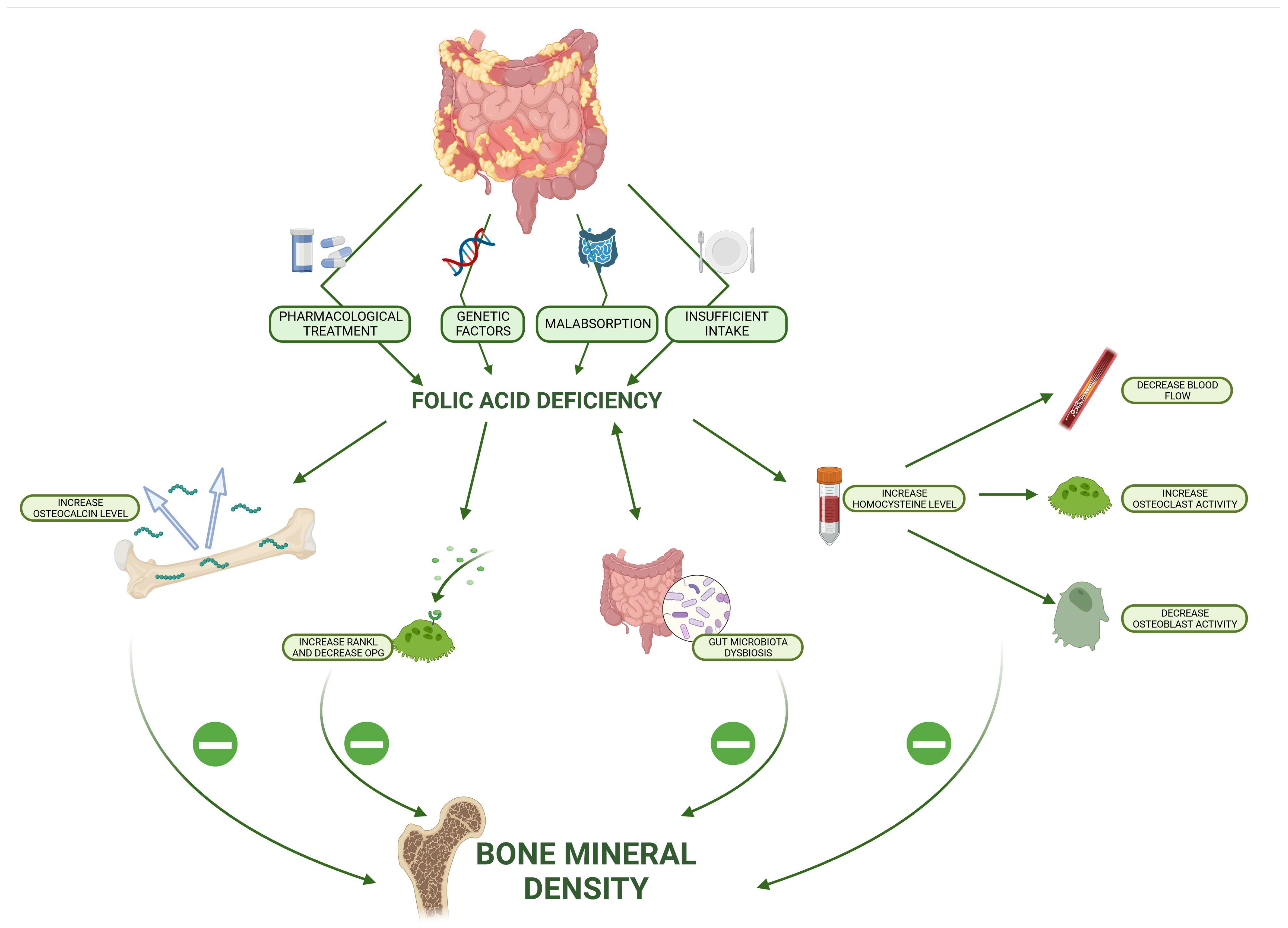
:1. Introduction
2. Materials and Methods
Statistical Analysis
3. Results
4. Discussion
5. Conclusions
Author Contributions
Funding
Institutional Review Board Statement
Informed Consent Statement
Data Availability Statement
Acknowledgments
Conflicts of Interest
References
- Ganguly, P.; Alam, S.F. Role of Homocysteine in the Development of Cardiovascular Disease. Nutr. J. 2015, 14, 6. [Google Scholar] [CrossRef] [PubMed]
- Khan, K.M.; Jialal, I. Folic Acid Deficiency. In StatPearls; StatPearls Publishing: Treasure Island, FL, USA, 2023. [Google Scholar]
- World Health Organization. Guideline: Daily Iron and Folic Acid Supplementation in Pregnant Women; WHO Guidelines Approved by the Guidelines Review Committee; World Health Organization: Geneva, Switzerland, 2012; ISBN 978-92-4-150199-6. [Google Scholar]
- Collaboration, H.L.T. Lowering Blood Homocysteine with Folic Acid Based Supplements: Meta-Analysis of Randomised Trials. Homocysteine Lowering Trialists’ Collaboration. BMJ 1998, 316, 894–898. [Google Scholar] [CrossRef] [PubMed]
- Kriebitzsch, C.; Verlinden, L.; Eelen, G.; van Schoor, N.M.; Swart, K.; Lips, P.; Meyer, M.B.; Pike, J.W.; Boonen, S.; Carlberg, C.; et al. 1,25-Dihydroxyvitamin D3 Influences Cellular Homocysteine Levels in Murine Preosteoblastic MC3T3-E1 Cells by Direct Regulation of Cystathionine β-Synthase. J. Bone Miner. Res. 2011, 26, 2991–3000. [Google Scholar] [CrossRef] [PubMed]
- Vacek, T.P.; Kalani, A.; Voor, M.J.; Tyagi, S.C.; Tyagi, N. The Role of Homocysteine in Bone Remodeling. Clin. Chem. Lab. Med. 2013, 51, 579–590. [Google Scholar] [CrossRef] [PubMed]
- Herrmann, M.; Widmann, T.; Herrmann, W. Homocysteine—A Newly Recognised Risk Factor for Osteoporosis. Clin. Chem. Lab. Med. 2005, 43, 1111–1117. [Google Scholar] [CrossRef] [PubMed]
- Clarke, M.; Ward, M.; Strain, J.J.; Hoey, L.; Dickey, W.; McNulty, H. B-Vitamins and Bone in Health and Disease: The Current Evidence. Proc. Nutr. Soc. 2014, 73, 330–339. [Google Scholar] [CrossRef]
- Salari, P.; Abdollahi, M.; Heshmat, R.; Meybodi, H.A.; Razi, F. Effect of Folic Acid on Bone Metabolism: A Randomized Double Blind Clinical Trial in Postmenopausal Osteoporotic Women. Daru 2014, 22, 62. [Google Scholar] [CrossRef]
- Hsieh, R.-L.; Huang, Y.-L.; Chen, W.-J.; Chen, H.-H.; Shiue, H.-S.; Lin, Y.-C.; Hsueh, Y.-M. Associations between Plasma Folate and Vitamin B12, Blood Lead, and Bone Mineral Density among Adults and Elderly Who Received a Health Examination. Nutrients 2022, 14, 911. [Google Scholar] [CrossRef]
- Pan, Y.; Liu, Y.; Guo, H.; Jabir, M.S.; Liu, X.; Cui, W.; Li, D. Associations between Folate and Vitamin B12 Levels and Inflammatory Bowel Disease: A Meta-Analysis. Nutrients 2017, 9, 382. [Google Scholar] [CrossRef]
- Ratajczak, A.E.; Rychter, A.M.; Zawada, A.; Dobrowolska, A.; Krela-Kaźmierczak, I. Nutrients in the Prevention of Osteoporosis in Patients with Inflammatory Bowel Diseases. Nutrients 2020, 12, 1702. [Google Scholar] [CrossRef]
- Lambert, K.; Pappas, D.; Miglioretto, C.; Javadpour, A.; Reveley, H.; Frank, L.; Grimm, M.C.; Samocha-Bonet, D.; Hold, G.L. Systematic Review with Meta-Analysis: Dietary Intake in Adults with Inflammatory Bowel Disease. Aliment. Pharmacol. Ther. 2021, 54, 742–754. [Google Scholar] [CrossRef]
- Weisshof, R.; Chermesh, I. Micronutrient Deficiencies in Inflammatory Bowel Disease. Curr. Opin. Clin. Nutr. Metab. Care 2015, 18, 576–581. [Google Scholar] [CrossRef] [PubMed]
- Bischoff, S.C.; Escher, J.; Hébuterne, X.; Kłęk, S.; Krznaric, Z.; Schneider, S.; Shamir, R.; Stardelova, K.; Wierdsma, N.; Wiskin, A.E.; et al. ESPEN Practical Guideline: Clinical Nutrition in Inflammatory Bowel Disease. Clin. Nutr. 2020, 39, 632–653. [Google Scholar] [CrossRef] [PubMed]
- Burr, N.E.; Hull, M.A.; Subramanian, V. Folic Acid Supplementation May Reduce Colorectal Cancer Risk in Patients With Inflammatory Bowel Disease: A Systematic Review and Meta-Analysis. J. Clin. Gastroenterol. 2017, 51, 247–253. [Google Scholar] [CrossRef] [PubMed]
- Gomollón, F.; Dignass, A.; Annese, V.; Tilg, H.; Van Assche, G.; Lindsay, J.O.; Peyrin-Biroulet, L.; Cullen, G.J.; Daperno, M.; Kucharzik, T.; et al. 3rd European Evidence-Based Consensus on the Diagnosis and Management of Crohn’s Disease 2016: Part 1: Diagnosis and Medical Management. J. Crohn’s Colitis 2017, 11, 3–25. [Google Scholar] [CrossRef] [PubMed]
- Krela-Kaźmierczak, I.; Michalak, M.; Szymczak-Tomczak, A.; Łykowska-Szuber, L.; Stawczyk-Eder, K.; Waszak, K.; Kucharski, M.A.; Dobrowolska, A.; Eder, P. Prevalence of Osteoporosis and Osteopenia in a Population of Patients with Inflammatory Bowel Diseases from the Wielkopolska Region. Pol. Arch. Intern. Med. 2018, 128, 447–454. [Google Scholar] [CrossRef]
- Targownik, L.E.; Bernstein, C.N.; Leslie, W.D. Inflammatory Bowel Disease and the Risk of Osteoporosis and Fracture. Maturitas 2013, 76, 315–319. [Google Scholar] [CrossRef]
- Bernstein, C.N.; Leslie, W.D. Therapy Insight: Osteoporosis in Inflammatory Bowel Disease--Advances and Retreats. Nat. Clin. Pract. Gastroenterol. Hepatol. 2005, 2, 232–239. [Google Scholar] [CrossRef]
- Bravenboer, N.; Oostlander, A.E.; van Bodegraven, A.A. Bone Loss in Patients with Inflammatory Bowel Disease: Cause, Detection and Treatment. Curr. Opin. Gastroenterol. 2021, 37, 128–134. [Google Scholar] [CrossRef]
- Abitbol, V.; Mary, J.Y.; Roux, C.; Soulé, J.C.; Belaiche, J.; Dupas, J.-L.; Gendre, J.P.; Lerebours, E.; Chaussade, S.; Groupe D’etudes Thérapeutiques des Affections Inflammatoires Digestives (GETAID). Osteoporosis in Inflammatory Bowel Disease: Effect of Calcium and Vitamin D with or without Fluoride. Aliment. Pharmacol. Ther. 2002, 16, 919–927. [Google Scholar] [CrossRef]
- Valentine, J.F.; Sninsky, C.A. Prevention and Treatment of Osteoporosis in Patients with Inflammatory Bowel Disease. Am. J. Gastroenterol. 1999, 94, 878–883. [Google Scholar] [CrossRef] [PubMed]
- Ratajczak, A.E.; Szymczak-Tomczak, A.; Rychter, A.M.; Zawada, A.; Dobrowolska, A.; Krela-Kaźmierczak, I. Does Folic Acid Protect Patients with Inflammatory Bowel Disease from Complications? Nutrients 2021, 13, 4036. [Google Scholar] [CrossRef] [PubMed]
- Weng, Y.J.; Gan, H.Y.; Li, X.; Huang, Y.; Li, Z.C.; Deng, H.M.; Chen, S.Z.; Zhou, Y.; Wang, L.S.; Han, Y.P.; et al. Correlation of Diet, Microbiota and Metabolite Networks in Inflammatory Bowel Disease. J. Dig. Dis. 2019, 20, 447–459. [Google Scholar] [CrossRef] [PubMed]
- Pietrzykowska-Kuncman, M.; Zasina-Olaszek, D.; Łukasz, K.; Niedźwiecka, M.; Szaflik, K.; Maroszyńska, I. Intake of Folic Acid by Polish Women with Higher Education—A Survey Research: Can We Do More? Ginekol. Pol. 2017, 88, 428–433. [Google Scholar] [CrossRef] [PubMed]
- Torres, J.; Chaparro, M.; Julsgaard, M.; Katsanos, K.; Zelinkova, Z.; Agrawal, M.; Ardizzone, S.; Campmans-Kuijpers, M.; Dragoni, G.; Ferrante, M.; et al. European Crohn’s and Colitis Guidelines on Sexuality, Fertility, Pregnancy, and Lactation. J. Crohns Colitis 2023, 17, 1–27. [Google Scholar] [CrossRef]
- Castellanos-Sinco, H.B.; Ramos-Peñafiel, C.O.; Santoyo-Sánchez, A.; Collazo-Jaloma, J.; Martínez-Murillo, C.; Montaño-Figueroa, E.; Sinco-Ángeles, A. Megaloblastic Anaemia: Folic Acid and Vitamin B12 Metabolism. Rev. Med. Hosp. Gen. Mex. 2015, 78, 135–143. [Google Scholar] [CrossRef]
- Sugihara, K.; Morhardt, T.L.; Kamada, N. The Role of Dietary Nutrients in Inflammatory Bowel Disease. Front. Immunol. 2018, 9, 3183. [Google Scholar] [CrossRef]
- Kaye, A.D.; Jeha, G.M.; Pham, A.D.; Fuller, M.C.; Lerner, Z.I.; Sibley, G.T.; Cornett, E.M.; Urits, I.; Viswanath, O.; Kevil, C.G. Folic Acid Supplementation in Patients with Elevated Homocysteine Levels. Adv. Ther. 2020, 37, 4149–4164. [Google Scholar] [CrossRef]
- Gao, X.; Li, J.; Chen, M. Effect of Homocysteine on the Differentiation of CD4+ T Cells into Th17 Cells. Dig. Dis. Sci. 2018, 63, 3339–3347. [Google Scholar] [CrossRef]
- Ratajczak, A.E.; Szymczak-Tomczak, A.; Michalak, M.; Rychter, A.M.; Zawada, A.; Dobrowolska, A.; Krela-Kaźmierczak, I. The Associations between Vitamin D, Bone Mineral Density and the Course of Inflammatory Bowel Disease in Polish Patients. Pol. Arch. Intern. Med. 2022, 132, 16329. [Google Scholar] [CrossRef]
- Rychter, A.M.; Ratajczak, A.E.; Szymczak-Tomczak, A.; Michalak, M.; Eder, P.; Dobrowolska, A.; Krela-Kaźmierczak, I. Associations of Lifestyle Factors with Osteopenia and Osteoporosis in Polish Patients with Inflammatory Bowel Disease. Nutrients 2021, 13, 1863. [Google Scholar] [CrossRef] [PubMed]
- Harbord, M.; Annese, V.; Vavricka, S.R.; Allez, M.; Barreiro-de Acosta, M.; Boberg, K.M.; Burisch, J.; De Vos, M.; De Vries, A.-M.; Dick, A.D.; et al. The First European Evidence-Based Consensus on Extra-Intestinal Manifestations in Inflammatory Bowel Disease. J. Crohns Colitis 2016, 10, 239–254. [Google Scholar] [CrossRef] [PubMed]
- Yin, Y.; Lu, X.; Li, Z.; Liu, S.; Shao, L.; Cao, L.; Liu, R.-Q.; Huang, L.-Y.; Zhu, Z.-X.; Guo, Z.; et al. Risk Factors for Worsening of Bone Loss in Patients Newly Diagnosed with Inflammatory Bowel Disease. Gastroenterol. Res. Pract. 2022, 2022, e1498293. [Google Scholar] [CrossRef] [PubMed]
- Komaki, Y.; Komaki, F.; Micic, D.; Ido, A.; Sakuraba, A. Risk of Fractures in Inflammatory Bowel Diseases: A Systematic Review and Meta-Analysis. J. Clin. Gastroenterol. 2019, 53, 441–448. [Google Scholar] [CrossRef]
- Palatianou, M.E.; Karamanolis, G.; Tsentidis, C.; Gourgiotis, D.; Papaconstantinou, I.; Vezakis, A.; Tzouvala, M. Signaling Pathways Associated with Bone Loss in Inflammatory Bowel Disease. Ann. Gastroenterol. 2023, 36, 132–140. [Google Scholar] [CrossRef] [PubMed]
- Ma, Q.; Liang, M.; Tang, X.; Luo, F.; Dou, C. Vitamin B5 Inhibit RANKL Induced Osteoclastogenesis and Ovariectomy Induced Osteoporosis by Scavenging ROS Generation. Am. J. Transl. Res. 2019, 11, 5008–5018. [Google Scholar] [PubMed]
- Fathi Maroufi, N.; Ghorbanihaghjo, A.; Sayyah Melli, M.; Vaezi, M.; Hekmati Azar Mehrabani, Z.; Bannazadeh Amirkhiz, M.; Rashtchizadeh, N. Effects of High and Low Doses of Folic Acid on the Soluble Receptor Activator of Nuclear Factor-Kappa B Ligand/Osteoprotegerin Ratio during Pregnancy. Iran. J. Public. Health 2017, 46, 517–524. [Google Scholar] [PubMed]
- Raine, T.; Bonovas, S.; Burisch, J.; Kucharzik, T.; Adamina, M.; Annese, V.; Bachmann, O.; Bettenworth, D.; Chaparro, M.; Czuber-Dochan, W.; et al. ECCO Guidelines on Therapeutics in Ulcerative Colitis: Medical Treatment. J. Crohns Colitis 2022, 16, 2–17. [Google Scholar] [CrossRef]
- Dignass, A.U.; Gasche, C.; Bettenworth, D.; Birgegård, G.; Danese, S.; Gisbert, J.P.; Gomollon, F.; Iqbal, T.; Katsanos, K.; Koutroubakis, I.; et al. European Consensus on the Diagnosis and Management of Iron Deficiency and Anaemia in Inflammatory Bowel Diseases. J. Crohns Colitis 2015, 9, 211–222. [Google Scholar] [CrossRef]
- Zimmer, M.; Sieroszewski, P.; Oszukowski, P.; Huras, H.; Fuchs, T.; Pawłosek, A. Polish Society of Gynecologists and Obstetricians recommendations on supplementation in pregnancy. Ginekol. I Perinatol. Prakt. 2020, 5, 170–181. [Google Scholar] [CrossRef]
- Lima, C.A.; Lyra, A.C.; Mendes, C.M.C.; Lopes, M.B.; Coqueiro, F.G.; Rocha, R.; Santana, G.O. Bone Mineral Density and Inflammatory Bowel Disease Severity. Braz. J. Med. Biol. Res. 2017, 50, e6374. [Google Scholar] [CrossRef] [PubMed]
- Engevik, M.A.; Morra, C.N.; Röth, D.; Engevik, K.; Spinler, J.K.; Devaraj, S.; Crawford, S.E.; Estes, M.K.; Kalkum, M.; Versalovic, J. Microbial Metabolic Capacity for Intestinal Folate Production and Modulation of Host Folate Receptors. Front. Microbiol. 2019, 10, 2305. [Google Scholar] [CrossRef] [PubMed]
- Gomes de Lima, K.V.; Maio, R. Nutritional Status, Systemic Inflammation and Prognosis of Patients with Gastrointestinal Cancer. Nutr. Hosp. 2012, 27, 707–714. [Google Scholar] [CrossRef] [PubMed]
- Clements, M.; Heffernan, M.; Ward, M.; Hoey, L.; Doherty, L.C.; Hack Mendes, R.; Clarke, M.M.; Hughes, C.F.; Love, I.; Murphy, S.; et al. A 2-Year Randomized Controlled Trial with Low-Dose B-Vitamin Supplementation Shows Benefits on Bone Mineral Density in Adults with Lower B12 Status. J. Bone Min. Res. 2022, 37, 2443–2455. [Google Scholar] [CrossRef] [PubMed]
- Li, S.; Mao, Y.; Zhou, F.; Yang, H.; Shi, Q.; Meng, B. Gut Microbiome and Osteoporosis. Bone Jt. Res. 2020, 9, 524–530. [Google Scholar] [CrossRef]
- Pandey, H.; Jain, D.; Tang, D.W.T.; Wong, S.H.; Lal, D. Gut Microbiota in Pathophysiology, Diagnosis, and Therapeutics of Inflammatory Bowel Disease. Intest. Res. 2023, 9, 2247. [Google Scholar] [CrossRef]
- Hiel, S.; Bindels, L.B.; Pachikian, B.D.; Kalala, G.; Broers, V.; Zamariola, G.; Chang, B.P.I.; Kambashi, B.; Rodriguez, J.; Cani, P.D.; et al. Effects of a Diet Based on Inulin-Rich Vegetables on Gut Health and Nutritional Behavior in Healthy Humans. Am. J. Clin. Nutr. 2019, 109, 1683–1695. [Google Scholar] [CrossRef]
- Raghubeer, S.; Matsha, T.E. Methylenetetrahydrofolate (MTHFR), the One-Carbon Cycle, and Cardiovascular Risks. Nutrients 2021, 13, 4562. [Google Scholar] [CrossRef]
- Ratajczak-Pawłowska, A.E.; Hryhorowicz, S.; Szymczak-Tomczak, A.; Wysocka, E.; Michalak, M.; Kaczmarek-Ryś, M.; Lis-Tanaś, E.; Bielawska, L.; Pławski, A.; Słomski, R.; et al. Genetic Variants of MTHFR Gene in Relation to Folic Acid Levels and Bone Mineral Density in Polish Patients with Inflammatory Bowel Disease. J. Appl. Genet. 2023; ahead of print. [Google Scholar] [CrossRef]
- Zheng, Z.; Luo, H.; Xu, W.; Xue, Q. Association between Dietary Folate Intake and Bone Mineral Density in a Diverse Population: A Cross-Sectional Study. J. Orthop. Surg. Res. 2023, 18, 684. [Google Scholar] [CrossRef]
- Kim, D.E.; Cho, S.H.; Park, H.M.; Chang, Y.K. Relationship between Bone Mineral Density and Dietary Intake of β-Carotene, Vitamin C, Zinc and Vegetables in Postmenopausal Korean Women: A Cross-Sectional Study. J. Int. Med. Res. 2016, 44, 1103–1114. [Google Scholar] [CrossRef]
- Kalimeri, M.; Leek, F.; Wang, N.X.; Koh, H.R.; Roy, N.C.; Cameron-Smith, D.; Kruger, M.C.; Henry, C.J.; Totman, J.J. Folate and Vitamin B-12 Status Is Associated With Bone Mineral Density and Hip Strength of Postmenopausal Chinese-Singaporean Women. JBMR Plus 2020, 4, e10399. [Google Scholar] [CrossRef] [PubMed]
- Bozkurt, N.; Erdem, M.; Yilmaz, E.; Erdem, A.; Biri, A.; Kubatova, A.; Bozkurt, M. The Relationship of Homocyteine, B12 and Folic Acid with the Bone Mineral Density of the Femur and Lumbar Spine in Turkish Postmenopausal Women. Arch. Gynecol. Obs. 2009, 280, 381–387. [Google Scholar] [CrossRef] [PubMed]
- Marini, H.; Minutoli, L.; Polito, F.; Bitto, A.; Altavilla, D.; Atteritano, M.; Gaudio, A.; Mazzaferro, S.; Frisina, A.; Frisina, N.; et al. Effects of the Phytoestrogen Genistein on Bone Metabolism in Osteopenic Postmenopausal Women: A Randomized Trial. Ann. Intern. Med. 2007, 146, 839–847. [Google Scholar] [CrossRef] [PubMed]
- Arcoraci, V.; Atteritano, M.; Squadrito, F.; D’Anna, R.; Marini, H.; Santoro, D.; Minutoli, L.; Messina, S.; Altavilla, D.; Bitto, A. Antiosteoporotic Activity of Genistein Aglycone in Postmenopausal Women: Evidence from a Post-Hoc Analysis of a Multicenter Randomized Controlled Trial. Nutrients 2017, 9, 179. [Google Scholar] [CrossRef] [PubMed]
- Wu, Z.-Y.; Sang, L.-X.; Chang, B. Isoflavones and Inflammatory Bowel Disease. World J. Clin. Cases 2020, 8, 2081–2091. [Google Scholar] [CrossRef] [PubMed]
- Basson, A.R.; Ahmed, S.; Almutairi, R.; Seo, B.; Cominelli, F. Regulation of Intestinal Inflammation by Soybean and Soy-Derived Compounds. Foods 2021, 10, 774. [Google Scholar] [CrossRef]
- Bhattacharya, A.; Pal, B.; Mukherjee, S.; Roy, S.K. Assessment of Nutritional Status Using Anthropometric Variables by Multivariate Analysis. BMC Public. Health 2019, 19, 1045. [Google Scholar] [CrossRef] [PubMed]
- Stone, K.L.; Lui, L.-Y.; Christen, W.G.; Troen, A.M.; Bauer, D.C.; Kado, D.; Schambach, C.; Cummings, S.R.; Manson, J.E. Effect of Combination Folic Acid, Vitamin B6, and Vitamin B12 Supplementation on Fracture Risk in Women: A Randomized, Controlled Trial. J. Bone Min. Res. 2017, 32, 2331–2338. [Google Scholar] [CrossRef]
- Ratajczak-Pawłowska, A.E.; Michalak, M.; Szymczak-Tomczak, A.; Rychter, A.M.; Zawada, A.; Skoracka, K.; Dobrowolska, A.; Krela-Kaźmierczak, I. Physical Activity, Quality of Diet and Bone Mineral Density in Patients with Inflammatory Bowel Disease. J. Hum. Nutr. Diet. 2023, 36, 1692–1700. [Google Scholar] [CrossRef]
- Marini, H.R. Mediterranean Diet and Soy Isoflavones for Integrated Management of the Menopausal Metabolic Syndrome. Nutrients 2022, 14, 1550. [Google Scholar] [CrossRef]


| Parameter | CD (n = 26) | UC (n = 30) | CG (n = 31) | p-Value |
|---|---|---|---|---|
| Age [years] | 30.50 (25.40; 37.20) | 28.20 (21.90; 37.60) | 32.90 (27.10; 42.90) | 0.11 |
| Body mass [kg] | 59.70 (50.80; 65.90) | 58.60 (53.30; 69.30) | 67.60 (61.50; 78.80) | <0.01 <0.01 a 0.02 b 0.99 c |
| BMI [kg/m2] | 19.95 (19.10; 23.10) | 19.90 (17.70; 22.40) | 23.00 (20.90; 25.90) | <0.01 0.04 a <0.01 b 0.99 c |
| BMD (L1–L4) [g/cm2] | 1.136 (1.077; 1.235) | 1.160 (0.984; 1.259) | 1.280 (1.172; 1.367) | <0.001 <0.01 a <0.01 b 0.99 c |
| T-score (L1–L4) | −0.400 (−1.000; 0.500) | −0.200 (−1.700; 0.700) | 0.700 (−0.100; 1.300) | <0.001 <0.01 a <0.01 b 0.99 c |
| Z-score (L1–L4) | −0.300 (−0.600; 0.600) | −0.400 (−1.300; 0.900) | 0.400 (0.100; 1.400) | <0.001 <0.01 a <0.01 b 0.99 c |
| BMD (femoral neck) [g/cm2] | 0.987 (0.938; 1.135) | 1.038 (0.837; 1.147) | 1.097 (1.048; 1.192) | <0.01 0.02 a 0.02 b 0.99 c |
| T-score (femoral neck) | −0.450 (−0.800; 0.600) | 0.050 (−1.200; 1.00) | 0.400 (0.100; 1.100) | <0.01 0.03 a 0.02 b 0.99 c |
| Z-score (femoral neck) | 0.050 (−0.400; 0.600) | 0.050 (−1.200; 1.000) | 0.600 (0.200; 1.200) | <0.01 0.04 a 0.02 b 0.99 c |
| Folate intake [mg] | 226.77 (191.27; 352.86) | 273.50 (205.42; 415.75) | 302.63 (226.93; 393.92) | 0.23 |
| Meet the RDA [%] | 56.69 (47.82; 88.21) | 68.38 (51.35; 103.95) | 75.66 (56.73; 98.48) | 0.23 |
| Energy intake [kcal] | 1720.415 (1401.15; 2151.87) | 1837.10 (1423.77; 2287.49) | 1738.74 (1504.08; 2274.53) | 0.58 |
| Folate/1000 kcal [mg/kcal] | 145.89 (123.04; 163.93) | 171.05 (120.67; 185.68) | 155.058 (131.98; 181.57) | 0.43 |
| CRP | 5.85 (1.40; 21.80) | 2.70 (1.10; 10.30) | 0.70 (0.30; 1.70) | <0.0001 <0.0001 a <0.001 b 0.97 c |
| Sex | Parameter | CD | UC | CG | p-Value |
|---|---|---|---|---|---|
| Women | Number of patients [n] | 16 | 16 | 19 | |
| Meet the RDA [%] | 56.69 (48.68; 84.26) | 67.25 (52.68; 86.86) | 63.50 (53.45; 90.41) | 0.78 | |
| Energy intake [kcal] | 1552.23 (1320.17; 1770.53) | 1551.89 (1378.58; 1772.36) | 1601 (1421.58; 1721.16) | 0.73 | |
| Folate/1000 kcal [mg/kcal] | 153.66 (139.16; 196.74) | 184.52 (157.82; 192.18) | 155.06 (131.98; 189.44) | 0.46 | |
| Men | Number of patients [n] | 10 | 14 | 12 | |
| Meet the RDA [%] | 62.21 (47.28; 88.56) | 78.06 (51.35; 104.02) | 88.23 (63.82; 102.56) | 0.21 | |
| Energy intake [kcal] | 2120.23 (1866.95; 2272.05) | 2304.79 (2003.21; 2423.36) | 2229.51 (1946.44; 2573.28) | 0.57 | |
| Folate/1000 kcal [mg/kcal] | 119.96 (106.91; 139.86) | 139.81 (114.51; 183.29) | 149.34 (130.89; 175.36) | 0.17 |
| Sex | Inadequate Intake (below 100% of RDA) | Adequate Intake (at least 100% of RDA) | p-Value | |
|---|---|---|---|---|
| Women | ||||
| Supplementation | yes | 12 (85.71%) | 2 (14.29%) | 0.70 |
| no | 30 (81.08%) | 7 (18.92%) | ||
| Men | ||||
| Supplementation | yes | 7 (63.64%) | 4 (36.36%) | 0.30 |
| no | 20 (80.00%) | 5 (20.00%) | ||
| CD | UC | CG | |||||||
|---|---|---|---|---|---|---|---|---|---|
| Insufficient (n = 24) | Normal (n = 2) | p-Value | Insufficient (n = 21) | Normal (n = 9) | p-Value | Insufficient (n = 24) | Normal (n = 7) | p-Value | |
| BMI [kg/m2] | 19.85 (19.00; 22.15) | 23.90 (23.10; 24.70) | 0.16 | 19.60 (17.70; 21.50) | 20.40 (19.70; 24.90) | 0.15 | 22.60 (20.85; 25.50) | 23.60 (22.00; 24.70) | 0.63 |
| BMD (L1–L4) [g/cm2] | 1.136 (1.047; 1.218) | 1.243 (1.095; 0.391) | 0.56 | 1.154 (0.989; 1.267) | 1.165 (0.984; 1.246) | 0.98 | 1.276 (1.172; 1.335) | 1.335 (1.226; 1.545) | 0.18 |
| T-score (L1–L4) | −0.400 (−1.200; 0.350) | 0.400 (−1.000; 1.800) | 0.60 | −0.200 (−1.700; 0.700) | −0.100 (−1.900; 0.200) | 0.98 | 0.550 (−0.200; 1.250) | 1.300 (0.100; 3.000) | 0.15 |
| Z-score (L1–L4) | −0.300 (−0.550; 0.400) | 0.500 (−0.900; 1.900) | 0.74 | −0.400 (−1.300; 0.900) | −0.400 (−1.300; 0.200) | 0.87 | 0.400 (−0.050; 1.150) | 0.800 (0.200; 3.200) | 0.20 |
| BMD (femoral neck) [g/cm2] | 0.987 (0.934; 1.129) | 1.158 (0.963; 1.353) | 0.36 | 1.032 (0.837; 1.148) | 1.063 (0.868; 1.109) | 0.96 | 1.095 (1.036; 1.174) | 1.102 (1.093; 1.255) | 0.21 |
| T-score (femoral neck) | −0.450 (−0.900; 0.550) | 0.750 (−0.800; 2.300) | 0.56 | −0.300 (−1.700; 0.800) | 0.200 (−1.600; 0.300) | 0.91 | 0.300 (0.000; 0.850) | 0.500 (0.200; 1.600) | 0.19 |
| Z-score (femoral neck) | 0.050 (−0.350; 0.550) | 0.850 (−1.000; 2.700) | 0.89 | 0.100 (−1.200; 1.200) | 0.000 (−1.200; 0.200) | 0.72 | 0.450 (0.200; 1.100) | 0.800 (0.400; 2.300) | 0.21 |
| CRP | 5.85 (1.45; 19.95) | 11.10 (0.40; 21.80) | 0.67 | 3.10 (1.10; 10.80) | 2.30 (1.80; 7.30) | 0.87 | 0.80 (0.40; 1.70) | 0.30 (0.20; 0.50) | 0.10 |
| Folic Acid Supplementation | ||||||
|---|---|---|---|---|---|---|
| CD | UC | |||||
| Yes (n = 11) | No (n = 15) | p-Value | Yes (n = 13) | No (n = 17) | p-Value | |
| BMI [kg/m2] | 22.10 (18.80; 23.40) | 19.40 (19.10; 20.80) | 0.30 | 19.70 (18.60; 21.30) | 20.20 (17.70; 22.40) | 0.88 |
| BMD (L1–L4) [g/cm2] | 1.136 (1.096; 1.287) | 1.126 (1.011; 1.235) | 0.31 | 1.154 (1.072; 1.259) | 1.165 (0.972; 1.246) | 0.52 |
| T-score (L1–L4) | −0.400 (−1.000; 0.700) | −0.500 (−1.700; 0.500) | 0.41 | −0.200 (−1.200; 0.700) | −0.200 (−1.800; 0.500) | 0.62 |
| Z-score (L1–L4) | −0.100 (−0.600; 0.600) | −0.400 (−1.200; 0.600) | 0.36 | −0.200 (−0.500; 0.900) | −0.500 (−1.300; 0.500) | 0.33 |
| BMD (neck) [g/cm2] | 0.998 (0.942; 1.145) | 0.980 (0.903; 1.074) | 0.41 | 1.043 (0.857; 1.131) | 0.968 (0.837; 1.232) | 0.93 |
| T-score (neck) | −0.500 (−0.700; 0.800) | −0.400 (−1.00; 0.300) | 0.48 | 0.000 (−1.600; 0.600) | −0.500 (−1.800; 1.400) | 0.97 |
| Z-score (neck) | 0.200 (−0.100; 0.800) | 0.000 (−0.700; 0.200) | 0.45 | 0.100 (−1.200; 0.900) | −0.400 (−1.400; 1.500) | 0.98 |
| CRP | 5.40 (1.30; 11.60) | 6.30 (2.30; 26.80) | 0.41 | 1.30 (0.60; 7.30) | 6.00 (2.00; 10.30) | 0.19 |
Disclaimer/Publisher’s Note: The statements, opinions and data contained in all publications are solely those of the individual author(s) and contributor(s) and not of MDPI and/or the editor(s). MDPI and/or the editor(s) disclaim responsibility for any injury to people or property resulting from any ideas, methods, instructions or products referred to in the content. |
© 2023 by the authors. Licensee MDPI, Basel, Switzerland. This article is an open access article distributed under the terms and conditions of the Creative Commons Attribution (CC BY) license (https://creativecommons.org/licenses/by/4.0/).
Share and Cite
Ratajczak-Pawłowska, A.E.; Szymczak-Tomczak, A.; Michalak, M.; Rychter, A.M.; Zawada, A.; Skoracka, K.; Dobrowolska, A.; Krela-Kaźmierczak, I. Impact of Folate Intake on Bone Mineral Density in Patients with Inflammatory Bowel Disease. Nutrients 2024, 16, 6. https://doi.org/10.3390/nu16010006
Ratajczak-Pawłowska AE, Szymczak-Tomczak A, Michalak M, Rychter AM, Zawada A, Skoracka K, Dobrowolska A, Krela-Kaźmierczak I. Impact of Folate Intake on Bone Mineral Density in Patients with Inflammatory Bowel Disease. Nutrients. 2024; 16(1):6. https://doi.org/10.3390/nu16010006
Chicago/Turabian StyleRatajczak-Pawłowska, Alicja Ewa, Aleksandra Szymczak-Tomczak, Michał Michalak, Anna Maria Rychter, Agnieszka Zawada, Kinga Skoracka, Agnieszka Dobrowolska, and Iwona Krela-Kaźmierczak. 2024. "Impact of Folate Intake on Bone Mineral Density in Patients with Inflammatory Bowel Disease" Nutrients 16, no. 1: 6. https://doi.org/10.3390/nu16010006
APA StyleRatajczak-Pawłowska, A. E., Szymczak-Tomczak, A., Michalak, M., Rychter, A. M., Zawada, A., Skoracka, K., Dobrowolska, A., & Krela-Kaźmierczak, I. (2024). Impact of Folate Intake on Bone Mineral Density in Patients with Inflammatory Bowel Disease. Nutrients, 16(1), 6. https://doi.org/10.3390/nu16010006