Review

The Beneficial Effects of Dietary Interventions on Gut Microbiota—An Up-to-Date Critical Review and Future Perspectives

by
Carmen Purdel
1,
Denisa Margină
2,*,
Ines Adam-Dima
1 and
Anca Ungurianu
2
1
Department of Toxicology, Faculty of Pharmacy, “Carol Davila” University of Medicine and Pharmacy, Traian Vuia 6, 020956 Bucharest, Romania
2
Department of Biochemistry, Faculty of Pharmacy, “Carol Davila” University of Medicine and Pharmacy, Traian Vuia 6, 020956 Bucharest, Romania
*
Author to whom correspondence should be addressed.
Nutrients 2023, 15(23), 5005; https://doi.org/10.3390/nu15235005
Submission received: 7 November 2023 / Revised: 22 November 2023 / Accepted: 2 December 2023 / Published: 3 December 2023
(This article belongs to the Special Issue Intermittent Fasting on Human Health and Disease)

Abstract

:
Different dietary interventions, especially intermittent fasting, are widely used and promoted by physicians; these regimens have been studied lately for their impact on the gut microbiota composition/function and, consequently, on the general physiopathological processes of the host. Studies are showing that dietary components modulate the microbiota, and, at the same time, the host metabolism is deeply influenced by the different products resulting from nutrient transformation in the microbiota compartment. This reciprocal relationship can potentially influence even drug metabolism for chronic drug regimens, significantly impacting human health/disease. Recently, the influence of various dietary restrictions on the gut microbiota and the differences between the effects were investigated. In this review, we explored the current knowledge of different dietary restrictions on animal and human gut microbiota and the impact of these changes on human health.

1. Introduction

The human body is home to a great number of microbial cells found in diverse locations; their highest density is in the intestinal compartment, where they generate a complex microbial population known as the gut microbiota. This microbial community starts to develop after birth, evolves, and becomes more and more complex through infancy to eventually reach its adult form [1,2,3,4,5,6]. Its diversity is subject to different environmental factors, such as pH, oxygen levels/redox state, dietary nutrients, water activity, and temperature [5,6]. At birth, the gut microbiota is mainly composed of Proteobacteria and Actinobacteria; depending on the type of milk and then on food and lifestyle, it evolves through a stage where Enterobacteriaceae, Bacteroides, and Bifidobacterium are present. These are, in time, replaced mainly by members of the Lachnospiraceae and Ruminococcaceae families, and, in adults, the intestinal colonies are dominated by anaerobic bacteria from the major phyla of Firmicutes (predominantly Lachnospiraceae and Ruminococcaceae), Bacteroidetes, Actinobacteria, Proteobacteria, and Verrucomicrobia (Akkermansia) [3,5,6,7,8,9,10,11,12,13,14,15]. Furthermore, adults develop a personalised microbiota. Therefore, comparing microbiota composition between communities is challenging unless one population contrasts with different food cultures [16,17].
The gut microbiota is clearly involved in digestion, the fermentation of dietary fibers, and the production of some vitamins. In recent years, literature data have also shown that it is a central regulator of the host’s nutritional and metabolic homeostasis, influencing immunological functions. Dysbiosis is correlated not only with digestive issues or irritable bowel syndrome but is also involved in the etiopathology of obesity, type 2 diabetes mellitus (T2DM), and other metabolic imbalances, with a high incidence in the general population [18,19,20,21,22]. Interestingly, gut microbiota impairment is also associated with cardiovascular disease, neurodegeneration, nonalcoholic fatty liver disease, colon cancer, etc. For example, Odoribacter is negatively correlated with blood pressure [7,23], while Escherichia coli abundance is associated with endothelial dysfunction and metabolic syndrome [24,25,26].
Short-chain fatty acids (SCFAs) generated from the microbiota’s metabolism of nutrients are involved in numerous biochemical processes in the host [27]. Non-digestible carbohydrates (fibers) can be used in the colon as energy sources for local bacteria, who transform them into SCFAs (e.g., acetate, butyrate, and propionate), recognised for their involvement in different physiologic processes such as modulation of energy homeostasis, glucose/lipid metabolism, inflammation, and immunity [28,29,30]. Studies show that Akkermansia municiphilla species are important propionate-producing mucin-degrading organisms, while butyrate production is mainly attributed to the fermentation of resistant starches under the influence of Ruminococcus bromii but also to the metabolism of Faecalibacterium prausnitzii, Eubacterium rectale, and Eubacterium hallii; on the other hand, acetate production is not specific to certain species, being a pathway widely distributed among several bacterial groups [29,31,32,33,34].
Interestingly, the literature shows that SCFAs produced by microbial fermentation induce the same type of effects as increased plasma glucagon-like peptide-1 (GLP-1) and peptide YY (PYY) levels and also stimulate the secretion of gut peptides by acting on specific G-protein-coupled receptors expressed in cells abundant in the colon and terminal ileum [35,36,37,38,39,40,41].
Dietary interventions, as well as therapies aiming to correct microbiota imbalances, improve the metabolic function of humans [18,42]. Preclinical studies show that animals fed a diet rich in saturated lipids (lard) are characterised by a lower abundance and diversity of beneficial bacteria (Akkermansia muciniphila, Lactobacillus, and Bifidobacterium) compared to animals fed an isocaloric high-fat diet containing fish oil, rich in n-3 PUFA; this is associated with an increase in insulin resistance, inflammation, and consequent metabolic impairment [43]. These reports show that the interaction between the nutrients and the host microbiota is complex, with both components influencing each other. The microbiota can be altered by diet, but also the host metabolism can be modulated by the different metabolism products resulted from the microbiome transformation of nutrients since gut microbiota can perform many processes that cannot be supported by the host [44].
Knowledge about the repercussions of dietary changes on the animal and human gut microbiota is still insufficient. Furthermore, it is still unclear if the differences between animal models and humans regarding gut microbiota composition and dynamics limit the use of preclinical results. Although in clinical practise caloric restriction regimens are known to provide efficacy in reducing bodyweight and adiposity, reducing the risk of non-communicable comorbid disease [45,46], it is unclear if the results on gut microbiota composition observed after these regimens are more robust compared to time-restricted fasting. It is also unknown if the effects observed on gut microbiota composition are only temporary and how they impact the renewal of the intestinal mucosa. Furthermore, only a few studies have explored the interaction between the immune system and gut microbiota under different fasting conditions [47].
The present paper aims to review the recent literature data concerning the ability of dietary interventions currently used/recommended by the international medical community to influence/alter the taxonomic composition of gut microbiota and identify gaps in knowledge and potential avenues for a targeted, personalised diet.

2. Updated Concept of Dietary Intervention and Gut Microbiome Links

Various dietary caloric and/or time restriction regimens have been studied for their potential health benefits in healthy subjects, and in recent years, studies have also been performed in patients with different pathologies. In this narrative review, the term dietary intervention is used as an umbrella term for several strategies for restricting the time period of food consumption or the total caloric intake. The most common types of dietary interventions include time-restricted fasting (TRF), such as intermittent fasting (IF) or Ramadan fasting, and caloric restriction (CR) programs, such as alternate-day fasting (ADF), Buchinger fasting programs, water-only fasting, and fasting-mimicking diets (FMD) [48,49,50].
IF involves a fasting pattern at all times. The most popular versions of IF include (1) time-restricted fasting (e.g., the 16:8 method, which includes fasting for 16 h a day and having all meals within an 8 h window, or Ramadan fasting); (2) eat-stop-eat, which involves a 24 h fast once or twice per week; or (3) weekly fasting, like the 5:2 method, which involves unrestricted eating for five days of the week and restricting calories (usually to 500–600 kcal/day) for two non-consecutive days [51]. Alternate-day fasting involves alternating between days of no food restriction and days where food intake is severely restricted (<500 kcal/day) [52]. Many religious traditions, such as Ramadan in Islam (sunrise to sunset fasting) or Yom Kippur in Judaism, incorporate periods of fasting and are also considered IF [23].
There are several forms of CR where the energy intake is reduced to a fraction of the regular ad libitum intake or even to only water fasting. A particular CR is the Buchinger fasting protocol, which involves a primary fasting phase (200–300 kcal/day) that can last for several days (generally 5–7 days) to several weeks, depending on the individual’s health status and the specific goals of the fasting program. After this phase, participants gradually reintroduce solid foods into their diet (the transition phase). Furthermore, a new CR approach, the FMD, involves a low-calorie, low-protein, and high-fat diet to provide the beneficial effects of fasting while still allowing food intake [53,54,55].
In the last decades, studies on the impact of restriction programmes on several markers of metabolic health have been conducted; some of these studies also identified changes in gut microbiota composition. The potential effects of dietary restrictions on the gut microbiota could include shifts in microbial diversity (changes in the abundance and type of microbes) and metabolic alterations. Fasting or caloric restriction may alter the gut environment by changing the nutrient availability and energy sources, which can impact the growth of certain microbial phylum and influence SCFAs production.
The main health-beneficial effects as a result of microbiota changes are illustrated in Figure 1.

3. Methodology

For this narrative review, a literature survey was performed in PubMed to find the most relevant articles reporting interactions between dietary caloric restriction and gut microbiota. Articles were limited to those published in English, focusing on the most recent works between 2015 and 2023 (80.30% of the cited material). Additionally, the literature was reviewed to ascertain the key aspects regarding gut microbiota characteristics. For preclinical data, the publishing year was narrowed to a 2021–2023 interval, and the keywords and MeSH terms used were “gut microbiota” AND “fasted” AND “caloric restriction” AND “mice”, “rats” OR "animals." For clinical data, the applied search terms were “gut microbiota” AND “fasting” OR “intermittent fasting” OR “Ramadan fasting” OR “dietary caloric restriction” AND “clinical trial”.
Two researchers independently screened the titles and abstracts first, and disagreements were solved by discussion and consensus. Protocols, case reports, and studies where the main text was unavailable were excluded. Then, the full text of this papers was reviewed to retrieve the relevant information, and the most relevant papers were selected. The following information about each study was recorded: name of the first author, year of publication, study design, tested sample and analysing method, and main findings. A total of 17 papers regarding nonclinical data and 26 papers on clinical studies were selected after eligibility analysis, cross-checking, and removing duplicates.
In order to evaluate the gut microbiota alterations in various dietary conditions, the clinical studies were classified into two main groups: (1) Time-restricted fasting, including intermittent fasting or Ramadan fasting; and (2) Caloric restriction programs, such as alternate-day fasting, the Buchinger fasting program, water-only fasting, or a very low-calorie diet.

4. Effects of Dietary Intervention on the Animal Gut Microbiota

The repercussions of dietary intervention on the animal gut microbiota are still insufficiently known, although numerous published studies exist. As some excellent reviews have already been published [24], we strived to update and correlate the state-of-the-art data. The most recent research, published between 2021 and 2023, that investigated the alteration of gut microbiota in different animal models is summarised in Table 1.
Most fasting research is conducted using different animal models, and some interesting studies also involve hibernating animals. Song et al. investigated the gut microbiota of hibernating asiatic toads and observed temporal remodelling in the small and large intestines with discrepant patterns. For instance, Proteobacteria assembled in a higher proportion in the small intestine, while Firmicutes and Bacteroidetes assembled in a higher percentage in the large intestine. Also, the large intestine microbiota exhibited higher diversity than the small intestine one [60].

4.1. Studies Conducted on Healthy Animals

The microbial community in trout intestines can be significantly influenced by two weeks of feed deprivation [59]. Dysbiosis was characterised by a significant increase in Bacteroidetes and Firmicutes and a reduction in the Actinobacteria population. The effect of the feed restriction on relative diversity was more evident when considering family and genera than when considering phylum levels. The feed deprivation allowed the development of different commensal or pathogenic bacteria, such as Helicobacter species, responsible for several animal diseases [73]. Resuming normal feeding reestablished the microbiota balance and restored digestive functions.
Li et al. observed that gut microbiota composition changed when healthy mice were given a high-fat or CR diet (60% calories of the control group) and that the Parabacteroides distasonis population was significantly reduced by both diets. The CR diet induced a modified bile acid serum profile with a low fraction of non-12α-hydroxylated bile acids, ursodeoxycholic and lithocholic, which is linked to P. distasonis low abundance [56].
According to Zhang et al., which investigated fasting and refeeding in mice, Akkermansia, Parabacteroides, and Muribaculaceae abundance increased during fasting, whereas Lactobacillus and Bifidobacterium populations decreased. All the microbiota variations that occurred during fasting were reversible when refeeding ensued. Akkermansia was negatively correlated with plasma unconjugated primary and secondary bile acids and glucose levels but positively correlated with plasma conjugated secondary bile acids and faecal unconjugated forms of both bile acids. In contrast, Lactobacillus and Bifidobacterium were positively correlated with plasma levels of unconjugated primary bile acids and unconjugated secondary bile acids [57]. These results are supported by other authors who demonstrated the involvement of Lactobacillus and Bifidobacterium in bile acid metabolism, including deconjugation, via bile salt hydrolases or 7α-dehydroxylation [74].
Another study on rats revealed that TRF programs, such as once-a-day feeding for 28 weeks, significantly lowered the alpha diversity of the microbiota, which is an index of the overall community heterogenicity, reduced Actinobacteria and Patescibacteria abundance, and increased Verrucomicrobia abundance. TRF induced these qualitative and populational microbiota variations in young and old animals, irrespective of the feeding diet used (ketogenetic or regular) [58].

4.2. Studies Conducted on Different Animal Models

The protective effects of preoperative fasting were observed in a mouse model of intestinal ischemia and reperfusion that caused an increased abundance of Helicobacteraceae and Ruminococcaceae and decreased populations of Muribaculaceae and Bacteroidaceae. The 24 h fasting helped preserve the microbiota diversity, which was reduced by the intestinal injury, significantly increasing the abundance of Akkermansia, Ruminococcaceae-UCG-014 and Parabacteroides and decreasing the Helicobacter population, while a 6 h fasting may not be effective in changing the microbiota [67].
An IF (18:6) 5-week study on healthy rats increased alpha diversity while the Firmicutes/Bacteroidetes ratio was significantly diminished. From a qualitative point of view, the proportion of Proteobacteria was halved in the IF group versus the control group, while the Lachnospiraceae and Lactobacillaceae abundances were significantly higher [68].
In a mouse model of induced colitis, two IF programmes (short-term and long-term) determined different changes in the gut microbiota composition. Short-term IF increased the proportion of Muribaculum, Bacteroides, and Akkermansia and decreased the abundance of Ruminiclostridium, while long-term fasting decreased the abundance of Akkermansia and increased the abundance of Lactobacillus. Significantly, long-term IF reduced the severity of the colitis inflammation via SCFAs production. Additionally, the increased levels of inosine and secondary bile acids produced by Akkermansia are likely to contribute to the anti-colitis effects of IF [65].
Zhang et al. observed that the systemic inflammation in streptozotocin (STZ)-induced T2DM model rats was reduced by the CR program, as proven by decreased IL-6 and TNF-α levels. CR also modulated gut microbiota, increasing Alistipes, Allobacum, and Lachnospiraceae abundance, decreasing Bacteroides, Lachnoclostridium, and Bifidobacterium, and ameliorating hyperglycemia, possibly due to modulation of the overall structure of gut microbiota [66].
There are published data on obese animals, especially mice. When obese mice were subjected to the TRF programme for 12 weeks, significant variation of the faeces microbiota was noticed, as was the increased presence of Lactobacillus and Verrucomicrobiaceae represented by A. muciniphila, together with the reduction of lipid absorption related to the inhibition of the phosphoinositide 3-kinases/protein kinase B (PI3K/AKT) signaling pathway [69]. The increase in Lactobacillus abundance is associated with beneficial effects through multiple mechanisms, such as lactate synthesis or bile salt-hydrolase activity [75], while Akkermansia abundance is negatively correlated with insulin resistance and obesity [76].
In another study, a TRF regimen (16:8) for 16 weeks was found to be effective in reducing the negative effects of an obesogenic Western diet, such as hyperlipidemia, by preventing excessive hepatic lipid droplet accumulation and fibrosis, and also by reinstating cyclical fluctuations of gut genera such as Lactobacillus, Faecalibacterium, Clostridia_UCG-014, and by reducing Helicobacter abundance. Additionally, TRF helped restore the gut microbiota’s natural diurnal rhythms under different feeding conditions [63].
IF influences microbiome metabolism. In a rat model with pulmonary arterial hypertension, microbiome metabolites related to right ventricle mitochondrial dysfunction, such as bile acids and aromatic amino acid metabolites, were increased in the non-IF groups, unlike the IF group, where a normalisation in their concentrations was observed after 24 days of fasting. Furthermore, IF increased the abundance of Lactobacillus and balanced microbiome metabolite levels, suggesting that IF reversed the implications of the disease-induced alteration of the microbiome [70].
Zhang et al. compared, in C57BL/6 male mice, the effects of CR and IF with different dietary regimens on metabolic health and gut microbiota composition. The microbiota’s qualitative and quantitative composition varied with fasting and a rapid response. CR created a stable and unique gut microbial community, while IF resulted in dynamic gut microbiota changes during fasting-refeeding cycles. The IF regimen (5:2) with normal amounts of food on the feeding days positively affected glucose and lipid metabolism, similar to the CR regimen. The abundance of Lactobacillus murinus OTU2 increased regardless of dietary regimens [61]. Lactobacillus murinus abundance is correlated with multiple metabolic improvements and decreased levels of inflammatory markers [77,78].
When assessing the effects of different IF programmes on the gut microbiota in a mouse animal model of allergy, Alistipes and Odoribacter were the most abundant genera among all the species. However, their populations increased differently: Alistipes in the IF (24:24) group and Odoribacter in the IF (16:8) group. The IF (24:24) regimen reshaped the gut microbiota, with a higher abundance of Alistipes and Rikenellaceae strains than the other regimen. Both IF regimens can reduce the production of IgE and IgG1, a type of antibody that plays a crucial role in allergic reactions [62]. Furthermore, Odoribacter contributes to the increased production of SCFAs and exhibits a protective anti-alergic effect [79,80]. These results can create premises for similar research in humans with allergies, although an IF (24:24) regimen might be challenging to implement.
Nowadays, it is well established that the gut microbiota is linked to brain functioning, and the gut microbiome is a component of the gut-brain axis. Communication between the gut microbiota and the central nervous system can occur through several signalling molecules, including SCFAs, folate, and tryptophan metabolites [81,82].
Changes in the gut microbiota may affect cognitive function, but the exact mechanisms are not fully understood.
Some authors tried to investigate whether longer TRF influences gut microbiome diversity and enhances cognitive function in young and old rats. In a 13-month TRF regimen, rat microbiota diversity significantly changed, with a particular decrease in the Allobaculum population, together with better cognitive performance. TRF had the most significant influence on cognitive performance in aged rats compared to young animals [83]. As Allobaculum is a SCFA producer, including butyric acid [84,85], the correlation between its abundance and increased cognitive task performance is plausible. In humans, there is evidence that manipulating the gut microbiota may enhance cognitive flexibility and executive function [86].
Furthermore, whether combined or separately, IF and aerobic exercise can impact the gut-brain axis. Specifically, Soare et al. evaluated rats that underwent IF or “trained” IF (IF and aerobic exercise) for four weeks and observed improvements in somatic and behavioural anxiety and depression parameters. Trained IF demonstrated a tendency towards an anxiolytic effect, while sedentary IF showed a potential antidepressant effect. Also, the trained IF group noticed lower counts of beneficial bacteria such as Bifidobacterium and Lactobacillus, while sedentary IF showed higher Bifidobacterium and Enterococcus counts [71].
Liang et al. observed that maternal long-term IF in mice for 12 weeks has negative metabolic consequences for the offspring in adulthood, mainly due to the marked reduction of the Lactobacillus intestinalis population. The reduction of L. intestinalis suppressed the expression of the intestinal tight junction protein Claudin-1, disrupted intestinal barrier integrity, and generated metabolic disturbances such as impaired glucose tolerance, adiposity, and advanced hepatic steatosis [64].
Interestingly, Graef et al. observed that fasting led to changes in the gut microbiota, which were associated with a reduction in Salmonella pathogen load, specifically in the cecum, but not in the stomach or small intestine of fasted mice. The fasting altered the gut microbiota by increasing the Akkermansia population and reducing the relative abundance of Bacilli and Erysipelotrichia. Fasting was found to protect hosts from intestinal bacterial infections, in part through the actions of the gut microbiome [72].
The main findings on the animal models are illustrated in Table 2.
Comparing the information regarding the variation of different types of bacteria in animals, the most frequently studied phylum was Firmicutes. However, the results are contradictory, as some studies reported an increase in the abundance of Firmicutes [56,59,60,71], but others mentioned the reduction of its proportion in the gut microbiota after 24:24 h or 16:8 h IF regimens [62,65,67]. Another phylum of great interest is Bacteroidetes, which is often assessed in conjunction with Firmicutes. Among the studies that refer to the Bacteroidetes levels, three have observed a corresponding increase [59,60,65,71], whereas one study also indicated a decrease in Firmicutes [65]. The decreased Firmicutes/Bacteroidetes ratio linked to IF was previously identified by other researchers [68,87].
The Lactobacillus genus, which is considered highly relevant for the microbiome composition and is generally linked to improved health status, was increased by TRF in five reports [63,65,68,69,70], while in the other three, it was lowered [57,64,71]. The results obtained for the Akkermansia population are also heterogeneous. IF resulted in increased Akkermansia abundance in five studies, mainly comprising short-term IF [57,65,67,69,72], but at lower levels during long-term IF [65]. Similar inconsistencies are observed in studies that reported variations of other genera, such as Clostridium or Allobaculum [58,66].
As a result, the results of animal studies indicate that gut microbiota composition is adaptive to feeding regimen modifications. Nevertheless, the results seem to lack homogeneity and coherence regarding individual bacteria strain/group dynamics. Moreover, some data suggests that different fasting regimens exhibit different effects on the microbiota, and not always long-term changes. Drawing specific conclusions about the consequences stemming from particular feeding regimens is challenging, given that the experimental protocols encompass a variety of feeding restrictions, different durations, and diverse diets. Additionally, examinations were conducted on various animal models, which may exhibit specific sensitivity to CR. The microbiota was analysed using faecal samples obtained from different digestive tract segments, with varying properties in different animal species. Therefore, interpretation of the preclinical results should be made with caution and in conjunction with human data.

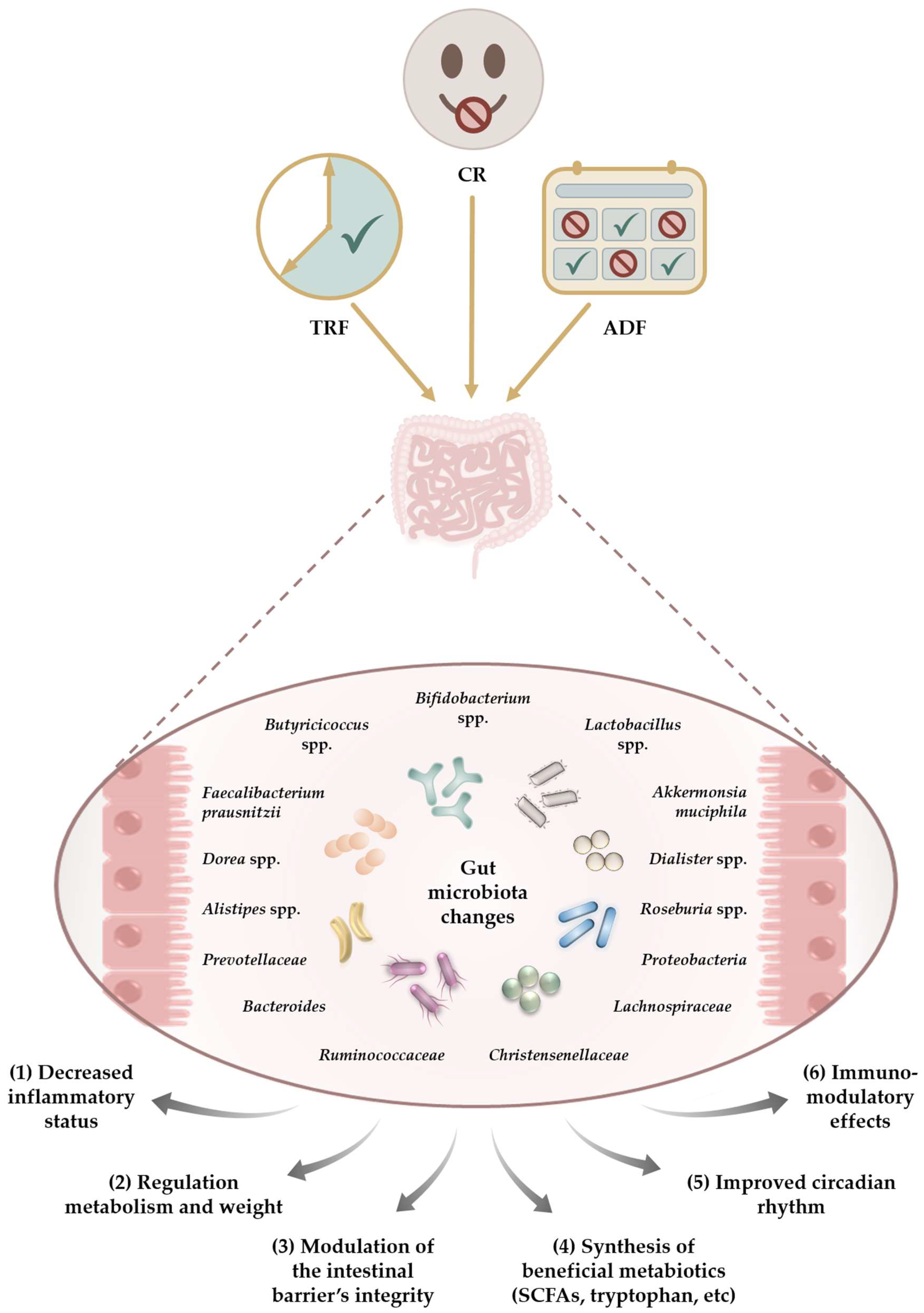
5. Effect of Dietary Interventions on the Human Gut Microbiota

The impact of different restriction regimens on the gut microbiota was investigated in healthy volunteers as well as in overweight, hypertensive, or metabolic syndrome patients. A total of 11 studies were identified in healthy volunteers, of which four evaluated the effects of CR regimens and seven investigated the influence of TRF, mainly Ramadan IF. In patients, 10 studies were identified, of which 6 evaluated the effects of CR and 4 investigated the influence of TRF regimens. The main findings are summarised in Table 3.

5.1. Studies Conducted on Healthy Volunteers

5.1.1. CR Regimens

In a randomised, controlled, single-blinded trial, Lilja et al. compared the gut microbiome before and after a five-day Buchinger fasting programme in healthy individuals with three months of supplementation containing sirtuin (SIRT)-activating nutraceuticals (SIRTFOOD intervention), SIRTs being at the forefront of energy metabolism, autophagy, and senescence [107,108]. Gut composition and metabolites were investigated using Illumnia sequencing and mass spectrometry of stool samples. In the Buchinger fasting group, Proteobacteria distribution increased while the Firmicutes/Bacteroidetes ratio decreased and was correlated with the body mass index (BMI) change. No changes for Actinobacteria were seen after the fasting intervention, but after the SIRTFOOD intervention, a different pattern was observed as Actinobacteria increased [88].
Later, the same authors observed that the Buchinger fasting programme also affects SIRT expression in healthy humans. Following fasting, the Firmicutes/Bacteroidetes ratio decreased. The participants with a higher abundance of Prevotella or Lactobacillus had higher levels of SIRT1 expression at baseline, but the correlation was not observed after the intervention. The abundance of Christensenellaceae species, which are butyrate producers, increased after fasting and was positively correlated with SIRT3 expression [89]. SIRT1 and SIRT3 are longevity-related proteins linked with mitochondrial biogenesis and mitophagy for damaged mitochondria [109,110]; therefore, fasting may have beneficial outcomes for human health and influence some longevity-associated mechanisms.
Mesnage et al. investigated the effects of a ten-day Buchinger fast on the gut microbiota of fifteen healthy volunteers and the subsequent 3-month refeeding effects. The stool was assessed with 16S rRNA gene amplicon sequencing. There were no differences in alpha diversity. Fasting caused an initial decrease in the abundance of Firmicutes, such as Lachnospiraceae and Ruminococcaceae, bacteria known to degrade dietary polysaccharides. There was a concomitant increase in Bacteroides and Proteobacteria abundance (Escherichia coli and Bilophila wadsworthia). The inversion of the Firmicutes/Bacteroidetes ratio was due to a significant decrease in the relative abundance of Firmicutes. The gut microbiota changes were reversed after three months. The levels of faecal branched-chain amino acids (BCAA) significantly increased during fasting, returned to baseline after refeeding, and declined significantly after three months [91]. The association between Bacteroidetes abundance and faecal BCAA levels suggests that using host-derived compounds, such as desquamated cells, could sustain energy requirements during fasting.
He et al. investigated the effects of two fasting models, water-only and juice-fast, for seven days in 16 healthy individuals. The stools were assessed using 16S rRNA gene sequencing. The water-only fasting dramatically changed the bacterial community, and participants developed a more homogenous gut microbiome and reduced the relative abundance of Fusobacterium. Furthermore, post-fasting Fusobacterium remained consistently low in all water-only fasting participants. Although the authors anticipated an increase in the relative abundance of Akkermansia in the water-only fast group, this was not observed since Akkermansia uses mucin as its sole substrate. The lack of this effect suggests that other bacteria could utilise mucin, which may compete with Akkermansia [90].

5.1.2. TRF Regimens

In a cohort trial, Su et al. evaluated the effect of the 30-day Ramadan fasting on the gut microbiota in healthy young and middle-aged individuals. Fasting led to substantial remodelling of the gut microbiota in both cohorts, supporting increased alpha diversity only in the younger group. Increased microbiome diversity was specifically associated with a reversible upregulation of the Clostridiales order-derived Lachnospiraceae and Ruminococcaceae families, while the abundance of the Prevotellaceae family decreased. As Lachnospiraceae species are linked to intestinal butyrate production, their increase positively affects blood glucose, body weight, and body fat mass. Microbiome composition returned to baseline during the follow-up phase [7].
Ozkul et al. investigated the microbiota alterations in nine volunteers undergoing Ramadan IF using 16S sequencing. Microbial richness increased, and elevated levels of SCFA producers, including Faecalibacterium prausnitzii, Roseburia, Eubacterium, and Akkermansia, were observed after the fasting. The most affected was Butyricicoccus pullicaecorum, one of the main butyrate-producing bacterial species. Although the Firmicutes/Bacteroidetes ratio remained increased, a higher abundance of Bacteroidetes was noticed compared to baseline [92]. These findings are in line with another similar Ramadan-based study published by the same researchers that reported an increased abundance of A. muciniphila and the Bacteroides fragilis group [23]. Furthermore, fasting significantly reduced fasting serum glucose and total cholesterol levels in all volunteers.
Ali et al. investigated microbiota changes throughout Ramadan IF in two cohorts of Chinese and Pakistani individuals using 16S sequencing of stool samples [93]. No effect on alpha diversity was observed, and other fast-related changes differ substantially between the subcohorts. Specifically, Prevotella and Faecalibacterium drove the predominance of Bacteroidetes and Firmicutes in the Pakistani group, while Bacteroides were the most prevalent among Chinese individuals. Dorea, Klebsiella, and Faecalibacterium were abundant in the Chinese group after fasting, while Sutterella, Parabacteroides, and Alistipes were significantly enriched after fasting in the Pakistani group. The prevalence of Sutterella suggests a potential metabolic benefit based on the positive impacts on glucose levels, while Parabacteroides are considered a potential factor associated with the inhibition of weight gain. Also, the genera Coprococcus, Clostridium_XlV, and Lachnospiracea significantly decreased after fasting. Within the Chinese individuals, Faecalibacterium increased after fasting, which is comparable with the findings of Ozkul et al. [92].
Although Mindikoglu et al. found no differences in bacterial richness and alpha diversity throughout IF Ramadan in fourteen healthy subjects, serum proteomic profiling showed that fasting upregulated the gene-protein products associated with glucose and lipid metabolism and insulin signaling. The upregulation of PKM M1/2 (pyruvate kinase M1/2, an enzyme involved in glycolysis) and PLIN4 (perilipin 4, a protein linked with cell lipid storage) expression indicates that IF may be highly effective in managing metabolic syndrome. Furthermore, NR1D1 (nuclear receptor subfamily 1 group D member 1) and ASGR2 GP (asialoglycoprotein receptor) levels increased significantly after the Ramadan fasting period, which may suggest that IF can also have a protective role in hepatic steatosis [111].
In a comparative cross-sectional study, Mohammadzadeh et al. investigated the consequences of Ramadan IF in thirty healthy subjects on gut microbiota and serum butyrate concentration. The study showed that gut Bacteroides and Firmicutes increased by 21% and 13% after fasting compared to baseline. The increment in Bacteroides occurred in both sexes, but Firmicutes significantly increased only in women. Also, the serum levels of butyrate significantly increased after IF. Furthermore, the BMI decrease after Ramadan fasting was associated with an increased abundance of Bacteroides in the gut microbiota [94]. In a cohort study on two ethnic groups (Chinese and Pakistani), Chen et al. observed shifts in faecal metabolite profiles after Ramadan fasting. Some metabolite levels (L-histidine, cordycepin, and lycofawcine) were significantly higher in the Chinese cohort, while brucine increased in the Pakistani group after IF. Several bacterial taxa were significantly correlated with specific metabolites unique to each ethnic group. The authors suggested that the observed changes in faecal metabolite profiles could be influenced by associated shifts in the gut microbiota [95].
Zeb et al. divided thirty healthy men into TRF and a control group (non-TRF) and investigated the effects of TRF (16:8) over 12 weeks on the gut microbiota. TRF significantly changed microbial diversity, increasing the Bacteroidetes phylum (Prevotella_9, Faecalibacterium, and Dialister) compared to the control. In the TRF group, Bacteroidetes was the most abundant phylum, followed by Firmicutes; this ratio suggests the beneficial effect of TRF on gut flora [96]. The same researchers also found that enhanced gut microbiota richness, such as Prevotellaceae, is linked to the activation of SIRT1 [112].

5.2. Studies Conducted on Obese Patients

5.2.1. CR Regimens

In a pilot study on 13 obese patients, Remely et al. [97] investigated the effect of a 1-week Buchinger fasting programme with laxative treatment followed by a 6-week intervention with an additional probiotic mixture of Lactobacillus sp., Streptococus thermophiles, and Bifidobacterium sp. Gut microbiota (the relative abundance or composition) were analysed based on 16s rDNA with a quantitative real-time polymerase chain reaction (qPCR). No significant changes in the abundance of Prevotella, Bacteroidetes, Clostridium cluster XIVa, or Clostridium cluster IV were found. However, SCFAs producing mucus-associated Faecalibacterium prausnitzii, Bifidobacteria, and Akkermanisa increased in abundance over this study period and influenced the integrity of the intestinal epithelial barrier. The inflammation-associated gut microbes Lactobacilli and Enterobacteria increased during the first week but then declined by the end of the intervention. The addition of probiotics increased gut microbial populations. The findings on Faecalibacterium prausnitzii and Akkermansia are comparable with Ozkul et al.’s results in healthy volunteers [92].
Maifeld et al. investigated, in a randomised controlled trial, the effect of a 5-day Buchinger fasting program, followed by a Mediterranean-like diet for three months, in hypertensive patients with metabolic syndrome. Fasting modified the gut microbiota, decreasing SCFA producers such as Bifidobacterium, Coprococcus comes, and Roseburia, increasing F. prausnitzii, Bacteroides, and Anaerotruncus, and also reducing propionate production capacity as well as mucin degradation gene modules. These changes had beneficial effects on body weight and blood pressure. No significant changes in alpha diversity were observed, and most of the microbiome alterations were reversed after three months of refeeding [98].
Guo et al. observed gut microbiota changes after eight weeks of IF (5:2) compared with the control group in a randomised clinical trial in 39 adult patients with metabolic syndrome. IF significantly increased the relative abundances of SCFA producers, such as Ruminococcaceae, Roseburia, and Clostridium, at the genus and family levels. Significant alterations of the gut microbiota were found at the species level. Intermittent fasting mainly induced a shift in Firmicutes but also decreased the circulating levels of lipopolysaccharides [99].
Alemán et al. assessed the gut microbiome composition through 16S rRNA sequencing of faecal microbiota in 10 obese postmenopausal women during a very low-calorie diet (800 kcal/day) for 46 days. An overall parallel shift in community structure was observed, corresponding to a reduced abundance of Faecalibacterium prausnitzii and the Roseburia genus and increased Christensenellaceae. Furthermore, changes in microbial taxa were correlated with faecal bile acid composition variations. A strong negative correlation between Clostridiaceae and lithocholic acid and positive correlations between Eubacterium and lithocholic acid were observed. Isolithocholic correlated negatively with Clostridiaceae and Ruminococcus, while Faecalibacterium showed strong positive correlations with murocholic acid and ursodeoxycholic acid [101]. The authors also observed that during this intervention, the increased lipolysis led to higher beta-hydroxybutyrate levels, an SCFA negatively correlated with F. prausnitzii and the genus Roseburia. These findings contrast with the results of other studies, but the reduced number of participants should be taken into account.
In a randomised trial of a group of obese patients, a 6-week VLCD (very low-calorie diet) resulted in an altered faecal microbiota, particularly Bacteroides spp., which decreased approximately two logs compared to baseline, and the Lactobacillus group with a reduction of approximately 1 log, but the change was transient. VLCD was also associated with a decrease in body weight. The normalisation of microbiota observed at the 12-month follow-up suggested that microbiota modifications were associated with dietary intake rather than with body weight variations [104]. Also, the effect of a long-term CR regimen was studied. Ruiz et al. observed that one year of CR impacts gut microbial composition in obese/overweight adolescents [105]. Long-term CR induced significant shifts in total and β-galactosidase-containing species, reduced Actinobacteria and the Firmicutes/Bacteroidetes ratio, and enhanced beneficial Bacteroides, Roseburia, Faecalibacterium, and Clostridium XIVa, all playing an essential functional role in the polysaccharide trophic chain. Moreover, the post-CR gut microbial community of obese adolescents exhibited similar metabolic performances compared with the lean control, suggesting a high level of metabolic plasticity [113].

5.2.2. TRF Regimens

Khan et al. included fourteen women and thirty-one men in a TRF regimen (16:8) for 16 days. The volunteers were divided based on their weight into overweight/obese, normal, or underweight groups [100]. The human faecal bacterial diversity exhibited significant changes with increased alpha diversity. The number of Bifidobacteria and Lactobacillus increased in all groups. In the female overweight/obese group, Firmicutes, Bacteroidetes, and Actinobacteria decreased, while Proteobacteria increased. In contrast, in the normal or underweight female groups, Firmicutes, Proteobacteria, Bacteroidetes, and Actinobacteria exhibited the same pattern as in the obese group. The effect of intermittent fasting on male volunteers is unique for both groups. In the male normal group, Firmicutes and Actinobacteria decreased, while Proteobacteria and Bacteroidetes increased, while in the underweight group, Firmicutes, Bacteroidetes, and Actinobacteria increased, and Proteobacteria decreased in response to TRF. These results suggest that IFs impact could be gender-specific, but more in-depth studies are needed to confirm this hypothesis. Furthermore, the ameliorated blood lipid profile is positively correlated with increased Lactobacillus and Bifidobacterium spp., which exhibit bile salt hydrolases that influence cholesterol absorption [18].
Gabel et al. examined in a 12-week pilot study the changes in the gut microbiome in obese adults after IF [114]. Participants underwent a TRF (16:8) intervention, and the faecal microbiota was determined by 16S rRNA gene sequencing. At baseline, the two most common phyla were Bacteroidetes and Firmicutes at 26.9% and 61.2%, respectively. No significant alterations in the abundance of common phyla were observed after 12 weeks of TRF. These findings contradict other published data [103,115], which have all reported changes in gut microbiota composition and/or diversity after 6 or 12 weeks of caloric restriction. The authors concluded that the CR used in their study was insufficient to have a beneficial impact on gut microbiota composition.
Dao et al. evaluated the effect of a 6-week CR and a 6-week weight stabilisation diet on faecal Akkermansia muciniphila abundance and faecal microbial gene richness in 49 obese adults [102]. At baseline, there were two groups: one with a high A. muciniphila abundance (Akk HI group) and the other with a lower abundance (Akk LO group). The Akk HI group exhibited the healthiest metabolic status, assessed by fasting plasma glucose, plasma triglycerides, and body fat distribution. While in the Akk HI group, A. muciniphila abundance decreased after CR and the total intervention period but remained significantly higher compared with the Akk LO group (more than 100-fold).
Frost et al. investigated in a pilot study in obese type 2 diabetics the effect of a 6-week VLCD followed by a nine-week food reintroduction and stabilisation period. The tests were performed on the faecal microbiota using 16S rRNA gene sequencing. From the reported data, alpha diversity increased, along with the depletion of some pathobiont taxa, like Collinsella, which decreased 8.4-fold, Roseburia, and Lachnospiraceae spp. As the Collinsella genus has been associated with poor metabolic status and T2DM, the reduced abundance of Collinsella could merely represent a biomarker indicating an improved metabolic state. Most microbiome changes observed had reverted until the end of this study [103].
Also, Jian et al. observed that eight weeks of caloric restriction (800–1200 kcal/day) in obese patients with pre-diabetes induced significant changes in gut microbiota, including increased levels of Akkermansia and Christensenellaceae R-7 groups and decreased levels of Blautia and Bifidobacterium spp. The changes in microbiota composition were significantly associated with weight loss [106].

6. Comparability between Dietary Intervention Regimens

A few studies investigate if different dietary programmes may have a common pattern of gut microbiota changes. These are summarised in Table 4.
Stanislawski et al. investigated the gut microbiota changes in overweight subjects recruited for the DRIFT2 trial. DRIFT2 compares the weight loss achieved by IF to that by CR. Gut microbiome analysis through 16S sequencing was performed for the first three months. The overall microbiota community structure (beta diversity) shifted significantly from baseline to three months. Collinsella and Subdoligranulum decreased in relative abundance, while Parabacteroides, Alistipes, and Bacteroides increased. Akkermansia increased significantly among IF participants but showed no significant change in daily CR [116].
Gutiérrez-Repiso et al. included 61 individuals who followed three different weight loss strategies (bariatric surgery, the Mediterranean diet, and a ketogenic very-low-calorie ketogenic diet, VLCKD), and the gut microbiota profile was assessed by next-generation sequencing. While microbiome changes occurred in each type of intervention, a common taxon could not be found. At the family level, Clostiridiaceae significantly increased their abundance with the Mediterranean diet and VLCKD, while Porphyromonadacean and Rikenellaceae significantly increased their abundance with VLCKD and bariatric surgery. At the genus level, Parabacteroides and Alistipes significantly increased their abundance, while Lactobacillus decreased with a caloric restriction diet and bariatric surgery. Predicted metagenome analysis suggested that most changes after the CR diet focused on biosynthesis and degradation/utilisation pathways, while enrichment characterised the Mediterranean diet in several pathways related of the fermentation to SCFAs. Bariatric surgery seems to decrease most of the biosynthesis pathways as a sign of extreme caloric restriction [118]. The same authors revealed that VLCKD changes in the microbiota are more significant if probiotics (such as Bifidobacterium lactis) and prebiotic fibers are supplemented during the diet [121].
Recently, in a real-life study, Ferrocino et al. compared the effects of a 12-week TRF regimen (<12 h feeding) with a CR (500–1000 kcal/day) in 49 obese patients. No differences in alpha and beta diversity or gut microbiota composition were observed. Lachnospiraceae, Parasutterella, and Romboutsia frequencies significantly increased in the TRE group [117]. The upregulation of Lachnospiraceae has already been mentioned after Ramadan fasting [7] and interpreted as a possible explanation for the health benefits of TRE, considering their butyric-acid-producing capacity.
Siebert et al. investigated the effect of continuous caloric restriction, or IF, on overweight or obese patients who participated in a 1-year behavioural weight loss intervention. Although the authors did not report between-group differences, they identified four gut microbes as predictors of change in weight: improved weight loss was correlated with Ruminococcaeae NK4A214 and Coprococcus 3 baseline abundance. At the same time, Bacteroides and Lachnospiraceae levels were considered disadvantageous for weight loss. Faecalibacterium and Blautia baseline abundances were associated with a decrease in triglicerides [119].
These findings regarding Blautia and Faecalibacterium dynamics and their beneficial effects can be seen in conjunction with another study on patients with other pathologies. Cignarella et al. initiated a 15-day randomised controlled pilot trial for multiple sclerosis subjects experiencing relapse; seventeen subjects were equally randomised to alternate-day fasting vs. an ad libitum diet [122]. In the stool samples, no bacteria were significantly different at day 15 between the two groups, but the abundance of Lachnospiraceae, Blautia, and Faecalibacterium showed an increasing trend in the IF group. As Faecalibacterium and Blautia, which belong to the Clostridia clusters XIV and XIVa, play an important role in producing butyrate in the gut, the increase of these genera might counterbalance the gut dysbiosis usually detected in multiple sclerosis [123].
Sowah et al. analysed the effects of intermittent CR (5:2) versus continuous CR on the faecal microbiota of 147 obese adults in a 50-week parallel-arm randomised controlled study. Except for Lactobacillales being enriched after intermittent CR, post-intervention microbiome composition did not significantly differ between groups. Despite the lack of effects on microbiota composition, the linear mixed effect analysis, which leveraged data across all time points and participants, revealed associations between HOMA-IR and Akkermansiaceae, Christensenellaceae, and Tanerellaceae, underscoring the potential importance of these bacteria for host metabolic status [120].

7. The Impact of CR Programs on the Gut Microbiota—Common Findings

The main findings of studies involving human subjects are illustrated in Table 5.
Comparing the above data, the most frequent change reported is an enrichment of Faecalibacterium prausnitzii, a member of Firmicutes, a well-known SCFA producer. The enrichment was observed in five studies [7,92,93,96,105,118], but also decreased in the other two trials [91,101]. F. prausnitzii exhibits beneficial effects by producing anti-inflammatory molecules such as shikimic and salicylic acids [108] and modulating the intestinal mucus barrier by modifying goblet cells and mucin glycosylation [124]. It is also positively correlated with obesity as it provides additional energy for gut epithelial cells and has a role in adipose tissue expansion [125]. Also, a low-fat fat/high-carbohydrate diet increased the abundance of F. prausnitzii in obese patients [126], but not in patients with metabolic syndrome [127], which suggests that the degree of dysbiosis associated with the disease may be a determinant factor in response to a specific diet-based treatment. Although changes in F. prausnitzii abundance were not consistent among the studies, there is sufficient evidence to use F. prausnitzii as an indicator or biomarker of human intestinal health.
Furthermore, other SCFA producers, such as Roseburia, decreased in four CR programs [91,101,103,118], and increased during refeeding in the other three studies [92,99,105]. Also, Butyricicoccus pullicaecorum abundance displayed a similar variable pattern, increasing during fasting [92,99]. As it is well known that Roseburia, F. prausnitzii, and B. pullicaecorum influence the integrity of the intestinal epithelial barrier and support immunity, these beneficial changes should be taken into account in further research.
The relative abundance of Akkermansia muciniphila, another SCFA producer and mucin-degrading bacterium, increased during different restriction programmes in five clinical trials [23,92,97,106,116]. As the level of A. muciniphila is inversely correlated with body weight and directly correlated with the weight loss observed in obese patients after CR restrictions and improvements in multiple indicators of cardio-metabolic health [128,129,130,131], it can be proposed as a prognostic tool in the context of predicting CR success. Further studies are needed to confirm this, as the effect was not consistently observed in all trials. The difference between restriction programs, especially the type and fibre content of the diet, can partially explain controversial results. The abundance of Akkermansia muciniphila increases in the absence of fibre polysaccharides [132], and this may explain Ramadan IF results since the Ramadan diet includes low fibre intake [23]. Recently, it was observed that, in addition to its metabolic and anti-inflammatory effects, Akkermansia muciniphila influences cognitive functions in patients with type 2 diabetes; therefore, increasing its abundance via fasting or the use of metformin [133] may be protective against cognitive decline in elderly type 2 diabetics.
Gut alpha diversity increased only in some studies after Ramadan IF or VLCD, but fasting in general did not influence it. As alpha diversity is considered a marker of gut health and homeostasis, the results indicate that dietary restrictions are not expected to have a negative impact on gut microbiota diversity.
Although an increase in Alisipes abundance was observed in a significant number of studies, considering that the role of Alisipes is not fully elucidated, these results have limited value. Alisipes exhibit protective effects against colitis or cardiovascular disease but are also considered to have a pathogenic role in colorectal cancer and are positively correlated with signs of anxiety or depression [134].
Bacteroides increased after TRF, including Ramadan IF or long-term CR, but decreased during refeeding. Furthermore, the Bacteroides abundance is linked to the Firmicutes/Bacteroides ratio, a marker of normal intestinal homeostasis. This ratio decreased in three trials after the Buchinger fasting programmes or long-term CR but did not change after VLCD. Interestingly, in one study, it remained increased after Ramadan IF [92].
Several studies reported a positive impact on butyrate-producing bacteria (such as Ruminococcaceae or Lachnospiraceae), while others showed no significant change in the relative abundances of such species. Therefore, a clear trend cannot be set, and further data are needed.
Also, Bifidobacterium and Lactobacillus species increased in obese patients after the CR regimen [97,120] but also decreased [106], with inconsistent results.
Prevotella abundance is also positively influenced by Ramadan IF, and considering it is linked to the activation of SIRT1, together with Diallister and Bacteroidia, it may influence and control circadian rhythmicity that regulates intestinal physiology [112,135].
The enrichment of the Parabacteriodes population observed in some studies should be mentioned because it is also linked with its capacity to conjugate bile acids and influence their biological functions, including controlling metabolic dysfunctions [136].
Although no clear trend or common pattern of gut microbiota changes can be established as a result of all different dietary approaches, some health-beneficial effects as a result of microbiota changes are apparent, including a reduction of risk factors for a range of age-related diseases and increased lifespan. Dietary restriction influences the gut microbiota by decreasing proinflammatory cytokines such as IFN-gamma and TNF-α [137] or enhancing SCFA production [108]. It also increases intestinal barrier integrity by stimulating mucin production and exhibits immunomodulatory effects by influencing the abundance of Roseburia and F. prausnitzii [138]. Furthermore, dietary restriction improves circadian rhythm via enhanced SCFA production or activation of SIRT1 [112,135]. It protects against metabolic illness via Lactobacillus and Oscillibacter spp., which regulate body weight and glucose and lipid metabolism by influencing the release of GLP-1 and PYY [139].
A beneficial and noteworthy effect induced by dietary changes—the counteracting of aging—was highlighted in some studies. The pro-longevity and regenerative effects of IF or CR could be partially connected to gut microbiota changes. In humans, ageing is linked with a decrease in Bifidobacteria, Ruminococcaceae, Lachnospiraceae, and Bacteridaceae [140,141]. Simultaneously, extremely long-lived individuals exhibit an increase in several beneficial taxa, particularly Akkermansia, Bifidobacterium, and Christensenellaceae, which are now considered a “signature of longevity” [142,143]. Therefore, the increased abundance of Akkermansia muciniphila observed after Ramadan IF or CR or the positive impact on Ruminococcaceae or Lachnospiraceae observed in some studies supports this hypothesis. In addition, the pro-longevity effects induced by IF or CR are also linked with other mechanisms, such as reducing circulating insulin-like growth factor-1 (IGF-1) levels, protein-kinase A activity, or mTOR pathways [144,145,146].
The heterogeneity of the human results is correlated with the fact that the studies were conducted either in healthy volunteers or in overweight patients. The microbiota of an overweight patient is significantly different from that of a normal-weight individual and characterised by a lower alpha diversity and increased abundance of Actinobacteria, Bacteroidetes, and Firmicutes phylum species [147,148,149]. This different status explains, for example, why, only in obese patients, Actinobacteria decreased in response to IF.
The impact of dietary caloric restrictions in the context of everyday life is still under debate, as many dietary restrictions are indicated by physicians in patients with different pathologies (obesity, hypertension, T2DM, cancer), which are also under specific medication, and gut microbiota can, directly and indirectly, affect drug metabolism [150]. Furthermore, drugs may directly alter the abundance of specific bacterial species [151]. This bidirectional interaction should be further investigated, especially regarding the commonly used drugs and their impact on their efficacy.

8. Conclusions

Considering that in humans, fasting and/or caloric restriction are often exercised on a daily basis for different reasons, and there is a significant interest worldwide in its influence on general health, we reviewed the recent literature data concerning the ability of different dietary interventions to influence the taxonomic composition of the gut microbiota. Although the research in the field is not abundant, most studies suggest beneficial effects, but the available data are insufficient to establish a typical pattern of gut microbiota changes induced by different dietary approaches. Furthermore, it is impossible to conclude if a certain type of restriction showed more consistent beneficial results on the gut microbiota than the others. This was expected due to the heterogeneity and low power of the studies, microbiota baseline variability, and varying individual responses. Furthermore, many published studies do not consider common confounders, such as smoking or physical exercise. Only a few studies used a control group or investigated if the beneficial effects may be reversible and require continued restricted eating behaviour. In addition, these beneficial effects should be carefully weighed against the risks induced by fasting, especially by Ramadan IF, which perturbs the human circadian rhythms and may negatively influence human health in the long run. Further research in this area is necessary, particularly on obese and metabolically compromised patients, as they show a low fluctuation of circadian rhythm. Also, the risk of developing an eating disorder due to IF should not be neglected, as recent reports suggest a substantial correlation [152]. IF was shown to decrease testosterone and the free androgen index among males [153], which, in theory, could negatively affect metabolic health and libido.
Although further data are needed, especially from well-designed randomised controlled studies, it is obvious that different dietary fasting or caloric restriction regimens significantly impact the gut microbiota, both qualitatively and quantitatively. This should be seen in conjunction with other beneficial effects regarding health span parameters, mainly reported during CR and less during IF regimens.
Since more and more CR programmes are developed, further studies are needed to identify and differentiate between the long-lasting microbiota changes that could be used for a targeted and tailored dietary intervention. Also, future research should assess the different molecules produced by the gut microbiota as the result of specific types of CR programs or may investigate the combined effects of physical exercise and caloric restriction or of fasting and caloric restriction.
We posit that dietary restriction programmes could become important non-pharmacological interventions in the treatment of various diseases.

Author Contributions

Conceptualisation, writing—original draft preparation, C.P. and D.M.; methodology, C.P. and I.A.-D.; formal analysis, investigation, writing—review and editing, A.U., C.P. and I.A.-D.; visualisation, A.U.; supervision, D.M. All authors have read and agreed to the published version of the manuscript.

Funding

This research received no external funding.

Institutional Review Board Statement

Not applicable.

Informed Consent Statement

Not applicable.

Data Availability Statement

Data are contained within the article.

Conflicts of Interest

The authors declare no conflict of interest.

References

  1. Yassour, M.; Vatanen, T.; Siljander, H.; Hamalainen, A.M.; Harkonen, T.; Ryhanen, S.J.; Franzosa, E.A.; Vlamakis, H.; Huttenhower, C.; Gevers, D.; et al. Natural history of the infant gut microbiome and impact of antibiotic treatment on bacterial strain diversity and stability. Sci. Transl. Med. 2016, 8, 343ra381. [Google Scholar] [CrossRef] [PubMed]
  2. Spor, A.; Koren, O.; Ley, R. Unravelling the effects of the environment and host genotype on the gut microbiome. Nat. Rev. Microbiol. 2011, 9, 279–290. [Google Scholar] [CrossRef] [PubMed]
  3. Hill, C.J.; Lynch, D.B.; Murphy, K.; Ulaszewska, M.; Jeffery, I.B.; O’Shea, C.A.; Watkins, C.; Dempsey, E.; Mattivi, F.; Tuohy, K.; et al. Evolution of gut microbiota composition from birth to 24 weeks in the INFANTMET Cohort. Microbiome 2017, 5, 4. [Google Scholar] [CrossRef] [PubMed]
  4. Arboleya, S.; Sanchez, B.; Milani, C.; Duranti, S.; Solis, G.; Fernandez, N.; de los Reyes-Gavilan, C.G.; Ventura, M.; Margolles, A.; Gueimonde, M. Intestinal microbiota development in preterm neonates and effect of perinatal antibiotics. J. Pediatr. 2015, 166, 538–544. [Google Scholar] [CrossRef] [PubMed]
  5. Ottman, N.; Smidt, H.; de Vos, W.M.; Belzer, C. The function of our microbiota: Who is out there and what do they do? Front. Cell. Infect. Microbiol. 2012, 2, 104. [Google Scholar] [CrossRef] [PubMed]
  6. Milani, C.; Duranti, S.; Bottacini, F.; Casey, E.; Turroni, F.; Mahony, J.; Belzer, C.; Delgado Palacio, S.; Arboleya Montes, S.; Mancabelli, L.; et al. The First Microbial Colonizers of the Human Gut: Composition, Activities, and Health Implications of the Infant Gut Microbiota. Microbiol. Mol. Biol. Rev. 2017, 81, 10–1128. [Google Scholar] [CrossRef] [PubMed]
  7. Su, J.; Wang, Y.; Zhang, X.; Ma, M.; Xie, Z.; Pan, Q.; Ma, Z.; Peppelenbosch, M.P. Remodeling of the gut microbiome during Ramadan-associated intermittent fasting. Am. J. Clin. Nutr. 2021, 113, 1332–1342. [Google Scholar] [CrossRef]
  8. Zoetendal, E.G.; Smith, A.H.; Sundset, M.A.; Mackie, R.I. The BaeSR two-component regulatory system mediates resistance to condensed tannins in Escherichia coli. Appl. Environ. Microbiol. 2008, 74, 535–539. [Google Scholar] [CrossRef]
  9. Zoetendal, E.G.; de Vos, W.M. Effect of diet on the intestinal microbiota and its activity. Curr. Opin. Gastroenterol. 2014, 30, 189–195. [Google Scholar] [CrossRef]
  10. Zoetendal, E.G.; Smidt, H. Endothelial dysfunction: What is the role of the microbiota? Gut 2018, 67, 201–202. [Google Scholar] [CrossRef]
  11. Zoetendal, E.G.; Rajilic-Stojanovic, M.; de Vos, W.M. High-throughput diversity and functionality analysis of the gastrointestinal tract microbiota. Gut 2008, 57, 1605–1615. [Google Scholar] [CrossRef]
  12. Qin, J.; Li, R.; Raes, J.; Arumugam, M.; Burgdorf, K.S.; Manichanh, C.; Nielsen, T.; Pons, N.; Levenez, F.; Yamada, T.; et al. A human gut microbial gene catalogue established by metagenomic sequencing. Nature 2010, 464, 59–65. [Google Scholar] [CrossRef]
  13. Rajilic-Stojanovic, M.; de Vos, W.M. The first 1000 cultured species of the human gastrointestinal microbiota. FEMS Microbiol. Rev. 2014, 38, 996–1047. [Google Scholar] [CrossRef]
  14. Li, J.; Jia, H.; Cai, X.; Zhong, H.; Feng, Q.; Sunagawa, S.; Arumugam, M.; Kultima, J.R.; Prifti, E.; Nielsen, T.; et al. An integrated catalog of reference genes in the human gut microbiome. Nat. Biotechnol. 2014, 32, 834–841. [Google Scholar] [CrossRef] [PubMed]
  15. de Vos, W.M.; Tilg, H.; Van Hul, M.; Cani, P.D. Gut microbiome and health: Mechanistic insights. Gut 2022, 71, 1020–1032. [Google Scholar] [CrossRef] [PubMed]
  16. Heiman, M.L.; Greenway, F.L. A healthy gastrointestinal microbiome is dependent on dietary diversity. Mol. Metab. 2016, 5, 317–320. [Google Scholar] [CrossRef] [PubMed]
  17. Wu, G.D.; Chen, J.; Hoffmann, C.; Bittinger, K.; Chen, Y.Y.; Keilbaugh, S.A.; Bewtra, M.; Knights, D.; Walters, W.A.; Knight, R.; et al. Linking long-term dietary patterns with gut microbial enterotypes. Science 2011, 334, 105–108. [Google Scholar] [CrossRef]
  18. Purdel, C.; Ungurianu, A.; Adam-Dima, I.; Margina, D. Exploring the potential impact of probiotic use on drug metabolism and efficacy. Biomed. Pharmacother. 2023, 161, 114468. [Google Scholar] [CrossRef] [PubMed]
  19. Ejtahed, H.S.; Angoorani, P.; Hasani-Ranjbar, S.; Siadat, S.D.; Ghasemi, N.; Larijani, B.; Soroush, A.R. Adaptation of human gut microbiota to bariatric surgeries in morbidly obese patients: A systematic review. Microb. Pathog. 2018, 116, 13–21. [Google Scholar] [CrossRef]
  20. Ejtahed, H.S.; Soroush, A.R.; Angoorani, P.; Larijani, B.; Hasani-Ranjbar, S. Gut Microbiota as a Target in the Pathogenesis of Metabolic Disorders: A New Approach to Novel Therapeutic Agents. Horm. Metab. Res. 2016, 48, 349–358. [Google Scholar] [CrossRef]
  21. Ejtahed, H.S.; Hasani-Ranjbar, S.; Larijani, B. Human Microbiome as an Approach to Personalized Medicine. Altern. Ther. Health Med. 2017, 23, 8–9. [Google Scholar]
  22. Ejtahed, H.S.; Angoorani, P.; Soroush, A.R.; Siadat, S.D.; Shirzad, N.; Hasani-Ranjbar, S.; Larijani, B. Our Little Friends with Big Roles: Alterations of the Gut Microbiota in Thyroid Disorders. Endocr. Metab. Immune Disord. Drug Targets 2020, 20, 344–350. [Google Scholar] [CrossRef]
  23. Ozkul, C.; Yalinay, M.; Karakan, T. Islamic fasting leads to an increased abundance of Akkermansia muciniphila and Bacteroides fragilis group: A preliminary study on intermittent fasting. Turk. J. Gastroenterol. 2019, 30, 1030–1035. [Google Scholar] [CrossRef] [PubMed]
  24. Angoorani, P.; Ejtahed, H.S.; Hasani-Ranjbar, S.; Siadat, S.D.; Soroush, A.R.; Larijani, B. Gut microbiota modulation as a possible mediating mechanism for fasting-induced alleviation of metabolic complications: A systematic review. Nutr. Metab. 2021, 18, 105. [Google Scholar] [CrossRef]
  25. Ruiz, L.; Margolles, A.; Sanchez, B. Bile resistance mechanisms in Lactobacillus and Bifidobacterium. Front. Microbiol. 2013, 4, 396. [Google Scholar] [CrossRef]
  26. Li, J.; Zhao, F.; Wang, Y.; Chen, J.; Tao, J.; Tian, G.; Wu, S.; Liu, W.; Cui, Q.; Geng, B.; et al. Gut microbiota dysbiosis contributes to the development of hypertension. Microbiome 2017, 5, 14. [Google Scholar] [CrossRef] [PubMed]
  27. Chen, Y.; Zhou, J.; Wang, L. Role and Mechanism of Gut Microbiota in Human Disease. Front. Cell. Infect. Microbiol. 2021, 11, 625913. [Google Scholar] [CrossRef] [PubMed]
  28. Cani, P.D.; Jordan, B.F. Gut microbiota-mediated inflammation in obesity: A link with gastrointestinal cancer. Nat. Rev. Gastroenterol. Hepatol. 2018, 15, 671–682. [Google Scholar] [CrossRef] [PubMed]
  29. Louis, P.; Flint, H.J. Formation of propionate and butyrate by the human colonic microbiota. Environ. Microbiol. 2017, 19, 29–41. [Google Scholar] [CrossRef] [PubMed]
  30. Chambers, E.S.; Preston, T.; Frost, G.; Morrison, D.J. Role of Gut Microbiota-Generated Short-Chain Fatty Acids in Metabolic and Cardiovascular Health. Curr. Nutr. Rep. 2018, 7, 198–206. [Google Scholar] [CrossRef]
  31. Derrien, M.; Vaughan, E.E.; Plugge, C.M.; de Vos, W.M. Akkermansia muciniphila gen. nov., sp. nov., a human intestinal mucin-degrading bacterium. Int. J. Syst. Evol. Microbiol. 2004, 54, 1469–1476. [Google Scholar] [CrossRef]
  32. Louis, P.; Young, P.; Holtrop, G.; Flint, H.J. Diversity of human colonic butyrate-producing bacteria revealed by analysis of the butyryl-CoA:acetate CoA-transferase gene. Environ. Microbiol. 2010, 12, 304–314. [Google Scholar] [CrossRef]
  33. Morrison, D.J.; Preston, T. Formation of short chain fatty acids by the gut microbiota and their impact on human metabolism. Gut Microbes 2016, 7, 189–200. [Google Scholar] [CrossRef]
  34. den Besten, G.; van Eunen, K.; Groen, A.K.; Venema, K.; Reijngoud, D.J.; Bakker, B.M. The role of short-chain fatty acids in the interplay between diet, gut microbiota, and host energy metabolism. J. Lipid Res. 2013, 54, 2325–2340. [Google Scholar] [CrossRef]
  35. Wang, X.; Gibson, G.R. Effects of the in vitro fermentation of oligofructose and inulin by bacteria growing in the human large intestine. J. Appl. Bacteriol. 1993, 75, 373–380. [Google Scholar] [CrossRef]
  36. Wang, T.; Rijnaarts, I.; Hermes, G.D.A.; de Roos, N.M.; Witteman, B.J.M.; de Wit, N.J.W.; Govers, C.; Smidt, H.; Zoetendal, E.G. Fecal Microbiota Signatures Are Not Consistently Related to Symptom Severity in Irritable Bowel Syndrome. Dig. Dis. Sci. 2022, 67, 5137–5148. [Google Scholar] [CrossRef]
  37. Wang, T.; van Dijk, L.; Rijnaarts, I.; Hermes, G.D.A.; de Roos, N.M.; Witteman, B.J.M.; de Wit, N.J.W.; Govers, C.; Smidt, H.; Zoetendal, E.G. Methanogen Levels Are Significantly Associated with Fecal Microbiota Composition and Alpha Diversity in Healthy Adults and Irritable Bowel Syndrome Patients. Microbiol. Spectr. 2022, 10, e0165322. [Google Scholar] [CrossRef] [PubMed]
  38. Aziz, A.A.; Kenney, L.S.; Goulet, B.; Abdel-Aalel, S. Dietary starch type affects body weight and glycemic control in freely fed but not energy-restricted obese rats. J. Nutr. 2009, 139, 1881–1889. [Google Scholar] [CrossRef] [PubMed]
  39. Keenan, M.J.; Zhou, J.; McCutcheon, K.L.; Raggio, A.M.; Bateman, H.G.; Todd, E.; Jones, C.K.; Tulley, R.T.; Melton, S.; Martin, R.J.; et al. Effects of resistant starch, a non-digestible fermentable fiber, on reducing body fat. Obesity 2006, 14, 1523–1534. [Google Scholar] [CrossRef] [PubMed]
  40. Zhou, J.; Martin, R.J.; Tulley, R.T.; Raggio, A.M.; McCutcheon, K.L.; Shen, L.; Danna, S.C.; Tripathy, S.; Hegsted, M.; Keenan, M.J. Dietary resistant starch upregulates total GLP-1 and PYY in a sustained day-long manner through fermentation in rodents. Am. J. Physiol. Endocrinol. Metab. 2008, 295, E1160–E1166. [Google Scholar] [CrossRef] [PubMed]
  41. Brown, A.J.; Goldsworthy, S.M.; Barnes, A.A.; Eilert, M.M.; Tcheang, L.; Daniels, D.; Muir, A.I.; Wigglesworth, M.J.; Kinghorn, I.; Fraser, N.J.; et al. The Orphan G protein-coupled receptors GPR41 and GPR43 are activated by propionate and other short chain carboxylic acids. J. Biol. Chem. 2003, 278, 11312–11319. [Google Scholar] [CrossRef]
  42. Schoeler, M.; Caesar, R. Dietary lipids, gut microbiota and lipid metabolism. Rev. Endocr. Metab. Disord. 2019, 20, 461–472. [Google Scholar] [CrossRef]
  43. Flach, J.; van der Waal, M.B.; Kardinaal, A.F.M.; Schloesser, J.; Ruijschop, R.M.A.J.; Claassen, E. Probiotic research priorities for the healthy adult population: A review on the health benefits of Lactobacillus rhamnosus GG and Bifidobacterium animalis subspecies lactis BB-12. Cogent Food Agric. 2018, 4, 1452839. [Google Scholar] [CrossRef]
  44. Caesar, R.; Tremaroli, V.; Kovatcheva-Datchary, P.; Cani, P.D.; Backhed, F. Crosstalk between Gut Microbiota and Dietary Lipids Aggravates WAT Inflammation through TLR Signaling. Cell Metab. 2015, 22, 658–668. [Google Scholar] [CrossRef] [PubMed]
  45. Fontana, L.; Partridge, L. Promoting health and longevity through diet: From model organisms to humans. Cell 2015, 161, 106–118. [Google Scholar] [CrossRef] [PubMed]
  46. Pifferi, F.; Terrien, J.; Perret, M.; Epelbaum, J.; Blanc, S.; Picq, J.L.; Dhenain, M.; Aujard, F. Promoting healthspan and lifespan with caloric restriction in primates. Commun. Biol. 2019, 2, 107. [Google Scholar] [CrossRef] [PubMed]
  47. Mohr, A.E.; Gumpricht, E.; Sears, D.D.; Sweazea, K.L. Recent advances and health implications of dietary fasting regimens on the gut microbiome. Am. J. Physiol. Gastrointest. Liver Physiol. 2021, 320, G847–G863. [Google Scholar] [CrossRef]
  48. Attinà, A.; Leggeri, C.; Paroni, R.; Pivari, F.; Dei Cas, M.; Mingione, A.; Dri, M.; Marchetti, M.; Di Renzo, L. Fasting: How to Guide. Nutrients 2021, 13, 1570. [Google Scholar] [CrossRef] [PubMed]
  49. Forslund, S.K. Fasting intervention and its clinical effects on the human host and microbiome. J. Intern. Med. 2023, 293, 166–183. [Google Scholar] [CrossRef] [PubMed]
  50. Schmidt, N.S.; Lorentz, A. Dietary restrictions modulate the gut microbiota: Implications for health and disease. Nutr. Res. 2021, 89, 10–22. [Google Scholar] [CrossRef]
  51. Patterson, R.E.; Sears, D.D. Metabolic Effects of Intermittent Fasting. Annu. Rev. Nutr. 2017, 37, 371–393. [Google Scholar] [CrossRef] [PubMed]
  52. Stockman, M.C.; Thomas, D.; Burke, J.; Apovian, C.M. Intermittent Fasting: Is the Wait Worth the Weight? Curr. Obes. Rep. 2018, 7, 172–185. [Google Scholar] [CrossRef] [PubMed]
  53. Longo, V.D.; Mattson, M.P. Fasting: Molecular mechanisms and clinical applications. Cell Metab. 2014, 19, 181–192. [Google Scholar] [CrossRef] [PubMed]
  54. de Cabo, R.; Mattson, M.P. Effects of Intermittent Fasting on Health, Aging, and Disease. N. Engl. J. Med. 2019, 381, 2541–2551. [Google Scholar] [CrossRef]
  55. Mattson, M.P.; Longo, V.D.; Harvie, M. Impact of intermittent fasting on health and disease processes. Ageing Res. Rev. 2017, 39, 46–58. [Google Scholar] [CrossRef] [PubMed]
  56. Li, M.; Wang, S.; Li, Y.; Zhao, M.; Kuang, J.; Liang, D.; Wang, J.; Wei, M.; Rajani, C.; Ma, X.; et al. Gut microbiota-bile acid crosstalk contributes to the rebound weight gain after calorie restriction in mice. Nat. Commun. 2022, 13, 2060. [Google Scholar] [CrossRef]
  57. Zhang, Y.; Qi, H.; Wang, L.; Hu, C.; Gao, A.; Wu, Q.; Wang, Q.; Lin, H.; Chen, B.; Wang, X.; et al. Fasting and refeeding triggers specific changes in bile acid profiles and gut microbiota. J. Diabetes 2023, 15, 165–180. [Google Scholar] [CrossRef]
  58. Hernandez, A.R.; Kemp, K.M.; Burke, S.N.; Buford, T.W.; Carter, C.S. Influence of Aging, Macronutrient Composition and Time-Restricted Feeding on the Fischer344 x Brown Norway Rat Gut Microbiota. Nutrients 2022, 14, 1758. [Google Scholar] [CrossRef]
  59. Messina, M.; Iacumin, L.; Pascon, G.; Tulli, F.; Tibaldi, E.; Cardinaletti, G. Effect of feed restriction and refeeding on body condition, digestive functionality and intestinal microbiota in rainbow trout (Oncorhynchus mykiss). Fish. Physiol. Biochem. 2023, 49, 169–189. [Google Scholar] [CrossRef]
  60. Song, X.; Zhai, Y.; Song, J.; Zhang, J.; Li, X. The structural discrepancy between the small and large gut microbiota of Asiatic toad (Bufo gargarizans) during hibernation. Folia Microbiol. 2023, 68, 537–546. [Google Scholar] [CrossRef]
  61. Zhang, Z.; Chen, X.; Loh, Y.J.; Yang, X.; Zhang, C. The effect of calorie intake, fasting, and dietary composition on metabolic health and gut microbiota in mice. BMC Biol. 2021, 19, 51. [Google Scholar] [CrossRef]
  62. Ma, R.X.; Hu, J.Q.; Fu, W.; Zhong, J.; Cao, C.; Wang, C.C.; Qi, S.Q.; Zhang, X.L.; Liu, G.H.; Gao, Y.D. Intermittent fasting protects against food allergy in a murine model. Front. Immunol. 2023, 14, 1167562. [Google Scholar] [CrossRef]
  63. Xia, J.; Guo, W.; Hu, M.; Jin, X.; Zhang, S.; Liu, B.; Qiu, H.; Wang, K.; Zhuge, A.; Li, S.; et al. Resynchronized rhythmic oscillations of gut microbiota drive time-restricted feeding induced nonalcoholic steatohepatitis alleviation. Gut Microbes 2023, 15, 2221450. [Google Scholar] [CrossRef]
  64. Liang, Y.; Yin, W.; Luo, C.; Sun, L.; Feng, T.; Zhang, Y.; Yin, Y.; Zhang, W. Maternal intermittent fasting in mice disrupts the intestinal barrier leading to metabolic disorder in adult offspring. Commun. Biol. 2023, 6, 30. [Google Scholar] [CrossRef]
  65. Wu, J.; Man, D.; Shi, D.; Wu, W.; Wang, S.; Wang, K.; Li, Y.; Yang, L.; Bian, X.; Wang, Q.; et al. Intermittent Fasting Alleviates Risk Markers in a Murine Model of Ulcerative Colitis by Modulating the Gut Microbiome and Metabolome. Nutrients 2022, 14, 5311. [Google Scholar] [CrossRef]
  66. Zhang, L.; Zhang, T.; Sun, J.; Huang, Y.; Liu, T.; Ye, Z.; Hu, J.; Zhang, G.; Chen, H.; He, Y.; et al. Calorie restriction ameliorates hyperglycemia, modulates the disordered gut microbiota, and mitigates metabolic endotoxemia and inflammation in type 2 diabetic rats. J. Endocrinol. Investig. 2023, 46, 699–711. [Google Scholar] [CrossRef]
  67. Huang, W.; Yan, Y.; Wu, M.; Hu, J.; Zhao, J.; Chen, X.; Liu, W.; Liu, K.; Li, C. Preoperative fasting confers protection against intestinal ischaemia/reperfusion injury by modulating gut microbiota and their metabolites in a mouse model. Br. J. Anaesth. 2022, 128, 501–512. [Google Scholar] [CrossRef]
  68. Teker, H.T.; Ceylani, T. Intermittent fasting supports the balance of the gut microbiota composition. Int. Microbiol. 2023, 26, 51–57. [Google Scholar] [CrossRef]
  69. Yang, H.; Li, C.; Che, M.; Li, Y.; Feng, R.; Sun, C. Gut microbiota mediates the anti-obesity effect of intermittent fasting by inhibiting intestinal lipid absorption. J. Nutr. Biochem. 2023, 116, 109318. [Google Scholar] [CrossRef] [PubMed]
  70. Prisco, S.Z.; Eklund, M.; Moutsoglou, D.M.; Prisco, A.R.; Khoruts, A.; Weir, E.K.; Thenappan, T.; Prins, K.W. Intermittent Fasting Enhances Right Ventricular Function in Preclinical Pulmonary Arterial Hypertension. J. Am. Heart Assoc. 2021, 10, e022722. [Google Scholar] [CrossRef] [PubMed]
  71. Soares, N.L.; Dorand, V.A.M.; Cavalcante, H.C.; Batista, K.S.; de Souza, D.M.; Lima, M.D.S.; Salvadori, M.G.D.S.; Magnani, M.; Alves, A.F.; Aquino, J.S. Does intermittent fasting associated with aerobic training influence parameters related to the gut-brain axis of Wistar rats? J. Affect. Disord. 2021, 293, 176–185. [Google Scholar] [CrossRef]
  72. Graef, F.A.; Celiberto, L.S.; Allaire, J.M.; Kuan, M.T.Y.; Bosman, E.S.; Crowley, S.M.; Yang, H.; Chan, J.H.; Stahl, M.; Yu, H.; et al. Fasting increases microbiome-based colonization resistance and reduces host inflammatory responses during an enteric bacterial infection. PLoS Pathog. 2021, 17, e1009719. [Google Scholar] [CrossRef] [PubMed]
  73. Ménard, A.; Smet, A. Review: Other Helicobacter species. Helicobacter 2019, 24 (Suppl. S1), e12645. [Google Scholar] [CrossRef] [PubMed]
  74. Jia, W.; Xie, G. Bile acid-microbiota crosstalk in gastrointestinal inflammation and carcinogenesis. Nat. Rev. Gastroenterol. Hepatol. 2018, 15, 111–128. [Google Scholar] [CrossRef] [PubMed]
  75. Heeney, D.D.; Gareau, M.G.; Marco, M.L. Intestinal Lactobacillus in health and disease, a driver or just along for the ride? Curr. Opin. Biotechnol. 2018, 49, 140–147. [Google Scholar] [CrossRef]
  76. Xu, Y.; Wang, N.; Tan, H.Y.; Li, S.; Zhang, C.; Feng, Y. Function of Akkermansia muciniphila in Obesity: Interactions With Lipid Metabolism, Immune Response and Gut Systems. Front. Microbiol. 2020, 11, 219. [Google Scholar] [CrossRef] [PubMed]
  77. Pan, F.; Zhang, L.; Li, M.; Hu, Y.; Zeng, B.; Yuan, H.; Zhao, L.; Zhang, C. Predominant gut Lactobacillus murinus strain mediates anti-inflammaging effects in calorie-restricted mice. Microbiome 2018, 6, 54. [Google Scholar] [CrossRef]
  78. Zhang, C.; Li, S.; Yang, L.; Huang, P.; Li, W.; Wang, S.; Zhao, G.; Zhang, M.; Pang, X.; Yan, Z.; et al. Structural modulation of gut microbiota in life-long calorie-restricted mice. Nat. Commun. 2013, 4, 2163. [Google Scholar] [CrossRef]
  79. Luu, M.; Monning, H.; Visekruna, A. Exploring the Molecular Mechanisms Underlying the Protective Effects of Microbial SCFAs on Intestinal Tolerance and Food Allergy. Front. Immunol. 2020, 11, 1225. [Google Scholar] [CrossRef]
  80. Zhao, Y.; Chen, F.; Wu, W.; Sun, M.; Bilotta, A.J.; Yao, S.; Xiao, Y.; Huang, X.; Eaves-Pyles, T.D.; Golovko, G.; et al. GPR43 mediates microbiota metabolite SCFA regulation of antimicrobial peptide expression in intestinal epithelial cells via activation of mTOR and STAT3. Mucosal Immunol. 2018, 11, 752–762. [Google Scholar] [CrossRef]
  81. O’Mahony, S.M.; Clarke, G.; Borre, Y.E.; Dinan, T.G.; Cryan, J.F. Serotonin, tryptophan metabolism and the brain-gut-microbiome axis. Behav. Brain Res. 2015, 277, 32–48. [Google Scholar] [CrossRef]
  82. Osadchiy, V.; Labus, J.S.; Gupta, A.; Jacobs, J.; Ashe-McNalley, C.; Hsiao, E.Y.; Mayer, E.A. Correlation of tryptophan metabolites with connectivity of extended central reward network in healthy subjects. PLoS ONE 2018, 13, e0201772. [Google Scholar] [CrossRef] [PubMed]
  83. Hernandez, A.R.; Watson, C.; Federico, Q.P.; Fletcher, R.; Brotgandel, A.; Buford, T.W.; Carter, C.S.; Burke, S.N. Twelve Months of Time-Restricted Feeding Improves Cognition and Alters Microbiome Composition Independent of Macronutrient Composition. Nutrients 2022, 14, 3977. [Google Scholar] [CrossRef]
  84. Janssen, A.W.F.; Katiraei, S.; Bartosinska, B.; Eberhard, D.; Willems van Dijk, K.; Kersten, S. Loss of angiopoietin-like 4 (ANGPTL4) in mice with diet-induced obesity uncouples visceral obesity from glucose intolerance partly via the gut microbiota. Diabetologia 2018, 61, 1447–1458. [Google Scholar] [CrossRef]
  85. Li, S.; Qi, Y.; Ren, D.; Qu, D.; Sun, Y. The Structure Features and Improving Effects of Polysaccharide from. Antibiotics 2019, 9, 8. [Google Scholar] [CrossRef]
  86. Tooley, K.L. Effects of the Human Gut Microbiota on Cognitive Performance, Brain Structure and Function: A Narrative Review. Nutrients 2020, 12, 3009. [Google Scholar] [CrossRef]
  87. Li, G.; Xie, C.; Lu, S.; Nichols, R.G.; Tian, Y.; Li, L.; Patel, D.; Ma, Y.; Brocker, C.N.; Yan, T.; et al. Intermittent Fasting Promotes White Adipose Browning and Decreases Obesity by Shaping the Gut Microbiota. Cell Metab. 2017, 26, 672–685.e674. [Google Scholar] [CrossRef]
  88. Lilja, S.; Bäck, H.; Duszka, K.; Hippe, B.; Suarez, L.; Höfinger, I.; Debebe, T.; König, J.; Haslberger, A. Fasting and fasting mimetic supplementation address sirtuin expression, miRNA and microbiota composition. Funct. Foods Health Dis. 2020, 10, 439–455. [Google Scholar] [CrossRef]
  89. Lilja, S.; Stoll, C.; Krammer, U.; Hippe, B.; Duszka, K.; Debebe, T.; Höfinger, I.; König, J.; Pointner, A.; Haslberger, A. Five Days Periodic Fasting Elevates Levels of Longevity Related. Int. J. Mol. Sci. 2021, 22, 2331. [Google Scholar] [CrossRef] [PubMed]
  90. He, Y.; Yin, J.; Lei, J.; Liu, F.; Zheng, H.; Wang, S.; Wu, S.; Sheng, H.; McGovern, E.; Zhou, H. Fasting challenges human gut microbiome resilience and reduces Fusobacterium. Med. Microecol. 2019, 1–2, 100003. [Google Scholar] [CrossRef]
  91. Mesnage, R.; Grundler, F.; Schwiertz, A.; Le Maho, Y.; Wilhelmi de Toledo, F. Changes in human gut microbiota composition are linked to the energy metabolic switch during 10 d of Buchinger fasting. J. Nutr. Sci. 2019, 8, e36. [Google Scholar] [CrossRef]
  92. Ozkul, C.; Yalinay, M.; Karakan, T. Structural changes in gut microbiome after Ramadan fasting: A pilot study. Benef. Microbes 2020, 11, 227–233. [Google Scholar] [CrossRef] [PubMed]
  93. Ali, I.; Liu, K.; Long, D.; Faisal, S.; Hilal, M.G.; Huang, X.; Long, R. Ramadan Fasting Leads to Shifts in Human Gut Microbiota Structured by Dietary Composition. Front. Microbiol. 2021, 12, 642999. [Google Scholar] [CrossRef] [PubMed]
  94. Mohammadzadeh, A.; Roshanravan, N.; Mesri Alamdari, N.; Safaiyan, A.; Mosharkesh, E.; Hadi, A.; Barati, M.; Ostadrahimi, A. The interplay between fasting, gut microbiota, and lipid profile. Int. J. Clin. Pract. 2021, 75, e14591. [Google Scholar] [CrossRef] [PubMed]
  95. Chen, S.; Ali, I.; Li, X.; Long, D.; Zhang, Y.; Long, R.; Huang, X. Shifts in Fecal Metabolite Profiles Associated With Ramadan Fasting Among Chinese and Pakistani Individuals. Front. Nutr. 2022, 9, 845086. [Google Scholar] [CrossRef]
  96. Zeb, F.; Wu, X.; Chen, L.; Fatima, S.; Chen, A.; Xu, C.; Jianglei, R.; Feng, Q.; Li, M. Time-restricted feeding is associated with changes in human gut microbiota related to nutrient intake. Nutrition 2020, 78, 110797. [Google Scholar] [CrossRef] [PubMed]
  97. Remely, M.; Hippe, B.; Geretschlaeger, I.; Stegmayer, S.; Hoefinger, I.; Haslberger, A. Increased gut microbiota diversity and abundance of Faecalibacterium prausnitzii and Akkermansia after fasting: A pilot study. Wien. Klin. Wochenschr. 2015, 127, 394–398. [Google Scholar] [CrossRef]
  98. Maifeld, A.; Bartolomaeus, H.; Löber, U.; Avery, E.G.; Steckhan, N.; Markó, L.; Wilck, N.; Hamad, I.; Šušnjar, U.; Mähler, A.; et al. Fasting alters the gut microbiome reducing blood pressure and body weight in metabolic syndrome patients. Nat. Commun. 2021, 12, 1970. [Google Scholar] [CrossRef]
  99. Guo, Y.; Luo, S.; Ye, Y.; Yin, S.; Fan, J.; Xia, M. Intermittent Fasting Improves Cardiometabolic Risk Factors and Alters Gut Microbiota in Metabolic Syndrome Patients. J. Clin. Endocrinol. Metab. 2021, 106, 64–79. [Google Scholar] [CrossRef]
  100. Khan, M.N.; Khan, S.I.; Rana, M.I.; Ayyaz, A.; Khan, M.Y.; Imran, M. Intermittent fasting positively modulates human gut microbial diversity and ameliorates blood lipid profile. Front. Microbiol. 2022, 13, 922727. [Google Scholar] [CrossRef]
  101. Alemán, J.O.; Bokulich, N.A.; Swann, J.R.; Walker, J.M.; De Rosa, J.C.; Battaglia, T.; Costabile, A.; Pechlivanis, A.; Liang, Y.; Breslow, J.L.; et al. Fecal microbiota and bile acid interactions with systemic and adipose tissue metabolism in diet-induced weight loss of obese postmenopausal women. J. Transl. Med. 2018, 16, 244. [Google Scholar] [CrossRef] [PubMed]
  102. Dao, M.C.; Everard, A.; Aron-Wisnewsky, J.; Sokolovska, N.; Prifti, E.; Verger, E.O.; Kayser, B.D.; Levenez, F.; Chilloux, J.; Hoyles, L.; et al. Akkermansia muciniphila and improved metabolic health during a dietary intervention in obesity: Relationship with gut microbiome richness and ecology. Gut 2016, 65, 426–436. [Google Scholar] [CrossRef] [PubMed]
  103. Frost, F.; Storck, L.J.; Kacprowski, T.; Gärtner, S.; Rühlemann, M.; Bang, C.; Franke, A.; Völker, U.; Aghdassi, A.A.; Steveling, A.; et al. A structured weight loss program increases gut microbiota phylogenetic diversity and reduces levels of Collinsella in obese type 2 diabetics: A pilot study. PLoS ONE 2019, 14, e0219489. [Google Scholar] [CrossRef]
  104. Simões, C.D.; Maukonen, J.; Scott, K.P.; Virtanen, K.A.; Pietiläinen, K.H.; Saarela, M. Impact of a very low-energy diet on the fecal microbiota of obese individuals. Eur. J. Nutr. 2014, 53, 1421–1429. [Google Scholar] [CrossRef]
  105. Ruiz, A.; Cerdó, T.; Jáuregui, R.; Pieper, D.H.; Marcos, A.; Clemente, A.; García, F.; Margolles, A.; Ferrer, M.; Campoy, C.; et al. One-year calorie restriction impacts gut microbial composition but not its metabolic performance in obese adolescents. Environ. Microbiol. 2017, 19, 1536–1551. [Google Scholar] [CrossRef] [PubMed]
  106. Jian, C.; Silvestre, M.P.; Middleton, D.; Korpela, K.; Jalo, E.; Broderick, D.; de Vos, W.M.; Fogelholm, M.; Taylor, M.W.; Raben, A.; et al. Gut microbiota predicts body fat change following a low-energy diet: A PREVIEW intervention study. Genome Med. 2022, 14, 54. [Google Scholar] [CrossRef] [PubMed]
  107. Ungurianu, A.; Zanfirescu, A.; Margina, D. Sirtuins, resveratrol and the intertwining cellular pathways connecting them. Ageing Res. Rev. 2023, 88, 101936. [Google Scholar] [CrossRef]
  108. Ungurianu, A.; Zanfirescu, A.; Margina, D. Regulation of Gene Expression through Food-Curcumin as a Sirtuin Activity Modulator. Plants 2022, 11, 1741. [Google Scholar] [CrossRef]
  109. Kaeberlein, M.; McVey, M.; Guarente, L. The SIR2/3/4 complex and SIR2 alone promote longevity in Saccharomyces cerevisiae by two different mechanisms. Genes. Dev. 1999, 13, 2570–2580. [Google Scholar] [CrossRef]
  110. Nakagawa, T.; Guarente, L. Sirtuins at a glance. J. Cell Sci. 2011, 124, 833–838. [Google Scholar] [CrossRef]
  111. Mindikoglu, A.L.; Abdulsada, M.M.; Jain, A.; Choi, J.M.; Jalal, P.K.; Devaraj, S.; Mezzari, M.P.; Petrosino, J.F.; Opekun, A.R.; Jung, S.Y. Intermittent fasting from dawn to sunset for 30 consecutive days is associated with anticancer proteomic signature and upregulates key regulatory proteins of glucose and lipid metabolism, circadian clock, DNA repair, cytoskeleton remodeling, immune system and cognitive function in healthy subjects. J. Proteomics 2020, 217, 103645. [Google Scholar] [CrossRef]
  112. Zeb, F.; Wu, X.; Chen, L.; Fatima, S.; Haq, I.U.; Chen, A.; Majeed, F.; Feng, Q.; Li, M. Effect of time-restricted feeding on metabolic risk and circadian rhythm associated with gut microbiome in healthy males. Br. J. Nutr. 2020, 123, 1216–1226. [Google Scholar] [CrossRef] [PubMed]
  113. Comte, J.; Fauteux, L.; Del Giorgio, P.A. Links between metabolic plasticity and functional redundancy in freshwater bacterioplankton communities. Front. Microbiol. 2013, 4, 112. [Google Scholar] [CrossRef] [PubMed]
  114. Gabel, K.; Marcell, J.; Cares, K.; Kalam, F.; Cienfuegos, S.; Ezpeleta, M.; Varady, K.A. Effect of time restricted feeding on the gut microbiome in adults with obesity: A pilot study. Nutr. Health 2020, 26, 79–85. [Google Scholar] [CrossRef] [PubMed]
  115. Heinsen, F.A.; Fangmann, D.; Müller, N.; Schulte, D.M.; Rühlemann, M.C.; Türk, K.; Settgast, U.; Lieb, W.; Baines, J.F.; Schreiber, S.; et al. Beneficial Effects of a Dietary Weight Loss Intervention on Human Gut Microbiome Diversity and Metabolism Are Not Sustained during Weight Maintenance. Obes. Facts 2016, 9, 379–391. [Google Scholar] [CrossRef] [PubMed]
  116. Stanislawski, M.A.; Frank, D.N.; Borengasser, S.J.; Ostendorf, D.M.; Ir, D.; Jambal, P.; Bing, K.; Wayland, L.; Siebert, J.C.; Bessesen, D.H.; et al. The Gut Microbiota during a Behavioral Weight Loss Intervention. Nutrients 2021, 13, 3248. [Google Scholar] [CrossRef] [PubMed]
  117. Ferrocino, I.; Pellegrini, M.; D’Eusebio, C.; Goitre, I.; Ponzo, V.; Fadda, M.; Rosato, R.; Mengozzi, G.; Beccuti, G.; Merlo, F.D.; et al. The Effects of Time-Restricted Eating on Metabolism and Gut Microbiota: A Real-Life Study. Nutrients 2022, 14, 2569. [Google Scholar] [CrossRef] [PubMed]
  118. Gutiérrez-Repiso, C.; Molina-Vega, M.; Bernal-López, M.R.; Garrido-Sánchez, L.; García-Almeida, J.M.; Sajoux, I.; Moreno-Indias, I.; Tinahones, F.J. Different Weight Loss Intervention Approaches Reveal a Lack of a Common Pattern of Gut Microbiota Changes. J. Pers. Med. 2021, 11, 109. [Google Scholar] [CrossRef]
  119. Siebert, J.C.; Stanislawski, M.A.; Zaman, A.; Ostendorf, D.M.; Konigsberg, I.R.; Jambal, P.; Ir, D.; Bing, K.; Wayland, L.; Scorsone, J.J.; et al. Multiomic Predictors of Short-Term Weight Loss and Clinical Outcomes During a Behavioral-Based Weight Loss Intervention. Obesity 2021, 29, 859–869. [Google Scholar] [CrossRef]
  120. Sowah, S.A.; Milanese, A.; Schübel, R.; Wirbel, J.; Kartal, E.; Johnson, T.S.; Hirche, F.; Grafetstätter, M.; Nonnenmacher, T.; Kirsten, R.; et al. Calorie restriction improves metabolic state independently of gut microbiome composition: A randomized dietary intervention trial. Genome Med. 2022, 14, 30. [Google Scholar] [CrossRef]
  121. Gutiérrez-Repiso, C.; Hernández-García, C.; García-Almeida, J.M.; Bellido, D.; Martín-Núñez, G.M.; Sánchez-Alcoholado, L.; Alcaide-Torres, J.; Sajoux, I.; Tinahones, F.J.; Moreno-Indias, I. Effect of Synbiotic Supplementation in a Very-Low-Calorie Ketogenic Diet on Weight Loss Achievement and Gut Microbiota: A Randomized Controlled Pilot Study. Mol. Nutr. Food Res. 2019, 63, e1900167. [Google Scholar] [CrossRef]
  122. Cignarella, F.; Cantoni, C.; Ghezzi, L.; Salter, A.; Dorsett, Y.; Chen, L.; Phillips, D.; Weinstock, G.M.; Fontana, L.; Cross, A.H.; et al. Intermittent Fasting Confers Protection in CNS Autoimmunity by Altering the Gut Microbiota. Cell Metab. 2018, 27, 1222–1235.e1226. [Google Scholar] [CrossRef] [PubMed]
  123. Bronzini, M.; Maglione, A.; Rosso, R.; Matta, M.; Masuzzo, F.; Rolla, S.; Clerico, M. Feeding the gut microbiome: Impact on multiple sclerosis. Front. Immunol. 2023, 14, 1176016. [Google Scholar] [CrossRef] [PubMed]
  124. Leylabadlo, H.E.; Ghotaslou, R.; Feizabadi, M.M.; Farajnia, S.; Moaddab, S.Y.; Ganbarov, K.; Khodadadi, E.; Tanomand, A.; Sheykhsaran, E.; Yousefi, B.; et al. The critical role of Faecalibacterium prausnitzii in human health: An overview. Microb. Pathog. 2020, 149, 104344. [Google Scholar] [CrossRef] [PubMed]
  125. Cani, P.D.; Delzenne, N.M. Benefits of bariatric surgery: An issue of microbial-host metabolism interactions? Gut 2011, 60, 1166–1167. [Google Scholar] [CrossRef]
  126. Seganfredo, F.B.; Blume, C.A.; Moehlecke, M.; Giongo, A.; Casagrande, D.S.; Spolidoro, J.V.N.; Padoin, A.V.; Schaan, B.D.; Mottin, C.C. Weight-loss interventions and gut microbiota changes in overweight and obese patients: A systematic review. Obes. Rev. 2017, 18, 832–851. [Google Scholar] [CrossRef] [PubMed]
  127. Haro, C.; Garcia-Carpintero, S.; Alcala-Diaz, J.F.; Gomez-Delgado, F.; Delgado-Lista, J.; Perez-Martinez, P.; Rangel Zuñiga, O.A.; Quintana-Navarro, G.M.; Landa, B.B.; Clemente, J.C.; et al. The gut microbial community in metabolic syndrome patients is modified by diet. J. Nutr. Biochem. 2016, 27, 27–31. [Google Scholar] [CrossRef] [PubMed]
  128. Everard, A.; Belzer, C.; Geurts, L.; Ouwerkerk, J.P.; Druart, C.; Bindels, L.B.; Guiot, Y.; Derrien, M.; Muccioli, G.G.; Delzenne, N.M.; et al. Cross-talk between Akkermansia muciniphila and intestinal epithelium controls diet-induced obesity. Proc. Natl. Acad. Sci. USA 2013, 110, 9066–9071. [Google Scholar] [CrossRef]
  129. Santacruz, A.; Collado, M.C.; García-Valdés, L.; Segura, M.T.; Martín-Lagos, J.A.; Anjos, T.; Martí-Romero, M.; Lopez, R.M.; Florido, J.; Campoy, C.; et al. Gut microbiota composition is associated with body weight, weight gain and biochemical parameters in pregnant women. Br. J. Nutr. 2010, 104, 83–92. [Google Scholar] [CrossRef]
  130. Cao, M.Z.; Wei, C.H.; Wen, M.C.; Song, Y.; Srivastava, K.; Yang, N.; Shi, Y.M.; Miao, M.; Chung, D.; Li, X.M. Clinical efficacy of weight loss herbal intervention therapy and lifestyle modifications on obesity and its association with distinct gut microbiome: A randomized double-blind phase 2 study. Front. Endocrinol. 2023, 14, 1054674. [Google Scholar] [CrossRef]
  131. Hill, E.B.; Konigsberg, I.R.; Ir, D.; Frank, D.N.; Jambal, P.; Litkowski, E.M.; Lange, E.M.; Lange, L.A.; Ostendorf, D.M.; Scorsone, J.J.; et al. The Microbiome, Epigenome, and Diet in Adults with Obesity during Behavioral Weight Loss. Nutrients 2023, 15, 3588. [Google Scholar] [CrossRef] [PubMed]
  132. Desai, M.S.; Seekatz, A.M.; Koropatkin, N.M.; Kamada, N.; Hickey, C.A.; Wolter, M.; Pudlo, N.A.; Kitamoto, S.; Terrapon, N.; Muller, A.; et al. A Dietary Fiber-Deprived Gut Microbiota Degrades the Colonic Mucus Barrier and Enhances Pathogen Susceptibility. Cell 2016, 167, 1339–1353.e1321. [Google Scholar] [CrossRef] [PubMed]
  133. Ke, H.; Li, F.; Deng, W.; Li, Z.; Wang, S.; Lv, P.; Chen, Y. Metformin Exerts Anti-inflammatory and Mucus Barrier Protective Effects by Enriching Akkermansia muciniphila in Mice With Ulcerative Colitis. Front. Pharmacol. 2021, 12, 726707. [Google Scholar] [CrossRef] [PubMed]
  134. Parker, B.J.; Wearsch, P.A.; Veloo, A.C.M.; Rodriguez-Palacios, A. The Genus. Front. Immunol. 2020, 11, 906. [Google Scholar] [CrossRef]
  135. Huang, Y.J.; Pai, Y.C.; Yu, L.C. Host-Microbiota Interaction and Intestinal Epithelial Functions under Circadian Control: Implications in Colitis and Metabolic Disorders. Chin. J. Physiol. 2018, 61, 325–340. [Google Scholar] [CrossRef] [PubMed]
  136. Wang, K.; Liao, M.; Zhou, N.; Bao, L.; Ma, K.; Zheng, Z.; Wang, Y.; Liu, C.; Wang, W.; Wang, J.; et al. Parabacteroides distasonis Alleviates Obesity and Metabolic Dysfunctions via Production of Succinate and Secondary Bile Acids. Cell Rep. 2019, 26, 222–235.e225. [Google Scholar] [CrossRef] [PubMed]
  137. Lew, L.C.; Hor, Y.Y.; Yusoff, N.A.A.; Choi, S.B.; Yusoff, M.S.B.; Roslan, N.S.; Ahmad, A.; Mohammad, J.A.M.; Abdullah, M.F.I.L.; Zakaria, N.; et al. Probiotic Lactobacillus plantarum P8 alleviated stress and anxiety while enhancing memory and cognition in stressed adults: A randomised, double-blind, placebo-controlled study. Clin. Nutr. 2019, 38, 2053–2064. [Google Scholar] [CrossRef]
  138. Hiippala, K.; Jouhten, H.; Ronkainen, A.; Hartikainen, A.; Kainulainen, V.; Jalanka, J.; Satokari, R. The Potential of Gut Commensals in Reinforcing Intestinal Barrier Function and Alleviating Inflammation. Nutrients 2018, 10, 988. [Google Scholar] [CrossRef]
  139. Martin, A.M.; Sun, E.W.; Rogers, G.B.; Keating, D.J. The Influence of the Gut Microbiome on Host Metabolism Through the Regulation of Gut Hormone Release. Front. Physiol. 2019, 10, 428. [Google Scholar] [CrossRef]
  140. Du, Y.; Gao, Y.; Zeng, B.; Fan, X.; Yang, D.; Yang, M. Effects of anti-aging interventions on intestinal microbiota. Gut Microbes 2021, 13, 1994835. [Google Scholar] [CrossRef]
  141. Jackson, M.A.; Jeffery, I.B.; Beaumont, M.; Bell, J.T.; Clark, A.G.; Ley, R.E.; O’Toole, P.W.; Spector, T.D.; Steves, C.J. Signatures of early frailty in the gut microbiota. Genome Med. 2016, 8, 8. [Google Scholar] [CrossRef] [PubMed]
  142. Biagi, E.; Franceschi, C.; Rampelli, S.; Severgnini, M.; Ostan, R.; Turroni, S.; Consolandi, C.; Quercia, S.; Scurti, M.; Monti, D.; et al. Gut Microbiota and Extreme Longevity. Curr. Biol. 2016, 26, 1480–1485. [Google Scholar] [CrossRef] [PubMed]
  143. Kong, F.; Hua, Y.; Zeng, B.; Ning, R.; Li, Y.; Zhao, J. Gut microbiota signatures of longevity. Curr. Biol. 2016, 26, R832–R833. [Google Scholar] [CrossRef] [PubMed]
  144. Cheng, C.W.; Adams, G.B.; Perin, L.; Wei, M.; Zhou, X.; Lam, B.S.; Da Sacco, S.; Mirisola, M.; Quinn, D.I.; Dorff, T.B.; et al. Prolonged fasting reduces IGF-1/PKA to promote hematopoietic-stem-cell-based regeneration and reverse immunosuppression. Cell Stem Cell 2014, 14, 810–823. [Google Scholar] [CrossRef] [PubMed]
  145. Longo, V.D.; Di Tano, M.; Mattson, M.P.; Guidi, N. Intermittent and periodic fasting, longevity and disease. Nat. Aging 2021, 1, 47–59. [Google Scholar] [CrossRef]
  146. Yilmaz, Ö.H.; Katajisto, P.; Lamming, D.W.; Gültekin, Y.; Bauer-Rowe, K.E.; Sengupta, S.; Birsoy, K.; Dursun, A.; Yilmaz, V.O.; Selig, M.; et al. mTORC1 in the Paneth cell niche couples intestinal stem-cell function to calorie intake. Nature 2012, 486, 490–495. [Google Scholar] [CrossRef] [PubMed]
  147. Duan, M.; Wang, Y.; Zhang, Q.; Zou, R.; Guo, M.; Zheng, H. Characteristics of gut microbiota in people with obesity. PLoS ONE 2021, 16, e0255446. [Google Scholar] [CrossRef]
  148. Pinart, M.; Dötsch, A.; Schlicht, K.; Laudes, M.; Bouwman, J.; Forslund, S.K.; Pischon, T.; Nimptsch, K. Gut Microbiome Composition in Obese and Non-Obese Persons: A Systematic Review and Meta-Analysis. Nutrients 2021, 14, 12. [Google Scholar] [CrossRef]
  149. Sanmiguel, C.; Gupta, A.; Mayer, E.A. Gut Microbiome and Obesity: A Plausible Explanation for Obesity. Curr. Obes. Rep. 2015, 4, 250–261. [Google Scholar] [CrossRef]
  150. Wilson, I.D.; Nicholson, J.K. Gut microbiome interactions with drug metabolism, efficacy, and toxicity. Transl. Res. 2017, 179, 204–222. [Google Scholar] [CrossRef]
  151. Weersma, R.K.; Zhernakova, A.; Fu, J. Interaction between drugs and the gut microbiome. Gut 2020, 69, 1510–1519. [Google Scholar] [CrossRef] [PubMed]
  152. Blumberg, J.; Hahn, S.L.; Bakke, J. Intermittent fasting: Consider the risks of disordered eating for your patient. Clin. Diabetes Endocrinol. 2023, 9, 4. [Google Scholar] [CrossRef] [PubMed]
  153. Cienfuegos, S.; Corapi, S.; Gabel, K.; Ezpeleta, M.; Kalam, F.; Lin, S.; Pavlou, V.; Varady, K.A. Effect of Intermittent Fasting on Reproductive Hormone Levels in Females and Males: A Review of Human Trials. Nutrients 2022, 14, 2343. [Google Scholar] [CrossRef] [PubMed]
Figure 1. Potential beneficial effects of dietary interventions on the gut as the results of microbiota changes include TRF—time-restricted fasting; CR—caloric restriction; and ADF—alternate-day fasting.
Nutrients 15 05005 g001
Table 1. Preclinical studies regarding the potential effects of dietary intervention on the gut microbiota.
Animal ModelInterventionBiospecimenMicrobiota Variations Assessment MethodMain Outcome on MicrobiotaReferences
C57BL/6J mice,
6-week-old
12 weeks
Control: food and water ad libitum(n  =  8)
HF(n = 8)
CR (n = 8)
cecumwhole-genome, shotgun metagenomic sequencing↑ Firmicutes, ↑ Actinobacteria,
↑ Firmicutes: Bacteroidetes ratio (in HF and CR)
↓ Bacteroidetes; ↓ Parabacteroides (in HF and CR)
Bifidobacteriaceae,↑ Lactobacillus johnsonii, ↑ Bifidobacterium pseudolongum, ↑ Faecalibaculum (in CR)
[56]
Male C57BL/6 mice,
6-week-old
(a) Control
(b) mice starved for 24 h
(c) mice starved for 24 h and then fed ad libitum for 24 h
faeces16S rRNA gene sequencingIn (b) and (c):
Akkermansia, ↑ Parabacteroides
Muribaculum, ↑ Muribaculaceae
Eubacterium coprostanoligenes
↓ Lactobacillus, ↓ Bifidobacterium
[57]
Male Fisher 344 x Brown Norway hybrid F1 ratsControl: food and water ad libitum (n = 10)
TRF Keto (n = 11)
TRF Control (n = 12)
feces16S rRNA gene sequencing↓ Actinobacteria, ↓ Patescibacteria in TRF Keto and TRF Control (young and old)
↑ Verrucomicrobia in TRF Keto and TRF Control (young and old)
[58]
Asian toadsHibernating
Asian toads from 2 cities (TJ, n = 22; XZ, n = 23) [59]
small and/or large gut16S rRNA gene amplicon sequencing↑ Proteobacteria in the small intestine
↑ Bacteroidetes and Firmicutes in the large intestine
[60]
Rainbow trout5 weeks
Control: food and water ad libitum (n = 32)
CR for 3 weeks, followed by 2 weeks of feeding (n = 32)
IF for 3 weeks followed by 2 weeks of feeding (n = 32)
proximal intestine16S rRNA
gene sequencing (V3 region)
In CR and IF groups:
Helicobacter, ↑ Bacteroidetes, ↑ Firmicutes
↓ Actinobacteria
[59]
C57BL/6 male mice,
7-week-old
11 weeks
n = 6–7/group;
(1) control
(2) CR group (30% calorie restriction)
(3) IF: 2-day fasting + 5-day ad libitum
(4) IFCtrl: 2-day fasting + 5-day feeding, with normal amounts of food
faeces16S rRNA gene V3-V4 regionLactobacillus murinus was the predominant bacterium in the CR, IF, and IFCtrl groups but barely present after fasting[61]
Female Balb/c mice with allergies
6–8 weeks old
56 days
Control: food and water ad libitum (n = 10)
16:8 IF (n = 10)
24: 24 IF (n = 10)
faeces16s rRNA sequencing24:24 group: ↑ Alistipes, ↑ Eubacterium, ↑ Rikenellaceae-RC9-gut-group, ↑ Alloprevotella
16:8 group: ↑ Lachnospiraceae-UCG-006
↓ Firmicutes in both IF groups
[62]
NASH male C57BL/6 mice,
6-week-old
16 weeks
WDAL: western diet ad libitum (n = 10)
WDTRF: western diet time-restricted
feeding (16:8) (n = 10)
NDAL: normal chow diet ad libitum (n = 10)
NDTRF: normal chow diet time-restricted feeding (16:8) (n= 10)
faeces16S rRNA gene sequencingWD treated mice (WDAL and WDTRF):
Bacteroidota, Proteobacteria
↓ Cyanobacteria
↑ Verrucomicrobia in WDTRF vs WDAL
NDTRF group: ↑ Lactobacillus
Muribaculaceae
Dubosiella, ↑ Clostridia
Faecalibacterium,
↓ Helicobacter, ↓ Mucispirillum
[63]
C57BL/6 mice,
4-week-old
M-IF female mice (n = 10), 12 weeks
Control ad libitum (n = 10)
O-IF ND: offspring from M-IF feed with a normal diet (n = 10)
O-AL ND: offspring from ad libitum feed with a normal diet (n = 10)
ileum16S rRNA gene sequencingLactobacillus intestinalis in
O-IF ND and O-AL ND
[64]
C57BL/6 mice
with induced colitis
SIF, 2 weeks (n = 8)
LIF, 20 weeks (n = 8)
IF = fed every other day (n = 8)
SAL (n = 8)
LAL (n = 8)
feces16S rRNA sequencingSIF vs. SAL: ↓ Firmicutes, ↑ Bacteroidetes
Verrucomicrobia in both SIF and LIF
At family level (SIF vs. SAL):
Muribaculaceae,↑ Akkermansiaceae,
Lachnospiraceae,↓ Ruminococcaceae
At genus level (SIF vs. SAL):
Bacteroides, ↑ Muribaculum, ↑ Akkermansia,
Ruminiclostridium
LIF vs. LAD: ↓ Akkermansiaceae,
Lactobacillaceae
[65]
Sprague–Dawley rats,
6–8 weeks old
T2DM group: fed ad libitum with HFD (n = 5)
T2DM + CR (30% calories of the HFD) (n = 5)
Control: food and water ad libitum (n = 5)
feces16S rRNA gene sequencingIn T2DM + CR vs. T2DM:
Alistipes, ↑Allobacum
Lachnospiraceae_NK4A136_group,
Ruminococcaceae_9,
Bacteroides, ↓ Lachnoclostridium,
↓ Bifidobacterium
[66]
Male C57BL/6 mice -intestinal ischemia/reperfusion model
6–8 weeks old
Preoperative fasting for 6, 12 or 24 h
Control: food and water ad libitum (n= not mentioned)
faeces16S rDNA sequencing, metabolomic analysisFasting group: ↓ Helicobacter
Ruminococcaceae-UCG-014, ↑ Akkermansia, ↑ Parabacteroides, ↑ Desulfovibrio,↑ Oceanisphaera,
Psychrobacter
[67]
Male Wistar rats, 12-month-old, high-fat diet-induced obesity5 weeks
IF(18/6F) (n = 7);
Control: food and water ad libitum (n = 7)
cecumGenomic DNA isolation↓ Firmicutes: Bacteroidetes ratio, ↓ Bacillus velezensis, ↑ Lachnospiraceae, ↑ Lactobacillaceae (in IF group)[68]
Male C57BL/6J mice
6-week-old
HF (60% fat) (n = 6)
IF- fed every other day, 12 weeks (n = 6),
Control: food and water ad libitum (n = 6)
ileum16S rRNA gene V4 ampliconLactobacillus in IF
Akkermansia muciniphila
[69]
Male Sprague-Dawley rats (7–8 weeks) monocrotaline model of pulmonary arterial hypertension24 days
Control (n = 10)
Monocrotaline-ad libitum feeding (n = 10)
Monocrotaline-IF (every other day feeding) (n = 10)
faeces16S ribosomal RNA gene V4-ampliconin monocrotaline-IF:
Lactobacillus
[70]
Male Wistar rats, 20 weeks old4 weeks
SC (n = 10)
TC (n = 10)
SIF (n = 10)
TIF (n = 10)
fecesIncubation of colonies Bifidobacterium, ↓ Lactobacillus,
Enterococcus (in TIF group)
Bifidobacterium, ↑ Enterococcus (in SIF group)
[71]
C57BL/6N female mice,
8–10-week-old
SPF: female mice in specific pathogen-free conditions, 48 fasting or not (n = 5)
GF: Germfree mice, 24 h fasting or not (n = 5)
+ oral infections with S. Typhimurium in SPF or GF mice
cecum16S rRNA gene sequencingAkkermansia, ↓ Bacilli, ↓ Erysipelotrichia in the fasted mice [72]
HF—high-fat diet; CR—caloric restriction; IF—intermittent fasting; (16:8)—16 h fasting/8 h feeding; (24:24)—24 h fasting/24 h feeding; WDAL—western diet ad libitum; WDTRF—western diet time-restricted feeding; NASH—notably alleviated obesity and nonalcoholic steatohepatitis; NDAL—normal chow diet ad libitum; NDTRF—normal chow diet time-restricted feeding; TRF—time-restricted feeding; M-IF—mother submitted to intermittent fasting; M-AL—mother fed ad libitum; O-IF ND—offspring from M-IF; normal diet; O-AL ND—offspring from M-AL; fed with a normal diet; SPF—specific pathogen-free; SIF—short-term intermittent fasting; LIF—long-term intermittent fasting; SAL—short-term ad libitum; LAL—long-term ad libitum; TJ—Tianjin (city); XZ—Xuzhou (city); T2DM—type 2 diabetes mellitus; STZ—streptozocin; SC—sedentary rats with access to feed ad libitum; TC—trained rats; SIF—sedentary rats submitted to intermittent fasting; TIF—trained rats were submitted to intermittent fasting.
Table 2. Main changes in the microbiota composition observed in the animal models.
Healthy Mice [56]Healthy Mice [57]Healthy Mice [64]Mice with Induced-Colitis (Short-Term IF) [65]Mice with Induced- Colitis (Long-Term IF) [65]Mice Ischemia/Reperfusion Model [67]NASH Mice [63]Mice Infected with S. Typhimurium [72]Mice with Allergy (16:8 IF) [62]Mice with Allergy (24:24 IF) [62]Asian Toads [60]Rainbow Trouts [59]Healthy Rats [58]Rats with HTA [70]Trained Rats [71]Obese Rats [68]Obese Rats [69]T2DM Model Rats [66]
Actinobacteria
Akkermansia
Alistipes
Allobaculum
Alloprevotella
Bacilli
Bacteroides
Bacteroidetes
Bifidobacterium
Butyricicoccus
Clostridium spp.
Dubosiella
Enterococcus
Eubacterium spp.
Faecalibacterium
Firmicutes
Helicobacter
Lachnospiraceae
Lactobacillus
Ligactobacillus
Muribaculaceae
Parabacteroides
Prevotella
Proteobacteria
Rhudospirillacea
Rikenellaceae
Ruminococcaceae
Verrucomicrobia
Lachnoclostridium
▲ Increased ▼ Decreased.
Table 3. Clinical studies regarding the potential effects of dietary intervention on the gut microbiota.
DesignStudy Population(s)Dietary InterventionMain Outcome on MicrobiotaReferences
A randomised, controlled, single-blinded study151 healthy volunteers5 days, Buchinger fasting program (n = 20)
Fasting mimetic (n = 100)
Control (n = 31)
Buchinger fasting:
↑ Proteobacteria
↓ Firmicutes/Bacteroides ratio
Sirtfood intervention:
↑ Actinobacteria
[88]
A randomised, controlled, single-blinded trial51 healthy volunteers5 days, Buchinger fasting program (n = 20)
Control (n = 31)
↓ Firmicutes/Bacteroides ratio
Christensenellaceae species
[89]
Intervention pre-and post-design16 healthy volunteerswater-only fast (n = 6)
juice fast (n = 10)
7 days
The water-only fasting:
↓ Fusobacterium
↑ homogenous gut microbiota
[90]
Longitudinal physiologic data in 2 cohorts sampled in 2 different years67 young and middle age healthy volunteersRamadan IF young (n = 30)
Ramadan IF middle age (n = 27)
Control (n = 10)
30 days
↑ alpha diversity only in the younger group
Lachnospiraceae, ↑ Ruminococcaceae
↓ Bacteroidales (Prevotellaceae)
[7]
Pilot trial15 healthy volunteers10 days, Buchinger fasting programme,
and subsequent 3-month refeeding
↓ Firmicutes (Lachnospiraceae and Ruminococcaceae)
↑ Bacteroides, Proteobacteria (Escherichia coli, Bilophila wadsworthia)
[91]
Pilot trial9 volunteersRamadan IF, 29 daysFaecalibacterium prausnitzii, Roseburia, Eubacterium, and Akkermansia, Bacteroides
Butyricicoccus pullicaecorum
[92]
Pilot trial9 volunteersRamadan IF, 29 daysA. muciniphila and Bacteroides fragilis[23]
Cohort trial34 volunteers (16 Chineseand 18 Pakistaniadults)Ramadan IF, 29 daysCoprococcus, Clostridium_XlV spp., Lachnospiracea (both groups)
Dorea, Klebsiella, Faecalibacterium (Chinese group)
Sutterella, Parabacteroides, Alistipes, Bacteroides (Pakistani group)
[93]
Cross-sectional study30 healthy subjectsRamadan IF, 29 daysBacteroides (both sexes)
↑ Firmicutes (only women)
↑ serum levels of butyrate
[94]
Cohort trial34 volunteers (16 Chinese; 18 Pakistanis)Ramadan IF, 29 days↑ L-histidine, cordycepin, lycofawcine (Chinese group)
↑ brucine (Pakistani group)
[95]
Cohort trial30 healthy menTRF (n = 15)
non-TRF (n = 15).
↑ Bacteroidetes phylum (Prevotella_9, Faecalibacterium, Dialister) in TRF group[96]
Pilot trial13 obese patients7 days Buchinger fasting program and laxative followed by 6 week probiotic formula↑ Faecalibacterium prausnitzii,
↑ Akkermanis, ↑Bifidobacteria
[97]
A randomised controlled trial35 hypertensive patients with metabolic syndrome5-days Buchinger fasting program, followed by a Mediteran-like diet for 3 monthsBifidobacterium, Coprococcus comes, Roseburia
Faecalibacterium prausnitzii, Bacteroides, Anaerotruncus
↑ propionate production capacity and mucin degradation gene modules
[98]
A randomised controlled trial39 patients with metabolic syndromeIF group (n = 21)
Control group (n = 18)
8 weeks
↑ SCFA levels
Ruminococcaceae, Roseburia, and Clostridium
↓ lipopolysaccharides
[99]
Intervention pre-and post-design71 underweight, normal, or obese volunteers Women (n = 40)
Men (n = 31)
TRF regimen (16:8) for 16 days.
↑ alpha diversity
Bifidobacteria, Lactobacillus (all groups)
↓ Firmicutes, (obese women)
↑ Proteobacteria, Bacteroidetes, Actinobacteria (all women groups)
↑ Firmicutes (normal/underweight female groups)
↓ Firmicutes, Actinobacteria (male normal group)
↑ Proteobacteria, Bacteroidetes (male normal group)
↑ Firmicutes, Bacteroidetes, and Actinobacteria (underweight group)
↓ Proteobacteria (underweight group)
[100]
Prospective cohort study10 obese postmenopausal womenVLCD (800 kcal/day) for 46 daysFaecalibacterium prausnitzii, Roseburia genus
Christensenellaceae
[101]
Cohort trial49 overweight and obese adults6-week caloric restriction and 6-week weight stabilisation dietAkkermansia muciniphila (Akk HI group)[102]
Pilot trial12 obese type 2 diabeticsVLCD, 6 weeks
15 week follow up
↑ alpha diversity
Collinsella genus, Roseburia, Lachnospiraceae spp.
changes observed had reverted until the end of the follow-up
[103]
Randomised trial16 obese patients6-week VLCD+ 12 months follow-up period Bacteroides spp., Lactobacillus, but the change was transient. [104]
Cross-sectional studyobese/overweight adolescent patients (n = 13)
Lean (n = 8)
Long-term CR (1700 kcal/day),
12 months
↓ Actinobacteria, ↓ Firmicutes:Bacteroidetes ratio
Bacteroides, Roseburia, Faecalibacterium and Clostridium XIVa
[105]
Multicentre trial211 obese/pre-diabetic patientsCR (800–1200 kcal/day),
8 weeks
Akkermansia and Christensenellaceae R-7 group
Blautia, Bifidobacterium spp.
[106]
VLCD—very low-calorie diet; CR—caloric restriction; IF—intermittent fasting; TRF—time restricted fasting.
Table 4. Summary of human studies investigating comparability between various dietary interventions and effects on gut microbiota.
DesignPopulationDietary RegimensMain FindingsReferences
Randomised trial59 overweight
or obese adults
CR (n = 25)
IF (n = 34)
3 months
  • Subdoligranulum and Collinsella
  • Parabacteroides, Alistipes, and Bacteroides (CR and IF)
  • Akkermansia (only in IF)
[116]
Real-life study49 obese
patients
TRF (n = 25)
CR(n = 24)
12 weeks
  • No differences in α and β diversity.
  • Lachnospiraceae, Parasutterella, and Romboutsia (TRF group)
[117]
Controlled parallel design trial61 obese
patients
VLCKD (n = 18), 2 months
BS (n = 22)
MetDiet (n = 21)
6 months
  • Clostiridiaceae (MetDiet, VLCKD)
  • Parabacteroides and Alistipes (VLCKD, BS)
  • Lactobacillus (MetDiet, BS, VLCKD)
[118]
Randomised trial62 overweight or obese
patients
CCR (n = 27)
IF (n = 35)
12 months
  • Weight loss was correlated with Ruminococcaeae NK4A214 and Coprococcus 3 baseline abundance.
  • Bacteroides and Lachnospiraceae levels were considered disadvantageous for weight loss.
  • Faecalibacterium and Blautia baseline abundances were associated with a decrease in triglicerides
[119]
A parallel-arm randomised controlled study147 overweight or obese adultsIF 5:2 (n = 47)
CCR (n = 46)
Control (n = 51)
50 weeks
  • ↑ Lactobacillus (IF)
  • Associations between HOMA-IR and Akkermansiaceae, Christensenellaceae, and Tanerellaceae
[120]
BS—bariatric surgery; CCR—continuous caloric restriction; CR—caloric restriction; IF—intermitent fasting; MetDiet—hypocaloric Mediterranean diet; TRF—time restriction fasting; VLCKD—very-low-calorie ketogenic diet.
Table 5. Main changes in the microbiota composition observed in the human population.
Healthy Chinese; Ramadan [86] Healthy Pakistani; Ramadan [86] Healthy (Both Groups); Ramadan [86] Healthy; Buchinger [83] Healthy Volunteers; IF(16:8) [90]Healthy Volunteers; Buchinger [80]Healthy; Ramadan [88]Healthy Volunteers; Ramadan [23]Healthy Volunteers; Ramadan [85]Healthy Volunteers; Ramadan [7]Obese Women; VLCD [95]Obese Patients; TRF [106]Obese Patients; VLCD [102]Obese Patients; VLCKD [107]Metabolic Syndrome; IF(5:2) [94]Obese/Pre-Diabetic; CR [104]Obese Patients; IF(16:8) [99]HTA/metab. syn; Buchinger [93] Obese Patiemts; Buchinger [92]Obese Adolescents; CR [97]Overweight Patients; CR [105]Obese Patients; IF(5:2) [109]
Actinobacteria
Actinomyces
Akkermansia
Alistipes
Alpha diversity
Anaerotruncus
Bacteroides
Bifidobacterium
Blautia
Butyricoccus
Christensellaceae spp.
Clostridium XIV spp.
Collinsella
Coprococcus
Dialister
Dorea
Eggerthella
Eubacterium
Faecalibacterium
Lachnospiraceae spp.
Lactobacillus
Oscillibacter
Parabacteroides
Prevotella
Pseudoflavonifractor
Romboutsia
Roseburia
Ruminococcaceae spp.
Streptococcus
Subdoligranulum
▲ Increased ▼ Decrease.
Disclaimer/Publisher’s Note: The statements, opinions and data contained in all publications are solely those of the individual author(s) and contributor(s) and not of MDPI and/or the editor(s). MDPI and/or the editor(s) disclaim responsibility for any injury to people or property resulting from any ideas, methods, instructions or products referred to in the content.

