Article

Astragalus Shiitake—A Novel Functional Food with High Polysaccharide Content and Anti-Proliferative Activity in a Colorectal Carcinoma Cell Line

1
Advanced Marine Biomanufacturing Laboratory, Centre for Marine Bioproducts Development, College of Medicine and Public Health, Flinders University, Bedford Park, SA 5042, Australia
2
School of Basic Medical Science, Shanxi University of Traditional Chinese Medicine, Taiyuan 030071, China
*
Author to whom correspondence should be addressed.
Theses authors contributed equally to this work.
Nutrients 2022, 14(11), 2333; https://doi.org/10.3390/nu14112333
Submission received: 2 March 2022 / Revised: 1 April 2022 / Accepted: 7 April 2022 / Published: 2 June 2022

Abstract

:
The chemical and nutritional constituents of mushrooms can alter significantly when grown on different substrates. Based on this fact, an approach was made to cultivate a new type of mushroom, Hengshan Astragalus Shiitake, by growing Shiitake mushrooms on beds supplemented with the roots of an edible herbal plant, Astragalus membranaceus. In this study, three green extraction techniques, including microwave-enzyme assisted (MEA), ultrasound-enzyme assisted (UEA) and microwave-ultrasound-enzyme assisted (MUEA) extractions, were used to compare both the yield and antiproliferative activity of the polysaccharide-rich extracts (PREs) from HAS in human colorectal carcinoma cells (HCT 116). Both HAS-A and HAS-B extracts contain significantly higher amounts of polysaccharides when compared to the control (Shiitake extract), regardless of the extraction methods. The PREs from HAS-B have significantly higher anti-proliferative activity in HCT 116 compared to the control when using the UEA extraction method. Our findings demonstrate that HAS-B can become a novel functional food with anti-proliferative activities and the optimization of UEA extraction would help to develop new active extract-based health products.

1. Introduction

Lentinus edodes, commonly known as Shiitake, is one of the most popular and edible mushrooms native to East Asian countries like China and Japan [1]. Shiitake has high nutritional value and contains therapeutically active polysaccharides that benefit the immune system [2,3] and provide promising anti-cancer proliferative effects [4,5,6]. Numerous studies have provided solid evidence for their efficient bioactivities against breast cancer [7,8,9], lung cancer [10,11,12] and colorectal cancer [6,13]. However, studies have also shown that nutritional composition and biological activity of the constituents in mushrooms vary significantly when grown on different substrates [14]. Research was conducted where the spent mushroom substrate was supplied along with other nutrients for the cultivation of five different mushroom strains, including Shiitake [15]. This led to enhanced concentration of the polysaccharides and biomass of the mushrooms [15]. Similar effects were observed in the research conducted by Silwulski et al., where cultivation of mushrooms in different substrates (with different chemical composition) directly affected both the yield and nutritional composition [16]. Moreover, the immunomodulatory effect of Shiitake polysaccharides was also reported to be enhanced by growing Shiitake on Selenium (Se) supplemented substrate beds, where Se was transformed from the substrate into the mushroom polysaccharides with superior anti-cancer effects [17].
Based on the above facts, our industry collaborator (Shanxi Yulongxiang Agricultural Development Co. Ltd., Datong, China) developed a novel mushroom by growing Shiitake in the substrate beds supplemented with the roots of Astragalus membranaceus, a well-known edible herbal medicine with anti-cancer and immunomodulatory effects [18,19,20,21,22], and named it as Hengshan Astragalus Shiitake (HAS). Two different varieties of HAS, HAS-5% (HAS-A, growing on substrate with 5% A. membranaceus supplementation) and HAS-20% (HAS-B, growing on substrate with 20% A. membranaceus supplementation), were used in this study, which were proven to contain some bio-transformed components of Astragalus, determined by global metabolome analysis in our previous study [23]. According to the literature, one of the major active components providing anti-cancer effects in both Astragalus and Shiitake is polysaccharide [5,6,24,25]. Thus, the main objective of this study was to determine the changes in the extraction yield, chemical profile and anti-proliferative properties of the polysaccharide-rich extracts (PREs) from HAS in colorectal cancer cell line HCT-116 when compared to the PRE of Shiitake grown under the control condition. In addition, three different green extraction techniques were compared in this study for their extraction yield and bioactivity to understand the feasibility of developing health and nutritional products based the active HAS polysaccharide-rich extracts.
The enzymatic extraction method is considered as one of the highly efficient, environmentally friendly and simple techniques for the extraction of polysaccharides from mushrooms (Cheng et al. 2015). As the fungal cell wall is mainly composed of cellulose, proteins, chitin, β-glucans and glycoproteins, the use of enzymes such as cellulases, proteases and pectinases help to degrade the cell wall and thus release the internal cellular components into the surrounding medium [26,27]. In addition, microwave and ultrasound assisted extractions have been proven as highly efficient techniques for the extraction of bioactives [28,29,30,31]. Microwaves assist the extraction process through the electro-magnetic waves that directly heat the extraction solvent rather than heat transfer from the vessel. Microwave heat allows minimal temperature gradient during the extraction [32]. On the other hand, ultrasound assists the extraction by forming a cavitation on the surface of materials [33,34] that allows the liquid bubbles near the solid surfaces to collapse vigorously, creating strong micro-jets to disrupt the cells.

2. Materials and Methods

2.1. Materials

The dried sample powders of A. membranaceus, Shiitake (L. edodes), HAS 5% (HAS-A) and HAS 20% (HAS-B) were provided by Shanxi Yulongxiang Agricultural Development Co. Ltd., China. The cultivation and preparation conditions were described in our earlier study [23]. The enzymes Alcalase and VinoTaste® Pro were obtained as gift samples from Novozymes Pty Ltd., Sydney, Australia. Other enzymes and chemicals, including cellulase, pectinase, DEAE-Sephadex-CL 6B, dialysis membrane (molecular weight cut off 12,000 Da), glucose, Bio-Rad dye reagent based on Bradford assay and Bovine serum albumin (BSA), were purchased from Sigma Aldrich, Sydney, Australia.
The human colorectal carcinoma cells, HCT-116, were obtained from American Type Culture Collection (ATCC), and all other media and reagents including McCoy’s 5A medium, DMSO and MTT were purchased from Sigma Aldrich, Australia. The McCoy’s 5A media was supplemented with 10% fetal bovine serum (FBS), 1% penicillin (10,000 U/mL)—streptomycin (10,000 μg/mL) (Sigma Aldrich) and 1% glutamax (Thermo Fisher, Waltham, MA, USA). The apoptosis detection kit, Alexa Fluor 488 Annexin V/Apoptosis kit, was purchased from Invitrogen, Melbourne, Australia. All the reagents used in the experiments were of analytical or reagent grade.

2.2. Methods

2.2.1. Extraction of Crude Polysaccharides

Extraction was carried out using three different extraction methods, including microwave-enzyme assisted (MEA), ultrasound-enzyme assisted (UEA) and microwave-ultrasound-enzyme assisted (MUEA) extraction methods. All extraction conditions were optimized according to previous studies.
MEA extraction: The extraction was carried out based on the previously described method of [35], with some modifications. The mushroom powder sample (3 g) was mixed with 90 mL of distilled water (1:30 w/v) and vortexed to form a homogenous suspension. The sample–liquid mixture was then microwaved (Milestone start SYNTH microwave extractor) under the microwave power of 800 W for an initial 30 s to reach 60 °C, followed by 70 W for another 10 min, maintaining the same temperature [32]. Afterwards, the pH of the mixture was adjusted and maintained at 5.5 with 0.1 M citric acid and 0.2 M disodium hydrogen phosphate buffer [36]. Then, the mixture was hydrolysed at 50 °C for 20 min using an enzyme cocktail (Alcalase, Cellulase, Pectinase and VinoTaste® Pro in the ratio of 1:1:1:1) with a concentration of 1% (w/w) of mushroom powder. This is a new enzyme cocktail developed for this study; most of the previous studies used only the three-enzyme cocktail of Alcalase, Cellulase and Pectinase in a ratio of 1:1:1. After 20 min of hydrolysis, the enzymes were deactivated by heating them in a water bath to a high temperature of 90 °C for 5 min. The mixture was then cooled and centrifuged at 5000× g for 10 min, and then the supernatant was collected. The supernatant was further precipitated with three volumes of absolute ethanol and stored at 4 °C overnight. The precipitates were then collected by centrifugation at 5000× g for 10 min and freeze-dried to obtain the PREs.
UEA extraction: The extraction was carried out based on the previously described methods of [33,34,37], with some modifications. The mushroom powder sample (3 g) was mixed with 90 mL of distilled water (1:30 w/v) and vortexed to form a homogenous suspension. The sample–liquid mixture was then exposed to pulsed ultrasonic power (750 Watt & 20 kHz Ultrasonic Processor) with 60% amplitude for 10 min. Then, the pH of the mixture was adjusted and maintained at 5.5 and hydrolyzed using the same four-enzyme cocktail under the same extraction condition and freeze-dried as described for the MEA extraction to produce the PREs.
MUEA extraction: The extraction was carried out based on the previously described method of [38], with some modifications. The mushroom powder sample (3 g) was mixed with 90 mL of distilled water (1:30 w/v) and vortexed to form homogenous suspension. The sample–liquid mixture was first extracted using ultrasound for 10 min, followed by microwave-assisted extraction for another 10 min using the same parameters described above. Eventually, this mixture was further enzymatically hydrolyzed and processed using the same process as the above experiments to produce the PREs.

2.2.2. Characterization of the Polysaccharide-Rich Extracts

Total carbohydrate content: The total carbohydrate content (TCC) in each extract was determined using the phenol-sulfuric acid method, where D-Glucose was used as a reference [28]. The assay was conducted in a 96-well microplate, and 50 μL of sample/standard solution was taken for each analysis. To the above sample/standard, 30 μL of 5% Phenol solution was added, followed by rapid addition of 150 μL of 98% sulfuric acid. The reaction mixture was then heated in a water bath maintained at 90 °C for 5 min, and the absorbance was taken at 490 nm using a plate reader. Additionally, since the PREs were prepared by precipitation with alcohol, the majority of the precipitated carbohydrates would be polysaccharides, with only trace amounts of oligosaccharides and monosaccharides [32]. Hence, the TCC value was indicated as the total polysaccharide content and was calculated as below:
%   Polysaccharide = glucose   equivalent   concentration   μ g mL   in   test   sample mushroom   powder   sample   concentration   μ g mL   used × 100 %
Monosaccharides: The composition of monosaccharides in each sample was determined using HPLC with Phenomenex Kinetex C18 column (2.6 um 3 × 100 mm 100 A). Initially, the samples (10 mg) were hydrolysed with 100 μL of 72% sulfuric acid at 30 °C for 1 h. The concentration of sulfuric acid was diluted to 1 M and further incubated for 3–4 h at 100–110 °C for complete hydrolysis. After hydrolysis, the samples were spun at 16,000× g for 5 min to remove any insoluble components, then 20 μL of each sample and standards were transferred in a 1.5 mL Eppendorf tube. To each of the above tubes, 20 μL of 0.5 mM 2-deoxy Glucose was added as an internal standard. This was followed by the addition of phenyl methyl pyrazolone (PMP)/Ammonia mixture (0.5 M PMP and 1 M ammonium hydroxide solution), and the mixture was again incubated at 70 °C for 1 h. After incubation, 20 μL of 10 M formic acid was added to each tube, followed by 1 mL of di-butyl ether. This mixture was shaken vigorously for one minute, and later, the top di-butyl ether layer was removed carefully. The addition, vigorous mixing and removal of di-butyl ether were repeated twice-thrice until the reaction mixture was clear enough. Then, the above samples were transferred into a rotary evaporator for the removal of any traces of di-butyl ether. These samples were micro-centrifuged at 16,000× g for 5 min and then injected into the HPLC column for monosaccharide analysis [39,40].
Proteins: Protein content was estimated using the Bradford method. The assay was performed in a microplate, and bovine serum albumin (BSA) was used as a standard [41]. The Bio-Rad reagent was first diluted (1:4 dilutions in distilled water), and to every 5 μL of sample, 250 μL of diluted Bio-Rad reagent was added. The mixture was left to react for 15–45 min in the dark, and the absorbance was taken at 595 nm using a plate reader. The percentage of protein content was calculated as below:
%   Proteins = protein   equivalent   concentration   μ g mL   in   test   sample mushroom   powder   sample   concentration   μ g mL   used × 100 %
Molecular weight distribution: The molecular weight of polysaccharide extracts from each sample was determined by size exclusion chromatography using HPLC (PL aquagel-OH Mixed-H 8um 300 × 7.5 mm column). Dextran with a molecular weight ranging from 1–1100 kDa was taken as the reference standard, and 0.1 M sodium nitrate as a mobile phase. Initially, the samples were dissolved in MilliQ water at the concentration of 5 mg/mL and then micro-centrifuged at 13,000× g for 5 min. Then, 100 μL of the clear supernatant was mixed with 100 μL of 0.2 M sodium nitrate solution. This mixture was again spun at 13,000× g for 5 min, and 150 μL of this supernatant was injected into the HPLC column for molecular weight analysis [40].

2.2.3. Anti-Proliferative Activities in HCT-116 Cells

The in vitro antiproliferative activity was determined in colorectal carcinoma cells, HCT-116. The cells were maintained in McCoy’s medium at 37 °C with 5% CO2 supplemented with 10% Fetal Bovine Serum, 1% penicillin/streptomycin and 1% Glutamax. Cells were seeded into a 96-well plate at a density of 5 × 10³ cells per well and incubated for 24 h [42]. These cells were then treated with each sample extract with the concentration of 200–3200 μg/mL. During treatment, 5-Fluorouracil, 1% DMSO in media and media alone were taken as the positive control, solvent control and the media control, respectively. The treated cells were incubated for 48 h at 37 °C with 5% CO2 supplemented. Subsequently, 100 μL of MTT (5 mg/mL diluted in media in a ratio of 1:10) was added to each well and again incubated for 4 h. The formazan crystals formed were then dissolved with 100 μL of DMSO and analyzed in the plate reader at 570 nm [25]. The percentage inhibition (i.e., anti-proliferative activity) was calculated as below:
Percentage   %   inhibition = 1 test   sample   absorbance media   control   absorbance × 100 %

2.2.4. Statistical Analysis

The data for each experiment were collected as three independent replicates and expressed as mean ± standard errors. Data were calculated and analyzed by Microsoft Excel 2016 and GraphPad Prism 8.0. The values with p < 0.05 were determined as statistically significant values and analyzed by one-way ANOVA followed by Dunnett’s and Tukey’s test.

3. Results

3.1. Effect of Different Extraction Methods on PRE Yield and Content of Total Polysaccharides

The total yield of PREs from three different extraction methods for four different samples is shown in Figure 1A. The total PRE yield from HAS-A and HAS-B were significantly higher than that of Shiitake (p < 0.01 and p < 0.0001, respectively), and the yield from HAS-B was significantly higher when compared to HAS-A (p > 0.001) in every extraction method. Thus, the yield increased in the following order, HAS-B > HAS-A > Shiitake > A. membranaceus. The highest yield was shown by the UEA extraction method, where the yield for HAS-B, HAS-A, Shiitake and Astragalus was 11.1 ± 0.29%, 8.07 ± 0.35%, 6 ± 0.13% and 2.5 ± 0.08 % w/w, respectively (Figure 1A). This trend was similar in the MEA and MUEA extraction methods, but there was no statistical difference in yield among the UEA, MEA and MUEA techniques.
The total polysaccharide content from three different extraction methods for four different samples is shown in Figure 1B. The total polysaccharide contents from HAS-A and HAS-B were significantly higher (p < 0.05 and p < 0.01, respectively) when compared to Shiitake in the UEA and MUEA extraction methods. However, there was no significant difference in these values in MEA extracts and no statistical difference was observed between HAS-A and HAS-B in all the three extraction methods. The highest polysaccharide content in HAS-B, HAS-A, Shiitake and Astragalus was obtained by MUEA extraction, which was 66.2 ± 6.3%, 60.4 ± 1.6%, 48.5 ± 1.3% and 29.3 ± 4.05% w/w, respectively.

3.2. Effect of Different Extraction Methods on Monosaccharide Composition of Polysaccharide Extracts Analyzed by HPLC

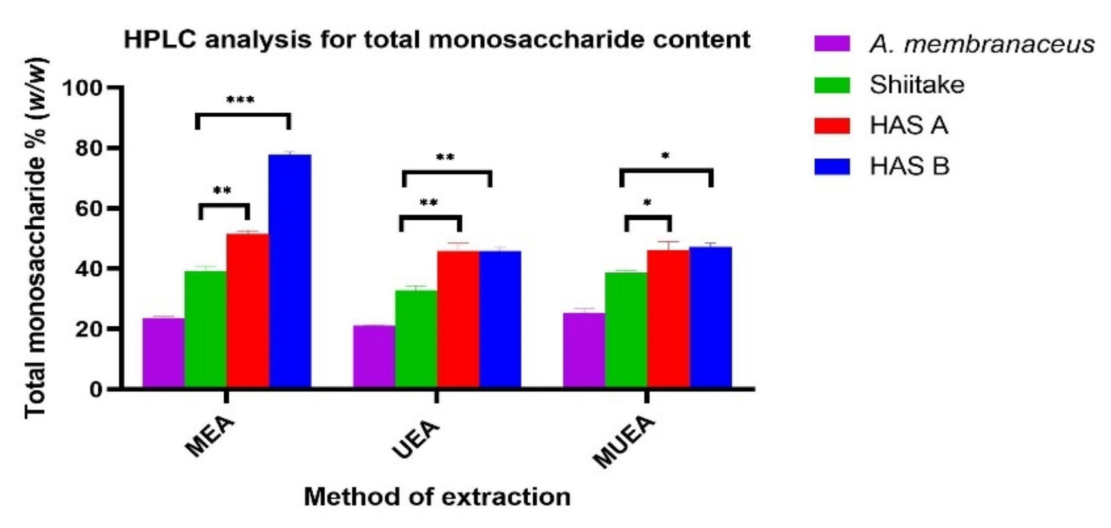
The total monosaccharide content of the PREs from three different extraction methods for four different samples is shown in Figure 2, and the composition and concentration of monosaccharides are shown in Table 1. In all the three methods, the total monosaccharide content in HAS-A and HAS-B were significantly higher than that in the control Shiitake (Figure 2). In UEA extracts of HAS-B and HAS-A, the amounts of GlcAc and Gluc were significantly higher (GlcAc ~ 551.1 ± 1.5 μg/mL and Gluc ~ 2084.1 ± 1.5 μg/mL in HAS-B and GlcAc ~ 487.3 ± 1.8 and Gluc ~ 1876.8 ± 2.6 μg/mL in HAS-A) compared to the control Shiitake (GlcAc ~ 346.6 ± 0.8 μg/mL and Gluc ~ 1131.6 ± 2.3 μg/mL) (p < 0.0001). Meanwhile, the amount of Mannose in the same extract was significantly low, with 127.1 ± 1.5 in HAS-B (p < 0.001) and 160.3 ± 1.8 μg/mL in HAS-A (p < 0.05), when compared to the control Shiitake (171.3 ± 1.8 μg/mL). A similar pattern was observed in all the three extraction methods for the above three extracts (Table 1).

3.3. Effect of Different Extraction Methods on the Protein Content

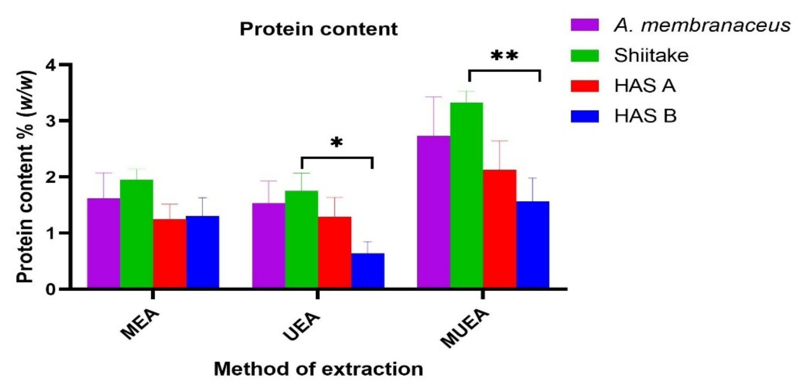
The protein contents in the PRE from the three different extraction methods for four different samples are shown in Figure 3. The protein content in HAS-B extracts (MUEA and UEA) was significantly lower than the Shiitake (p < 0.001 and p < 0.05, respectively). However, there was no statistical difference in these values between HAS-A and HAS-B extracts in all the extraction methods. The lowest protein content for HAS-B, HAS-A, Shiitake and Astragalus extracts was obtained from the UEA extraction, which was 0.637 ± 0.21%, 1.294 ± 0.34%, 1.756 ± 0.31% and 1.535 ± 0.39% w/w, respectively. There was no significant difference in these values with MEA extracts.

3.4. Effect of Different Extraction Methods on Molecular Weight Profile of Polysaccharide-Rich Extracts

The molecular weight profiles of polysaccharides in PREs from three different extraction methods for four different samples are shown in Table 2. The relative molecular weight of HAS-B polysaccharides ranged from 1.2–822.8 kDa for MUEA extracts, 1.2–814 kDa for MEA extracts and 1.1–833 kDa for UEA extracts. These values were similar for HAS-A (Table 2), but for Shiitake polysaccharides, these values ranged from 1.2–10.5 kDa for MUEA, 1.2–10.4 kDa for MEA and 1.05–12.7 kDa for UEA extracts (Table 2). HAS-A and HAS-B contain higher molecular weight polysaccharides than Shiitake. When comparing HAS-A and HAS-B, the percentage concentration of higher molecular weight polysaccharides in HAS-A was significantly less (p < 0.01) than that in HAS-B.

3.5. Effect of Different Extraction Methods on Anti-Proliferative Activity of PREs in HCT 116 Cells

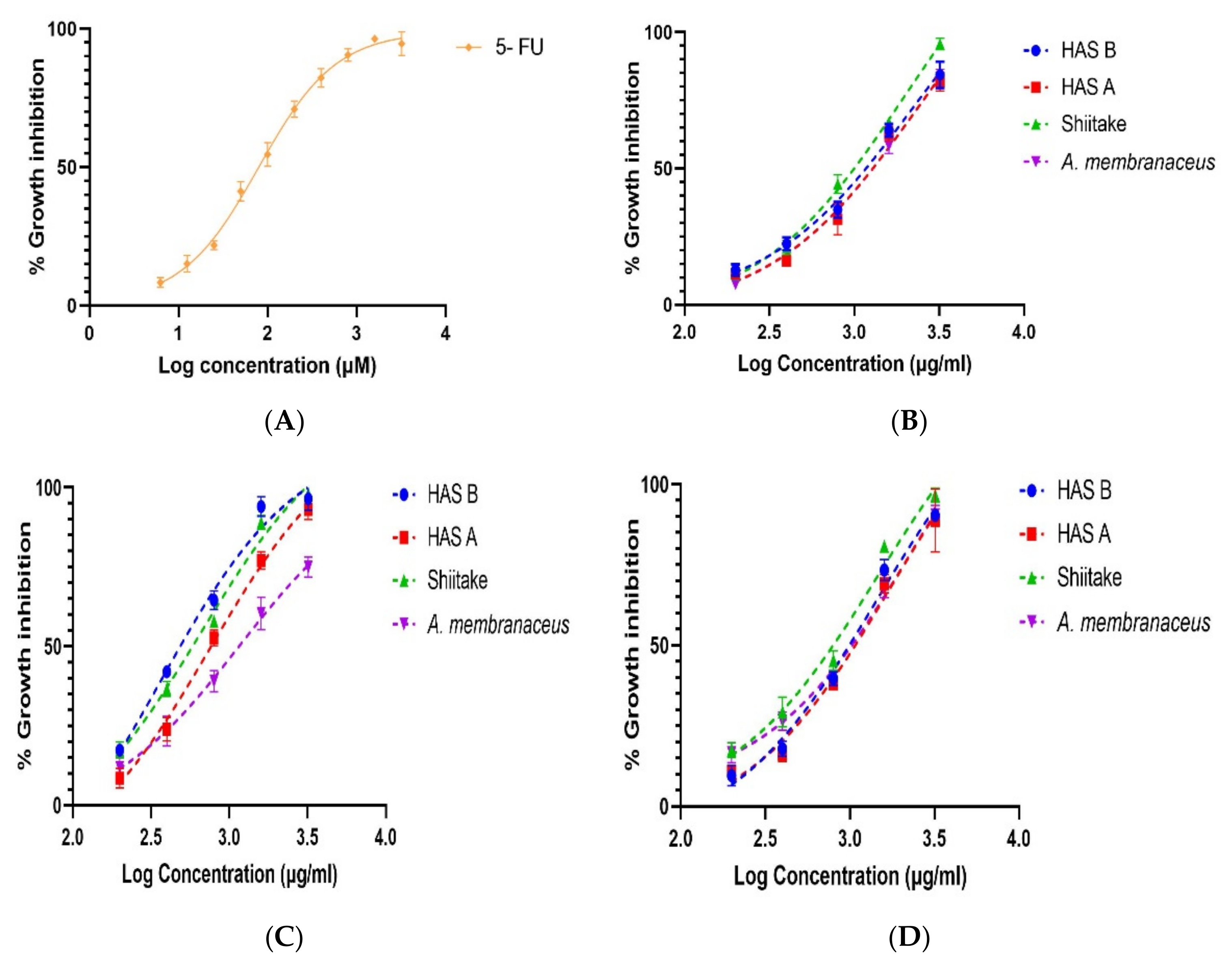
The anti-proliferative activity of PREs from three different extraction methods for four different samples is shown in Figure 4 and Table 3. The MTT results showed dose-dependent antiproliferative activity of HAS-A and HAS-B in HCT 116 colorectal carcinoma cells after treatment for 48 h (Figure 4). The highest anti-proliferative activity for HAS-A, HAS-B, Shiitake and A. membranaceus was obtained by UEA extraction, where the extracts at 1.6 mg/mL caused 77 ± 2.82%, 94 ± 3.14%, 89 ± 2.24% and 60.35 ± 5.1% of cell death, respectively (Figure 4C). The IC50 value was 0.76 ± 0.122 mg/mL, 0.367 ± 0.044 mg/mL, 0.659 ± 0.059 mg/mL and 1.181 ± 0.145 mg/mL, respectively (Table 3). UEA extracts of HAS-B had the lowest IC50 value (p < 0.05) and higher anti-proliferative activity compared to the control Shiitake, while HAS-A had similar IC50 to the control (not statistically difference). On the other hand, the anti-proliferative activity of HAS-A, HAS-B and Shiitake with MEA and MUEA extraction methods was very low, with IC50 values more than three times higher than that of the UEA extraction method (Table 3).

4. Discussion

In recent years, polysaccharides from biological origin are receiving greater attention in the medical field due to their strong therapeutic efficacy and effectiveness for a wide range of diseases such as cancer [4,6,43], cardiovascular diseases [44], diabetes [45,46], lung diseases [47] and kidney-associated diseases [48]. The polysaccharide components of both Astragalus and Shiitake have been proven to provide promising anti-proliferative properties in breast cancer [5,49], lung cancer [12,50] and colorectal cancers [6,51,52].
In this study, we investigated the yield, content and anti-proliferative activities of PREs (against colorectal cancer cells, HCT 116) from a new type of commercially produced Shiitake mushroom (HAS) using three green extraction methods to ensure the polysaccharide-rich extracts could be used for food and pharmaceutical industries. We found that the new approach of cultivating Shiitake in an edible medicinal plant Astragalus-supplemented bed helped in enhancing polysaccharide production in Shiitake mushrooms regardless of the extraction methods used. Taking the highest yield achieved by the UEA extraction method, the PRE yield for HAS-B and HAS-A increased by 85% and 35% when compared with the control Shiitake, respectively (Figure 1A). In contrast to our study, Yin et al. reported the highest yield, 9.38% w/w of Shiitake polysaccharides, using the MUEA extraction method [38], while we found only 6% w/w using MUEA in our study. The reason for lower yield in our study could be due to the modifications in previously reported extraction conditions. In the literature [35,38], the enzymatic hydrolysis was carried out for two hours before exposure to microwave or ultrasonic power. However, during our preliminary study, these conditions worked well only in terms of increasing the polysaccharide yield but were not promising in terms of providing higher anti-proliferative activity. For example, when the previously reported method of Yin et al. was used for UEA extraction of HAS-B [38], the yield was as high as 18.12% w/w, but its IC50 value against HCT 116 was ~2.56 mg/mL. Our modified UEA method had a comparatively lower yield of 11.1% w/w but had a significantly higher anti-proliferative activity in HCT 116 with IC50 value of 0.367 mg/mL. Hence, the modified condition in our experiment (exposing to microwave or ultrasonic power initially followed by enzyme hydrolysis for 20 min) was used with comparatively lower yield (Figure S1(A1)) but significantly improved anti-proliferative activity (p < 0.0001, in case of UEA HAS-B) compared to that of previously reported methods (Figure S1(A2)). Therefore, the enzymes were added in the sample/solvent mixture after exposing them to microwave/ultrasound in MEA, MUEA and UEA extractions in this study; these enzyme mixtures would have caused partial hydrolysis of the extracted polysaccharides, resulting in a decrease in PRE yield [35].
In addition, during our preliminary experiment, we compared the PRE yield and anti-proliferative effect of the extracts prepared from previously reported three-enzyme mixtures (Papain: Cellulase: Pectinase in a ratio 1:1:1) [27,38] with our new four-enzyme mixture (Alcalase: Cellulase; Pectinase: VinoTaste Pro® at a ratio of 1:1:1:1). This test was only conducted in the UEA–HAS-B sample. There was no statistically significant difference in the PRE yield (Figure S2(B1)). But interestingly, the extracts prepared from our new four-enzyme mixture had a significantly higher anti-proliferative activity (p < 0.0001, for UEA HAS-B) when compared to the extracts prepared from previously reported three- enzyme mixture (Figure S2(B2)). For example, the IC50 value of the UEA–HAS-B extract using the previously reported three-enzyme mixture was 1.8 mg/mL, while the same sample extracted with our new four-enzyme mixture had a IC50 value of 0.367 mg/mL (Figure S2(B2)). Therefore, the new four-enzyme mixture was chosen for all the enzyme-assisted extractions in this study. The higher anti-proliferative activity could be due to the use of extra enzyme VinoTaste Pro®, the exo-(1,3) β glucanase, which would have cut the long (1,3) β glucan chain into a relatively shorter chain. The relatively smaller molecular weight polysaccharides can easily cross the biological membranes and show improved biological activity when compared to the larger ones [53,54]. In contrast to the polysaccharide content, the protein contents were lower in both HAS-A and HAS-B compared to the control Shiitake in each extraction method. Thus, it can be inferred that the cultivation of Shiitake in the beds supplemented with A. membranaceus activates the production of polysaccharides in the mushroom while inhibiting the protein synthesis. The molecular mechanisms involved in these processes can be studied in the future.
When the anti-proliferative effects of PREs from Shiitake, HAS-A and HAS-B were compared with the literature, the extracts from this study showed comparatively higher activity than that of published reports. For example, [27] reported nearly 71% cell death in HCT 116 and nearly 73% cell death in Hela cells at 1.8 mg/mL of Shiitake polysaccharides. Another experiment conducted by [55] showed the highest inhibitory action of Shiitake polysaccharides at 5 mg/mL, where the percentage inhibition of S-180, HCT 116 and HT 29 with their best polysaccharide fraction was ~97%, ~64% and ~53%, respectively. Furthermore, Unursaikhan et al. generated an O-sulfonated derivative of mushroom polysaccharide (lentinan) for better solubility and anti-proliferative activity; the modified derivative had the highest inhibition of ~68% at 5 mg/mL in S-180 cells [56]. The results from our studies showed a better anti-proliferative activity, i.e., ~89% and ~77% inhibitory effect in HCT 116 with 1.6 mg/mL of UEA-Shiitake and HAS-A polysaccharides, respectively. This inhibitory effect was more prominent with UEA-HAS-B polysaccharides, i.e., ~94% of inhibition at 1.6 mg/mL (Figure 4C). These results indicate that the extraction modifications made in our experiments are effective for extraction of bioactive polysaccharides from Shiitake, HAS-A and HAS-B.
According to Zhang et al., the anti-proliferative activity of a polysaccharide molecule is directly related to its molecular weight and triple helix conformation [57]. Wang et al. further added that glucans with the molecular weights below 50 kDa cannot form a triple helix structure and hence cannot show prominent anti-proliferative activity [58]. Liu et al. also concluded that the glucans with a molecular weight between 290 and 2420 kDa exist as a triple helix conformation and those with a molecular weight of more than 1 × 10⁶ Da had better immune-enhancing properties [59]. However, our studies have shown significantly different results compared to the above-mentioned literature. In our study, the highest anti-proliferative activity of HAS-B, HAS-A and A. membranaceus in HCT 116 was obtained with the polysaccharide extracts of Mw ~ 1.1–832.7 kDa, ~1.2–976 kDa and ~2.5–113 kDa, respectively. Meanwhile, this molecular weight range was even lower in the case of Shiitake, where the highest inhibitory effect was shown by the polysaccharide extracts of Mw ~ 1.05–12.7 kDa (Table 3) which is completely different (extremely low) than what has been reported in the literature [58,60]. Therefore, in the future, determining the relationship between the molecular weight and bioactivities, including the anti-proliferative effects of these polysaccharides, would be critical for further optimization of extraction processes and bioactivities. Nonetheless, many studies have reported that high molecular weight polysaccharides showed better bio-activity only in vitro; when given in vivo, the immune system was highly triggered, and the molecules were engulfed by the phagocytes and macrophages and completely cleared off [61,62]. This suggests that the polysaccharide extracts with lower molecular weights in our study may have better anti-proliferative effects in vivo, which needs to be determined in future studies.

5. Conclusions

In conclusion, Shiitake mushrooms grown in the substrate beds supplemented with Astragalus membranaceus have a significantly higher amount of polysaccharides when compared to Shiitake grown under controlled conditions. The anti-proliferative activity of the PREs from HAS-A is not significantly different from that of the control Shiitake. However, the PREs from HAS-B have significantly higher anti-proliferative activity compared to the control Shiitake when optimum extraction conditions are used. Compared with MUEA and MEA extracts, UEA extracts had significantly higher anti-proliferative activity in HCT 116. In addition, the extracts from the UEA extraction method of the current study had significantly higher anti-proliferative activity in HCT 116 compared to previously reported studies. Hence, further investigation of the activities of HAS-B using the optimized extraction conditions and efficient purification would help to develop HAS as a promising functional food and/or a complementary medicine, particularly in the treatment and prevention of cancer.

6. Patents

The main research results from the work reported in this manuscript are involved in the filing of a patent; the authors will update this section once the process has been completed.

Supplementary Materials

The following supporting information can be downloaded at: https://www.mdpi.com/article/10.3390/nu14112333/s1, Figure S1: Effect of old and new UEA extraction techniques on the yield and anti-proliferative activity of HAS-B PREs against HCT 116; Figure S2: Effect of enzyme mixtures on yield and antiproliferative activity of HAS-B PREs against HCT 116.

Author Contributions

Conceptualization, W.Z., B.T. and Q.L.; methodology, B.T., B.B. and S.P.; software, B.T.; validation, B.T., Q.L. and W.Z.; formal analysis, B.T.; investigation, B.T.; resources, Q.L.; writing—original draft preparation, B.T. and Q.L.; writing—B.T., Q.L. and W.Z.; supervision, W.Z., B.B. and Q.L.; project administration, W.Z.; funding acquisition, W.Z. and Q.L. All authors have read and agreed to the published version of the manuscript.

Funding

This study was funded by the collaborative research project grant between Flinders University and Shanxi Yulongxiang Agricultural Development Co., Ltd. The project is entitled “Biorefinery process and health product development of Hengshan Astragalus Shiitake”.

Institutional Review Board Statement

Not applicable.

Informed Consent Statement

Not applicable.

Data Availability Statement

Data are available in a publicly accessible repository. The data presented in this study are openly available in FigShare at https://doi.org/10.6084/m9.figshare.19221252.v1 (accessed on 23 February 2022).

Acknowledgments

The authors are thankful to the Center for Marine Bioproducts and Development (CMBD) at Flinders University for providing the facility. The authors are thankful to the Shanxi Yulongxiang Agricultural Development Co. Ltd. for its collaboration with the CMBD and providing the materials for this research.

Conflicts of Interest

The authors declare no conflict of interest.

References

  1. Hobbs, C. Medicinal value of Lentinus edodes (Berk.) Sing.(Agaricomycetideae). A literature review. Int. J. Med. Mushrooms 2000, 2, 1–16. [Google Scholar] [CrossRef]
  2. Miyaji, C.; Poersch, A.; Ribeiro, L.; Eira, A.; Colus, I. Shiitake (Lentinula edodes (Berkeley) Pegler) extracts as a modulator of micronuclei induced in HEp-2 cells. Toxicol. In Vitro 2006, 20, 1555–1559. [Google Scholar] [CrossRef] [PubMed]
  3. Murphy, E.J.; Masterson, C.; Rezoagli, E.; O’Toole, D.; Major, I.; Stack, G.D.; Lynch, M.; Laffey, J.G.; Rowan, N.J. β-Glucan extracts from the same edible shiitake mushroom Lentinus edodes produce differential in-vitro immunomodulatory and pulmonary cytoprotective effects—Implications for coronavirus disease (COVID-19) immunotherapies. Sci. Total Environ. 2020, 732, 139330. [Google Scholar] [CrossRef]
  4. Finimundy, T.C.; Scola, G.; Scariot, F.J.; Dillon, A.J.; Moura, S.; Echeverrigaray, S.; Henriques, J.P.; Roesch-Ely, M. Extrinsic and intrinsic apoptotic responses induced by shiitake culinary-medicinal mushroom Lentinus edodes (Agaricomycetes) aqueous extract against a larynx carcinoma cell line. Int. J. Med. Mushrooms 2018, 20, 31–46. [Google Scholar] [CrossRef] [PubMed]
  5. Morales, D.; Rutckeviski, R.; Villalva, M.; Abreu, H.; Soler-Rivas, C.; Santoyo, S.; Iacomini, M.; Smiderle, F.R. Isolation and comparison of α-and β-D-glucans from shiitake mushrooms (Lentinula edodes) with different biological activities. Carbohydr. Polym. 2020, 229, 115521. [Google Scholar] [CrossRef] [PubMed]
  6. Wang, J.; Li, W.; Huang, X.; Liu, Y.; Li, Q.; Zheng, Z.; Wang, K. A polysaccharide from Lentinus edodes inhibits human colon cancer cell proliferation and suppresses tumor growth in athymic nude mice. Oncotarget 2017, 8, 610. [Google Scholar] [CrossRef] [PubMed] [Green Version]
  7. Li, W.; Wang, J.; Hu, H.; Li, Q.; Liu, Y.; Wang, K. Functional polysaccharide Lentinan suppresses human breast cancer growth via inducing autophagy and caspase-7-mediated apoptosis. J. Funct. Foods 2018, 45, 75–85. [Google Scholar] [CrossRef]
  8. Wang, Y.; Chen, J.; Han, Q.; Luo, Q.; Zhang, H.; Wang, Y. Construction of doxorubicin-conjugated lentinan nanoparticles for enhancing the cytotoxocity effects against breast cancer cells. Colloids Surf. Physicochem. Eng. Asp. 2019, 579, 123657. [Google Scholar] [CrossRef]
  9. Zhang, Y.; Li, Q.; Shu, Y.; Wang, H.; Zheng, Z.; Wang, J.; Wang, K. Induction of apoptosis in S180 tumour bearing mice by polysaccharide from Lentinus edodes via mitochondria apoptotic pathway. J. Funct. Foods 2015, 15, 151–159. [Google Scholar] [CrossRef]
  10. Yin, X.; Ying, J.; Li, L.; Zhang, H.; Wang, H. A meta-analysis of lentinan injection combined with chemotherapy in the treatment of nonsmall cell lung cancer. Indian J. Cancer 2015, 52, 29. [Google Scholar]
  11. Wang, X.-E.; Wang, Y.-H.; Zhou, Q.; Peng, M.; Zhang, J.; Chen, M.; Ma, L.-J.; Xie, G.-M. Immunomodulatory effect of lentinan on aberrant T subsets and cytokines profile in non-small cell lung cancer patients. Pathol. Oncol. Res. 2020, 26, 499–505. [Google Scholar] [CrossRef] [PubMed]
  12. Zhang, Y.; Li, Q.; Wang, J.; Cheng, F.; Huang, X.; Cheng, Y.; Wang, K. Polysaccharide from Lentinus edodes combined with oxaliplatin possesses the synergy and attenuation effect in hepatocellular carcinoma. Cancer Lett. 2016, 377, 117–125. [Google Scholar] [CrossRef] [PubMed]
  13. Del Buono, A.; Bonucci, M.; Pugliese, S.; D’ORTA, A.; Fioranelli, M. Polysaccharide from Lentinus edodes for integrative cancer treatment: Immunomodulatory effects on lymphocyte population. WCRJ 2016, 3, 1–7. [Google Scholar]
  14. Özçelik, E.; Pekşen, A. Hazelnut husk as a substrate for the cultivation of shiitake mushroom (Lentinula edodes). Bioresour. Technol. 2007, 98, 2652–2658. [Google Scholar] [CrossRef] [PubMed]
  15. Economou, C.N.; Diamantopoulou, P.A.; Philippoussis, A.N. Valorization of spent oyster mushroom substrate and laccase recovery through successive solid state cultivation of Pleurotus, Ganoderma, and Lentinula strains. Appl. Microbiol. Biotechnol. 2017, 101, 5213–5222. [Google Scholar] [CrossRef]
  16. Siwulski, M.; Rzymski, P.; Budka, A.; Kalač, P.; Budzyńska, S.; Dawidowicz, L.; Hajduk, E.; Kozak, L.; Budzulak, J.; Sobieralski, K.; et al. The effect of different substrates on the growth of six cultivated mushroom species and composition of macro and trace elements in their fruiting bodies. Eur. Food Res. Technol. 2019, 245, 419–431. [Google Scholar] [CrossRef] [Green Version]
  17. Kaleta, B.; Górski, A.; Zagożdżon, R.; Cieślak, M.; Kaźmierczak-Barańska, J.; Nawrot, B.; Klimaszewska, M.; Malinowska, E.; Górska, S.; Turło, J. Selenium-containing polysaccharides from Lentinula edodes—Biological activity. Carbohydr. Polym. 2019, 223, 115078. [Google Scholar] [CrossRef]
  18. Wu, C.-Y.; Ke, Y.; Zeng, Y.-F.; Zhang, Y.-W.; Yu, H.-J. Anticancer activity of Astragalus polysaccharide in human non-small cell lung cancer cells. Cancer Cell Int. 2017, 17, 115. [Google Scholar] [CrossRef] [Green Version]
  19. Fan, Y.; Hu, Y.; Wang, D.; Liu, J.; Zhang, J.; Zhao, X.; Liu, X.; Liu, C.; Yuan, J.; Ruan, S. Effects of Astragalus polysaccharide liposome on lymphocyte proliferation in vitro and adjuvanticity in vivo. Carbohydr. Polym. 2012, 88, 68–74. [Google Scholar] [CrossRef]
  20. Yang, B.; Xiao, B.; Sun, T. Antitumor and immunomodulatory activity of Astragalus membranaceus polysaccharides in H22 tumor-bearing mice. Int. J. Biol. Macromol. 2013, 62, 287–290. [Google Scholar] [CrossRef]
  21. Jin, M.; Zhao, K.; Huang, Q.; Shang, P. Structural features and biological activities of the polysaccharides from Astragalus membranaceus. Int. J. Biol. Macromol. 2014, 64, 257–266. [Google Scholar] [CrossRef] [PubMed]
  22. Liu, A.-J.; Yu, J.; Ji, H.-Y.; Zhang, H.-C.; Zhang, Y.; Liu, H.-P. Extraction of a novel cold-water-soluble polysaccharide from Astragalus membranaceus and its antitumor and immunological activities. Molecules 2018, 23, 62. [Google Scholar] [CrossRef] [PubMed] [Green Version]
  23. Balakrishnan, B.; Liang, Q.; Fenix, K.; Tamang, B.; Hauben, E.; Ma, L.; Zhang, W. Combining the Anticancer and Immunomodulatory Effects of Astragalus and Shiitake as an Integrated Therapeutic Approach. Nutrients 2021, 13, 2564. [Google Scholar] [CrossRef]
  24. Guo, S.; Ma, B.; Jiang, X.; Li, X.; Jia, Y. Astragalus polysaccharides inhibits tumorigenesis and lipid metabolism through miR-138-5p/SIRT1/SREBP1 pathway in prostate cancer. Front. Pharmacol. 2020, 11, 598. [Google Scholar] [CrossRef] [PubMed]
  25. Yang, S.; Sun, S.; Xu, W.; Yu, B.; Wang, G.; Wang, H. Astragalus polysaccharide inhibits breast cancer cell migration and invasion by regulating epithelial-mesenchymal transition via the Wnt/β-catenin signaling pathway. Mol. Med. Rep. 2020, 21, 1819–1832. [Google Scholar] [CrossRef] [PubMed] [Green Version]
  26. Zhu, Y.; Li, Q.; Mao, G.; Zou, Y.; Feng, W.; Zheng, D.; Wang, W.; Zhou, L.; Zhang, T.; Yang, J.; et al. Optimization of enzyme-assisted extraction and characterization of polysaccharides from Hericium erinaceus. Carbohydr. Polym. 2014, 101, 606–613. [Google Scholar] [CrossRef]
  27. Zhao, Y.-M.; Wang, J.; Wu, Z.-G.; Yang, J.-M.; Li, W.; Shen, L.-X. Extraction, purification and anti-proliferative activities of polysaccharides from Lentinus edodes. Int. J. Biol. Macromol. 2016, 93, 136–144. [Google Scholar] [CrossRef]
  28. Chen, Z.; Zhang, W.; Tang, X.; Fan, H.; Xie, X.; Wan, Q.; Wu, X.; Tang, J.Z. Extraction and characterization of polysaccharides from Semen Cassiae by microwave-assisted aqueous two-phase extraction coupled with spectroscopy and HPLC. Carbohydr. Polym. 2016, 144, 263–270. [Google Scholar] [CrossRef]
  29. Chen, Y.; Gu, X.; Huang, S.-Q.; Li, J.; Wang, X.; Tang, J. Optimization of ultrasonic/microwave assisted extraction (UMAE) of polysaccharides from Inonotus obliquus and evaluation of its anti-tumor activities. Int. J. Biol. Macromol. 2010, 46, 429–435. [Google Scholar] [CrossRef]
  30. Sun, H.; Li, C.; Ni, Y.; Yao, L.; Jiang, H.; Ren, X.; Fu, Y.; Zhao, C. Ultrasonic/microwave-assisted extraction of polysaccharides from Camptotheca acuminata fruits and its antitumor activity. Carbohydr. Polym. 2019, 206, 557–564. [Google Scholar] [CrossRef]
  31. Wang, Y.; Wang, C.; Guo, M. Effects of Ultrasound Treatment on Extraction and Rheological Properties of Polysaccharides from Auricularia Cornea var. Li. Molecules 2019, 24, 939. [Google Scholar] [CrossRef] [PubMed] [Green Version]
  32. Gil-Ramírez, A.; Smiderle, F.R.; Morales, D.; Iacomini, M.; Soler-Rivas, C. Strengths and weaknesses of the aniline-blue method used to test mushroom (1→ 3)-β-d-glucans obtained by microwave-assisted extractions. Carbohydr. Polym. 2019, 217, 135–143. [Google Scholar] [CrossRef] [PubMed] [Green Version]
  33. Alzorqi, I.; Sudheer, S.; Lu, T.-J.; Manickam, S. Ultrasonically extracted β-d-glucan from artificially cultivated mushroom, characteristic properties and antioxidant activity. Ultrason. Sonochem. 2017, 35, 531–540. [Google Scholar] [CrossRef] [PubMed]
  34. Zhao, Y.-M.; Yang, J.-M.; Liu, Y.-H.; Zhao, M.; Wang, J. Ultrasound assisted extraction of polysaccharides from Lentinus edodes and its anti-hepatitis B activity in vitro. Int. J. Biol. Macromol. 2018, 107, 2217–2223. [Google Scholar] [CrossRef] [PubMed]
  35. Cheng, Z.; Song, H.; Yang, Y.; Liu, Y.; Liu, Z.; Hu, H.; Zhang, Y. Optimization of microwave-assisted enzymatic extraction of polysaccharides from the fruit of Schisandra chinensis Baill. Int. J. Biol. Macromol. 2015, 76, 161–168. [Google Scholar] [CrossRef]
  36. Amigh, S.; Taghian Dinani, S. Combination of ultrasound-assisted aqueous enzymatic extraction and cooking pretreatment for date seed oil recovery. Heat Mass Transf. 2020, 56, 2345–2354. [Google Scholar] [CrossRef]
  37. Morales, D.; Smiderle, F.R.; Villalva, M.; Abreu, H.; Rico, C.; Santoyo, S.; Iacomini, M.; Soler-Rivas, C. Testing the effect of combining innovative extraction technologies on the biological activities of obtained β-glucan-enriched fractions from Lentinula edodes. J. Funct. Foods 2019, 60, 103446. [Google Scholar] [CrossRef]
  38. Yin, C.; Fan, X.; Fan, Z.; Shi, D.; Gao, H. Optimization of enzymes-microwave-ultrasound assisted extraction of Lentinus edodes polysaccharides and determination of its antioxidant activity. Int. J. Biol. Macromol. 2018, 111, 446–454. [Google Scholar] [CrossRef]
  39. Yang, X.; Zhao, Y.; Wang, Q.; Wang, H.; Mei, Q. Analysis of the monosaccharide components in Angelica polysaccharides by high performance liquid chromatography. Anal. Sci. 2005, 21, 1177–1180. [Google Scholar] [CrossRef] [Green Version]
  40. Lorbeer, A.J.; Lahnstein, J.; Fincher, G.B.; Su, P.; Zhang, W. Kinetics of conventional and microwave-assisted fucoidan extractions from the brown alga, Ecklonia radiata. J. Appl. Phycol. 2015, 27, 2079–2087. [Google Scholar] [CrossRef]
  41. Kruger, N.J. The Bradford method for protein quantitation. In The Protein Protocols Handbook; Springer: Berlin/Heidelberg, Germany, 2009; pp. 17–24. [Google Scholar]
  42. You, L.; Gao, Q.; Feng, M.; Yang, B.; Ren, J.; Gu, L.; Cui, C.; Zhao, M. Structural characterisation of polysaccharides from Tricholoma matsutake and their antioxidant and antitumour activities. Food Chem. 2013, 138, 2242–2249. [Google Scholar] [CrossRef] [PubMed]
  43. Qi, W.; Zhou, X.; Wang, J.; Zhang, K.; Zhou, Y.; Chen, S.; Nie, S.; Xie, M. Cordyceps sinensis polysaccharide inhibits colon cancer cells growth by inducing apoptosis and autophagy flux blockage via mTOR signaling. Carbohydr. Polym. 2020, 237, 116113. [Google Scholar] [CrossRef]
  44. Li, H.; Zhang, M.; Ma, G. Hypolipidemic effect of the polysaccharide from Pholiota nameko. Nutrition 2010, 26, 556–562. [Google Scholar] [CrossRef] [PubMed]
  45. Ghoneim, M.A.; Hassan, A.I.; Mahmoud, M.G.; Asker, M.S. Effect of polysaccharide from Bacillus subtilis sp. on cardiovascular diseases and atherogenic indices in diabetic rats. BMC Complementary Altern. Med. 2016, 16, 112. [Google Scholar] [CrossRef] [PubMed] [Green Version]
  46. Zhou, W.; Chen, G.; Chen, D.; Ye, H.; Zeng, X. The antidiabetic effect and potential mechanisms of natural polysaccharides based on the regulation of gut microbiota. J. Funct. Foods 2020, 75, 104222. [Google Scholar] [CrossRef]
  47. Cao, P.; Wu, S.; Wu, T.; Deng, Y.; Zhang, Q.; Wang, K.; Zhang, Y. The important role of polysaccharides from a traditional Chinese medicine-Lung Cleansing and Detoxifying Decoction against the COVID-19 pandemic. Carbohydr. Polym. 2020, 240, 116346. [Google Scholar] [CrossRef] [PubMed]
  48. Wu, T.; Shen, M.; Liu, S.; Yu, Q.; Chen, Y.; Xie, J. Ameliorative effect of Cyclocarya paliurus polysaccharides against carbon tetrachloride induced oxidative stress in liver and kidney of mice. Food Chem. Toxicol. 2020, 135, 111014. [Google Scholar] [CrossRef]
  49. Li, W.; Hu, X.; Wang, S.; Jiao, Z.; Sun, T.; Liu, T.; Song, K. Characterization and anti-tumor bioactivity of astragalus polysaccharides by immunomodulation. Int. J. Biol. Macromol. 2020, 145, 985–997. [Google Scholar] [CrossRef]
  50. Lai, X.; Xia, W.; Wei, J.; Ding, X. Therapeutic effect of Astragalus polysaccharides on hepatocellular carcinoma H22-bearing mice. Dose-Response 2017, 15, 1559325816685182. [Google Scholar] [CrossRef] [Green Version]
  51. Chao, L.; Zhou, J.; Zhu, X.-X.; Ni, W.-P.; Wang, H.-D.; Wang, Q. Effect of Astragalus Polysaccharides on Proliferation and Cell Cycle of Human Gastric Carcinoma Cell Line MKN45. Chin. Arch. Tradit. Chin. Med. 2012, 30, 2474–2477. [Google Scholar]
  52. Lv, J.; Zhang, Y.; Tian, Z.; Liu, F.; Shi, Y.; Liu, Y.; Xia, P. Astragalus polysaccharides protect against dextran sulfate sodium-induced colitis by inhibiting NF-κB activation. Int. J. Biol. Macromol. 2017, 98, 723–729. [Google Scholar] [CrossRef] [PubMed]
  53. Shang, H.; Chen, S.; Li, R.; Zhou, H.; Wu, H.; Song, H. Influences of extraction methods on physicochemical characteristics and activities of Astragalus cicer L. polysaccharides. Process Biochem. 2018, 73, 220–227. [Google Scholar] [CrossRef]
  54. Li, X.; Wang, L.; Wang, Y.; Xiong, Z. Effect of drying method on physicochemical properties and antioxidant activities of Hohenbuehelia serotina polysaccharides. Process Biochem. 2016, 51, 1100–1108. [Google Scholar] [CrossRef]
  55. Jeff, I.B.; Yuan, X.; Sun, L.; Kassim, R.M.; Foday, A.D.; Zhou, Y. Purification and in vitro anti-proliferative effect of novel neutral polysaccharides from Lentinus edodes. Int. J. Biol. Macromol. 2013, 52, 99–106. [Google Scholar] [CrossRef]
  56. Unursaikhan, S.; Xu, X.; Zeng, F.; Zhang, L. Antitumor activities of O-sulfonated derivatives of (1→ 3)-α-D-glucan from different Lentinus edodes. Biosci. Biotechnol. Biochem. 2006, 70, 38–46. [Google Scholar] [CrossRef]
  57. Zhang, L.; Li, X.; Xu, X.; Zeng, F. Correlation between antitumor activity, molecular weight, and conformation of lentinan. Carbohydr. Res. 2005, 340, 1515–1521. [Google Scholar] [CrossRef]
  58. Wang, Q.; Sheng, X.; Shi, A.; Hu, H.; Yang, Y.; Liu, L.; Fei, L.; Liu, H. β-Glucans: Relationships between modification, conformation and functional activities. Molecules 2017, 22, 257. [Google Scholar] [CrossRef] [Green Version]
  59. Liu, Y.; Tang, Q.; Zhang, J.; Xia, Y.; Yang, Y.; Wu, D.; Fan, H.; Cui, S.W. Triple helix conformation of β-d-glucan from Ganoderma lucidum and effect of molecular weight on its immunostimulatory activity. Int. J. Biol. Macromol. 2018, 114, 1064–1070. [Google Scholar] [CrossRef]
  60. Zhang, J.; Wen, C.; Gu, J.; Ji, C.; Duan, Y.; Zhang, H. Effects of subcritical water extraction microenvironment on the structure and biological activities of polysaccharides from Lentinus edodes. Int. J. Biol. Macromol. 2019, 123, 1002–1011. [Google Scholar] [CrossRef]
  61. Hao, L.; Sheng, Z.; Lu, J.; Tao, R.; Jia, S. Characterization and antioxidant activities of extracellular and intracellular polysaccharides from Fomitopsis pinicola. Carbohydr. Polym. 2016, 141, 54–59. [Google Scholar] [CrossRef]
  62. Lemmon, H.R.; Sham, J.; Chau, L.A.; Madrenas, J. High molecular weight polysaccharides are key immunomodulators in North American ginseng extracts: Characterization of the ginseng genetic signature in primary human immune cells. J. Ethnopharmacol. 2012, 142, 1–13. [Google Scholar] [CrossRef] [PubMed]
Figure 1. Effect of three different extraction methods on the PRE yield (A) and polysaccharide content (B) of A. membranaceus, Shiitake, HAS-A and HAS-B. The extraction methods are: MEA—Microwave/Enzyme assisted, UEA—Ultrasound/Enzyme assisted and MUEA—Microwave/Ultrasound/Enzyme assisted extractions. HAS-A and HAS-B significantly different from the control Shiitake with p < 0.05 indicated as *, p < 0.01 as **, and p < 0.0001 as ****, while # represents significant difference between HAS-A and HAS-B (p < 0.001 indicated as ### and p < 0.0001 as ####).
Nutrients 14 02333 g001
Figure 2. Effect of three different extraction methods on total monosaccharide content in the polysaccharide rich extracts of A. membranaceus, Shiitake, HAS-A and HAS-B, determined by HPLC. MEA—Microwave/Enzyme assisted, UEA—Ultrasound/Enzyme assisted, MUEA—Microwave/Ultrasound/Enzyme assisted extraction methods. HAS-A and HAS-B significantly different from the control Shiitake with p < 0.05 indicated as *, p < 0.01 as ** and p < 0.001 as ***.
Nutrients 14 02333 g002
Figure 3. Effect of three different extraction methods on the protein content of A. membranaceus, Shiitake, HAS-A and HAS-B PREs. Protein content in HAS-B significantly different from control Shiitake with p < 0.05 indicated as * and p < 0.01 as **.
Nutrients 14 02333 g003
Figure 4. Effect of three different extraction methods on growth inhibition of HCT 116 after 48 h treatment with (200–3200) μg/mL of HAS-A, HAS-B, Shiitake and A. membranaceus PREs, determined by MTT assay (n = 3 with mean ± SD). (A) Growth inhibition by positive control, 5-FU. (B) Growth inhibition by MEA extracts. (C) Growth inhibition by UEA extracts. (D) Growth inhibition by MUEA extracts.
Nutrients 14 02333 g004
Table 1. Composition and concentration of monosaccharides in MEA, UEA and MUEA extracts of A. membranaceus, Shiitake, HAS-A and HAS-B, determined by HPLC (concentration μg/mL ± S.D. n = 3).
Method of ExtractionExtractsMonosaccharides Concentration μg/mL ± S.D
ManRibRhaGlcAcGalAcGlucGalXylAraFuc
MEAA. membranaceus60.0 ± 0.814.8 ± 0.243.6 ± 1.1254.5 ± 7.161.1 ± 2730.4 ± 15303.6 ± 4.959.8 ± 1.1209.8 ± 318.5 ± 0.3
Shiitake194.5 ± 6.313.0 ± 1.59.6 ± 0.8392.8 ± 3722.2 ± 2.41352.7 ± 47785.1 ± 28.322.4 ± 0.53.2 ± 0.7109.2 ± 4.1
HAS-A158.6 ± 4.2 ##24.3 ± 1.811.8 ± 0.3566.4 ± 23.9 #13.7 ± 9.62235.4 ± 70 ###688.9 ± 2217.9 ± 3.55.7 ± 0.297.6 ± 2.7
HAS-B174.6 ± 0.8 *37.8 ± 3.611.1 ± 2.4858.0 ± 6.6 ***21.4 ± 12.13581.7 ± 27 ****911.1 ± 2.217.1 ± 4.63.6 ± 0.3156.0 ± 6
UEAA. membranaceus67.9 ± 0.68.5 ± 0.453.4 ± 1.2165.1 ± 149.5 ± 0.2634.8 ± 2.5374.9 ± 6.730.9 ± 0.1192.9 ± 3.219.0 ± 0.2
Shiitake171.3 ± 1.97.7 ± 0.68.9 ± 1.1346.6 ± 0.925.1 ± 1.51131.7 ± 2.4663.0 ± 3624.4 ± 1.93.4 ± 0.390.8 ± 4.7
HAS-A160.3 ± 1.9 #9.5 ± 1.310.1 ± 1.4487.3 ± 1.9 ####21.1 ± 3.91876.9 ± 2.6 ####736.6 ± 41.419.4 ± 0.74.7 ± 1.3105.8 ± 6.2
HAS-B127.1 ± 1.5 ***10.0 ± 0.113.9 ± 13.1551.1 ± 1.5 ****10.7 ± 1.22084.1 ± 1.5 ****557.1 ± 6312.8 ± 1.25.0 ± 0.6100.7 ± 12
MUEAA. membranaceus53.7 ± 2.99.1 ± 1.644.8 ± 2.5298.3 ± 25.945.1 ± 5901.7 ± 37.7313.1 ± 14.627.5 ± 0.48169.0 ± 9.116.3 ± 0.5
Shiitake193.4 ± 3.89.7 ± 2.59.2 ± 0.8385.7 ± 3.825.8 ± 0.21365.3 ± 23.8749.2 ± 15.121.5 ± 0.463.4 ± 1.3103.5 ± 2
HAS-A149.2 ± 7.3 #40.7 ± 5.57.0 ± 0.9501.4 ± 4319.0 ± 8.71892.1 ± 97 #697.0 ± 35.517.5 ± 2.23.9 ± 2.2101.9 ± 5.8
HAS-B94.8 ± 9.4 **29.2 ± 5.310.4 ± 0.2533.1 ± 40.8 *11.0 ± 2.52066.0 ± 61 *512.9 ± 45.310.2 ± 0.213.5 ± 0.889.8 ± 8
Note: Man (mannose), Rib (ribose), Rha (rhamnose), GlcAc (glucuronic acid), GalAc (galacturonic acid), Gluc (glucose), Gal (galactose), Xyl (xylose), Ara (arabinose) and Fuc (fucose). HAS-A and HAS-B significantly different from control Shiitake indicated as # and *, respectively. p < 0.05 indicated as #/*, p < 0.01 as ##/**, p < 0.001 as ###/*** and p < 0.0001 as ####/****.
Table 2. Relative molecular weight profiles of MEA, UEA and MUEA polysaccharide-rich extracts of HAS-B, HAS-A, Shiitake and A. membranaceus determined by size exclusion chromatography by HPLC.
ExtractsMajor Peaks for HAS-BMajor Peaks for HAS-AMajor Peaks for ShiitakeMajor Peaks for Astragalus
Rt, mins% Concen-trationMw, kDaRt, mins% Concen-trationMw, kDaRt, mins% Concen-trationMw, kDaRt, mins% Concen-trationMw, kDa
MEA12.88512.901 ***813.96712.640.0311092.56716.51480.82910.39915.0551.95360.021
16.01533.5618.9416.69372.5658.38717.60510.8732.80416.66556.5468.674
17.03722.1645.54717.60412.3392.80718.3076.8211.20617.8226.3992.165
17.65510.8482.6418.3157.681.195 18.31810.2291.19
18.31610.9731.193
UEA12.86612.983 **832.76312.7340.194975.88516.3520.50812.7214.6294.399100.135
16.02825.04418.64617.68778.2882.54117.1742.9884.7317.08829.4135.218
17.71936.9662.44518.32616.6251.17917.522.043.1917.70639.9372.483
18.34415.2171.154 18.4112.251.0518.33311.8241.169
MUEA12.87620.234 ****822.81712.7874.685 ##915.67911.37575.27610.515.3041.02944.501
15.95353.48420.40416.50370.53610.53716.50612.7632.69816.73357.157.993
17.6388.5812.69517.6049.5542.80717.63710.1991.21317.67125.8822.59
18.3056.1261.20918.294.4541.23118.302 18.31112.1521.2
Note: The percentage concentration of higher molecular weight polysaccharides was significantly higher in HAS-B (p < 0.01 indicated as **, p < 0.001 as *** and p < 0.0001 as ****) and HAS-A (p < 0.01 indicated as ##) when compared to Shiitake.
Table 3. Effect of three different extraction methods on IC50 values of HAS-A, HAS-B, Shiitake and Astragalus in HCT 116 determined by MTT assay.
ExtractsIC50 (mg/mL) ± S.D. (n = 3)
HAS-BHAS-AShiitakeA. membranaceus
MUEA1.444 ± 0.2441.78 ± 0.4431.491 ± 0.3792.417 ± 0.298
MEA2.224 ± 0.2062.395 ± 0.521.975 ± 0.32.346 ± 0.181
UEA0.367 ± 0.044 *0.76 ± 0.122 ##0.659 ± 0.0591.181 ± 0.145
Note: * IC50 value for UEA-HAS-B extract was significantly low (p < 0.05 indicated as *) in HCT 116 compared to UEA Shiitake, while ## indicates IC50 value of HAS-A is significantly higher than HAS-B (p < 0.01).
Publisher’s Note: MDPI stays neutral with regard to jurisdictional claims in published maps and institutional affiliations.