
Anti-Salmonella Activity and Peptidomic Profiling of Peptide Fractions Produced from Sturgeon Fish Skin Collagen (Huso huso) Using Commercial Enzymes

 ,
,  and
and 
Abstract
:1. Introduction
2. Materials and Methods
2.1. Chemicals
2.2. Enzymatic Hydrolysis of Fish Skin Collagen
2.3. Characterization of the Protein Hydrolysates
2.3.1. Degree of Hydrolysis (DH)
2.3.2. SDS Polyacrylamide Gel Electrophoresis (SDS-PAGE)
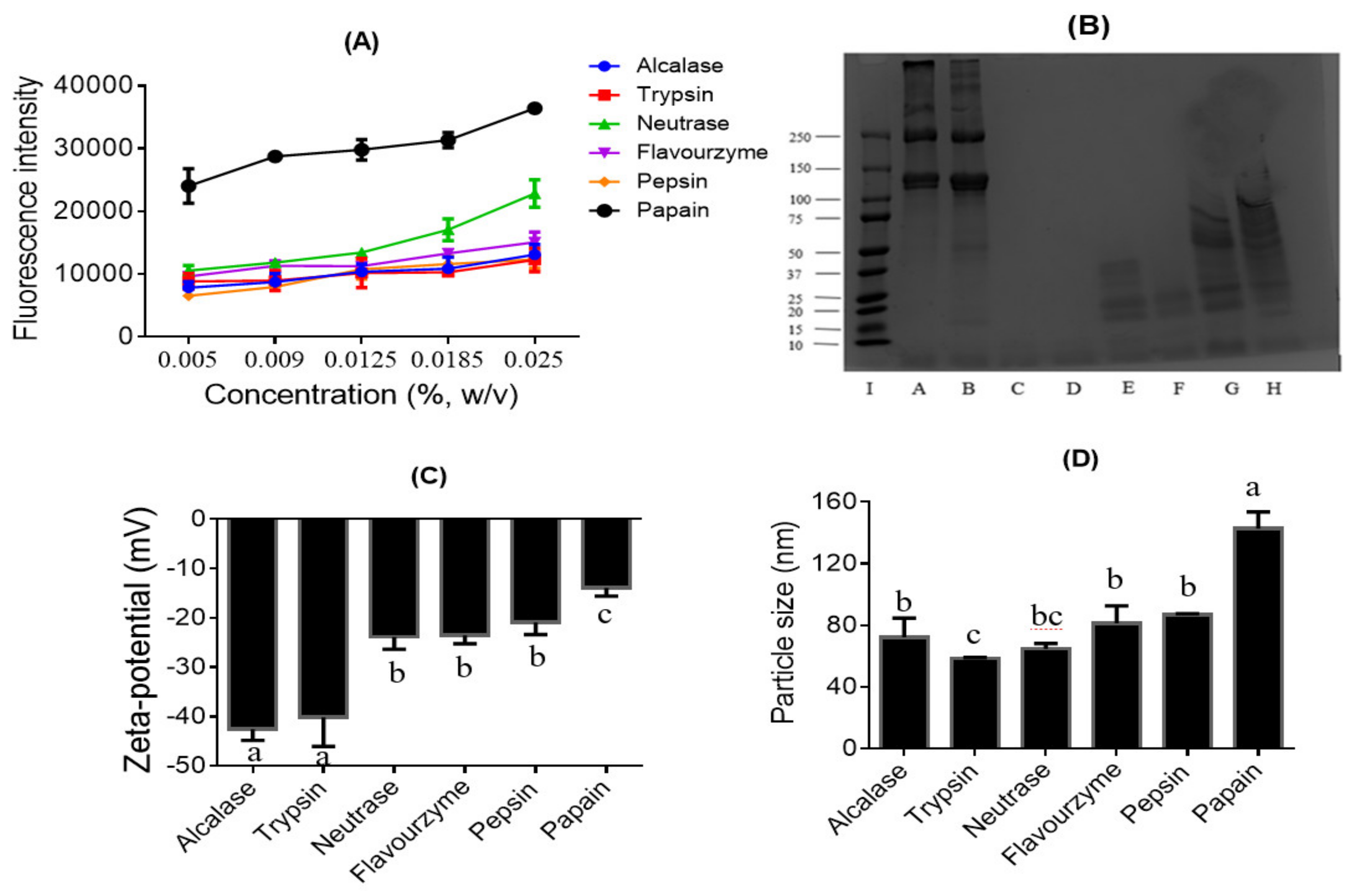
2.3.3. Surface Hydrophobicity (Ho)
2.3.4. Dynamic Light Scattering (DLS) Analysis
2.4. Solvent Fractionation of the Hydrolysates
2.5. Liquid Chromatography-Tandem Mass Spectrometry (LC-MS/MS) and Peptidomics Data Analysis
2.6. Antibacterial Activity of Collagen Peptides
2.7. Statistical Analysis
3. Results
3.1. Degree of Hydrolysis and Surface Hydrophobicity of the Hydrolysates
3.2. Molecular Weight Distribution of the Collagen Hydrolysates
3.3. Surface Charge and Particle Size of the Collagen Hydrolysates
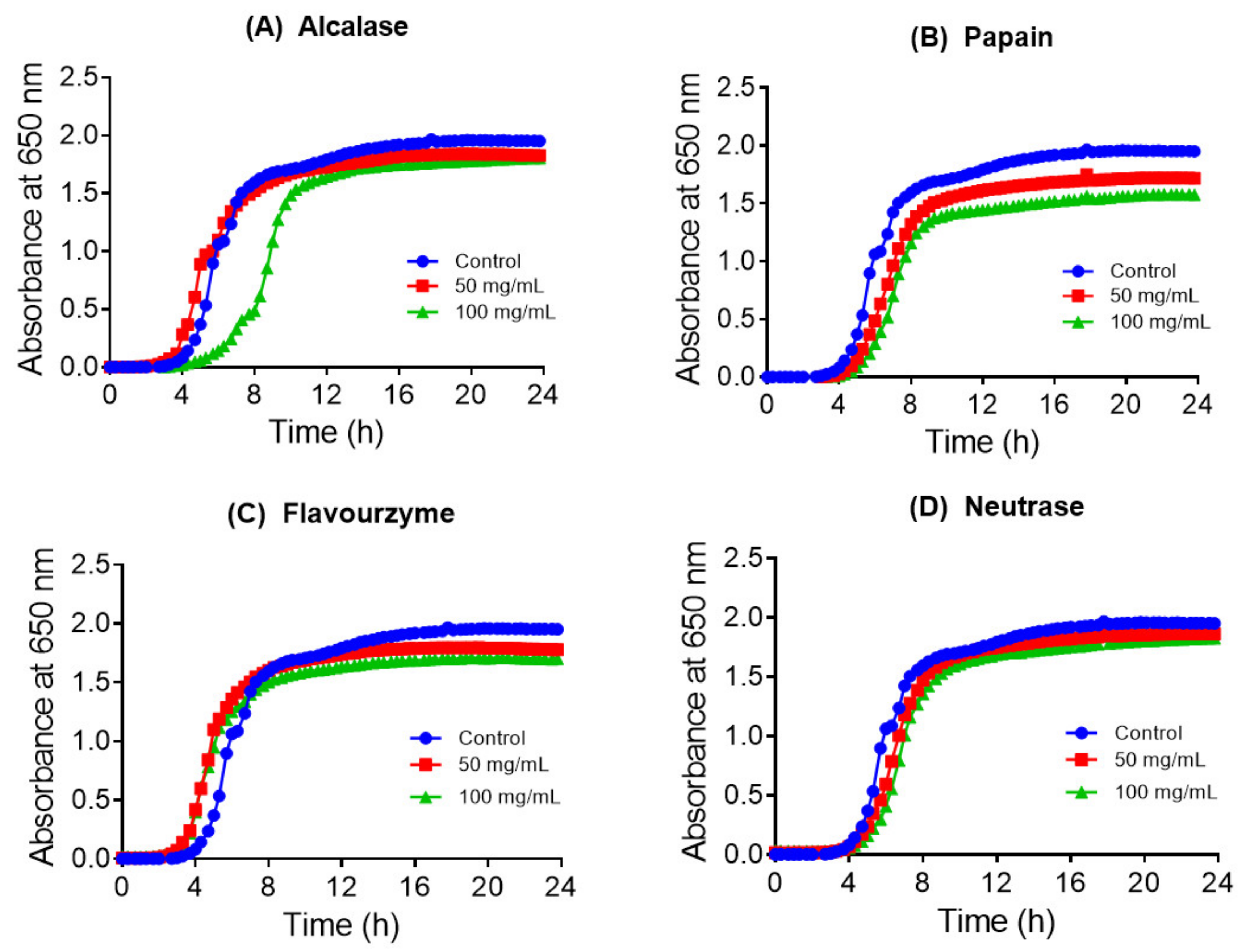
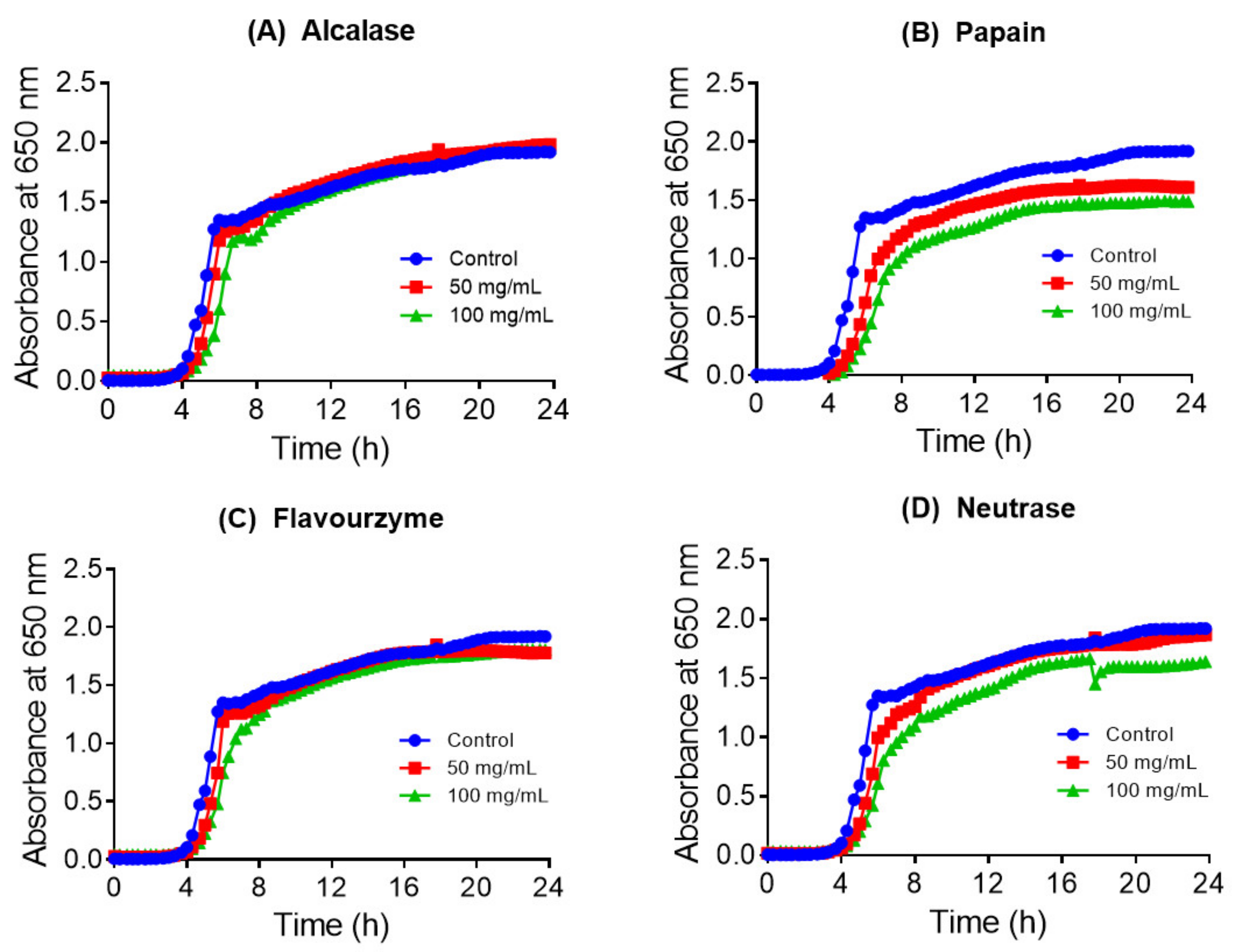
3.4. Antibacterial Activity of the Collagen Hydrolysates and Peptide Fractions
3.5. Prole of Peptides in the Acetone Fractions
4. Discussion
5. Conclusions
Author Contributions
Funding
Institutional Review Board Statement
Informed Consent Statement
Data Availability Statement
Acknowledgments
Conflicts of Interest
References
- Mor, A. Peptide-based antibiotics: A potential answer to raging antimicrobial resistance. Drug. Dev. Res. 2000, 50, 440–447. [Google Scholar] [CrossRef]
- Song, R.; Wei, R.-B.; Luo, H.-Y.; Wang, D.-F. Isolation and Characterization of an Antibacterial Peptide Fraction from the Pepsin Hydrolysate of Half-Fin Anchovy (Setipinna taty). Moleclues 2012, 17, 2980–2991. [Google Scholar] [CrossRef] [PubMed]
- Atef, M.; Ojagh, S.M. Health benefits and food applications of bioactive compounds from fish byproducts: A review. J. Funct. Foods 2017, 35, 673–681. [Google Scholar] [CrossRef]
- Pellegrini, A. Antimicrobial Peptides from Food Proteins. Curr. Pharm. Des. 2003, 9, 1225–1238. [Google Scholar] [CrossRef] [PubMed]
- Bellamy, W.; Takase, M.; Yamauchi, K.; Wakabayashi, H.; Kawase, K.; Tomita, M. Identification of the bactericidal domain of lactoferrin. Biochim. Biophys. Acta Protein Struct. Mol. Enzym. 1992, 1121, 130–136. [Google Scholar] [CrossRef]
- Ibrahim, H.R.; Iwamori, E.; Sugimoto, Y.; Aoki, T. Identification of a distinct antibacterial domain within the N-lobe of ovotransferrin. Biochim. Biophys. Acta Bioenerg. 1998, 1401, 289–303. [Google Scholar] [CrossRef] [Green Version]
- Zucht, H.-D.; Raida, M.; Adermann, K.; Mägert, H.-J.; Forssmann, W.-G. Casocidin-I: A casein-αs2derived peptide exhibits antibacterial activity. FEBS Lett. 1995, 372, 185–188. [Google Scholar] [CrossRef] [Green Version]
- Pellegrini, A.; Dettling, C.; Thomas, U.; Hunziker, P. Isolation and characterization of four bactericidal domains in the bovine β-lactoglobulin. Biochim. Biophys. Acta Gen. Subj. 2001, 1526, 131–140. [Google Scholar] [CrossRef]
- Rasmussen, R.S.; Morrissey, M.T. Marine Biotechnology for Production of Food Ingredients. Adv. Food Nutr. Res. 2007, 52, 237–292. [Google Scholar]
- Felician, F.F.; Yu, R.-H.; Li, M.-Z.; Li, C.-J.; Chen, H.-Q.; Jiang, Y.; Tang, T.; Qi, W.-Y.; Xu, H.-M. The wound healing potential of collagen peptides derived from the jellyfish Rhopilema esculentum. Chin. J. Traumatol. 2019, 22, 12–20. [Google Scholar] [CrossRef]
- Jemil, I.; Abdelhedi, O.; Nasri, R.; Mora, L.; Jridi, M.; Aristoy, M.-C.; Toldrá, F.; Nasri, M. Novel bioactive peptides from enzymatic hydrolysate of Sardinelle (Sardinella aurita) muscle proteins hydrolysed by Bacillus subtilis A26 proteases. Food Res. Int. 2017, 100, 121–133. [Google Scholar] [CrossRef]
- Ennaas, N.; Hammami, R.; Beaulieu, L.; Fliss, I. Production of antibacterial fraction from Atlantic mackerel (Scomber scombrus) and its processing by-products using commercial enzymes. Food Bioprod. Process. 2015, 96, 145–153. [Google Scholar] [CrossRef]
- Sila, A.; Hedhili, K.; Przybylski, R.; Ellouz-Chaabouni, S.; Dhulster, P.; Bougatef, A.; Nedjar-Arroume, N. Antibacterial activity of new peptides from barbel protein hydrolysates and mode of action via a membrane damage mechanism against Listeria monocytogenes. J. Funct. Foods 2014, 11, 322–329. [Google Scholar] [CrossRef]
- Sila, A.; Nedjar-Arroume, N.; Hedhili, K.; Chataigné, G.; Balti, R.; Nasri, M.; Dhulster, P.; Bougatef, A. Antibacterial peptides from barbel muscle protein hydrolysates: Activity against some pathogenic bacteria. LWT 2014, 55, 183–188. [Google Scholar] [CrossRef]
- Wald, M.; Schwarz, K.; Rehbein, H.; Bußmann, B.; Beermann, C. Detection of antibacterial activity of an enzymatic hydrolysate generated by processing rainbow trout by-products with trout pepsin. Food Chem. 2016, 205, 221–228. [Google Scholar] [CrossRef] [PubMed]
- Beaulieu, L.; Thibodeau, J.; Bonnet, C.; Bryl, P.; Carbonneau, M.-É. Detection of antibacterial activity in an enzymatic hydrolysate fraction obtained from processing of Atlantic rock crab (Cancer irroratus) by-products. PharmaNutrition 2013, 1, 149–157. [Google Scholar] [CrossRef]
- Beaulieu, L.; Thibodeau, J.; Desbiens, M.; Saint-Louis, R.; Zatylny-Gaudin, C.; Thibault, S. Evidence of Antibacterial Activities in Peptide Fractions Originating from Snow Crab (Chionoecetes opilio) By-Products. Probiotics Antimicrob. Proteins 2010, 2, 197–209. [Google Scholar] [CrossRef]
- Noga, E.J.; Stone, K.L.; Wood, A.; Gordon, W.L.; Robinette, D. Primary structure and cellular localization of callinectin, an antimicrobial peptide from the blue crab. Dev. Comp. Immunol. 2011, 35, 409–415. [Google Scholar] [CrossRef] [PubMed] [Green Version]
- Khoo, L.; Robinette, D.W.; Noga, E.J. Callinectin, an Antibacterial Peptide from Blue Crab, Callinectes sapidus, Hemocytes. Mar. Biotechnol. 1999, 1, 44–51. [Google Scholar] [CrossRef] [PubMed]
- Mu, C.; Zheng, P.; Zhao, J.; Wang, L.; Zhang, H.; Qiu, L.; Gai, Y.; Song, L. Molecular characterization and expression of a crustin-like gene from Chinese mitten crab, Eriocheir sinensis. Dev. Comp. Immunol. 2010, 34, 734–740. [Google Scholar] [CrossRef]
- Imjongjirak, C.; Amparyup, P.; Tassanakajon, A.; Sittipraneed, S. Molecular cloning and characterization of crustin from mud crab Scylla paramamosain. Mol. Biol. Rep. 2008, 36, 841–850. [Google Scholar] [CrossRef]
- Relf, J.M.; Chisholm, J.R.S.; Kemp, G.D.; Smith, V.J. Purification and characterization of a cysteine-rich 11.5-kDa antibacterial protein from the granular haemocytes of the shore crab, Carcinus maenas. JBIC J. Biol. Inorg. Chem. 1999, 264, 350–357. [Google Scholar] [CrossRef] [Green Version]
- Schnapp, D.; Kemp, G.D.; Smith, V.J. Purification and Characterization of a Proline-Rich Antibacterial Peptide, with Sequence Similarity to Bactenecin-7, from the Haemocytes of the Shore Crab, Carcinus Maenas. JBIC J. Biol. Inorg. Chem. 1996, 240, 532–539. [Google Scholar] [CrossRef] [PubMed]
- Zhang, Y.; Olsen, K.; Grossi, A.; Otte, J. Effect of pretreatment on enzymatic hydrolysis of bovine collagen and formation of ACE-inhibitory peptides. Food Chem. 2013, 141, 2343–2354. [Google Scholar] [CrossRef] [PubMed]
- Ennaas, N.; Hammami, R.; Gomaa, A.; Bédard, F.; Biron, Éric; Subirade, M.; Beaulieu, L.; Fliss, I. Collagencin, an antibacterial peptide from fish collagen: Activity, structure and interaction dynamics with membrane. Biochem. Biophys. Res. Commun. 2016, 473, 642–647. [Google Scholar] [CrossRef] [PubMed]
- Kittiphattanabawon, P.; Benjakul, S.; Visessanguan, W.; Kishimura, H.; Shahidi, F. Isolation and Characterisation of collagen from the skin of brownbanded bamboo shark (Chiloscyllium punctatum). Food Chem. 2010, 119, 1519–1526. [Google Scholar] [CrossRef]
- Nielsen, P.M.; Petersen, D.; Dambmann, C. Improved Method for Determining Food Protein Degree of Hydrolysis. J. Food Sci. 2001, 66, 642–646. [Google Scholar] [CrossRef]
- Sun, X.; Ohanenye, I.C.; Ahmed, T.; Udenigwe, C.C. Microwave treatment increased protein digestibility of pigeon pea (Cajanus cajan) flour: Elucidation of underlying mechanisms. Food Chem. 2020, 329, 127196. [Google Scholar] [CrossRef]
- Laemmli, U.K. Cleavage of structural proteins during the assembly of the head of bacteriophage T4. Nature 1970, 227, 680–685. [Google Scholar] [CrossRef]
- Mohan, A.; Udenigwe, C.C. Towards the design of hypolipidaemic peptides: Deoxycholate binding affinity of hydrophobic peptide aggregates of casein plastein. J. Funct. Foods 2015, 18, 129–136. [Google Scholar] [CrossRef]
- Tyanova, S.; Temu, T.; Cox, J. The MaxQuant computational platform for mass spectrometry-based shotgun proteomics. Nat. Protoc. 2016, 11, 2301–2319. [Google Scholar] [CrossRef]
- Hammami, R.; Zouhir, A.; Ben Hamida, J.; Neffati, M.; Vergoten, G.; Naghmouchi, K.; Fliss, I. Antimicrobial properties of aqueous extracts from three medicinal plants growing wild in arid regions of Tunisia. Pharm. Biol. 2009, 47, 452–457. [Google Scholar] [CrossRef]
- Kristinsson, H.G.; Rasco, B.A. Fish Protein Hydrolysates: Production, Biochemical, and Functional Properties. Crit. Rev. Food Sci. Nutr. 2000, 40, 43–81. [Google Scholar] [CrossRef] [PubMed]
- Jang, H.L.; Liceaga, A.M.; Yoon, K.Y. Purification, characterisation and stability of an antioxidant peptide derived from sandfish (Arctoscopus japonicus) protein hydrolysates. J. Funct. Foods 2016, 20, 433–442. [Google Scholar] [CrossRef]
- Klompong, V.; Benjakul, S.; Kantachote, D.; Shahidi, F. Antioxidative activity and functional properties of protein hydrolysate of yellow stripe trevally (Selaroides leptolepis) as influenced by the degree of hydrolysis and enzyme type. Food Chem. 2007, 102, 1317–1327. [Google Scholar] [CrossRef]
- Jia, J.; Zhou, Y.; Lu, J.; Chen, A.; Li, Y.; Zheng, G. Enzymatic hydrolysis of Alaska pollack (Theragra chalcogramma) skin and antioxidant activity of the resulting hydrolysate. J. Sci. Food Agric. 2010, 90, 635–640. [Google Scholar] [CrossRef] [PubMed]
- Wu, H.; Liu, Z.; Zhao, Y.; Zeng, M. Enzymatic preparation and characterization of iron-chelating peptides from anchovy (Engraulis japonicus) muscle protein. Food Res. Int. 2012, 48, 435–441. [Google Scholar] [CrossRef]
- Atef, M.; Ojagh, S.M.; Latifi, A.M.; Esmaeili, M.; Udenigwe, C.C. Biochemical and structural characterization of sturgeon fish skin collagen (Huso huso). J. Food Biochem. 2020, 44, e13256. [Google Scholar] [CrossRef]
- Suárez-Jiménez, G.M.; Robles-Sánches, R.M.; Yépiz-Plascencia, G.; Burgos-Hernández, A.; Ezquerra-Brauer, J.M. In vitro antioxidant, antimutagenic and antiproliferative activities of collagen hydrolysates of jumbo squid (Dosidicus gigas) byproducts. Food Sci. Technol. 2015, 35, 421–427. [Google Scholar] [CrossRef] [Green Version]
- Chi, C.; Hu, F.; Li, Z.; Wang, B.; Luo, H.; Hu, F. Influence of different hydrolysis processes by trypsin on the physicochemical, antioxidant, and functional properties of collagen hydrolysates from Sphyrna lewini, Dasyatis akjei, and Raja porosa. J. Aquat. Food Prod. Technol. 2015, 25, 616–632. [Google Scholar] [CrossRef]
- Wu, W.U.; Hettiarachchy, N.S.; Qi, M. Hydrophobicity, solubility, and emulsifying properties of soy protein peptides prepared by papain modification and ultrafiltration. J. Am. Oil Chem. Soc. 1998, 75, 845–850. [Google Scholar] [CrossRef]
- Boachie, R.T.; Okoro, F.L.; Imai, K.; Sun, L.; Elom, S.O.; Nwankwo, J.O.; Ejike, C.E.C.C.; Udenigwe, C.C. Enzymatic release of dipeptidyl peptidase-4 inhibitors (gliptins) from pigeon pea (Cajanus cajan) nutrient reservoir proteins: In silico and in vitro assessments. J. Food Biochem. 2019, 43, e13071. [Google Scholar] [CrossRef] [PubMed]
- Ahmed, T.; Hammami, R. Recent insights into structure-function relationships of antimicrobial peptides. J. Food Biochem. 2018, 43, e12546. [Google Scholar] [CrossRef] [PubMed] [Green Version]
- Cheng, X.; Tang, X.; Wang, Q.; Mao, X. Antibacterial effect and hydrophobicity of yak κ-casein hydrolysate and its fractions. Int. Dairy J. 2013, 31, 111–116. [Google Scholar] [CrossRef]
- Yasir, M.; Willcox, M.D.P.; Dutta, D. Action of Antimicrobial Peptides against Bacterial Biofilms. Materials 2018, 11, 2468. [Google Scholar] [CrossRef] [Green Version]
- Fox, J.L. Antimicrobial peptides stage a comeback. Nat. Biotechnol. 2013, 31, 379–382. [Google Scholar] [CrossRef]
- Ruangsri, J.; Fernandes, J.M.; Brinchmann, M.F.; Kiron, V. Antimicrobial activity in the tissues of Atlantic cod (Gadus morhua L.). Fish Shellfish Immunol. 2010, 28, 879–886. [Google Scholar] [CrossRef]
- Dashper, S.G.; Liu, S.W.; Reynolds, E.C. Antimicrobial Peptides and their Potential as Oral Therapeutic Agents. Int. J. Pept. Res. Ther. 2007, 13, 505–516. [Google Scholar] [CrossRef]
- Ilić, N.; Novkovic, M.; Guida, F.; Xhindoli, D.; Benincasa, M.; Tossi, A.; Juretić, D. Selective antimicrobial activity and mode of action of adepantins, glycine-rich peptide antibiotics based on anuran antimicrobial peptide sequences. Biochim. Biophys. Acta Biomembr. 2013, 1828, 1004–1012. [Google Scholar] [CrossRef] [Green Version]
- Dong, N.; Ma, Q.; Shan, A.; Lv, Y.; Hu, W.; Gu, Y.; Li, Y. Strand Length-Dependent Antimicrobial Activity and Membrane-Active Mechanism of Arginine- and Valine-Rich β-Hairpin-Like Antimicrobial Peptides. Antimicrob. Agents Chemother. 2012, 56, 2994–3003. [Google Scholar] [CrossRef] [Green Version]


| Enzyme | Optimum Conditions | DH (%) | ||||
|---|---|---|---|---|---|---|
| E/S | Time (h) | Temp. (°C) | pH | pH (Inactivation) | ||
| Trypsin | 1:100 | 3 | 37 | 8 | 3 | 40.35 ± 0.07 a |
| Alcalase | 1:100 | 3 | 50 | 8 | 4 | 24.37 ± 0.01 b |
| Neutrase | 1:100 | 3 | 50 | 8 | 4 | 19.63 ± 0.01 c |
| Flavourzyme | 1:100 | 3 | 50 | 7 | 4 | 14.43 ± 0.03 d |
| Pepsin | 1:100 | 3 | 37 | 2 | 6.5–8 | 12.35 ± 0.02 d |
| Papain | 1:100 | 3 | 37 | 6.5 | 3 | 7.38 ± 0.01 e |
| Strains | Control | Neutrase | Papain |
|---|---|---|---|
| S. abony (NCTC 6017) | 9.30 ± 0.22 a | 7.83 ± 0.12 b | 8.67 ± 0.10 a |
| S. chol (ATCC 10708) | 9.10 ± 0.17 a | 8.54 ± 0.10 a | 8.78 ± 0.21 a |
| S. typhimurium (ATCC 13311) | 9.15 ± 0.11 a | 6.87 ± 0.21 b | 7.89 ± 0.12 b |
| S. typhimurium (ATCC 14028) | 9.21 ± 0.12 a | 7.56 ± 0.12 b | 7.81 ± 0.12 b |
| No | Sequence | Chain Length | Mass (Da) | Net Charge | Fragment Position | Protein Name |
|---|---|---|---|---|---|---|
| 1 | AAGDAGKPGERG | 12 | 1084.5261 | −2;3 | 581–592 | Type1 collagen alpha1 chain |
| 2 | AAGPPGATGFPG | 12 | 998.48214 | −2 | 860–871 | Type1 collagen alpha1 chain |
| 3 | AAGPPGATGFPGAAGR | 16 | 1353.6789 | −2 | 860–875 | Type1 collagen alpha1 chain |
| 4 | AEIKAQY | 7 | 821.42832 | −2 | 274–280 | |
| 5 | AGEELWSLLAD | 11 | 1202.5819 | +2 | 562–572 | Afamin |
| 6 | AGPPGADGQAGAK | 13 | 1095.5309 | −2 | 807–819 | Type1 collagen alpha1 chain |
| 7 | ASGPAGPRGPA | 11 | 936.47773 | −2 | 1127–1137 | Type1 collagen alpha1 chain |
| 8 | ASGPAGPRGPAGPA | 14 | 1161.5891 | −2 | 1127–1140 | Type1 collagen alpha1 chain |
| 9 | ATGPAGARGSPGSPGND | 17 | 1467.6702 | −2 | 683–699 | Type1 collagen alpha1 chain |
| 10 | CHRWVSL | 7 | 956.46506 | +2 | 428–434 | |
| 11 | DAFLGSFLYEY | 11 | 1323.6023 | +2 | 347–357 | Serum albumin |
| 12 | DFGFVAQ | 7 | 782.3599 | −1 | 1190–1196 | Type1 collagen alpha1 chain |
| 13 | DGAKGDSGPAGPK | 13 | 1155.552 | −3 | 267–279 | Type1 collagen alpha1 chain |
| 14 | DGAKGDSGPAGPKGEPGSSGE | 21 | 1855.8184 | −2;3 | 267–287 | Type1 collagen alpha1 chain |
| 15 | DQLEGALQQ | 9 | 1000.4825 | +2 | 399–407 | Keratin, type II cytoskeletal 72 |
| 16 | DVNRDDACDLLV | 12 | 1403.635 | +2 | 256–267 | Inter-alpha-trypsin inhibitor heavy… |
| 17 | ENEVALRQSVE | 11 | 1272.631 | −2 | 239–249 | Keratin, type I cytoskeletal 10 |
| 18 | FSGLDGAKGDSGPAGPK | 17 | 1559.758 | +3 | 263–279 | Type1 collagen alpha1 chain |
| 19 | FSGLPGPTGEPGKQGPGGPSGE | 22 | 2008.949 | −3 | 965–986 | Type1 collagen alpha1 chain |
| 20 | FYAPELLYYANK | 12 | 1490.7446 | +2 | 172–183 | Serum albumin |
| 21 | GAAGDAGKPGE | 11 | 928.42502 | +2 | 580–590 | Type1 collagen alpha1 chain |
| 22 | GAAGDAGKPGERG | 13 | 1141.5476 | +2;3 | 580–592 | Type1 collagen alpha1 chain |
| 23 | GERGFPGERGGPG | 13 | 1271.6007 | +3 | 670–682 | Type1 collagen alpha1 chain |
| 24 | GFPGERGGPGA | 11 | 1000.4726 | −2 | 673–683 | Type1 collagen alpha1 chain |
| 25 | GGDGAPGKDGIRG | 13 | 1155.5632 | −3 | 745–757 | Type1 collagen alpha1 chain |
| 26 | GGDGAPGKDGIRGM | 14 | 1286.6037 | +3 | 745–758 | Type1 collagen alpha1 chain |
| 27 | GGPGAKGEVGPAGGRGSDGPQGARG | 25 | 2148.042 | −3 | 340–364 | Type1 collagen alpha1 chain |
| 28 | GGPGATGPAGAR | 12 | 967.48354 | +2 | 679–690 | Type1 collagen alpha1 chain |
| 29 | GKNGDRGESGPAGPA | 15 | 1368.6382 | −2 | 1054–1068 | Type1 collagen alpha1 chain |
| 30 | GKNGDRGESGPAGPAGPA | 18 | 1593.7495 | −2 | 1054–1071 | Type1 collagen alpha1 chain |
| 31 | GKNGDRGESGPAGPAGPAGPA | 21 | 1818.8609 | −2 | 1054–1074 | Type1 collagen alpha1 chain |
| 32 | GKNGDRGESGPAGPAGPAGPAGA | 23 | 1946.9195 | −2 | 1054–1076 | Type1 collagen alpha1 chain |
| 33 | GKNGDRGESGPAGPAGPAGPAGAR | 24 | 2103.0206 | −3 | 1054–1077 | Type1 collagen alpha1 chain |
| 34 | GKNGDRGESGPAGPAGPAGPAGARG | 25 | 2160.042 | −2;3 | 1054–1078 | Type1 collagen alpha1 chain |
| 35 | GPAGPAGARG | 10 | 809.4144 | +2 | 1069–1078 | Type1 collagen alpha1 chain |
| 36 | GPAGPRGPA | 9 | 778.40859 | +2 | 1129–1137 | Type1 collagen alpha1 chain |
| 37 | GPAGPRGPAGPA | 12 | 1003.5199 | −2 | 1129–1140 | Type1 collagen alpha1 chain |
| 38 | GPSGPQGAR | 9 | 825.40931 | −2 | 238–246 | Type1 collagen alpha1 chain |
| 39 | GSRGSPGERGESGPPGPAG | 19 | 1707.7925 | −2 | 787–805 | Type1 collagen alpha1 chain |
| 40 | IGPAGPPGTPGPPGPPGPPGGGFD | 24 | 2048.9956 | +2 | 1167–1190 | Type1 collagen alpha1 chain |
| 41 | IVGLPGQRGERG | 12 | 1237.6891 | +2 | 953–964 | Type1 collagen alpha1 chain |
| 42 | KPKYGLVTY | 9 | 1067.6015 | +2 | 305–313 | |
| 43 | LDGAKGDSGPAGPK | 14 | 1268.6361 | −2;3 | 266–279 | Type1 collagen alpha1 chain |
| 44 | LGRVVDP | 7 | 754.43374 | +2 | 443–449 | |
| 45 | LQAETEGL | 8 | 859.42871 | −2 | 333–340 | |
| 46 | LQMDYSK | 7 | 883.41095 | +2 | 255–261 | Thyroxine-binding globulin |
| 47 | LSGAPGEAGREG | 12 | 1099.5258 | +2 | 998–1009 | Type1 collagen alpha1 chain |
| 48 | LTGSPGSPGPDGKTGPAGPAGQ | 22 | 1904.9228 | +2 | 533–554 | Type1 collagen alpha1 chain |
| 49 | LTYTSNDSALFILPDKGKM | 19 | 2113.0765 | +2 | 259–277 | Serpin A3–4 |
| 50 | MESTEVFTKKT | 11 | 1299.6381 | +2 | 141–151 | |
| 51 | MNRDSNKNTLI | 11 | 1304.6507 | +2 | 1434–1444 | |
| 52 | NGDRGESGPAGPAGPAGPAGAR | 22 | 1917.9041 | −3 | 1056–1077 | Type1 collagen alpha1 chain |
| 53 | PGAAGPA | 7 | 539.27036 | −1 | 839–845 | |
| 54 | QDPVTGLTVN | 10 | 1042.5295 | +2 | 681–690 | Inter-alpha-trypsin inhibitor heavy… |
| 55 | QLQISVDQHGDNLKNTKSEI | 20 | 2266.1553 | +2 | 410–429 | Cytokeratin-4 |
| 56 | RADLERQ | 7 | 886.46208 | +2 | 377–383 | Keratin, type I cuticular Ha8 |
| 57 | RGDKGEAGEAGERG | 14 | 1387.644 | −2;3 | 1086–1099 | Type1 collagen alpha1 chain |
| 58 | RGESGPAGAPGAPGAPGA | 18 | 1475.7117 | −2 | 1029–1046 | Type1 collagen alpha1 chain |
| 59 | RGESGPPGPAGF | 12 | 1127.536 | −2 | 795–806 | Type1 collagen alpha1 chain |
| 60 | RGPPGPMGPPG | 11 | 1018.5018 | −2 | 987–997 | Type1 collagen alpha1 chain |
| 61 | RGPPGPMGPPGL | 12 | 1131.5859 | +2 | 987–998 | Type1 collagen alpha1 chain |
| 62 | SAGAQGARGDKGEAGE | 16 | 1459.6651 | +2 | 1079–1094 | Type1 collagen alpha1 chain |
| 63 | SAGAQGARGDKGEAGEAGER | 20 | 1872.8674 | +3 | 1079–1098 | Type1 collagen alpha1 chain |
| 64 | SGAPGEAGREG | 11 | 986.44174 | +2 | 999–1009 | Type1 collagen alpha1 chain |
| 65 | SGAPGEAGREGAAG | 14 | 1185.5374 | +2 | 999–1012 | Type1 collagen alpha1 chain |
| 66 | SRTSFSSVSRS | 11 | 1199.5895 | +2 | 28–38 | |
| 67 | TSGLLGAHASAITA | 14 | 1268.6725 | +2 | 1182–1195 | |
| 68 | VAGAPGALG | 9 | 711.39154 | −2 | 593–601 | Type1 collagen alpha1 chain |
| 69 | VGATGPKGSRG | 11 | 985.53049 | +2 | 849–859 | Type1 collagen alpha1 chain |
| 70 | VPGQRG | 6 | 612.33436 | −1 | 424–429 | |
| 71 | VRLCPG | 6 | 700.36903 | −2 | 347–352 | Alpha-2 HS-glycoprotein |
| No | Sequence | Chain Length | Mass (Da) | Net Charge | Fragment Position | Protein Name |
|---|---|---|---|---|---|---|
| 1 | AAGDAGKPGERG | 12 | 1084.5261 | −2;3 | 581–592 | Type1 collagen alpha1 chain |
| 2 | AGPAGPAGAR | 10 | 823.43005 | −2 | 1068–1077 | Type1 collagen alpha1 chain |
| 3 | AGPPGADGQAGA | 12 | 967.43592 | −2 | 807–818 | Type1 collagen alpha1 chain |
| 4 | AGPPGADGQAGAKGEPGDS | 19 | 1637.7281 | −2 | 807–825 | Type1 collagen alpha1 chain |
| 5 | AGRPGEPGPAGPPGPTGE | 18 | 1599.7641 | −2;3 | 909–926 | Type1 collagen alpha1 chain |
| 6 | AKGEPGDSGAKGDAG | 15 | 1315.6004 | −2;3 | 818–832 | Type1 collagen alpha1 chain |
| 7 | AKGETGPAGAPG | 12 | 1011.4985 | +2 | 701–712 | Type1 collagen alpha1 chain |
| 8 | AKIQLCPPPPQVPNACDMTTTV | 22 | 2437.1804 | +2 | 806–827 | Complement factor H |
| 9 | APDPFRHY | 8 | 1001.4719 | −2 | 1202–1209 | Type1 collagen alpha1 chain |
| 10 | APGEAGREGAAG | 12 | 1041.4839 | +2 | 1001–1012 | Type1 collagen alpha1 chain |
| 11 | APGEKGESGPAGPGGPTG | 18 | 1521.7059 | −2 | 770–787 | Type1 collagen alpha1 chain |
| 12 | APGEKGESGPAGPGGPTGS | 19 | 1608.738 | −2 | 770–788 | Type1 collagen alpha1 chain |
| 13 | APGFPGGPGA | 10 | 826.39735 | +2 | 335–344 | Type1 collagen alpha1 chain |
| 14 | ARGSPGSPGNDGAKGETGPAG | 21 | 1838.8507 | −2 | 689–709 | Type1 collagen alpha1 chain |
| 15 | ASGPAGPRGPAGPAGSSGKD | 20 | 1692.818 | −2;3 | 1127–1146 | Type1 collagen alpha1 chain |
| 16 | ASGPAGPRGPAGPAGSSGKDGVSG | 24 | 1992.9613 | −2;3 | 1127–1150 | Type1 collagen alpha1 chain |
| 17 | ATEAGHSAAAWLLTAQGSGTHSPL | 24 | 2333.14 | +3 | 53–76 | Peptidoglycan recognition protein 2 |
| 18 | DEGQDDRPKVGLG | 13 | 1384.6583 | +2 | 34–46 | Fibrinogen beta chain |
| 19 | DGAKGDSGPAGPKGEPGSSGE | 21 | 1855.8184 | −2;3 | 267–287 | Type1 collagen alpha1 chain |
| 20 | DGHARGDSVSQGTGLAPGSP | 20 | 1864.8664 | +3 | 270–289 | Fibrinogen alpha chain |
| 21 | DKGRLQSELKTMQD | 14 | 1647.825 | +3 | 279–292 | Cytokeratin-4 |
| 22 | DSALQLQDFYQEVANPLMTSVAF | 23 | 2586.2312 | +3 | 446–468 | Inter-alpha-trypsin inhibitor heavy... |
| 23 | DSGGPLACEKNG | 12 | 1203.519 | +2 | 576–587 | |
| 24 | EKGESGPAGPGGPT | 14 | 1239.5731 | +2 | 773–786 | Type1 collagen alpha1 chain |
| 25 | EKGEYFAFLETYGT | 14 | 1653.7563 | +2 | 336–349 | Complement component C9 |
| 26 | EKIGCSQPPQIDHG | 14 | 1564.7304 | +2 | 866–879 | Complement factor H |
| 27 | ENGLQQLTFPLSSE | 14 | 1561.7624 | +2 | 184–197 | Alpha-2-macroglobulin |
| 28 | ERGFPGE | 7 | 790.36097 | −2 | 671–677 | Type1 collagen alpha1 chain |
| 29 | EVVSLTVTCCAE | 12 | 1366.6109 | +2 | 66–77 | Vitamin D binding protein |
| 30 | EWNASQVLANLTW | 13 | 1530.7467 | +3 | 308–320 | Alpha-2-antiplasmin |
| 31 | FMQSVTGWNMGRAL | 14 | 1596.7541 | +2 | 245–258 | Angiotensinogen |
| 32 | GAAGDAGKPGERGVA | 15 | 1311.6531 | +3 | 580–594 | Type1 collagen alpha1 chain |
| 33 | GAAGPKGGPGE | 11 | 896.43519 | +2 | 493–503 | Type1 collagen alpha1 chain |
| 34 | GADGQAGAKGEPG | 13 | 1113.5051 | +2 | 811–823 | Type1 collagen alpha1 chain |
| 35 | GAKGDAGSPGPAGPTG | 16 | 1295.6106 | +2 | 826–841 | Type1 collagen alpha1 chain |
| 36 | GARGDKGEAGEAGE | 14 | 1302.58 | −2;3 | 1084–1097 | Type1 collagen alpha1 chain |
| 37 | GDRGESGPAG | 10 | 901.38897 | +2 | 1027–1036 | Type1 collagen alpha1 chain |
| 38 | GEPGDSGAKGDAGSPGPAGPTG | 22 | 1837.8078 | −2 | 820–841 | Type1 collagen alpha1 chain |
| 39 | GEPGPGGVQ | 9 | 796.37153 | −2 | 442–450 | Type1 collagen alpha1 chain |
| 40 | GEVGPAGGRGSDGPQGA | 17 | 1467.6702 | +2 | 346–362 | Type1 collagen alpha1 chain |
| 41 | GFPGADGAAGPKG | 13 | 1100.5251 | +2 | 487–499 | Type1 collagen alpha1 chain |
| 42 | GFPGPKGAAGDAGKP | 15 | 1325.6728 | +2 | 574–588 | Type1 collagen alpha1 chain |
| 43 | GGDGAPGKDGIR | 12 | 1098.5418 | +2 | 745–756 | Type1 collagen alpha1 chain |
| 44 | GGDGAPGKDGIRGM | 14 | 1286.6037 | +3 | 745–758 | Type1 collagen alpha1 chain |
| 45 | GGPGATGPAGA | 11 | 811.38243 | +2 | 679–689 | Type1 collagen alpha1 chain |
| 46 | GHRGFTGL | 8 | 843.43514 | +2 | 1102–1109 | Type1 collagen alpha1 chain |
| 47 | GIAGQRGIVG | 10 | 926.52976 | −2 | 946–955 | Type1 collagen alpha1 chain |
| 48 | GLVGPKGDTGE | 11 | 1028.5138 | +2 | 67–77 | Adiponectin |
| 49 | GMKGCPAVMPIDHVYGTLGI | 20 | 2115.0315 | +2 | 88–107 | periostin isoform X7 |
| 50 | GPAGPAGPAG | 10 | 750.36605 | −2 | 1063–1072 | Type1 collagen alpha1 chain |
| 51 | GPAGPAGSSGK | 11 | 884.43519 | +2 | 1135–1145 | Type1 collagen alpha1 chain |
| 52 | GPAGPRGPA | 9 | 778.40859 | −2 | 1129–1137 | Type1 collagen alpha1 chain |
| 53 | GPAGPRGPAGPAG | 13 | 1060.5414 | +2 | 1129–1141 | Type1 collagen alpha1 chain |
| 54 | GPMGPRGPPGPA | 12 | 1089.5389 | −2 | 175–186 | Type1 collagen alpha1 chain |
| 55 | GPMGPRGPPGPAG | 13 | 1146.5604 | −2 | 175–187 | Type1 collagen alpha1 chain |
| 56 | GPRGPPGPAG | 10 | 861.4457 | +1 | 178–187 | Type1 collagen alpha1 chain |
| 57 | GRSGRSGSFLYQ | 12 | 1313.6476 | +2 | 2406–2417 | Truncated profilaggrin |
| 58 | GRSRSFLYQVSSHE | 14 | 1651.8067 | +3 | 1436–1449 | Truncated profilaggrin |
| 59 | GSAGAQGARGDKGEAGE | 17 | 1516.6866 | −2 | 1078–1094 | Type1 collagen alpha1 chain |
| 60 | GSIQIENGYFVHYF | 14 | 1672.7886 | +3 | 251–264 | Inter-alpha-trypsin inhibitor heavy... |
| 61 | GSPGERGESGPPGPAG | 16 | 1407.6379 | +2 | 790–805 | Type1 collagen alpha1 chain |
| 62 | GSPGSPGNDGAKGETGPAG | 19 | 1611.7125 | −2 | 691–709 | Type1 collagen alpha1 chain |
| 63 | GVCISSLSCSRVGS | 14 | 1467.681 | +2 | 46–59 | |
| 64 | HRGFSGL | 7 | 772.39802 | +2 | 260–266 | Type1 collagen alpha1 chain |
| 65 | HRGFTGL | 7 | 786.41367 | −2 | 1103–1109 | Type1 collagen alpha1 chain |
| 66 | IRDVWGIEGPID | 12 | 1368.7038 | +2 | 192–203 | vitronectin |
| 67 | KGDAGSPGPAGPTG | 14 | 1167.552 | +2 | 828–841 | Type1 collagen alpha1 chain |
| 68 | KNGDRGESGPAGPAGPAGPA | 20 | 1761.8394 | −2 | 1055–1074 | Type1 collagen alpha1 chain |
| 69 | KNGDRGESGPAGPAGPAGPAG | 21 | 1818.8609 | −2 | 1055–1075 | Type1 collagen alpha1 chain |
| 70 | KNGDRGESGPAGPAGPAGPAGA | 22 | 1889.898 | −2 | 1055–1076 | Type1 collagen alpha1 chain |
| 71 | KNGDRGESGPAGPAGPAGPAGAR | 23 | 2045.9991 | −3 | 1055–1077 | Type1 collagen alpha1 chain |
| 72 | KSENARLVLQI | 11 | 1269.7405 | +3 | 158–168 | Keratin, type I cuticular Ha6 |
| 73 | LDGAKGDSGPAGPK | 14 | 1268.6361 | +3 | 266–279 | Type1 collagen alpha1 chain |
| 74 | LGIANPATDF | 10 | 1017.5131 | −2 | 728–737 | Inter-alpha-trypsin inhibitor heavy... |
| 75 | LMGEVARHSVQDGK | 14 | 1525.7671 | +3 | 103–116 | Peptidoglycan recognition protein 2 |
| 76 | LPGPTG | 6 | 540.29076 | −1 | 968–973 | |
| 77 | LPGPTGEPGKQGPGGPSGE | 19 | 1717.8271 | +2 | 968–986 | Type1 collagen alpha1 chain |
| 78 | LVDTELNCTVLQMD | 14 | 1649.7641 | +2 | 245–258 | Thyroxine-binding globulin |
| 79 | MHGLISDAEERGER | 14 | 1598.7471 | +2 | 409–422 | Keratin, type II cytoskeletal 1b |
| 80 | MSAPGPMGPMGPRGPPGPAG | 20 | 1817.8375 | +3 | 168–187 | Type1 collagen alpha1 chain |
| 81 | MSAPGPMGPMGPRGPPGPAGSN | 22 | 2018.9125 | +2 | 168–189 | Type1 collagen alpha1 chain |
| 82 | NGDRGESGPAGPAGPAGPAGA | 21 | 1761.803 | −2 | 1056–1076 | Type1 collagen alpha1 chain |
| 83 | NGDRGESGPAGPAGPAGPAGAR | 22 | 1917.9041 | −2;3 | 1056–1077 | Type1 collagen alpha1 chain |
| 84 | PAGPAGQDGRAGPPGPSGARG | 21 | 1828.8929 | +3 | 548–568 | Type1 collagen alpha1 chain |
| 85 | PGPTGEPGKQGPGGPSGE | 18 | 1604.7431 | −2 | 969–986 | Type1 collagen alpha1 chain |
| 86 | PYRVYCDMKTEKG | 13 | 1645.7592 | +2 | 269–281 | Fibrinogen beta chain |
| 87 | QLEPEE | 6 | 743.33375 | +1 | 234–239 | Complement C3 |
| 88 | RGDKGEAGEAGE | 12 | 1174.5214 | −2 | 1086–1097 | Type1 collagen alpha1 chain |
| 89 | RGEGGPAGAPGF | 12 | 1071.5098 | +2 | 624–635 | Type1 collagen alpha1 chain |
| 90 | RGESGPAGPAGPAGPAGA | 18 | 1475.7117 | +2 | 1059–1076 | Type1 collagen alpha1 chain |
| 91 | RGPPGPMGPPG | 11 | 1018.5018 | +2 | 987–997 | Type1 collagen alpha1 chain |
| 92 | RGSAGAQGARGDKGEAGE | 18 | 1672.7877 | +3 | 1077–1094 | Type1 collagen alpha1 chain |
| 93 | RGSAGAQGARGDKGEAGEA | 19 | 1743.8248 | −3 | 1077–1095 | Type1 collagen alpha1 chain |
| 94 | RGSPGSPGNDGAKGETGPAG | 20 | 1767.8136 | −2 | 690–709 | Type1 collagen alpha1 chain |
| 95 | SGAPGEAGREGAAGN | 15 | 1299.5804 | −2 | 999–1013 | Type1 collagen alpha1 chain |
| 96 | SGAPGEAGREGAAGNEGAPGRD | 22 | 1981.8838 | +2 | 999–1020 | Type1 collagen alpha1 chain |
| 97 | SGPAGPRGPAGPA | 13 | 1090.552 | +2 | 1128–1140 | Type1 collagen alpha1 chain |
| 98 | SGPPGPAG | 8 | 638.30239 | +1 | 798–805 | Type1 collagen alpha1 chain |
| 99 | SRGERGFPGERGGPGATGPAG | 21 | 1968.9514 | +3 | 668–688 | Type1 collagen alpha1 chain |
| 100 | SVMADATSVPVTE | 13 | 1305.6122 | +2 | 25–37 | Protein HP-25 homolog 2 |
| 101 | VAQPSQE | 7 | 757.36063 | −2 | 1194–1200 | Type1 collagen alpha1 chain |
| 102 | VKGGDGAPGKDGIRG | 15 | 1382.7266 | −2 | 743–757 | Type1 collagen alpha1 chain |
| 103 | VKGGDGAPGKDGIRGM | 16 | 1513.7671 | +3 | 743–758 | Type1 collagen alpha1 chain |
Publisher’s Note: MDPI stays neutral with regard to jurisdictional claims in published maps and institutional affiliations. |
© 2021 by the authors. Licensee MDPI, Basel, Switzerland. This article is an open access article distributed under the terms and conditions of the Creative Commons Attribution (CC BY) license (https://creativecommons.org/licenses/by/4.0/).
Share and Cite
Atef, M.; Chait, Y.A.; Ojagh, S.M.; Latifi, A.M.; Esmaeili, M.; Hammami, R.; Udenigwe, C.C. Anti-Salmonella Activity and Peptidomic Profiling of Peptide Fractions Produced from Sturgeon Fish Skin Collagen (Huso huso) Using Commercial Enzymes. Nutrients 2021, 13, 2657. https://doi.org/10.3390/nu13082657
Atef M, Chait YA, Ojagh SM, Latifi AM, Esmaeili M, Hammami R, Udenigwe CC. Anti-Salmonella Activity and Peptidomic Profiling of Peptide Fractions Produced from Sturgeon Fish Skin Collagen (Huso huso) Using Commercial Enzymes. Nutrients. 2021; 13(8):2657. https://doi.org/10.3390/nu13082657
Chicago/Turabian StyleAtef, Maryam, Yasmina Ait Chait, Seyed Mahdi Ojagh, Ali Mohammad Latifi, Mina Esmaeili, Riadh Hammami, and Chibuike C. Udenigwe. 2021. "Anti-Salmonella Activity and Peptidomic Profiling of Peptide Fractions Produced from Sturgeon Fish Skin Collagen (Huso huso) Using Commercial Enzymes" Nutrients 13, no. 8: 2657. https://doi.org/10.3390/nu13082657
APA StyleAtef, M., Chait, Y. A., Ojagh, S. M., Latifi, A. M., Esmaeili, M., Hammami, R., & Udenigwe, C. C. (2021). Anti-Salmonella Activity and Peptidomic Profiling of Peptide Fractions Produced from Sturgeon Fish Skin Collagen (Huso huso) Using Commercial Enzymes. Nutrients, 13(8), 2657. https://doi.org/10.3390/nu13082657