Article

Nerolidol Mitigates Colonic Inflammation: An Experimental Study Using both In Vivo and In Vitro Models

1
Department of Physiology, College of Medicine and Health Sciences, United Arab Emirates University, Al Ain 17666, UAE
2
Zayed Bin Sultan Center for Health Sciences, College of Medicine and Health Sciences, United Arab Emirates University, Al Ain 17666, UAE
3
Department of Pathology, College of Medicine and Health Sciences, United Arab Emirates University, Al Ain 17666, UAE
4
Department of Pharmacology and Therapeutics, College of Medicine and Health Sciences, United Arab Emirates University, Al Ain 17666, UAE
5
Department of Basic Medical Sciences, Mohamed Bin Rashid University of Medicine and Health Sciences, Dubai 505055, UAE
*
Author to whom correspondence should be addressed.
Nutrients 2020, 12(7), 2032; https://doi.org/10.3390/nu12072032
Submission received: 29 May 2020 / Revised: 1 July 2020 / Accepted: 2 July 2020 / Published: 8 July 2020
(This article belongs to the Special Issue Bioactive Compounds and Chronic Inflammation)

Abstract

:
Nerolidol (NED) is a naturally occurring sesquiterpene alcohol present in various plants with potent anti-inflammatory effects. In the current study, we investigated NED as a putative anti-inflammatory compound in an experimental model of colonic inflammation. C57BL/6J male black mice (C57BL/6J) were administered 3% dextran sodium sulfate (DSS) in drinking water for 7 days to induce colitis. Six groups received either vehicle alone or DSS alone or DSS with oral NED (50, 100, and 150 mg/kg body weight/day by oral gavage) or DSS with sulfasalazine. Disease activity index (DAI), colonic histology, and biochemical parameters were measured. TNF-α-treated HT-29 cells were used as in vitro model of colonic inflammation to study NED (25 µM and 50 µM). NED significantly decreased the DAI and reduced the inflammation-associated changes in colon length as well as macroscopic and microscopic architecture of the colon. Changes in tissue Myeloperoxidase (MPO) concentrations, neutrophil and macrophage mRNA expression (CXCL2 and CCL2), and proinflammatory cytokine content (IL-1β, IL-6, and TNF-α) both at the protein and mRNA level were significantly reduced by NED. The increase in content of the proinflammatory enzymes, COX-2 and iNOS induced by DSS were also significantly inhibited by NED along with tissue nitrate levels. NED promoted Nrf2 nuclear translocation dose dependently. NED significantly increased antioxidant enzymes activity (Superoxide dismutase (SOD) and Catalase (CAT)), Hemeoxygenase-1 (HO-1), and SOD3 mRNA levels. NED treatment in TNF-α-challenged HT-29 cells significantly decreased proinflammatory chemokines (CXCL1, IL-8, CCL2) and COX-2 mRNA levels. NED supplementation attenuates colon inflammation through its potent antioxidant and anti-inflammatory activity both in in vivo and in vitro models of colonic inflammation.

1. Introduction

Inflammatory bowel diseases (IBD) is an umbrella term that comprises the chronic inflammatory conditions of gastrointestinal tract, which are categorized as Crohn’s disease (CD) and ulcerative colitis (UC) [1,2].
Though the exact mechanisms underlying the pathophysiology of UC are not clearly understood, it is a multifactorial disease where an interplay between environmental factors and genetic susceptibility, together with a dysregulated immune response, could be the major cause for the onset of this inflammatory condition [3,4]. CD is categorized as inappropriate immune response of Th1 pathway and involvement of various mechanisms in different periods of disease progression [1]. UC is associated with impairment of gut barrier integrity [5], inflammatory and immune responses in the mucosa, and elevated oxidative stress [6,7,8,9,10]. Activation of macrophages with an elevated production of proinflammatory cytokines such as tumor necrosis factor α (TNF-α), interferon-γ (IFN-γ), interleukin (IL)-1β, IL-6, and IL-12 are important features of UC [11,12]. The current pharmacological therapy for UC includes sulfasalazine, corticosteroids, immunosuppressive agents such as azathioprine, and biological therapy represented by the anti-TNF-α antibody to suppress the aberrant immune responses and inflammatory reactions [13]. However, the adverse effects associated with these prescription drugs over prolonged treatment periods, and the high relapse rates, limits their use [11,14]. Moreover, it has been reported that 40 % of the patients with IBD use some form of herbal or dietary supplements in addition to their medications [15]. For centuries, medicinal plants have been an essential source for many pharmacologically active agents, and still, a large number of the present medications are plant-derived products or their subsidiaries [16,17,18,19].
Nerolidol (NED) [3,7,11-trimethyl-1,6,10-dodecatrien-3-ol] is an aliphatic sesquiterpene, commonly found in essential oils from plants with floral odor. High content of (E)-NED is found in Oolong tea (a traditional semi-oxidized Chinese tea) and is known to contribute to the floral aroma to tea (Camellia sinensis) [20]. Madeira wines from Portugal are known to contain NED [21] it also contributes to the flavor of kiwifruit [22] and the aroma of strawberries [23]. The US Food and Drug Administration (FDA) has categorized NED as “generally regarded as safe” (GRAS) and has approved its use as a flavor enhancer in food industry [24]. Scientific reports provided evidence for its therapeutic efficacy in neurodegenerative diseases. It also possesses anti-microbial, anti-biofilm, antioxidant, anti-parasitic, skin-penetration enhancer, skin-repellent, anti-nociceptive, anti-inflammatory, and anti-cancer properties [24,25]. A NED derivative prevents oxidative injury in human lung epithelial cells via the activation of nuclear factor erythroid 2-related factor 2 (Nrf2). The cyto-protective transcription factor Nrf-2 mediates the cellular antioxidant defense mechanism. [26]. Several recent studies have shown that NED has anti-inflammatory effects [24,27,28]. In the current study, we evaluated the effect of NED in colonic inflammation using both in vivo and in vitro models.

2. Materials and Methods

2.1. Chemicals and Reagents

Dextran Sulfate Sodium—DSS (Molecular weight 36,000–50,000 Da) and NED were purchased from Sigma-Aldrich, (St. Louis, MO, USA). MPO, IL-6, IL-1β, and TNF-α enzyme-linked immunosorbent assay (ELISA) kits were purchased from R&D systems (Minneapolis, MN, USA). The reverse transcription kit and SYBR select mastermix were purchased from Applied Biosystems (Foster City, CA, USA). Macrogen Inc. (Seoul, South Korea), supplied primers used in qPCR. Protease and phosphatase inhibitor cocktail tablets were purchased from Roche (Basel, Switzerland). Antibodies were purchased from Abcam (Cambridge, MN, USA). The PierceTM BCA protein estimation kit, PVDF membrane, TRIZOL reagent, cytoplasmic and nuclear fraction isolation NE-PER nuclear extraction kit, and West Pico super signal chemiluminescent substrate were purchased from Thermo-Scientific (Waltham, MA, USA). RIPA buffer was purchased from Millipore (St. Louis, MO, USA). Dulbecco’s modified Eagle’s medium was purchased from Pierce Hyclone (Fremont, CA, USA). Heat-inactivated fetal bovine serum was purchased from Gibco (Carlsbad, CA, USA). Cell Titer-Glo® Luminescent Cell Viability Assay Kit was purchased from Promega (Madison, WI, USA).

2.2. Animals

Twelve-week-old male C57BL/6J mice weighing 25–30 g were obtained from the central animal facility, College of Medicine and Health Sciences, UAE University. The animals were housed in groups of 6 per cage at a temperature of 23 ± 1 °C, a 12-h light–dark cycle, and a humidity of 50–60% for 1 week before the experiment. Food and water were provided ad libitum. All studies were approved by the Institutional Animal Ethics Committee of UAE University (approval # ERA_2017_5599).

2.3. Experimental Design

Mice were randomly allocated to 6 groups (10 animals per group). Untreated control, DSS alone, DSS + NED in three different doses (50, 100 and 150 mg/kg body weight/day by oral gavage). The 6th group received Sulfasalazine (SAZ) as the standard treatment at a dose of 50 mg/kg body weight/day by oral gavage in DSS treated animals. DSS (3%) was prepared with drinking water freshly every day and the animals were treated for seven days. At the end of the treatment (with NED and SAZ) period, mice were euthanized using pentobarbital overdose (100 mg/kg body weight). After laparotomy, the colon was excised and measured using graduated scale along with caecum. The colon was then flushed with ice cold saline to remove the fecal content and then cut into 1 cm pieces, which were immediately snap frozen using liquid nitrogen and then transferred to a −80 °C freezer. These tissues were used later for protein and RNA analysis. For histopathological scoring, the colon pieces from the junction of the cecum were fixed using 10% formalin for histopathology.

2.4. Evaluation of the Clinical Score for Colitis and Construction of Disease Activity Index (DAI)

Mice were weighed daily, and the occurrence of diarrhea and bleeding was recorded. Scores were constructed based on parameters highlighted in Table 1 [29]. The scores for weight loss, diarrhea, and bleeding were used to compile the disease activity index (DAI).

2.5. Quantification of Proinflammatory Cytokines by Enzyme-Linked Immunosorbent Assay (ELISA)

The concentrations of myeloperoxidase (MPO), TNF-α, IL-1β, and IL-6 in the colon homogenate of the mice were measured using ELISA kits according to the manufacturer’s instructions.

2.6. Histopathological Evaluation

After saline wash, the proximal segment of each colon was placed in a tissue cassette and immersed in 10% formaldehyde overnight. Tissues were then dehydrated by increasing concentrations of ethanol and embedded in paraffin. Slices 2 μm thick from the paraffin sections were stained with hematoxylin and eosin for the histological evaluation. Histologic scoring for each sample was made by a pathologist who was blinded to the experimental protocol. Samples were graded for colonic inflammation and crypt damage based on the scoring system highlighted in Table 2 and Table 3, respectively.

2.7. RNA Extraction and Real Time RT-PCR

Total RNA was extracted from colon using the TRIZOL reagent, and cDNA conversion was carried out by using the High-Capacity cDNA Reverse Transcription kit. Real-time polymerase chain reaction (PCR) was performed using the Quant Studio 7 Flex Real-Time PCR System (Thermo Fisher Scientific, MD, USA) with SYBR Select Master Mix. The obtained data were normalized using 18 s RNA as a reference gene, and the comparative CT (2-ΔΔCT) method was used as relative quantification for mRNA expression [30]. Primer sequences for each gene, PMID, and gene accession number are provided in Table 4.

2.8. Preparation of Cytosolic and Nuclear Fractions

For the investigation of Nrf2 and Lamin B1 nuclear translocation by Western blot analysis, cytosolic and nuclear proteins were extracted. To separate cytoplasmic and nuclear fractions, the NE-PER nuclear extraction kit was used following the manufacturer’s instructions. Nuclear and cytoplasmic fractions were stored at −80 °C until use. The protein concentration of nuclear and cytosolic fractions was measured using BCA protein assay reagent.

2.9. Measurement of Superoxide Dismutase, Catalase Enzyme Activity, and Tissue Nitrite Concentration

Superoxide dismutase (SOD) was assayed by using the method of Kakkar et al., [31] based on 50% inhibition of the formation of NADH–phenazine methosulphate–nitroblue tetrazolium (NBT) formazan at 520 nm. One unit of the enzyme taken as the amount of enzyme required for 50% inhibition of NBT reduction/min/mg protein. Catalase activity (CAT) was determined by the method of Sinha [32]. The values of CAT activity were expressed as moles of H2O2 utilized/min/mg protein. The levels of nitric oxide (NO) in colon homogenate were measured using a Griess reagent by the method of Lu et al. [33]; nitrite concentration, an indicator of NO production, was calculated from a NaNO2 standard curve and expressed as µmoles/mg protein. Snap frozen colon samples processed for biochemical estimation on the following day of sample collection.

2.10. Western Blot

Frozen colonic tissue samples were thawed, transferred to bead tubes from Biospec (Bartlesville, OK, USA), and homogenized in RIPA buffer supplemented with a protease and phosphatase inhibitor cocktail tablets using the Precellys homogenizer (Bertin instruments, Bretonneux, France) (three times for 15 s at 6500× g for 5 cycles). Then, the homogenates underwent brief sonication. Protein fractions were separated by sodium dodecyl sulphate-polyacrylamide gel electrophoresis (SDS-PAGE) gels, transferred to polyvinylidene difluoride PVDF membrane, and immune blotted for COX-2, iNOS, KEAP, and Nrf2 using specific antibodies. Blots were normalized to GAPDH for the cytoplasmic proteins, and Lamin-1 was used as an internal control for Nrf-2 expression in the nuclear fraction.

2.11. HT-29 Cell Culture

Human colorectal cancer cells (HT-29) cells were obtained from ATCC (Manassas, VA, USA). The cells were maintained under standard cell culture conditions at 37 °C and 5% CO2 in a humidified incubator using high-glucose Dulbecco’s modified Eagle’s medium, supplemented with 100 U/mL penicillin, 100 µg/mL streptomycin, and 10% (v/v) heat-inactivated fetal bovine serum. Before reaching confluence, cells were subcultured. Cells were seeded on 6-well plates 24 h before treatment with 1.5 × 10⁵ cells per well. To induce inflammatory conditions, cells were incubated with 1 ng/mL TNF-α with or without NED (25 and 50 μM, NED, MW 222.37 g/mol). After another 24 h, cells were harvested for inflammatory marker genes (CXCL1, CXCL2, and IL-8) and COX-2 gene expression analysis [34].

2.12. Cell Viability Assay

Cells were seeded onto 96 well plates at 5000 cells per well and were incubated with various concentrations of NED (0, 12.5, 50, 100, 150, and 200 µM) for 24 h. At the end of the incubation period, cell viability was determined using Cell Titer-Glo® Luminescent Cell Viability Assay (Promega Corporation, Madison, WI, USA), according to the manufacturer’s protocol. This estimates the amount of ATP used as a correlation for the number of metabolically viable cells in culture. The luminescent signal was measured using the GLOMAX Luminometer system. The data are presented as percent cell viability of experimental groups compared to that of control untreated cells.

2.13. Statistical Analysis

All statistical analysis was carried out using SPSS (Version 25, UAE University licensed) Software (Armonk, NY, USA). Comparisons between groups were performed by one-way analysis of variance (ANOVA), followed by Tukey’s post hoc test for multiple comparisons. Data are plotted as mean ± SEM in the figures. p values ˂ 0.05 were considered statistically significant.

3. Results

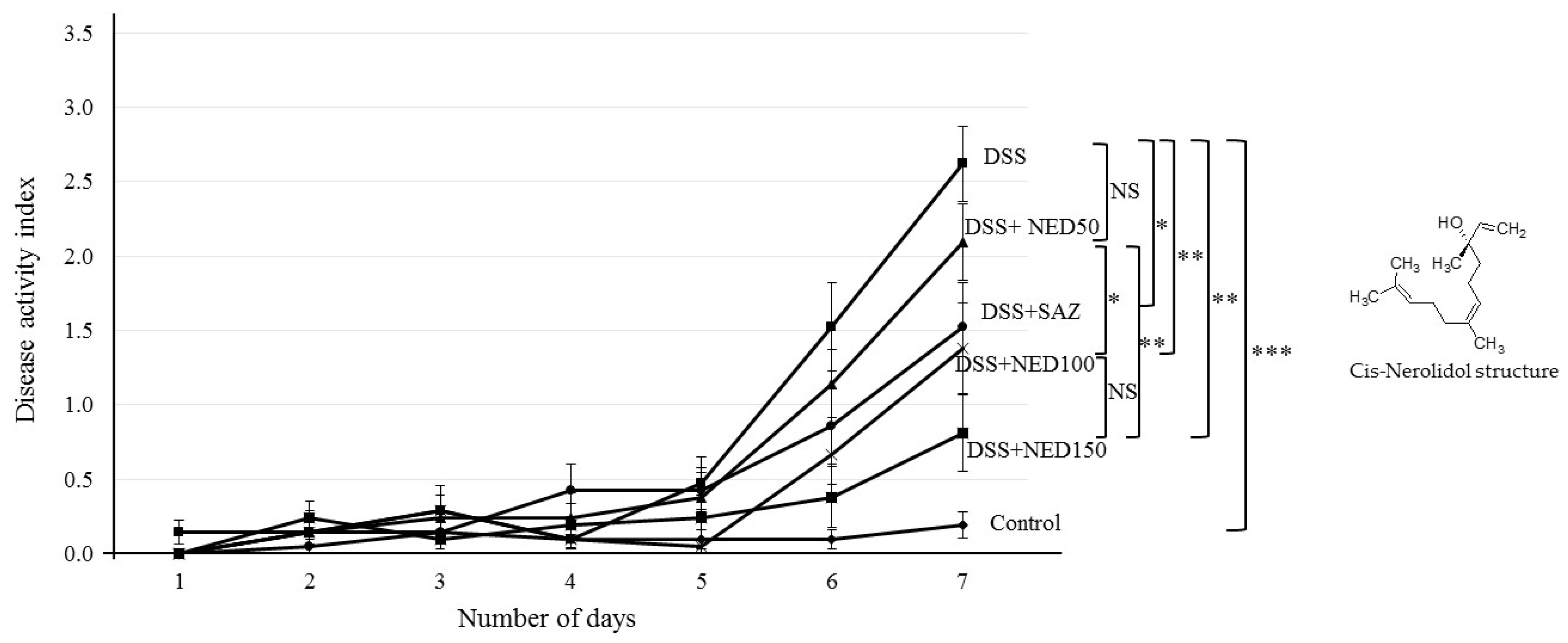
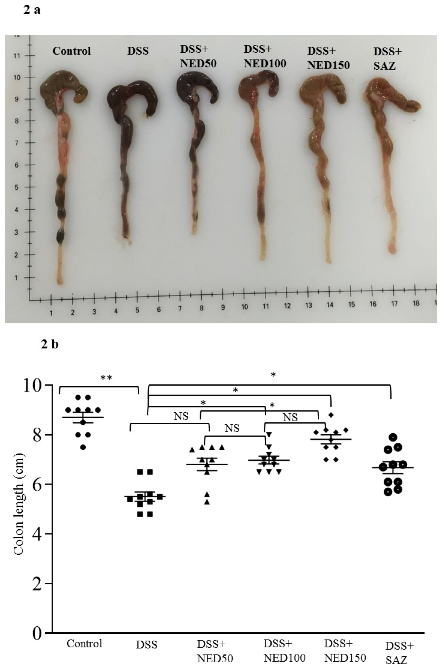
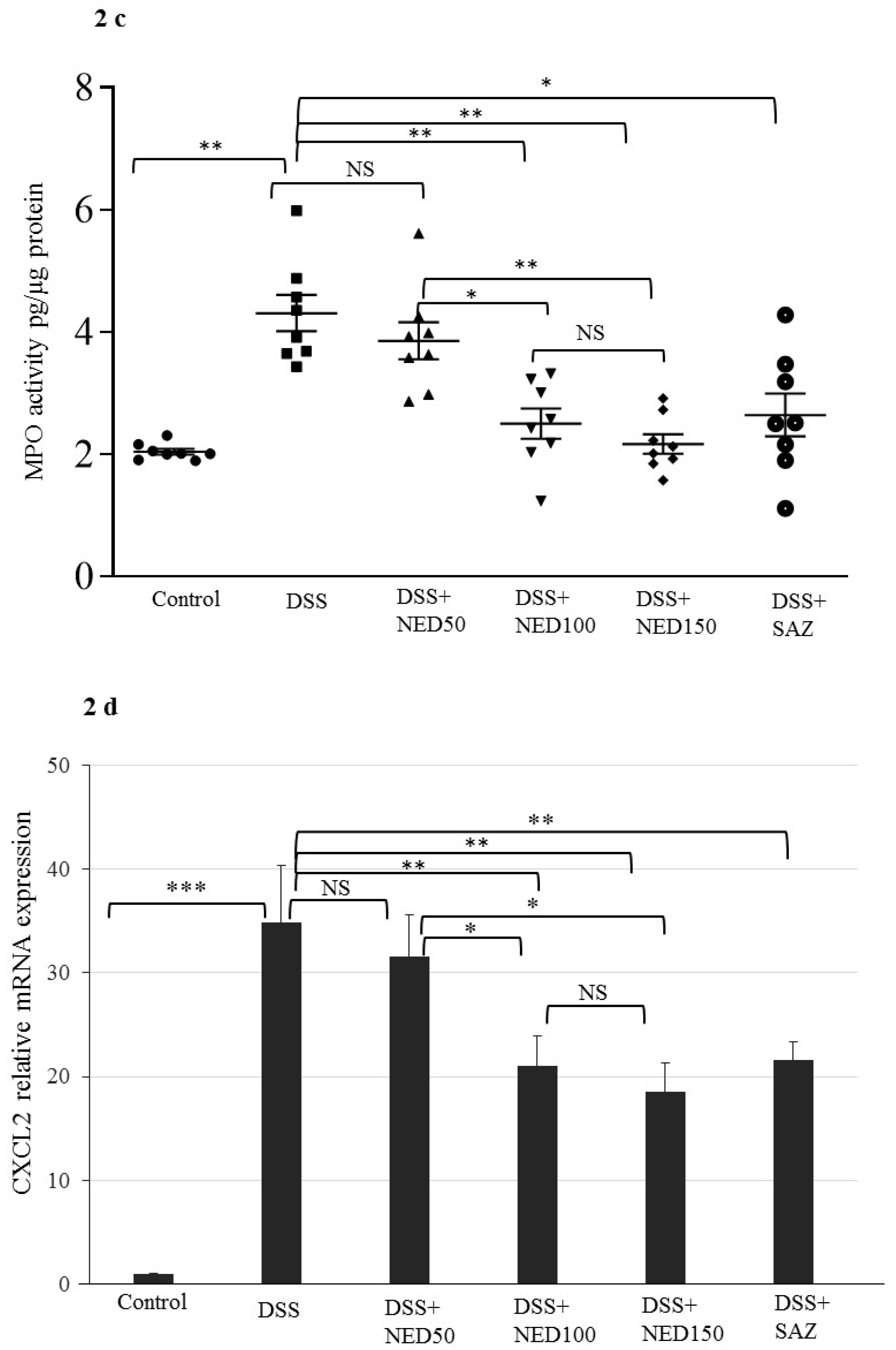
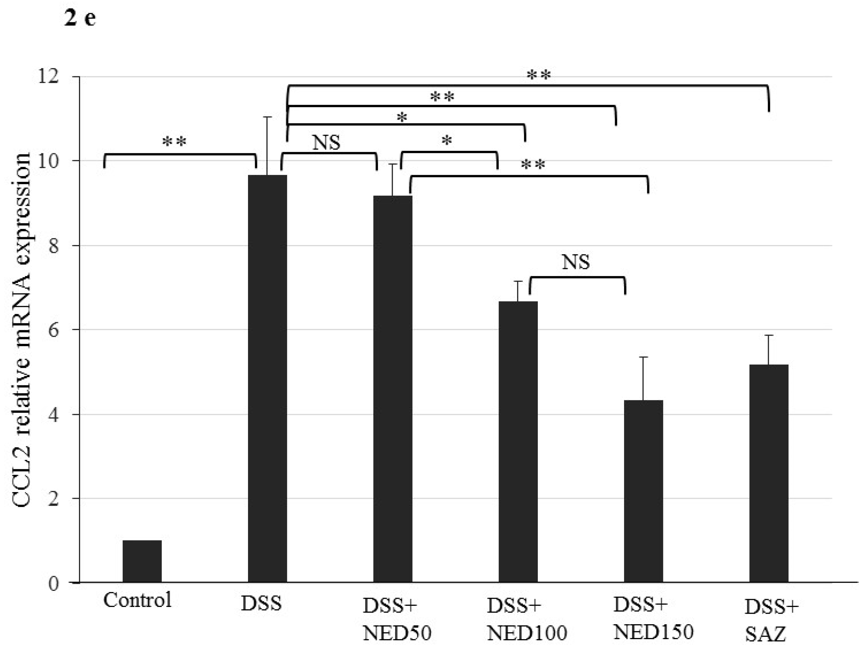
3.1. Effect of NED on Disease Activity Index (DAI), Colon Length, MPO Concentration, and CXCL2 and CCL2 mRNA

In C5BL/6J mice, 3% DSS in drinking water induced distinct features of ulcerative colitis. The DAI score was high in the DSS treated group compared to the control group (p < 0.001). NED treatment of DSS-colitis animals at the higher doses (100 and 150 mg/kg bd wt) significantly (p < 0.01) improved the DAI scores. However, at the lowest dose of 50 mg/kg bd wt, NED treatment had no significant effect. SAZ treatment, as a positive control, showed significant (p < 0.05) improvement in DAI score (Figure 1).
DSS treatment-induced inflammation resulted in significantly (p < 0.01) decreased colon length. SAZ and NED treatment at the two higher doses (100 mg and 150 mg/kg) significantly prevented shortening of colon length (p < 0.05, Figure 2a,b). However, the lowest dose of NED was not effective in preventing the shortening of colon length due to DSS treatment. In the preliminary experiments conducted, NED treatment alone did not affect the colon length (data not shown).
MPO is an enzyme secreted from activated neutrophils and is commonly used as a marker to assess the level of neutrophil infiltration in the submucosa, indicating the degree of inflammation. DSS treatment significantly (p < 0.01) increased MPO concentrations compared to the untreated control group (Figure 2c). NED treatment at the two higher doses and SAZ treatment significantly decreased MPO concentration (p < 0.01, Figure 2c). However, the lowest dose (50 mg/kg) of NED had no significant effect
We measured mRNA expression of the chemokines CXCL2 and CCL2 as markers of macrophage infiltration. DSS treatment significantly increased the expression of both CXCL2 (p < 0.01) and CXCL2 (p < 0.001), indicating macrophage infiltration (Figure 2d,e). NED treatment at the two higher doses (100 and 150 mg/kg) and SAZ treatment significantly decreased the expression of both CXCL2 and CCL2 (Figure 2d,e). However, the lowest dose (50 mg/kg) of NED did not affect CXCL2 and CCL2 expression.

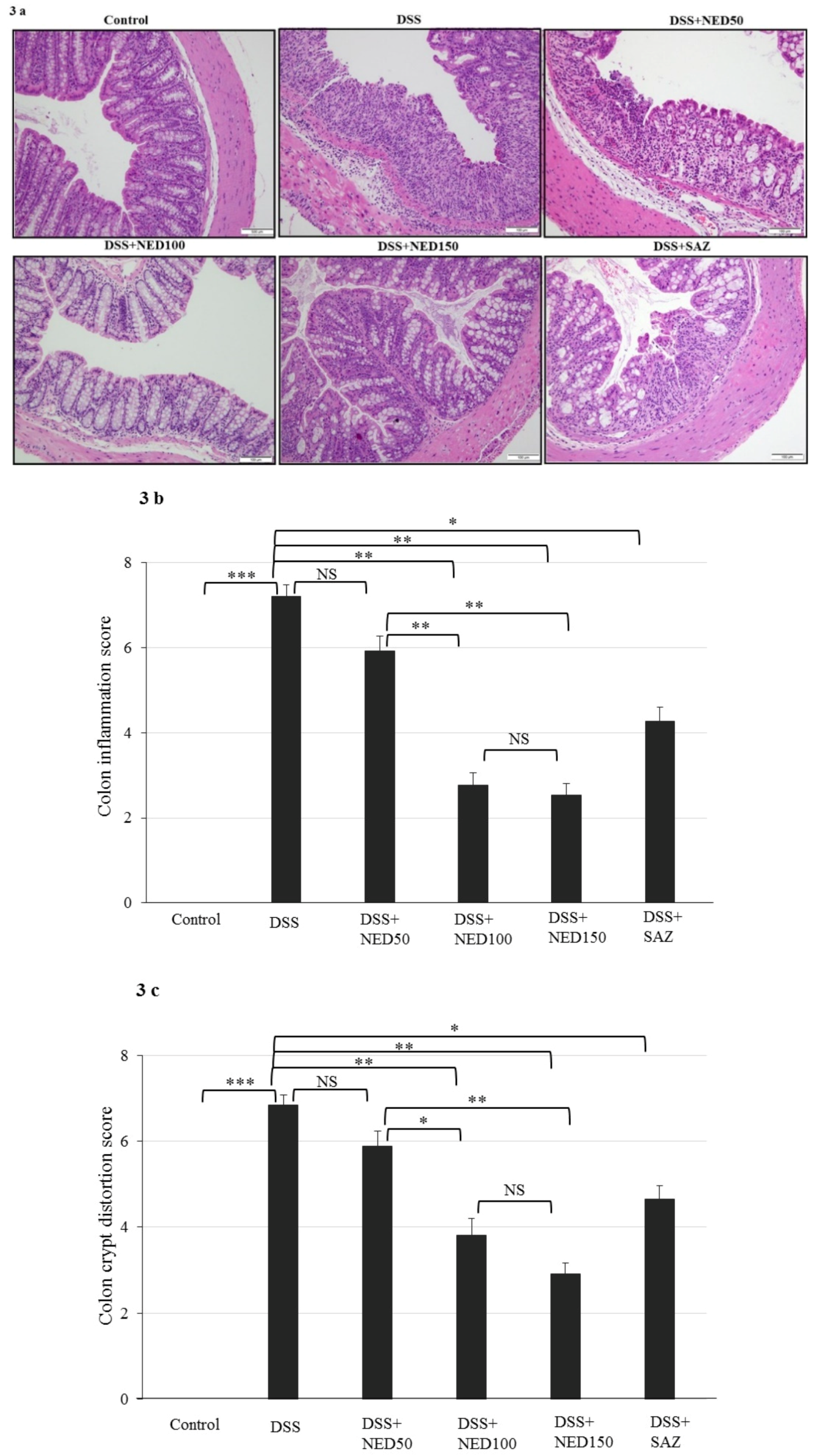
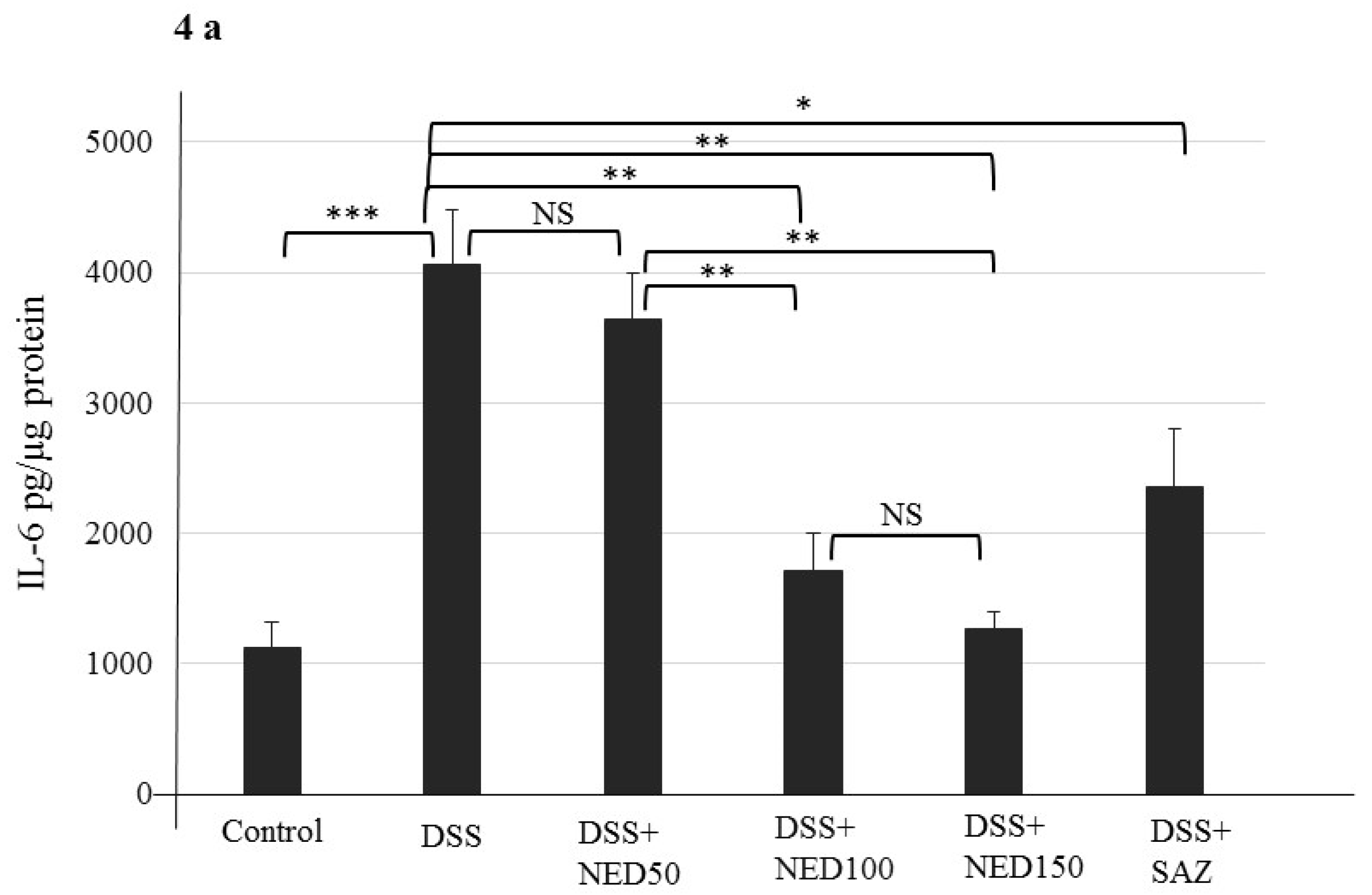
3.2. Effect of NED on Colon Microscopic Architecture

The control healthy group colon sections depicted typical architecture with normal thickness of the submucosa and muscle layers, as well as well-formed crypt structure in the mucosa. In the DSS-treated group, colonic inflammation penetrated the submucosa with focal loss of crypt and the surface epithelium. In contrast, the two higher dose NED-treated, DSS-induced inflammation groups, showed intact epithelium with minimum loss of crypt and inflammation. SAZ-treated DSS group also showed protection of colon microarchitecture. NED treatment alone did affect the microarchitecture of the colon (data not shown).
The most notable histological finding in the DSS-treated group was excessive crypt damage with edema and complete destruction of the colon surface epithelium (Figure 3a). The parameters used for scoring the colonic inflammation and crypt damage are highlighted in Table 2 and Table 3. As expected, the colonic inflammation score was statistically (p < 0.001) high in the DSS-treated group compared to the untreated control group (Figure 3b). NED treatment in DSS-induced inflammation significantly (p < 0.01) reduced the colonic inflammation score compared to DSS-treatment alone at the two higher doses (Figure 3b). As expected, SAZ treatment also reduced DSS-induced inflammation. However, the lowest dose of NED did not reduce inflammation. The crypt damage score was s higher in the DSS-treated group compared to the control group (p < 0.001, Figure 3c). NED-treatment protected crypt damage at the two higher doses (p < 0.01 Figure 3c). Significant protection of crypt damage was also observed in the SAZ-treated DSS group (p < 0.05). However, the lowest dose (50 mg/kg) of NED treatment did not provide any protection from crypt damage.

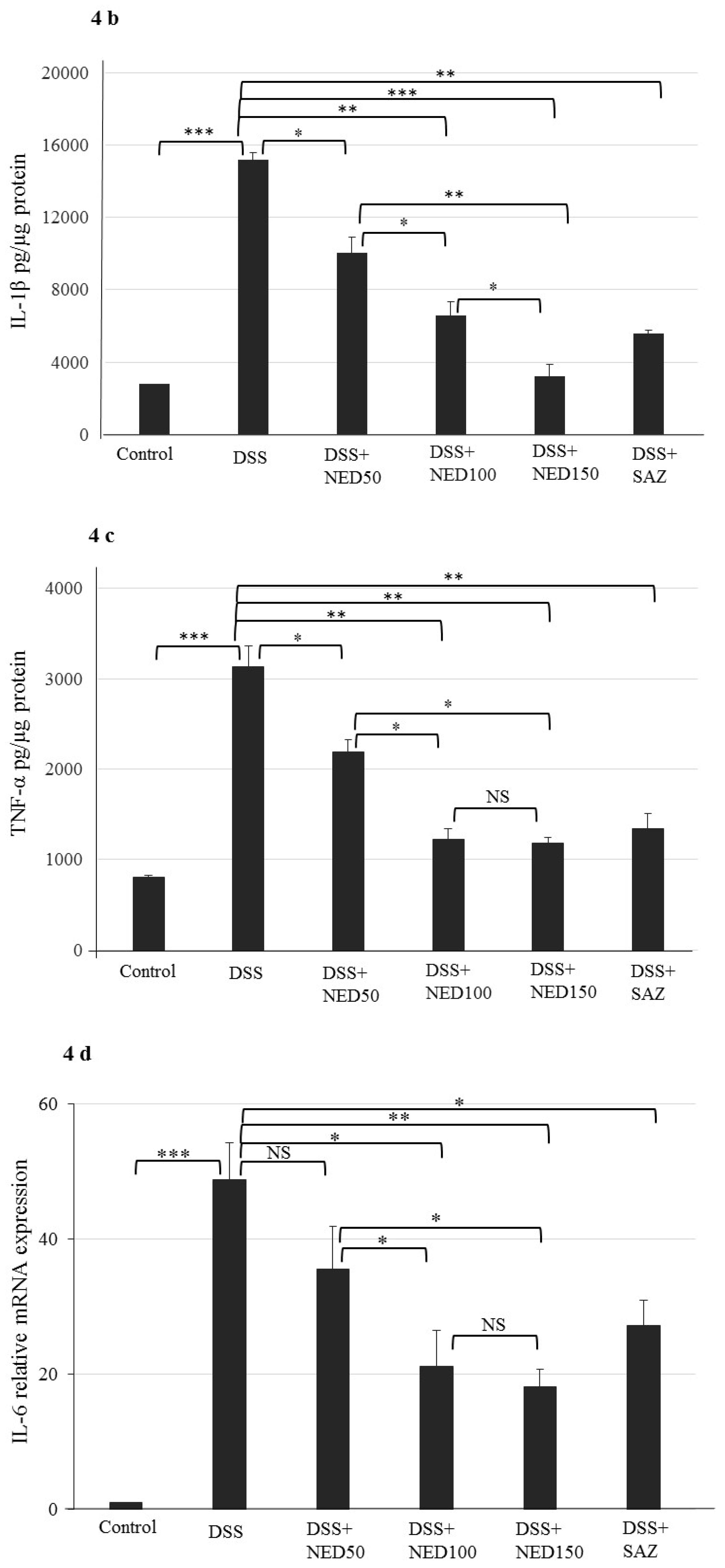
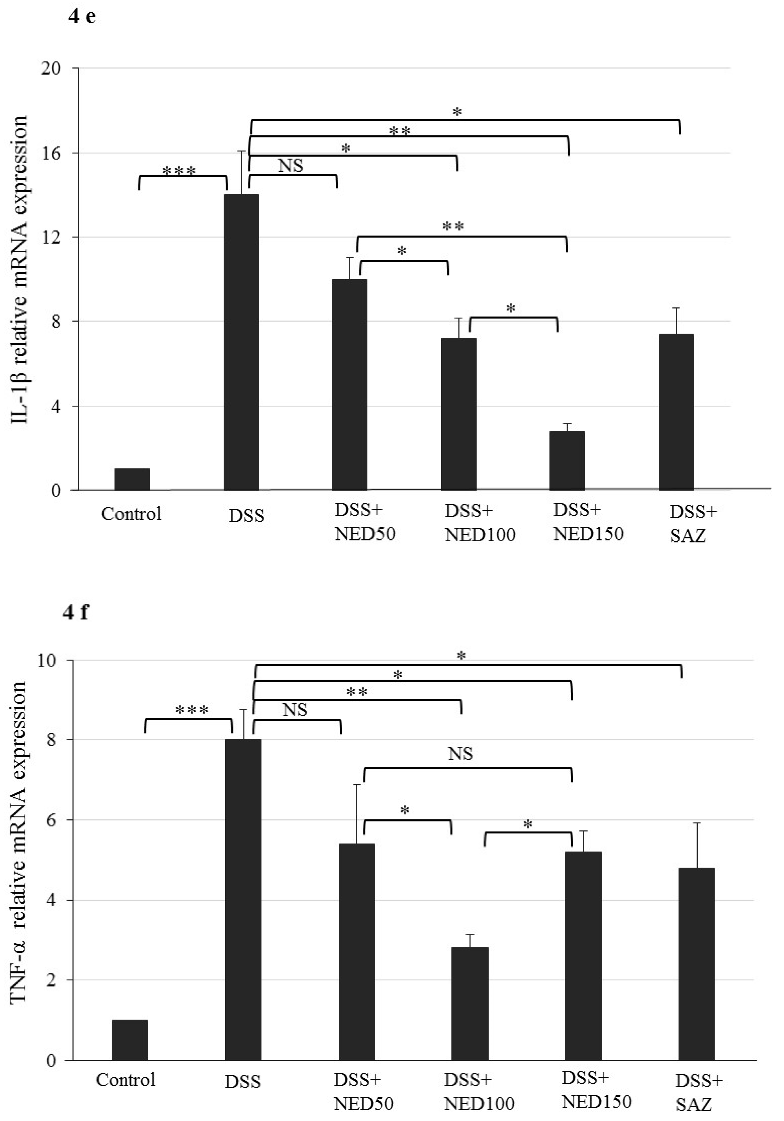
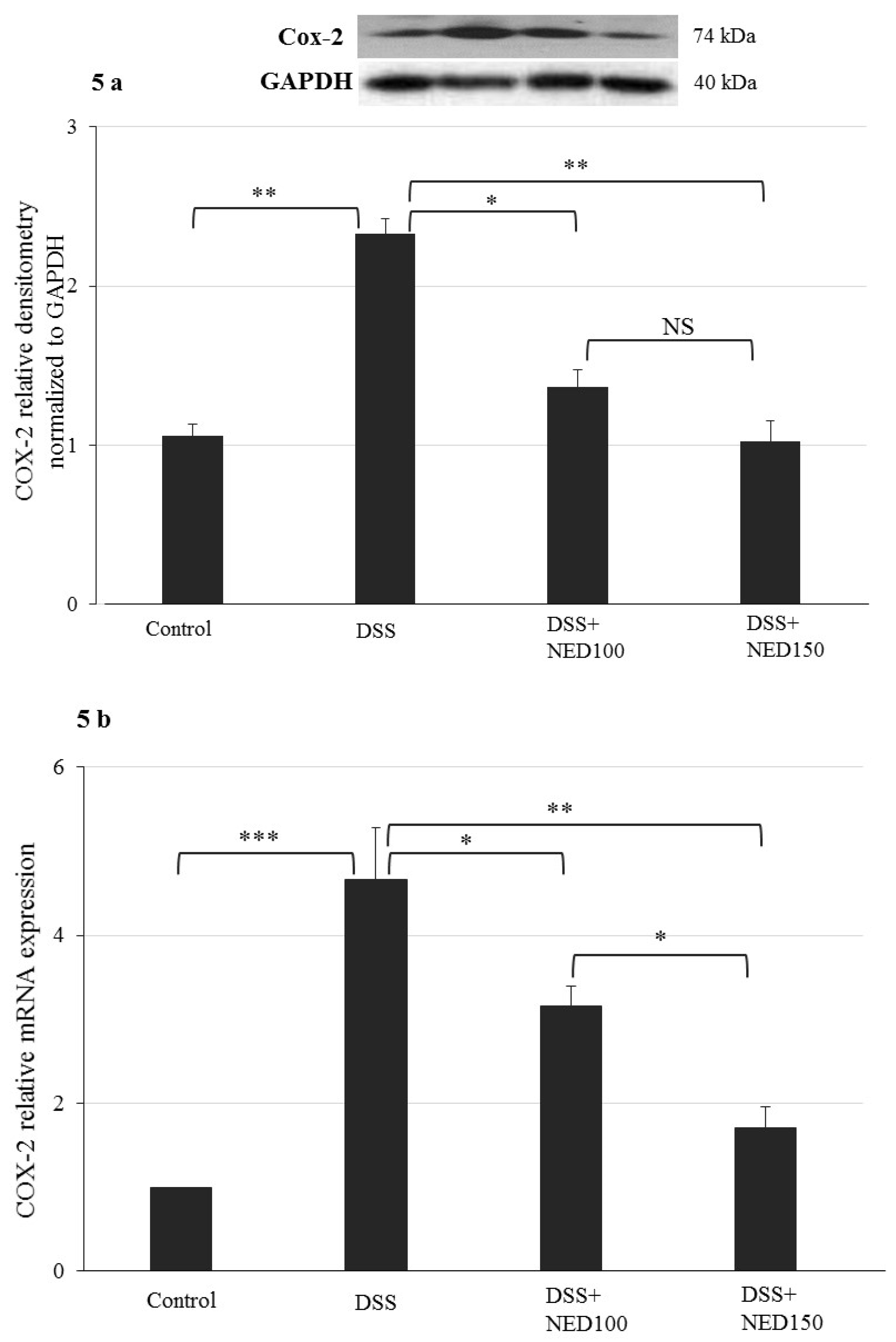
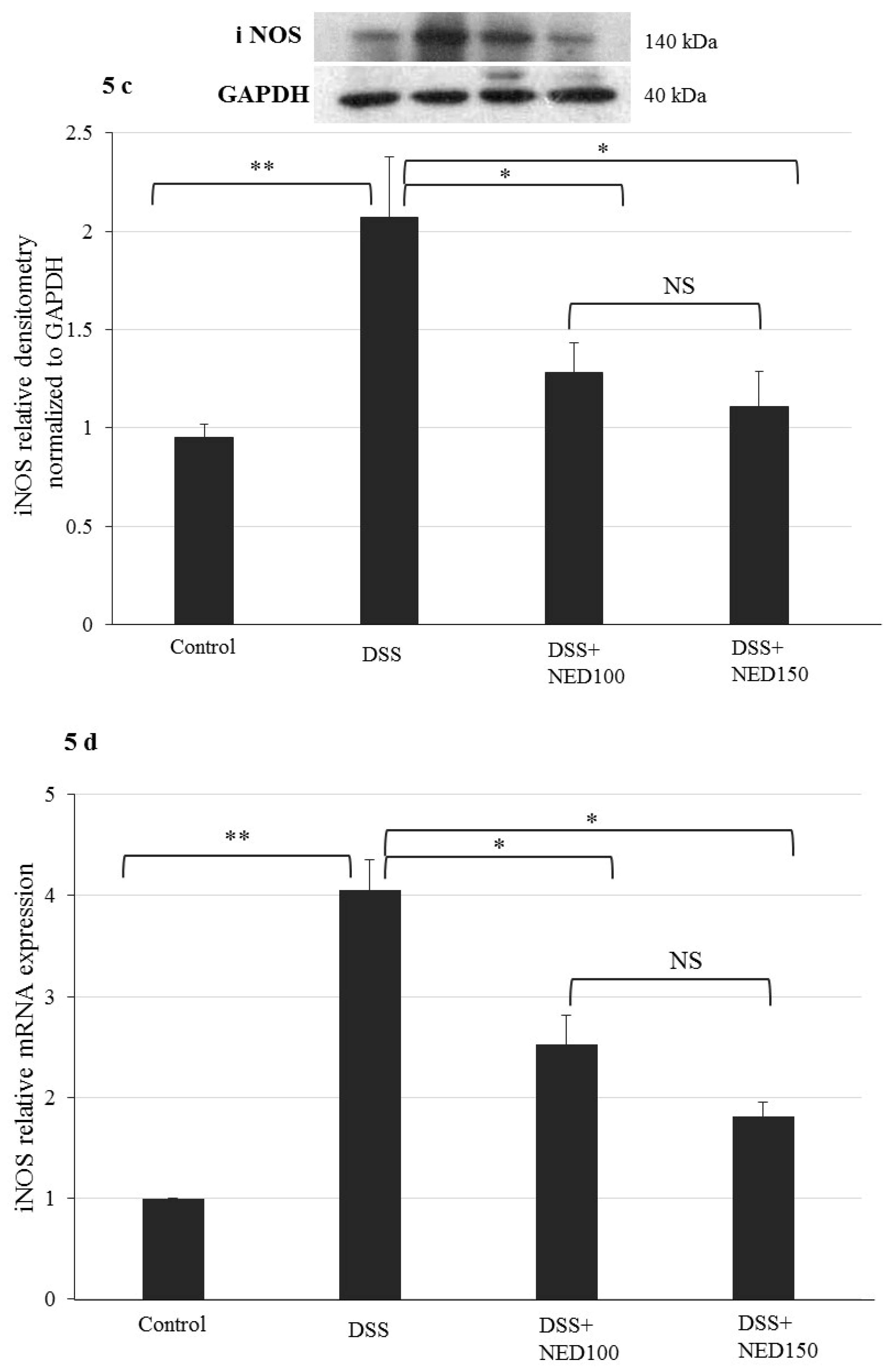
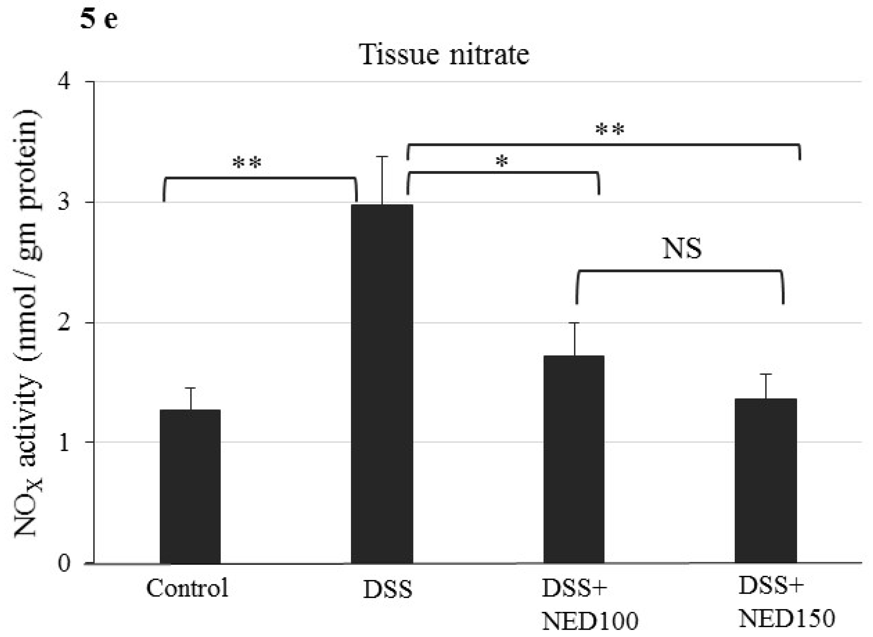
3.3. Effect of NED on Proinflammatory Cytokines and Proinflammatory Mediators

DSS treatment caused increases in concentrations of the proinflammatory cytokines statiscally: IL-6, IL-1β, and TNF-α, (all p < 0.001, Figure 4a–c). Concomitant increases in the relative expression of the mRNA for these respective cytokines (p < 0.001) were seen compared to the untreated control group (Figure 4d–f). NED-treated animals had significantly lower concentrations of these cytokines and their respective mRNAs than the DSS-treated control group (p < 0.01 for IL-6 and IL-1β at 100 mg and 150 mg/kg NED, p < 0.01 at 100 mg and p < 0.001 at 150 mg/kg for TNF-α) (Figure 4). However, the lowest dose of NED did not provide any protection from proinflammatory cytokine expression or release. SAZ treatment also reduced (p < 0.05) the proinflammatory cytokines expression and release (Figure 4). Since these results indicate that the lowest dose of NED (50 mg/kg) is not effective in limiting DSS-induced inflammatory responses, this group was omitted from subsequent studies. Similarly, the SAZ group was also removed from further studies.
The effect of NED in DSS treatment was also evaluated on the proinflammatory enzymes COX-2 and iNOS at both the protein and mRNA level. DSS treatment significantly increased COX-2 and iNOS protein and mRNA expression (COX-2; p < 0.01; COX-2 mRNA p < 0.001; iNOS; p < 0.01 and iNOS mRNA p < 0.01, Figure 5). NED treatment decreased COX-2 protein and mRNA expression (p < 0.05 at 100 mM NED and p < 0.01, at 150 mM NED) (Figure 5a,b). Similarly, NED decreased iNOS protein and mRNA expression (p < 0.05 at both 100 and 150 mM NED) (Figure 5c,d). A decreased in iNOS both at protein and mRNA level upon NED treatment was correlated with decreased in the tissue nitrate level significantly (p < 0.05 at both 100 and 150 mM NED) (Figure 5e).

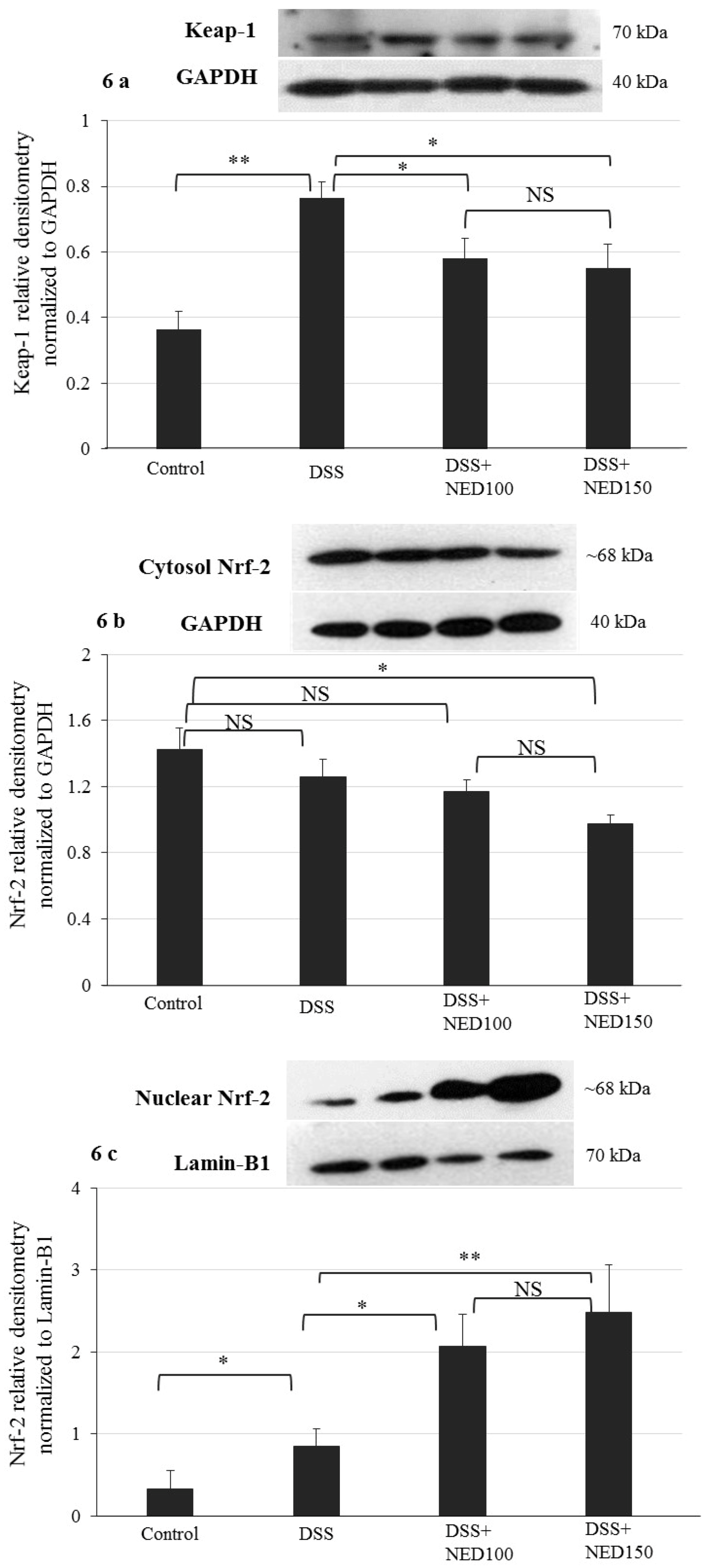
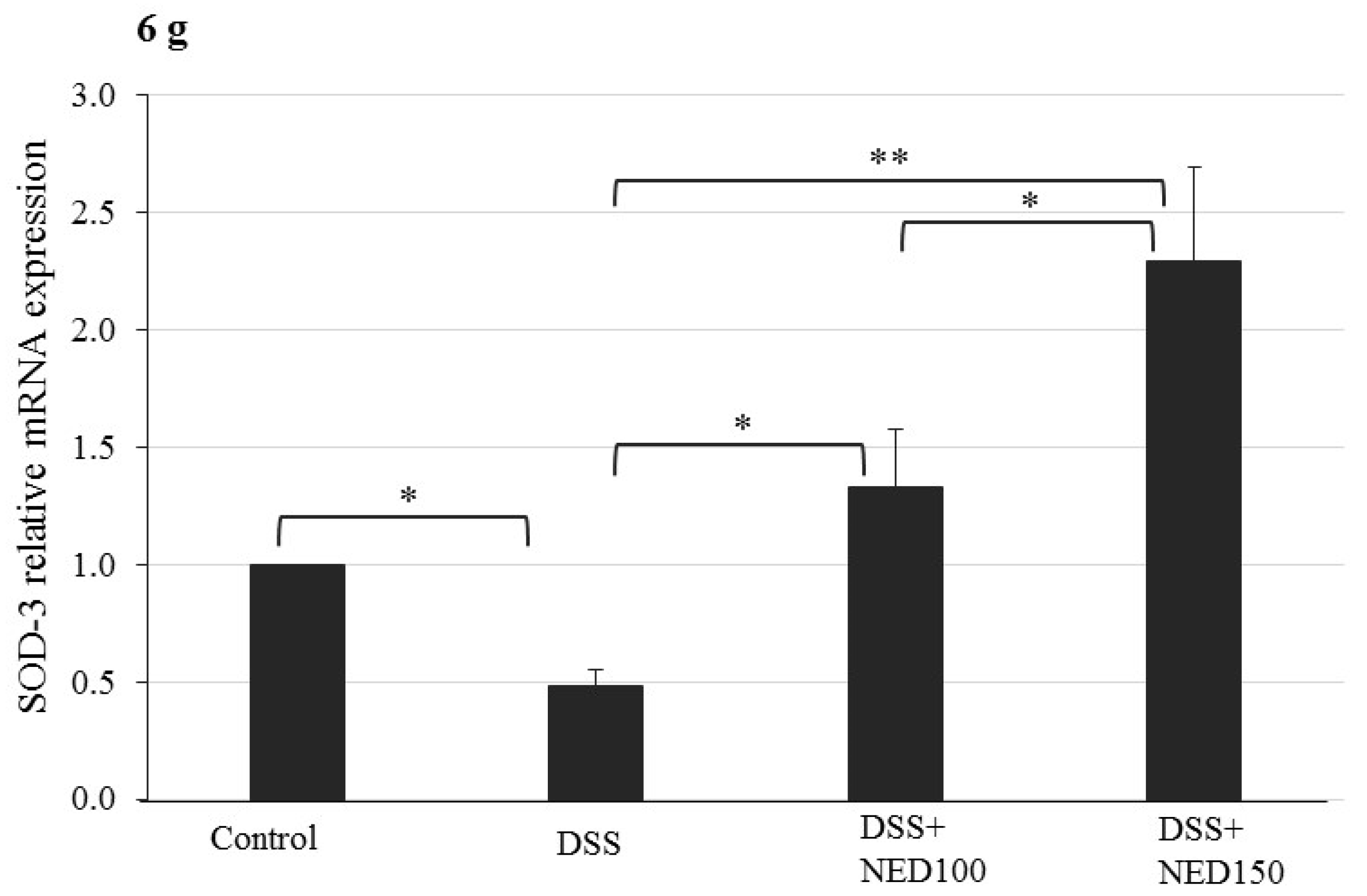
3.4. Effect of NED on Antioxidant Keap-1 and Nrf-2 Transcription Factor Protein, SOD, CAT Activity, and Downstream Target mRNA Expression

Nuclear factor-erythroid 2-related factor (Nrf-2) is a transcription factor of great significance because it is the major regulator of the expression of genes codes for antioxidant enzymes. Keap-1 is a repressor that binds to Nrf-2 and promotes its degradation via the ubiquitin proteasome pathway. We investigated the effect of NED on the Keap-1 and Nrf-2 system. DSS treatment increased Keap-1 expression in the cytoplasm, and NED treatment significantly decreased this response (p < 0.05) (Figure 6a). Under basal conditions, Keap-1 binds to Nrf-2 in the cytoplasm; however, upon activation, Nrf-2 translocates into the nucleus and binds to the antioxidant response element. Therefore, Nrf-2 expression carried out using both cytoplasm and the nuclear fraction of colonic epithelial cells. In the cytoplasm, DSS treatment did not affect the Nrf-2 protein level compared to control; however, the high dose of NED treatment significantly (p < 0.05) decreased the Nrf-2 level (Figure 6b). In contrast, the concentration of Nrf-2 in the nuclear fraction was modestly increased by DSS treatment (p < 0.05) and was statistically increased in a concentration-dependent manner by NED (p < 0.05 at 100 µM and p < 0.01 at 150 µM) (Figure 6c). Numerous cytoprotective genes are regulated by Nrf-2 when translocated into the nucleus [35]. Therefore, tissue superoxide dismutase (SOD) and catalase enzyme activity was determined in NED-treated DSS colitis animals along with expression of Heme Oxygenase-1 (HO-1) and SOD3 mRNA. NED treatment significantly increased colon mucosal SOD (p < 0.05 at 100 mM and p < 0.01 at 150 mM) activity (Figure 6e). However, the catalase activity was increased only at higher dose of NED in statistically significant manner (p < 0.01 at 150 µM) (Figure 6f). NED treatment also increased HO-1 mRNA (p < 0.05 at 100 mM and p < 0.01 at 150 mM) (Figure 6g) and SOD3 mRNA expression significantly (p < 0.05 at 100 mM and p < 0.01 at 150 mM) in DSS colitis colon (Figure 6g).

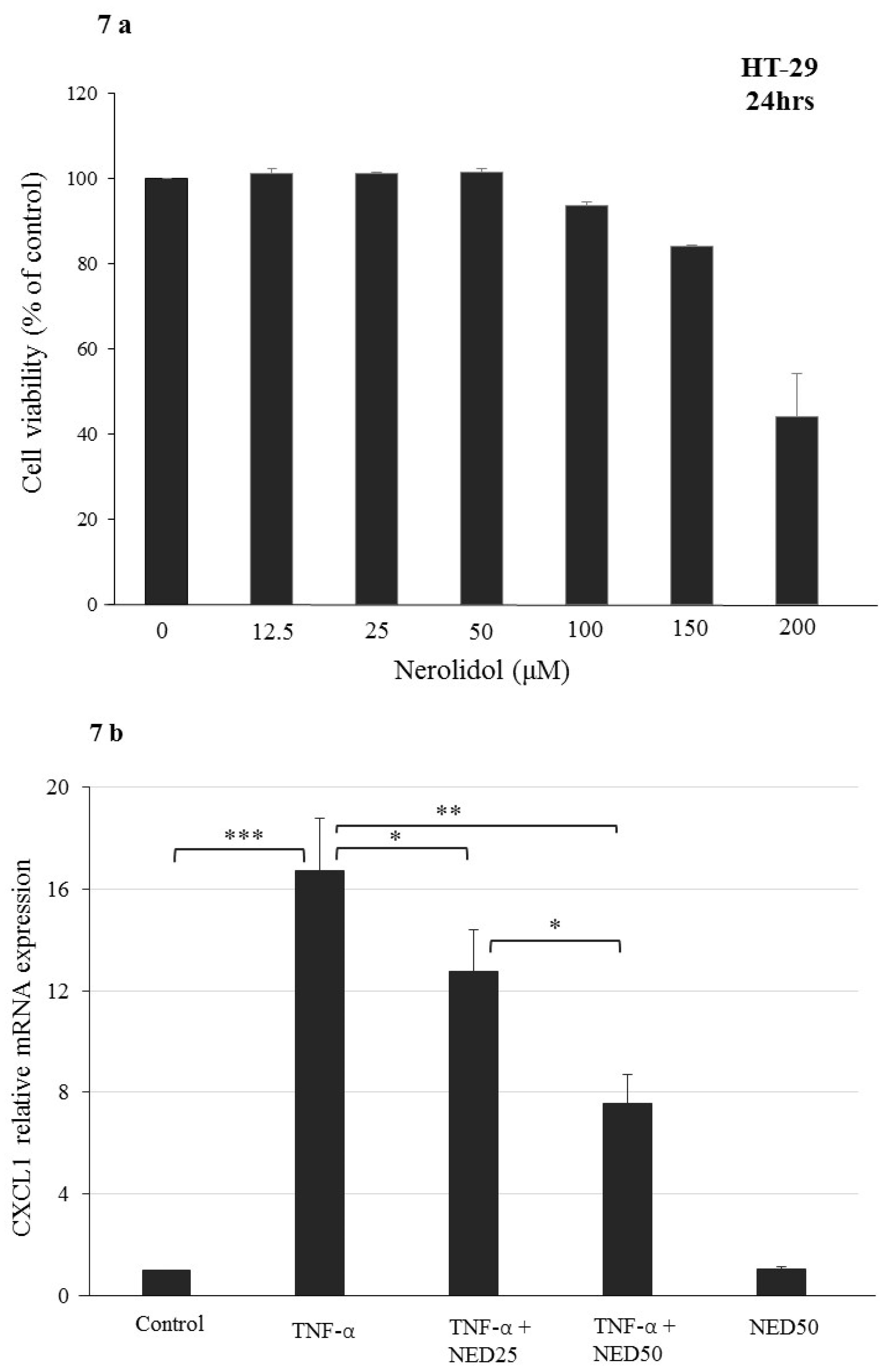
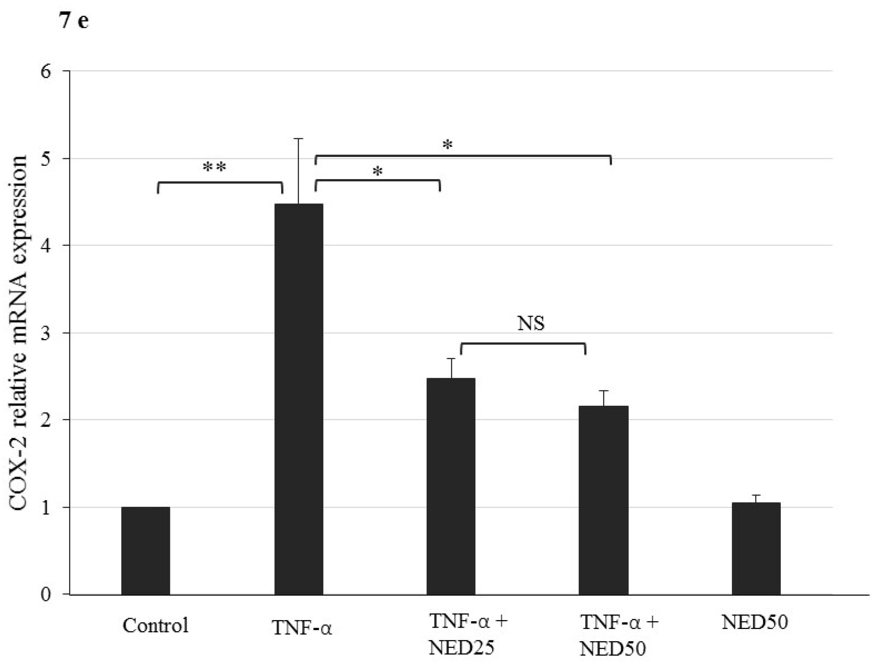
3.5. Effect of NED on TNF-α stimulated HT-29 Colon Cells

We evaluated the anti-inflammatory effect of NED in a disease-relevant cellular context by using TNF-α stimulated HT-29 colon cancer cells in culture, a well-established model for inflammatory process in the gut [34]. To eliminate the possibility of cytotoxicity of NED in HT-29 colon cells and to optimize treatment, we treated these cells with different concentrations (in micromolar range) of NED for 24 hr. No cytotoxic effect observed up to 150 µM concentration. Only at 200 µM, a slight but significant (p < 0.05) cytotoxic effect was observed (Figure 7a). Therefore, the subsequent experiments carried out using 25 to 50 µM NED, concentrations well within the non-cytotoxic range. HT-29 cells stimulated with TNF-α showed a significant increase in the mRNA of the inflammatory marker genes, CXCL1, CXCL2, IL-8 (p < 0.001), and COX-2 (p < 0.01). All these proinflammatory gene markers were significantly downregulated upon NED treatment in TNF-α stimulated HT-29 cells (Figure 7b–e). However, NED alone did not induce any change in gene expression profile compared to the control treatment. These results further confirm the potent anti-inflammatory properties of NED.

4. Discussion

In the present study, we investigated the role of NED in mitigating the colonic inflammation induced by DSS in mice; a well-accepted experimental model of ulcerative colitis. Oral administration of NED alleviated DSS-induced weight loss as well as clinical symptoms as reflected by the DAI score in C56B7/6J mice at a doses of 100 and 150 mg/kg/day PO, which were confirmed by macroscopic, histological and biochemical examinations. High DAI scores and a shortened colon represent the pathological state and the severity of colon in the colitis model group. At the two higher concentrations (100 and 150 mg/kg/day) NED was more effective than the control drug, sulfasalazine (50 mg/kg/day) in reducing inflammation as indicated by all of the studies undertaken. However, the lowest dose of NED (50 m/kg/day) had no significant effect on any inflammatory parameter.
NED significantly prevented the DSS-induced reduction of colon length, and a dose-dependent reduction in the DAI score was also observed with NED treatment. The DSS colitis mice colons exhibited marked histopathological changes, including epithelial erosion and ulceration, crypt abscess formation, mucosal erosion and edema, and substantial infiltration of macrophages. Although no significant effect was observed at 50 mg/kg dose, NED administration was able to preserve near to normal microscopic architecture of colon at the higher doses. Colonic MPO was increased in DSS colitis mice statistically. Upregulated MPO leads to weakened antioxidant defense mechanisms, which will facilitate oxidative injury [36]. NED remarkably reduced inflammation-induced neutrophil infiltration. CXCL2 and CCL2 are known as neutrophil and macrophage chemoattractant chemokines involved in immune responses [37,38]. The DSS-induced increase in mRNA levels of these chemoattractants were significantly prevented by NED.
Infiltration of neutrophils into colonic tissue causes mucosal damage by releasing reactive oxygen species (ROS), which eventually results in the induction of proinflammatory cytokine release, especially TNF-α [4,28]. TNF-α is a primary mediator of the inflammatory response and is closely linked to colonic inflammation of UC [29]. Excessive cytokine production due to immune response is implicated in the intestinal inflammation and the extraintestinal manifestations in IBD [39]. We observed that the elevated tissue levels of IL-6, IL-1β, and TNF-α in DSS treated animals were significantly reduced by higher doses (100 mg and 150 mg/kg bd wt) of NED treatment. These findings are compatible with previous reports, where NED was shown to decrease the influx of polymorphonuclear cells in carrageenan-induced peritonitis, reduce levels of TNF-α in the peritoneal lavage, and reduce production of IL-1β in LPS-stimulated, peritoneal macrophages [27]. These observations indicate that this sesquiterpene has potent anti-inflammatory properties.
Proinflammatory cytokines at the site of inflammation induce cyclooxygenase 2 (COX-2) expression which results in the increased synthesis of prostaglandins and amplification of mucosal inflammation in the intestinal epithelium of IBD patients [40]. Inflammatory cytokines, such as IL-6, are known to upregulate COX-2 expression [41]. Our results reveal increased expression of COX-2 at both the mRNA and protein levels in DSS-treated animals. This increase in COX-2 expression was significantly reduced by NED treatment. In a previous study, NED was shown to suppress TNF-α and IL-6 [42]; therefore, the observed inhibition of COX-2 activity may be facilitated through the suppression of these cytokines. Inducible nitric oxide synthase (iNOS) is expressed in response to bacterial/proinflammatory stimuli and results in nitric oxide (NO) production, which provides cytoprotection. Excessive iNOS expression leads the upregulated NO turnover that is associated with the pathogenesis of IBD [43]. The up-regulation of inducible oxide synthase (iNOS) expression is induced by proinflammatory cytokines in IBD [44]. Our results show that iNOS, a macrophage activation maker, was increased in colonic tissues consequent to DSS-induced injury, as demonstrated by quantitative RT-PCR and Western blotting. NED suppressed the expression of COX-2 and iNOS at both the mRNA and protein levels significantly. In lipopolysaccharide (LPS)-stimulated RAW macrophages model of inflammation, the essential oil from Lindera erythrocarpa, which contains NED, inhibits the expression of iNOS and COX-2 and decreases the subsequent production of NO and prostaglandin E2, respectively [45]. Similarly, NED inhibited iNOS and COX-2 protein expression in a rat model of neuro-inflammation [28]. To address the role of iNOS-derived NO in colitis, studies have been carried out using various iNOS specific inhibitors in in vivo colitis models. However, the results emanating from these studies have revealed conflicting results [46].
Under physiological conditions, a dimer of Nrf2 and Keap1 exists in the cytoplasm. In response to electrophiles or ROS, Nrf2 dissociates from Keap1, which is a negative modulator of Nrf2, and this results in translocation of Nrf2 into the nucleus, where it activates transcription of numerous genes coding for the enzymes involved in antioxidant defense mechanisms [47]. Our results indicate that NED promotes the nuclear translocation of Nrf2 and a decrease in the expression of Keap-1. These results are in agreement with previous studies where sesquiterpenes activate Nrf2 nuclear translocation to modulate antioxidant enzyme expression [48,49,50]. The mRNA expression of heme oxygenase-1 (HO-1) and superoxide dismutase-3 (SOD3), which are among the downstream targets of Nrf-2, were also analyzed. HO-1 is an important enzyme for catabolism of several heme-containing proteins including hemoglobin, myoglobin, and cytochrome p450. A basal level of expression is seen under physiological conditions, but HO-1 is highly expressed in response to stressful stimuli [35,51]. This increased HO-1 expression offers protection during intestinal inflammation [52,53]. Dose-dependent increases in expression of HO-1 mRNA observed in the NED-treated group may provide protection from inflammation. In the present study, SOD3 mRNA expression was decreased in the DSS group, but dose-dependently increased by NED treatment to levels higher than control group. Among the three isoforms of SOD, SOD-3 was shown to be decreased in intestinal epithelial cells (IECs) [54]. It is also reported that increased SOD3 mRNA expression resulted in apoptosis and death of cancer cells and suggestive of cell viability [55]. In a rat model of rotenone-induced Parkinson’s disease, NED maintained cellular SOD, CAT, and GSH and decreased the level of MDA significantly [28]. These results are in agreement with our findings.
The anti-inflammatory activity of NED was also examined using TNF-α-induced HT-29 cells, which are human colonic adenocarcinoma cells with an epithelial morphology. The effects of NED on the expression of COX-2, CXCL1, CXCL2, and IL8 mRNA were measured in cells exposed to TNF-α to induce an inflammatory state. Chemokine ligand-2 (CXCL2) is a chemotactic chemokine produced by colonic epithelial cells and macrophages in response to infection or injury [56]; IL8 is the most active neutrophil chemoattractant that is upregulated during inflammation [34,57]. Chemokine ligand-1 (CXCL1) plays a role in inflammation and tumorigenesis [58]. In colonic mucosa of IBD patients, increased IL-8 and CXCL1 are positively correlated with the disease activity [59]. The results of our study in TNF-α-treated HT-29 cells confirm that NED has a potent anti-inflammatory property. The reductions seen in mRNA expression may be partly attributed to the observations from a study where NED containing essential oil from Liquidambar formosana leaves the inflammatory response suppressed in LPS-induced macrophages [60].

5. Conclusions

NED, which has been shown to suppress inflammation in several different models, exhibited statistically significant anti-inflammatory effects in a well-studied model of inflammatory bowel disease. It was notable that the effects of this compound were at least as efficacious as those of sulfasalazine, a drug commonly used in treatment of Crohn’s disease and ulcerative colitis. Safety appears not to be an issue because NED is used widely as a flavor enhancer in the food industry and has approval from the US Food and Drug Administration for use in food. NED or a synthetic derivative may be valuable in the treatment of inflammatory bowel disease and perhaps other inflammatory conditions too.

Author Contributions

Conceptualization of the study, S.B.S. and S.K.O.; NED compound provision, S.K.O.; performance of the experiments, V.R., B.V., and S.C.; histology work, scoring, and picture provision, S.A.M.; analysis of all other data, V.R., B.V., S.C., T.E.A., and S.B.S., manuscript—first draft preparation, V.R., B.V., T.E.A., and S.B.S.; HT-29 cell line treatments and cell viability data, S.A.; final editing of the manuscript, S.B.S. and T.E.A. All authors have read and agreed to the published version of the manuscript.

Funding

S.B.S. was supported by United Arab Emirates University, CMHS faculty grant, 31M384.

Acknowledgments

The Authors thank Kholoud Arafat for her technical support. The authors would like to thank Bhoomendra Bongade Department of Pharmaceutical Chemistry, RAK College of Pharmaceutical Sciences, RAK Medical & Health Sciences University, Ras Al Khaimah, United Arab Emirates, for providing Cis-Nerolidol structure.

Conflicts of Interest

The authors declare no conflicts of interest.

References

  1. Ballester Ferre, M.P.; Bosca-Watts, M.M.; Minguez Perez, M. Crohn’s disease. Med. Clínica 2018, 151, 26–33. [Google Scholar] [CrossRef] [PubMed]
  2. Ungaro, R.; Mehandru, S.; Allen, P.B.; Peyrin-Biroulet, L.; Colombel, J.F. Ulcerative colitis. Lancet 2017, 389, 1756–1770. [Google Scholar] [CrossRef]
  3. Sairenji, T.; Collins, K.L.; Evans, D.V. An Update on Inflammatory Bowel Disease. Prim. Care 2017, 44, 673–692. [Google Scholar] [CrossRef] [PubMed]
  4. Zhang, Y.Z.; Li, Y.Y. Inflammatory bowel disease: Pathogenesis. World J. Gastroenterol. 2014, 20, 91–99. [Google Scholar] [CrossRef] [PubMed]
  5. Ghishan, F.K.; Kiela, P.R. Epithelial transport in inflammatory bowel diseases. Inflamm. Bowel Dis. 2014, 20, 1099–1109. [Google Scholar] [CrossRef] [Green Version]
  6. Xavier, R.J.; Podolsky, D.K. Unravelling the pathogenesis of inflammatory bowel disease. Nature 2007, 448, 427–434. [Google Scholar] [CrossRef]
  7. Barbosa, D.S.; Cecchini, R.; El Kadri, M.Z.; Rodriguez, M.A.; Burini, R.C.; Dichi, I. Decreased oxidative stress in patients with ulcerative colitis supplemented with fish oil omega-3 fatty acids. Nutrition 2003, 19, 837–842. [Google Scholar] [CrossRef]
  8. Kruidenier, L.; Kuiper, I.; Lamers, C.B.; Verspaget, H.W. Intestinal oxidative damage in inflammatory bowel disease: Semi-quantification, localization, and association with mucosal antioxidants. J. Pathol. 2003, 201, 28–36. [Google Scholar] [CrossRef]
  9. Monk, J.M.; Zhang, C.P.; Wu, W.; Zarepoor, L.; Lu, J.T.; Liu, R.; Pauls, K.P.; Wood, G.A.; Tsao, R.; Robinson, L.E.; et al. White and dark kidney beans reduce colonic mucosal damage and inflammation in response to dextran sodium sulfate. J. Nutr. Biochem. 2015, 26, 752–760. [Google Scholar] [CrossRef]
  10. Binienda, A.; Ziolkowska, S.; Ingvild, H.H.; Salaga, M. The Role of Immune and Epithelial Stem Cells in Inflammatory Bowel Disease Therapy. Curr. Drug Targets 2020. [Google Scholar] [CrossRef]
  11. De Souza, H.S.; Fiocchi, C. Immunopathogenesis of IBD: Current state of the art. Nat. Rev. Gastroenterol. Hepatol. 2016, 13, 13–27. [Google Scholar] [CrossRef] [PubMed]
  12. Shepherd, C.; Giacomin, P.; Navarro, S.; Miller, C.; Loukas, A.; Wangchuk, P. A medicinal plant compound, capnoidine, prevents the onset of inflammation in a mouse model of colitis. J. Ethnopharmacol. 2018, 211, 17–28. [Google Scholar] [CrossRef]
  13. Kwak, M.S.; Cha, J.M.; Ahn, J.H.; Chae, M.K.; Jeong, S.; Lee, H.H. Practical strategy for optimizing the timing of anti-tumor necrosis factor-alpha therapy in Crohn disease: A nationwide population-based study. Medicine 2020, 99, e18925. [Google Scholar] [CrossRef]
  14. Braus, N.A.; Elliott, D.E. Advances in the pathogenesis and treatment of IBD. Clin. Immunol. 2009, 132, 1–9. [Google Scholar] [CrossRef] [PubMed] [Green Version]
  15. Head, K.A.; Jurenka, J.S. Inflammatory bowel disease Part 1: Ulcerative colitis--pathophysiology and conventional and alternative treatment options. Altern. Med. Rev. 2003, 8, 247–283. [Google Scholar]
  16. Guo, B.J.; Bian, Z.X.; Qiu, H.C.; Wang, Y.T.; Wang, Y. Biological and clinical implications of herbal medicine and natural products for the treatment of inflammatory bowel disease. Ann. N. Y. Acad. Sci. 2017, 1401, 37–48. [Google Scholar] [CrossRef] [PubMed]
  17. Triantafyllidi, A.; Xanthos, T.; Papalois, A.; Triantafillidis, J.K. Herbal and plant therapy in patients with inflammatory bowel disease. Ann. Gastroenterol. 2015, 28, 210–220. [Google Scholar]
  18. Danciu, C.; Avram, S.; Pavel, I.Z.; Ghiulai, R.; Dehelean, C.A.; Ersilia, A.; Minda, D.; Petrescu, C.; Moaca, E.A.; Soica, C. Main Isoflavones Found in Dietary Sources as Natural Anti-inflammatory Agents. Curr. Drug Targets 2018, 19, 841–853. [Google Scholar] [CrossRef]
  19. Wu, Z.Y.; Sang, L.X.; Chang, B. Isoflavones and inflammatory bowel disease. World J. Clin. Cases 2020, 8, 2081–2091. [Google Scholar] [CrossRef]
  20. Zhou, Y.; Zeng, L.; Liu, X.; Gui, J.; Mei, X.; Fu, X.; Dong, F.; Tang, J.; Zhang, L.; Yang, Z. Formation of (E)-nerolidol in tea (Camellia sinensis) leaves exposed to multiple stresses during tea manufacturing. Food Chem. 2017, 231, 78–86. [Google Scholar] [CrossRef] [PubMed]
  21. Alves, R.F.; Nascimento, A.M.D.; Nogueira, J.M.F. Characterization of the aroma profile of Madeira wine by sorptive extraction techniques. Anal. Chim. Acta 2005, 546, 11–21. [Google Scholar] [CrossRef] [PubMed]
  22. Green, S.A.; Chen, X.; Nieuwenhuizen, N.J.; Matich, A.J.; Wang, M.Y.; Bunn, B.J.; Yauk, Y.K.; Atkinson, R.G. Identification, functional characterization, and regulation of the enzyme responsible for floral (E)-nerolidol biosynthesis in kiwifruit (Actinidia chinensis). J. Exp. Bot. 2012, 63, 1951–1967. [Google Scholar] [CrossRef] [Green Version]
  23. Yan, J.W.; Ban, Z.J.; Lu, H.Y.; Li, D.; Poverenov, E.; Luo, Z.S.; Li, L. The aroma volatile repertoire in strawberry fruit: A review. J. Sci. Food Agric. 2018, 98, 4395–4402. [Google Scholar] [CrossRef]
  24. Chan, W.K.; Tan, L.T.; Chan, K.G.; Lee, L.H.; Goh, B.H. Nerolidol: A Sesquiterpene Alcohol with Multi-Faceted Pharmacological and Biological Activities. Molecules 2016, 21, 529. [Google Scholar] [CrossRef] [Green Version]
  25. De Carvalho, R.B.F.; De Almeida, A.A.C.; Campelo, N.B.; Lellis, D.; Nunes, L.C.C. Nerolidol and its Pharmacological Application in Treating Neurodegenerative Diseases: A Review. Recent Pat. Biotechnol. 2018, 12, 158–168. [Google Scholar] [CrossRef]
  26. Zhou, M.X.; Li, G.H.; Sun, B.; Xu, Y.W.; Li, A.L.; Li, Y.R.; Ren, D.M.; Wang, X.N.; Wen, X.S.; Lou, H.X.; et al. Identification of novel Nrf2 activators from Cinnamomum chartophyllum H.W. Li and their potential application of preventing oxidative insults in human lung epithelial cells. Redox Biol. 2018, 14, 154–163. [Google Scholar] [CrossRef]
  27. Fonseca, D.V.; Salgado, P.R.; de Carvalho, F.L.; Salvadori, M.G.; Penha, A.R.; Leite, F.C.; Borges, C.J.; Piuvezam, M.R.; Pordeus, L.C.; Sousa, D.P.; et al. Nerolidol exhibits antinociceptive and anti-inflammatory activity: Involvement of the GABAergic system and proinflammatory cytokines. Fundam. Clin. Pharmacol. 2016, 30, 14–22. [Google Scholar] [CrossRef]
  28. Javed, H.; Azimullah, S.; Abul Khair, S.B.; Ojha, S.; Haque, M.E. Neuroprotective effect of nerolidol against neuroinflammation and oxidative stress induced by rotenone. BMC Neurosci. 2016, 17, 58. [Google Scholar] [CrossRef] [PubMed] [Green Version]
  29. Subramanya, S.B.; Chandran, S.; Almarzooqi, S.; Raj, V.; Al Zahmi, A.S.; Al Katheeri, R.A.; Al Zadjali, S.A.; Collin, P.D.; Adrian, T.E. Frondanol, a Nutraceutical Extract from Cucumaria frondosa, Attenuates Colonic Inflammation in a DSS-Induced Colitis Model in Mice. Mar. Drugs 2018, 16, 148. [Google Scholar] [CrossRef] [PubMed] [Green Version]
  30. Livak, K.J.; Schmittgen, T.D. Analysis of relative gene expression data using real-time quantitative PCR and the 2(-Delta Delta C(T)) Method. Methods 2001, 25, 402–408. [Google Scholar] [CrossRef]
  31. Kakkar, P.; Das, B.; Viswanathan, P.N. A modified spectrophotometric assay of superoxide dismutase. Indian J. Biochem. Biophys. 1984, 21, 130–132. [Google Scholar] [PubMed]
  32. Sinha, A.K. Colorimetric assay of catalase. Anal. Biochem. 1972, 47, 389–394. [Google Scholar] [CrossRef]
  33. Lu, S.C.; Wu, H.W.; Lin, Y.J.; Chang, S.F. The essential role of Oct-2 in LPS-induced expression of iNOS in RAW 264.7 macrophages and its regulation by trichostatin A. Am. J. Physiol. Cell Physiol. 2009, 296, C1133–C1139. [Google Scholar] [CrossRef] [Green Version]
  34. Wilson, C.A.; Browning, J.L. Death of HT29 adenocarcinoma cells induced by TNF family receptor activation is caspase-independent and displays features of both apoptosis and necrosis. Cell Death Differ. 2002, 9, 1321–1333. [Google Scholar] [CrossRef] [Green Version]
  35. Loboda, A.; Damulewicz, M.; Pyza, E.; Jozkowicz, A.; Dulak, J. Role of Nrf2/HO-1 system in development, oxidative stress response and diseases: An evolutionarily conserved mechanism. Cell. Mol. Life Sci. 2016, 73, 3221–3247. [Google Scholar] [CrossRef] [Green Version]
  36. Gautam, M.; Goel, S.; Ghatule, R.; Singh, A.; Nath, G.; Goel, R. Curative effect of Terminalia chebula extract on acetic acid-induced experimental colitis: Role of antioxidants, free radicals and acute inflammatory marker. Inflammopharmacology 2013, 21, 377–383. [Google Scholar] [CrossRef] [PubMed]
  37. Al-Alwan, L.A.; Chang, Y.; Mogas, A.; Halayko, A.J.; Baglole, C.J.; Martin, J.G.; Rousseau, S.; Eidelman, D.H.; Hamid, Q. Differential roles of CXCL2 and CXCL3 and their receptors in regulating normal and asthmatic airway smooth muscle cell migration. J. Immunol. 2013, 191, 2731–2741. [Google Scholar] [CrossRef] [Green Version]
  38. Deshmane, S.L.; Kremlev, S.; Amini, S.; Sawaya, B.E. Monocyte chemoattractant protein-1 (MCP-1): An overview. J. Interferon Cytokine Res. 2009, 29, 313–326. [Google Scholar] [CrossRef]
  39. Neurath, M.F. Cytokines in inflammatory bowel disease. Nat. Rev. Immunol. 2014, 14, 329–342. [Google Scholar] [CrossRef]
  40. Jurjus, A.; Eid, A.; Al Kattar, S.; Zeenny, M.N.; Gerges-Geagea, A.; Haydar, H.; Hilal, A.; Oueidat, D.; Matar, M.; Tawilah, J.; et al. Inflammatory bowel disease, colorectal cancer and type 2 diabetes mellitus: The links. BBA Clin. 2016, 5, 16–24. [Google Scholar] [CrossRef] [Green Version]
  41. Tsatsanis, C.; Androulidaki, A.; Venihaki, M.; Margioris, A.N. Signalling networks regulating cyclooxygenase-2. Int. J. Biochem. Cell Biol. 2006, 38, 1654–1661. [Google Scholar] [CrossRef]
  42. Zhang, L.; Sun, D.; Bao, Y.; Shi, Y.; Cui, Y.; Guo, M. Nerolidol Protects Against LPS-induced Acute Kidney Injury via Inhibiting TLR4/NF-kappaB Signaling. Phytother Res. 2017, 31, 459–465. [Google Scholar] [CrossRef]
  43. Minhas, R.; Bansal, Y.; Bansal, G. Inducible nitric oxide synthase inhibitors: A comprehensive update. Med. Res. Rev. 2019. [Google Scholar] [CrossRef]
  44. Soufli, I.; Toumi, R.; Rafa, H.; Touil-Boukoffa, C. Overview of cytokines and nitric oxide involvement in immuno-pathogenesis of inflammatory bowel diseases. World J. Gastrointest. Pharmacol. Ther. 2016, 7, 353–360. [Google Scholar] [CrossRef]
  45. Ko, Y.J.; Ahn, G.; Ham, Y.M.; Song, S.M.; Ko, E.Y.; Cho, S.H.; Yoon, W.J.; Kim, K.N. Anti-inflammatory effect and mechanism of action of Lindera erythrocarpa essential oil in lipopolysaccharide-stimulated RAW264.7 cells. EXCLI J. 2017, 16, 1103–1113. [Google Scholar] [CrossRef]
  46. Kolios, G.; Valatas, V.; Ward, S.G. Nitric oxide in inflammatory bowel disease: A universal messenger in an unsolved puzzle. Immunology 2004, 113, 427–437. [Google Scholar] [CrossRef]
  47. Yu, M.; Wei, Z.Y.; Xu, Z.H.; Pan, J.Q.; Chen, J.H. Oxidative Damage and Nrf2 Translocation Induced by Toxicities of Deoxynivalenol on the Placental and Embryo on Gestation Day 12.5 d and 18.5 d. Toxins 2018, 10, 370. [Google Scholar] [CrossRef] [Green Version]
  48. Peng, S.; Hou, Y.; Yao, J.; Fang, J. Activation of Nrf2 by costunolide provides neuroprotective effect in PC12 cells. Food Funct. 2019, 10, 4143–4152. [Google Scholar] [CrossRef]
  49. Liu, L.; Wu, W.; Li, J.; Jiao, W.H.; Liu, L.Y.; Tang, J.; Sun, F.; Han, B.N.; Lin, H.W. Two sesquiterpene aminoquinones protect against oxidative injury in HaCaT keratinocytes via activation of AMPKalpha/ERK-Nrf2/ARE/HO-1 signaling. Biomed. Pharmacother. 2018, 100, 417–425. [Google Scholar] [CrossRef]
  50. Park, J.H.; Choi, J.W.; Ju, E.J.; Pae, A.N.; Park, K.D. Antioxidant and Anti-Inflammatory Activities of a Natural Compound, Shizukahenriol, through Nrf2 Activation. Molecules 2015, 20, 15989–16003. [Google Scholar] [CrossRef] [Green Version]
  51. Intagliata, S.; Salerno, L.; Ciaffaglione, V.; Leonardi, C.; Fallica, A.N.; Carota, G.; Amata, E.; Marrazzo, A.; Pittala, V.; Romeo, G. Heme Oxygenase-2 (HO-2) as a therapeutic target: Activators and inhibitors. Eur. J. Med. Chem. 2019, 183, 111703. [Google Scholar] [CrossRef] [PubMed]
  52. Takagi, T.; Naito, Y.; Mizushima, K.; Hirai, Y.; Harusato, A.; Okayama, T.; Katada, K.; Kamada, K.; Uchiyama, K.; Handa, O.; et al. Heme oxygenase-1 prevents murine intestinal inflammation. J. Clin. Biochem. Nutr. 2018, 63, 169–174. [Google Scholar] [CrossRef] [PubMed] [Green Version]
  53. Takagi, T.; Naito, Y.; Mizushima, K.; Nukigi, Y.; Okada, H.; Suzuki, T.; Hirata, I.; Omatsu, T.; Okayama, T.; Handa, O.; et al. Increased intestinal expression of heme oxygenase-1 and its localization in patients with ulcerative colitis. J. Gastroenterol. Hepatol. 2008, 23, S229–S233. [Google Scholar] [CrossRef] [PubMed]
  54. Kruidenier, L.; Kuiper, I.; van Duijn, W.; Marklund, S.L.; van Hogezand, R.A.; Lamers, C.B.; Verspaget, H.W. Differential mucosal expression of three superoxide dismutase isoforms in inflammatory bowel disease. J. Pathol. 2003, 201, 7–16. [Google Scholar] [CrossRef]
  55. Laukkanen, M.O. Extracellular Superoxide Dismutase: Growth Promoter or Tumor Suppressor? Oxid. Med. Cell. Longev. 2016, 2016, 3612589. [Google Scholar] [CrossRef] [Green Version]
  56. Ohtsuka, Y.; Lee, J.; Stamm, D.S.; Sanderson, I.R. MIP-2 secreted by epithelial cells increases neutrophil and lymphocyte recruitment in the mouse intestine. Gut 2001, 49, 526–533. [Google Scholar] [CrossRef] [Green Version]
  57. Fuhr, L.; Rousseau, M.; Plauth, A.; Schroeder, F.C.; Sauer, S. Amorfrutins Are Natural PPARgamma Agonists with Potent Anti-inflammatory Properties. J. Nat. Prod. 2015, 78, 1160–1164. [Google Scholar] [CrossRef]
  58. Ogata, H.; Sekikawa, A.; Yamagishi, H.; Ichikawa, K.; Tomita, S.; Imura, J.; Ito, Y.; Fujita, M.; Tsubaki, M.; Kato, H.; et al. GROalpha promotes invasion of colorectal cancer cells. Oncol. Rep. 2010, 24, 1479–1486. [Google Scholar] [PubMed]
  59. Imada, A.; Ina, K.; Shimada, M.; Yokoyama, T.; Yokoyama, Y.; Nishio, Y.; Yamaguchi, T.; Ando, T.; Kusugami, K. Coordinate upregulation of interleukin-8 and growth-related gene product-alpha is present in the colonic mucosa of inflammatory bowel. Scand. J. Gastroenterol. 2001, 36, 854–864. [Google Scholar] [CrossRef]
  60. Hua, K.F.; Yang, T.J.; Chiu, H.W.; Ho, C.L. Essential oil from leaves of Liquidambar formosana ameliorates inflammatory response in lipopolysaccharide-activated mouse macrophages. Nat. Prod. Commun. 2014, 9, 869–872. [Google Scholar] [CrossRef] [PubMed] [Green Version]
Figure 1. Effect of Nerolidol (NED) on disease activity index (DAI). Dextran sodium sulfate (DSS) treatment significantly increased the DAI score. Sulfasalazine (SAZ) and the higher concentrations of NED significantly prevented the increase in DAI scores induced by DSS. However, the lowest dose of NED (50 mg/kg) treatment had no effect. Data were obtained from n = 10 animals in each group and are expressed as mean ± SEM. *** p ≤ 0.001. ** p ≤ 0.01. * p ≤ 0.05. P values were obtained by one-way ANOVA followed by Tukey’s multiple comparison test using SPSS software, and p ≤ 0.05 was considered statistically significant.
Nutrients 12 02032 g001
Figure 2. Effect of NED on colon length, MPO concentration, CXCL2 and CCL2 mRNA macrophage markers. The mean colon length was significantly decreased in the DSS-treated group. The higher doses of NED-treatment and SAZ-treatment significantly prevented the decrease in colon length compared to DSS group. The lowest concentration of NED had no effect (a,b). Data were obtained from n = 10 animals in each group and are expressed as means ± SEM. * p ≤ 0.05. DSS-treatment significantly increased MPO concentrations compared to untreated controls. The higher doses of NED-and SAZ reduced MPO concentrations compared to DSS treatment alone. However, the lowest dose of NED had no effect (c). Data were obtained from n = 10 animals in each group and are expressed as means ± SEM. ** p ≤ 0.01 * p ≤ 0.05. DSS-treatment significantly increased colonic CXCL2 and CCL2 mRNA levels compared to controls. The higher concentrations of NED and SAZ-treatment prevented the increase of both CXCL2 and CCL2 mRNA concentrations compared to DSS alone. However, the lowest concentration of NED-had no effect (d,e). Data were obtained from n = 8 animals in each group and are expressed as means ± SEM: *** p ≤ 0.001 ** p ≤ 0.01, and * p ≤ 0.05 were considered statistically significant, and NS indicates not significant.
Nutrients 12 02032 g002aNutrients 12 02032 g002bNutrients 12 02032 g002c
Figure 3. Effect of NED on colon histology. (a) Microscopic analysis shows typical architecture of the colon with normal thickness of the submucosa, muscle layer and regular crypt and villi structure in the mucosa in the control samples (Scale Bars: 100 µM). The DSS-induced colitis colon section shows focal loss of crypts and surface epithelium with inflammation reaching up to the submucosa. NED-treatment, at 100 and 150 mg/kg, protected the microscopic architecture in DSS-induced colitis. Crypt distortion score and colon inflammation scores were high in DSS-induced colitis group. NED treatment at 100 and 150 mg/kg significantly reduced both colon inflammation scores (b) and crypt distortion (c). The lowest concentration of NED (50 mg/kg) had no effect. Data were obtained from n = 8 animals in each group and are expressed as means ± SEM. *** p ≤ 0.001. ** p ≤ 0.01. * p ≤ 0.05. p values were obtained by one-way ANOVA followed by Tukey’s multiple comparison test using SPSS software. p ≤ 0.05 was considered statistically significant, and NS indicates not significant.
Nutrients 12 02032 g003
Figure 4. Effect of NED on proinflammatory cytokine protein and mRNA expression. NED-treatment, at 100 and 150 mg/kg, significantly inhibited the DSS-induced increase in concentrations of proinflammatory cytokines, at both the protein level (IL-6 (a), IL-1β (b), and TNF-α (c)) and mRNA expression levels (IL-6 (d), IL-1β (e), and TNF-α (f)) in DSS-induced colitis group. Data were obtained from n = 8 animals for ELISA and n = 6 animals for mRNA expression studies in each group and expressed as means ± SEM. *** p ≤ 0.001. ** p ≤ 0.01. * p ≤ 0.05. p values were obtained by one-way ANOVA followed by Tukey’s multiple comparison test using SPSS software. p ≤ 0.05 is considered statistically significant, and NS indicates not significant.
Nutrients 12 02032 g004aNutrients 12 02032 g004bNutrients 12 02032 g004c
Figure 5. Effect of NED on inflammatory mediator COX-2 and inducible oxide synthase (iNOS) protein and mRNA expression and tissue nitrate levels. NED-treatment significantly inhibited the DSS-induced expression of COX-2 and iNOS at both the protein (a,b) and mRNA levels (c,d) and also reduced the tissue nitrate levels (e). Data were obtained from n = 4 animals for Western blot and n = 6 animals for mRNA expression and tissue nitrate level studies in each group and expressed as means ± SEM. *** p ≤ 0.001. ** p ≤ 0.01. * p ≤ 0.05; p values were obtained by one-way ANOVA followed by Tukey’s multiple comparison test using SPSS software, and p ≤ 0.05 was considered statistically significant.
Nutrients 12 02032 g005aNutrients 12 02032 g005bNutrients 12 02032 g005c
Figure 6. Effect of NED on Keap-1 and Nrf-2 and antioxidant system. At the two higher concentrations, NED prevented the DSS-induced increase in Keap-1 protein expression (a). The nuclear translocation of the Nfr-2 protein was enhanced by NED treatment in DSS-induced colitis (b,c). Superoxide dismutase (SOD) and catalase enzyme activities were significantly increased in NED-treated DSS colitis animals (d,e). Heme Oxygenase-1 (HO-1) and SOD3 mRNA expression were also significantly increased in NED-treated DSS colitis in mice (f,g). Data were obtained from n = 4 animals for the Western blot studies, n = 6 for enzyme activity and mRNA level. Data expressed as means ± SEM. *** p ≤ 0.001. ** p ≤ 0.01. * p ≤ 0.05. p values were obtained by one-way ANOVA followed by Tukey’s multiple comparison test using SPSS software, and p ≤ 0.05 was considered statistically significant.
Nutrients 12 02032 g006aNutrients 12 02032 g006bNutrients 12 02032 g006c
Figure 7. Effect of NED on cell viability and proinflammatory cytokine mRNA expression in TNF-α-treated HT-29 human colonic cancer cells. The concentration-dependent cytotoxic effects of NED were investigated treatment of HT-29 cells with different concentrations of NED for 24 h. NED had no effect on HT-29 cell viability at concentrations up to 100 µM, so its anti-inflammatory effects were examined at concentrations of 25 and 50 µM in the subsequent experiments (a). To investigate the effect of NED on inflammatory cytokine production, cells were treated with 1 ng/mL TNF-α for 24 h. Real-time PCR was carried out to detect gene expression of the inflammatory markers CXCl1, CXCL2, IL-8, and COX-2 (be). Data are shown relative to DMSO vehicle-treated cells. Data expressed as mean ± SEM (n = 6). *** p ≤ 0.001. ** p ≤ 0.01. * p ≤ 0.05. p values were obtained by one-way ANOVA followed by Tukey’s multiple comparison test using SPSS software. p ≤ 0.05 was considered statistically significant, and NS indicates not significant.
Nutrients 12 02032 g007aNutrients 12 02032 g007bNutrients 12 02032 g007c
Table 1. Disease activity index score.
Weight LossScoreStool ConsistencyScoreRectal BleedingScore
No loss0Normal0No Blood0
1–5%1Loose stool2Heme occult + ve and visual pellet bleeding2
5–10%2Diarrhea4Gross bleeding and blood around anus4
10–20%3
>20%4
Table 2. Colon tissue histological examination Crypt aberration score.
Inflammation GradedPercentage of Inflammation Involvement of Mucosal Surface AreaHyperplastic Epithelium—Graded Based on Extent of Involvement
none0no inflammation0none
mild11–25%11–25%
moderate226–50%226–50%
severe351–75%351–75%
476–100%476–100%
Table 3. Colon tissue histological examination for inflammation Score.
Crypt GradeQuantification on Percentage of Crypt ChangeCrypt Distortion—Graded Based on Extent of Involvement
Grade 0Intact crypt11–25%0no crypt distortion
Grade 1Shortening and loss of basal 1/3 of crypts226–50%11–25%
Grade 2Loss of basal 2/3 of crypts351–75%226–50%
Grade 3Loss of entire crypt with intact surface epithelium476–100%351–75%
Grade 4Loss of both entire crypt and surface epithelium (erosion) 476–100%
Table 4. Primers list.
GeneForwardReversePMID NoGene Accession Number
CXCL2 (mouse)5′-GGATGGCTTTCATGGAAGGAG-3′5′-TTGCTAAGCAAGGCACTGTGC-3′22326488NM_009140.2
CCL2 (mouse)5′-CAGCCAGATGCAGTTAACGC-3′5′-GCCTACTCATTGGGATCATCTTG-3′10953027NM_011333.2
IL-6 (mouse)5′-ACAAGTCGGAGGCTTAATTACACAT-3′5′-TTGCCATTGCACAACTCTTTTC-3′21735552X06203
TNF-α (mouse)5′-AGGCTGCCCCGACTACGT-3′5′-GACTTTCTCCTGGTATGAGATAGCAAA-3′21705622NM_013693.2
IL-1β (mouse)5′-TCGCTCAGGGTCACAAGAAA-3′5′-CATCAGAGGCAAGGAGGAAAC-3′21735552NM_008361.4
COX-2 (mouse)5′-AACCGCATTGCCTCTGAAT-3′5′-CATGTTCCAGGAGGATGGAG-3′22158945NM_011198.4
iNOS (mouse)5′-CGAAACGCTTCACTTCCAA-3′5′-TGAGCCTATATTGCTGTGGCT-3′22158945BC062378.1
NRF-2 (mouse)5′-GAGCTAGATAGTGCCCCTGG-3′5′-CAGGACTCACGGGAACTTCT-3′29162986U20532.1
HO-1 (mouse)5′-AAGCCGAGAATGCTGAGTTCA-3′5′-GCCGTGTAGATATGGTACAAGGA-3′25112868BC010757.1
SOD3 (mouse)5′-TTCTACGGCTTGCTACTGGC-3′5′-GCTAGGTCGAAGCTGGACTC-3′26513461NM_011435.3
18S (mouse)5′-CCCCTCGATGACTTTAGCTGAGTGT-3′5′-CGCCGGTCCAAGAATTTCACCTCT-3′22427817NR_003278
CXCL1 (human)5′-GCGGAAAGCTTGCCTCAATC-3′5′-GGTCAGTTGGATTTGTCACTGT-3′25938459BC011976.1
IL-8 (human)5′-CTGATTTCTGCAGCTCTGTG-3′5′-GGGTGGAAAGGTTTGGAGTATG-3′20150959BC013615.1
CXCL2 (human)5′-TTTATTGTGGGCTTCACACG-3′5′-GATTTGCGCACACAGACAAC-3′17591792NM_004591.3
COX2 (human)5′-ACAGTGTGTGGTCAACATTTCTC-3′5′-TCGAAACCTCTCTGCTCTAACAC-3′25938459BC015753.1
18S (human)5′-GTGGAGCGATTTGTCTGGTT-3′5′-AACGCCACTTGTCCCTCTAA-3′25369870NR_003286

