Review

Vitamin D and Immunity in Infants and Children

by
Geneviève Mailhot
1,2,* and
John H. White
3,4,*
1
Department of Nutrition, University of Montreal, Montreal, QC H3T 1A8, Canada
2
Centre Hospitalier Universitaire Sainte-Justine, Montreal, QC H3T 1C5, Canada
3
Department of Physiology, McGill University, Montreal, QC H3G 1Y6, Canada
4
Department of Medicine, McGill University, Montreal, QC H4A 3J1, Canada
*
Authors to whom correspondence should be addressed.
Nutrients 2020, 12(5), 1233; https://doi.org/10.3390/nu12051233
Submission received: 8 April 2020 / Revised: 24 April 2020 / Accepted: 25 April 2020 / Published: 27 April 2020
(This article belongs to the Special Issue Vitamin D on Immune Function)

Abstract

:
The last couple of decades have seen an explosion in our interest and understanding of the role of vitamin D in the regulation of immunity. At the molecular level, the hormonal form of vitamin D signals through the nuclear vitamin D receptor (VDR), a ligand-regulated transcription factor. The VDR and vitamin D metabolic enzymes are expressed throughout the innate and adaptive arms of the immune system. The advent of genome-wide approaches to gene expression profiling have led to the identification of numerous VDR-regulated genes implicated in the regulation of innate and adaptive immunity. The molecular data infer that vitamin D signaling should boost innate immunity against pathogens of bacterial or viral origin. Vitamin D signaling also suppresses inflammatory immune responses that underlie autoimmunity and regulate allergic responses. These findings have been bolstered by clinical studies linking vitamin D deficiency to increased rates of infections, autoimmunity, and allergies. Our goals here are to provide an overview of the molecular basis for immune system regulation and to survey the clinical data from pediatric populations, using randomized placebo-controlled trials and meta-analyses where possible, linking vitamin D deficiency to increased rates of infections, autoimmune conditions, and allergies, and addressing the impact of supplementation on these conditions.

1. Introduction

Vitamin D is obtained from several sources: The diet, supplements, or seasonal exposure of skin to adequate solar ultraviolet irradiation via photochemical and thermal conversion of the cholesterol precursor 7-dehydrocholesterol. However, many diets are quite poor in vitamin D, and in populous temperate regions, notably in northern Europe and Scandinavia, vitamin D winter, the period where cutaneous vitamin D synthesis cannot occur, is substantially longer than 6 months of the year [1]. Thus, in epidemiological studies, residence at higher latitudes and lower sun exposure have become proxies for risk of a poor vitamin D status. In addition, sun avoidance and conservative dress in more southern regions leads to vitamin D deficiency in several populations worldwide [2]. The risk of vitamin D deficiency occurs at all stages of life, including during pregnancy, in infants and children, as well as in adults. In infants, it can be exacerbated by sun avoidance habits that are usually recommended for very young children, limited body stores, the risk of poor vitamin D content of human milk in exclusively breastfed infants, and a lack of knowledge regarding systematic vitamin D supplementation in some cultures [3].
Vitamin D is best known for its role as a key regulator of calcium homeostasis and bone health in both children and adults [4]. However, it has been the subject of increasing interest in both the scientific literature and the popular press over the last couple of decades, largely because of its “non-classical” actions in tissues unrelated to calcium homeostasis, particularly in the regulation of several aspects of immune system function. Ours goals here are to first provide an overview of the molecular basis for the evidence of a physiological role of vitamin D in tissues unrelated to calcium homeostasis, with a focus on the immune system. We will then detail the clinical evidence for links between vitamin D deficiency and immune system dysfunction in infants and children, with an emphasis on intervention trials, where available. Associations between deficiency and rates of infection; autoimmune conditions, such as type 1 diabetes (T1D) and multiple sclerosis (MS); and allergic conditions, including asthma, will be surveyed. We will also analyze the potential links between vitamin D deficiency and pediatric inflammatory bowel disease and dental caries, both of which are linked to immune system function. Based on the studies detailed below, there is a compelling need for further clinical analysis of the potential benefit of vitamin D supplementation in pediatric populations at risk for several immune-related disorders.

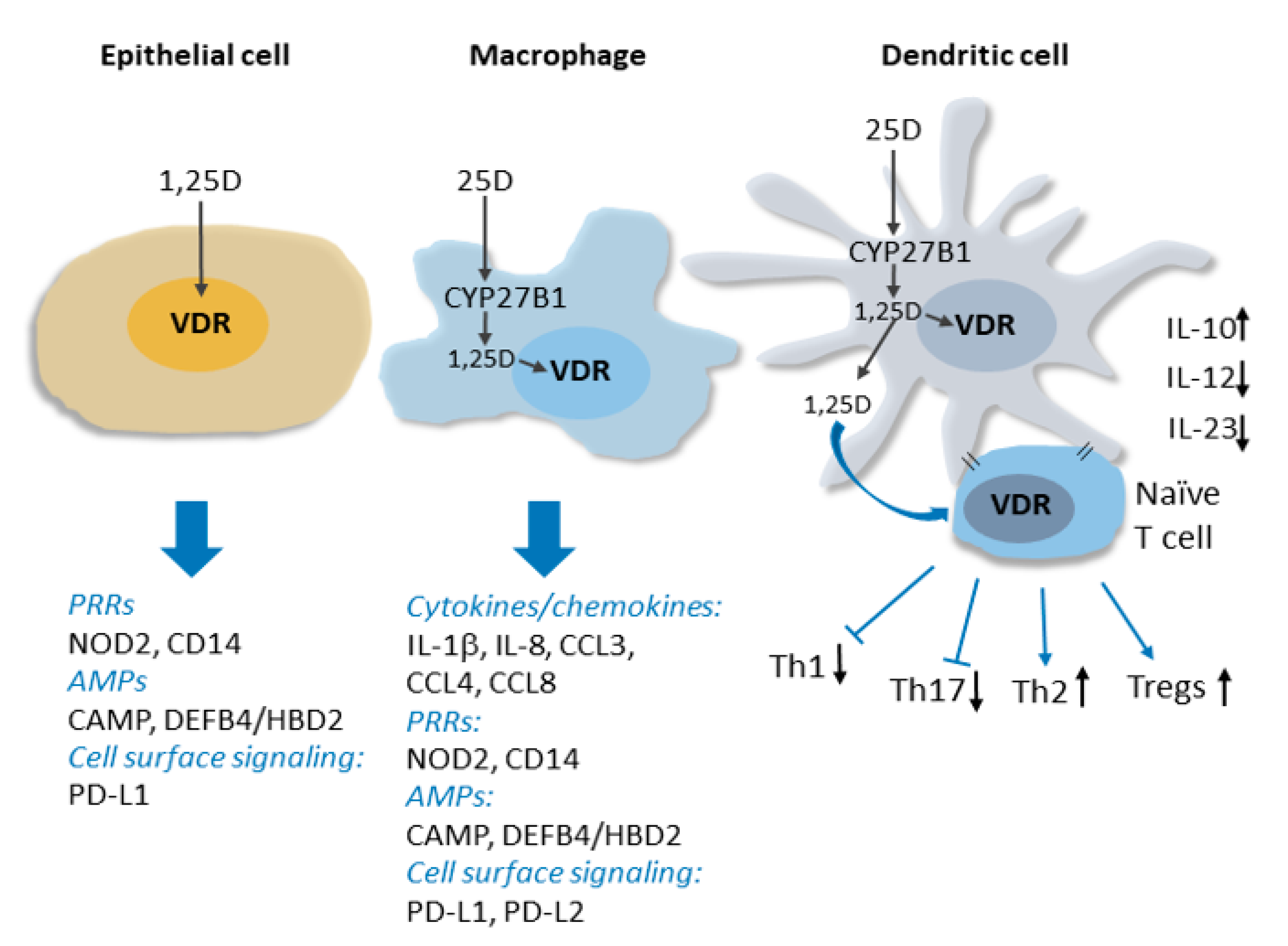
2. Overview of the Molecular Basis for the Non-Classical Actions of Vitamin D in the Immune System

The active hormonal form of vitamin D, 1,25-dihydroxyvitamin D (1,25(OH)2D3), signals through the vitamin D receptor (VDR), a member of the nuclear receptor family of ligand-regulated transcription factors [4]. 1,25(OH)2D3 is produced by sequential hydroxylations of vitamin D, first by largely hepatic 25-hydroxylation catalyzed by cytochrome P450 2 R1 (CYP2R1) and other enzymes, followed by tightly regulated 1α-hydroxylation by CYP27B1 in peripheral tissues. The classical view of vitamin D action in calcium homeostasis is based on the conversion of 25-hydroxyvitamin D (25OHD), the major circulating form, into 1,25(OH)2D3 in the kidney. Renal CYP27B1 expression is controlled by key regulators of calcium homeostasis, such as parathyroid hormone and fibroblast growth factor 23 (FGF23) [4]. In the classical model, renal 1,25(OH)2D3 released into the circulation acts in an endocrine manner to maintain circulating calcium concentrations, most notably by enhancing the intestinal uptake of dietary calcium. Indeed, severe neonatal vitamin D deficiency leads to hypocalcemia and rickets, with defects in skeletal growth associated with softening and weakening of the bones. Descriptions of rickets, along with paleontological evidence, date back millennia [5]. Cod liver oil and sun exposure were proposed as treatments for nutritional rickets as early as the beginning of the 19th century [6]. Unfortunately, 200 years later, nutritional rickets remains a clinical problem and is almost certainly under-reported [7,8]. Moreover, rickets diagnosed in the clinic represents the tip of the iceberg both in terms of the extent of pediatric vitamin D deficiency and because it is often associated with an increased prevalence of diseases unrelated to disrupted calcium homeostasis [5]. These include a number of immune-related disorders, such as increased rates of infections and conditions associated with compromised innate immunity; autoimmune diseases, such as type 1 diabetes (T1D) and multiple sclerosis (MS); and allergic conditions, such as atopic dermatitis [4,9,10].
Clinical interest in the links between vitamin D deficiency and these non-classical immune indications has been bolstered by rapidly expanding evidence from laboratory and translational studies that vitamin D signaling is a key regulator of both the innate and adaptive arms of the immune system [3]. The VDR and vitamin D metabolic enzymes are present throughout the immune system, and, importantly, CYP27B1 production in immune cells is induced by pathogen detection and regulated by a complex cytokine network [11,12], and is independent of calcium homeostatic signals. Thus, hormonal 1,25(OH)2D3 can be produced and act locally in the immune system under conditions of pathogen threat. The discovery of induced CYP27B1 expression in myeloid cells in vitro provides a molecular basis for observations of elevated levels of 1,25(OH)2D3 produced by macrophages in granulomatous diseases like sarcoidosis, which in extreme cases can lead to hypercalcemia [13] (see Figure 1 for details).
Innate immune responses are initiated by the detection of pathogen-associated antigens (pathogen-associated molecular patterns; PAMPS) by families of so-called pattern recognition receptors (PRRs). PRR activation and signaling leads to the production of antimicrobial peptides and a network of downstream responses, including an array of cytokines and chemokines, which propagate the signal to other components of the immune system. Studies of 1,25(OH)2D3-regulated gene expression in innate immune cells have revealed that vitamin D signaling functions upstream and downstream of PRRs to enhance immune responses. This includes inducing the expression of CD14 [14], a cofactor of toll-like receptor 4 PRR signaling, as well as that of NOD2 (nucleotide-binding oligomerization domain protein 2), a PRR defective in a subset of patients with Crohn’s disease [15]. The hormone-bound VDR also directly stimulates the transcription of genes encoding antimicrobial peptides cathelicidin antimicrobial peptide (CAMP/LL37) and human beta/defensin 2 defensin B4 (HBD2/DEFB4) [15,16,17], and readily detectable antibacterial activity is secreted into the media of 1,25(OH)2D3-treated cells in vitro [16,18]. Importantly, antimicrobial activity in pulmonary surface airway fluid was significantly higher in vitamin D-supplemented patients in a placebo-controlled double-blind randomized trial (RCT) [19]. In the presence of 1,25(OH)2D3, the expression of several cytokines, including interleukin 1β (IL-1β), a core component of innate immune responses, and the neutrophil chemokine IL-8/CXCL8 are induced, notably in macrophages infected with Mycobacterium tuberculosis [20]. Vitamin D signaling also regulates the innate-adaptive immune interface by rendering dendritic cells less inflammatory [4,21,22]. This contributes to suppression by 1,25(OH)2D3 of peripheral inflammatory T cell responses and enhanced development of T-regulatory (Treg) cells [21,23,24,25]. In addition to the above, genome-wide analyses of vitamin D signaling have revealed that the VDR regulates the transcription of numerous other genes implicated in immune system function [26]. Thus, we are physiologically wired to produce 1,25(OH)2D3 locally in immune cells in response to pathogens, and vitamin D signaling is a key component of many aspects of immune responses.

Antiviral Activity of Vitamin D Signaling: Specific Reference to COVID-19

At this writing, the world is in the grips of the COVID-19 pandemic, which is caused by the SARS-CoV-2 (severe acute respiratory syndrome-Covonavirus-2) virus. As such, along with SARS and MERS (Middle East respiratory syndrome), it represents the third and most severe coronavirus outbreak of this century. Notably, a recent British Medical Journal editorial on COVID-19 led to an extended discussion of vitamin D deficiency as a potential risk factor [27]. While COVID-19 is particularly severe in elderly populations, all age groups, including pediatric populations, are susceptible. One study provided evidence that pediatric COVID-19 was associated with coinfections [28], and fears of the spread of SARS-COV-2 in children will grow in many countries with a return to school. Clinical trials have yet to be registered to test the effects of vitamin D supplementation in the prevention/treatment of COVID-19 in children, although they are sure to come. However, clinical evidence is presented below that vitamin D supplementation reduces the rates of respiratory tract infections many of which are viral in nature. There is molecular evidence to support such antiviral activity. The antimicrobial peptide CAMP/LL37, whose expression is strongly inducible by 1,25(OH)2D3, has antiviral activity against enveloped viruses in vitro and influenza A in vivo [29]. 1,25(OH)2D3 also enhances the antiviral activity of bronchial epithelial cells in vitro and diminishes rhinovirus replication [30]. While these findings support the notion that hormonal vitamin D induces antiviral activity, it should also be noted that vitamin D signaling acts as a negative regulator of the renin-angiotensin system [31], which includes ACE2 (angiotensin converting enzyme 2), the receptor for SARS-COV-2 ACE2 [32]. ACE2 itself functions as a negative regulator of the renin-angiotensin cascade, and in an animal model, a 1,25(OH)2D3 analogue enhanced ACE2 expression in vitro [33]. This may not be beneficial in the context of a SARS-COV-2 infection; it has been hypothesized that patients being treated with ACE inhibitors for hypertension, which enhance ACE2 expression, may be at an increased risk for the development of severe COVID-19 [34].

3. Vitamin D and Infectious Diseases in Pediatric Populations

As developed above, there is extensive molecular evidence supporting vitamin D supplementation of deficient populations as a means to combat both the incidence and severity of infectious diseases. This would not be of substantial clinical relevance if populations were generally vitamin D sufficient. However, observations of widespread vitamin D deficiency [2] suggest that supplementation would be of clinical benefit. Notably, a survey of 1006 adolescents in 10 cities in 9 geographically dispersed European countries found that 80% of the subjects had 25OHD levels of less than 75 nM, considered the threshold of sufficiency, and that ~42% were either deficient (27.5–49.99 nM; 27%) or severely deficient (<27.5 nM; 15%) [35]. Moreover, poor vitamin D status in adolescents can be exacerbated during vitamin D winter and by high body mass index (BMI) [36]. The observations of widespread deficiency in European adolescents is consistent with estimated dietary intakes, which were found to be deficient in vitamin D [37]. They are also in line with the general European population; an analysis of 14 population studies in 55,844 European individuals concluded that 40% had 25OHD levels below 50 nM and that poor vitamin D status was elevated in dark-skinned subgroups [38]. This has led to calls for the measurement of 25OHD levels in risk populations, including pregnant women, young children, and adolescents, using standardized protocols [39].
Poor vitamin D status is also rampant in more southerly countries, such as India, where it ranged from ~40%–70% in pregnant women and infants to over 80% in school-aged children [40]. In addition, it is a predictor of infections in Indian children [41]. In a Yemeni study, a diagnosis of rickets was a predictor of a poor response to therapy in children hospitalized with severe pneumonia [42]. Strong support for the benefits of vitamin D supplementation to reduce the risk of infections came from meta-analyses of RCTs by Martineau and colleagues, which revealed that daily or weekly vitamin D supplementation, but not bolus dosing, reduced the rates of upper respiratory tract infections (URTIs). Moreover, the effect was most pronounced in subjects who were the most vitamin D deficient [43,44]. In a subgroup analysis (8 studies), vitamin D supplementation reduced the risk of URTIs in a mixed population of 513 children (1.1–15.9 years; adjusted OR: 0.60; 95%-CI 0.46 to 0.77). One of these studies was an RCT in Japanese schoolchildren [45], which provided evidence that vitamin D supplementation reduced the rates of seasonal influenza A. The results were most striking in the sub-group who had not previously received supplements. Similarly, in an RCT using cluster randomization, classrooms of 247 Mongolian children were randomly assigned during winter to daily supplementation with unfortified regular milk or milk fortified with vitamin D3 [46]. Supplementation halved the risk of parent-reported acute respiratory infections. This study was remarkable for several reasons: Notably, median serum 25OHD levels were very low at baseline (7 ng/mL; 17.5 nM), and the daily vitamin D dose used was low (300 IU/d), although supplementation raised the median 25OHD levels to 19 ng/mL (47.5 nM). Another RCT evaluated whether vitamin D supplementation was effective in reducing the number of cases of episodes of recurrent acute otitis media (AOM) in 116 otitis-prone children [47]. Subjects received daily doses of 1000 IU of vitamin D daily or placebo over a 4-month period, which led to a significant reduction in the number of children suffering a single AOM episode (26/58 vs. 38/58; p < 0.03). In contrast, an RCT using bolus dosing (100,000IU every 3 months for 18 months) as an approach to reduce the risk of pneumonia in children in Kabul, Afghanistan did not show any benefit of supplementation [48]. In a related RCT, no benefit was observed when a single dose of 100,000IU was used along with antibiotics to reduce the recurrence of pneumonia in children [49]. Similarly, 300,000IU bolus doses in children under five had no significant beneficial effect on the prevention of recurrent pneumonia [50].
A recent systematic review and meta-analysis of 7434 pediatric patients investigated the association of vitamin D status with mortality in children with acute or critical conditions [51]. The study found high levels of 25OHD deficiency (47% < 50 nM) in the group as a whole and higher levels (64%) in children with sepsis. Moreover, vitamin D deficiency was associated with increased mortality. These findings were consistent with those from a Canadian study, which did not find a difference in serum 25OHD levels in children with acute lower respiratory tract infections and controls but did see more frequent vitamin D deficiency in children admitted to intensive care with severe disease [52]. Collectively, the above results are striking because they support the hypothesis that the maintenance of vitamin D sufficiency should reduce rates of infections in pediatric populations. They are also striking because of the apparent lack of efficacy of bolus dosing. Bolus dosing is practical from a clinical perspective because it eliminates concerns about compliance, and, given the relatively long half-life of circulating 25OHD, should ensure that serum 25OHD remains above the level of sufficiency for several weeks. It can be speculated that daily or weekly dosing may lead to elevated levels of intracellular vitamin D, perhaps stored in lipid droplets, which may serve as a source of local production.

4. Crohn’s Disease

Crohn’s disease (CD), whose incidence is rising [53], is a relapsing-recurring inflammatory bowel condition resulting from defective intestinal innate immune homeostasis [54]. CD is notable because its incidence tends to rise with increasing latitude and for its association with low sun exposure early in life [55,56]. Although about one quarter of cases are diagnosed during childhood, pediatric CD is less well studied than adult disease. This is unfortunate as a cohort study identified 404 pediatric CD patients (0-17 years at diagnosis) between 1998 and 2002 in a population base of 1,312,141 in northern France. Disease complications occurred in a high percentage of these patients and 34% required surgery during the 5 years after diagnosis [57]. Based on the studies detailed below, there is compelling evidence that vitamin D supplementation may be of therapeutic benefit in pediatric Crohn’s patients [58].
There is a strong molecular and genetic basis for a potential therapeutic role of vitamin D signaling control of CD. The hormone-bound VDR regulates the expression of multiple human CD susceptibility loci, including the pattern recognition receptor NOD2, also known as IBD1 (inflammatory bowel disease 1) [15,59]. The VDR also directly induces expression of gene encoding the cell surface glycoprotein programmed cell death ligand 1 (PD-L1/B7-H1/CD274) [60], which interacts with PD1 on the surface of T cells to suppress T cell-mediated inflammatory responses. In a mouse model, defective intestinal epithelial Pd-l1 expression led to gut inflammation, underlining the key role of PD-L1 in suppressing pathological inflammation in the periphery [61]. Genome-wide association studies have revealed that several CD susceptibility loci encode proteins active in innate immune signaling and autophagy (which is critical for the control of intracellular pathogens) [54], both of which are stimulated by vitamin D signaling [26,58].
Several clinical studies, including generally small intervention trials, have provided evidence that vitamin D supplementation reduces disease activity [58]. While studies with children are limited, one examining dosing optimization of pediatric CD patients found that inflammatory markers were lower with high-dose vitamin D treatment (31 children receiving 1000 IU daily in summer and 2000 IU in winter and spring) [62]. The findings of recent meta-analyses of observational studies and RCTs [63,64] collectively confirm that a poor vitamin D status is associated with increased disease activity and that supplementation lowers it. However, based on subgroup analysis, the study of RCTs noted it was too early to make the same conclusions for pediatric CD patients [63]. It would therefore be worthwhile to conduct an RCT examining the effects of robust vitamin D supplementation on disease activity in a pediatric population. In this regard, there is an ongoing RCT at eight Canadian institutions looking at the impact of vitamin D supplementation of newly diagnosed children with CD on the rate of CD relapse (Clinicaltrials.gov: NCT03999580; unpublished clinical trials cited are listed in Table 1).
Although the above results provide support for the therapeutic benefit of vitamin D supplementation in CD, the efficacy of oral supplementation may be complicated by intestinal malabsorption, particularly in the case of patients who have undergone bowel resections and ostomy procedures. In one study, two patients who had recently undergone bowel resections and ileostomies, one of which had CD, failed to respond to aggressive oral vitamin D3 supplementation [65]. However, the patients did respond to high-dose sublingual vitamin D2 treatment. An alternative in such cases is moderate UVB exposure. Notably, in a CD patient with malabsorption due to multiple bowel resections, exposure to a series of non-erythemic doses of UVB on a tanning bed dramatically improved her vitamin D status [66].

5. Vitamin D Deficiency and Dental Caries

Dental caries (cavities) arise through the breakdown of teeth by acidic secretions from bacteria in dental plaque. Numerous studies have linked vitamin D deficiency during pregnancy and infancy to an increased rate of early childhood caries [67,68,69]. An association between vitamin D deficiency and dental caries was brought to the forefront by a systematic review and meta-analysis published in 2013 [70]. It surveyed 24 trials that included 2827 children, which generated a pooled relative rate estimate of 0.53 (95%-CI, 0.43–0.65) for the effect of vitamin D supplementation. There was substantial heterogeneity of the results between trials and some evidence of publication bias, generating a low-certainty conclusion when all trials were included. However, when the review was limited to higher-quality studies, it reduced the heterogeneity between trials and provided evidence that vitamin D was highly efficacious. The more restrictive analysis increased the estimated reduction in caries from 47% to 54% [70].
Vitamin D sufficiency may protect against caries through its role in tooth development [71,72], and molar-incisor mineralization [73]. However, vitamin D sufficiency may also be protective through its capacity to boost antimicrobial innate immunity and suppress inflammation. An inverse correlation was observed in 6700 adults between gingival inflammation and serum 25OHD levels [74]. A Swedish study of children found a correlation between poor vitamin D status and increased risk of caries. Notably, vitamin D status in this study correlated positively with levels of the 1,25(OH)2D3-inducible antimicrobial peptide CAMP (LL37) in saliva [75]. CAMP/LL37 is efficacious against major bacterial species found in plaque, such as Streptococcus mutans [76]. In a recent RCT, vitamin D supplementation enhanced the levels of antimicrobial activity in lung surface airway fluid [19]. This suggests that enhanced secreted antimicrobial activity should be detectable in an RCT of vitamin D supplementation and is consistent with the induced secretion of antimicrobial activity by 1,25(OH)2D3-treated cells in vitro [16]. Given these results, it would be of interest to conduct an RCT to determine whether vitamin D supplementation enhances antimicrobial activity in saliva against major bacterial components of dental plaque.

6. Links between Vitamin D Deficiency and Autoimmune Conditions in Pediatric Populations

There is extensive preclinical and clinical literature on the links between vitamin D deficiency and autoimmune conditions, in particular type 1 diabetes (T1D) and multiple sclerosis (MS). The effects of 1,25(OH)2D3 on T cell function are likely key to its potential capacity to limit autoimmunity. Expression of the VDR in T lymphocytes was discovered in the 1980s, and 1,25(OH)2D3 attenuates T cell activation and proliferation [77,78], regulating the function of several T cell subsets [26]. Briefly, vitamin D suppresses T cell-driven inflammation while enhancing the action of suppressive Treg cells. Vitamin D signaling in dendritic cells alters their metabolic profile, producing a more tolerogenic phenotype and enhancing interleukin-10 secretion, which promotes Treg production [26,79,80]. There is also support for the potential of vitamin D supplementation in attenuating autoimmunity from studies in mouse models of human autoimmune diseases [81]. At present, clinical use is limited to psoriasis, which is treated with topical application of vitamin D or its analogues [82]. However, overall, there is no consensus for the therapeutic benefit of vitamin D supplementation in the treatment of autoimmune conditions, and, unfortunately, many studies are limited by group sizes.

6.1. Juvenile-Onset Type 1 Diabetes

The incidence of T1D, a multifactorial autoimmune condition leading to destruction of the insulin-producing β-cells of the pancreas, is increasing worldwide [83]. There is intriguing preclinical evidence supporting a preventive role of vitamin D, most of which has been generated in the non-obese diabetic (NOD) mouse model of human T1D [84]. In work done by the Mathieu group with the NOD model, vitamin D deficiency in early life increased the incidence and accelerated onset of the disease [85]. Moreover, 1,25(OH)2D3 and its analogues are more efficacious at preventing insulitis and diabetes in NOD mice when administered early, prior to the onset of immune-mediated beta cell attack [86,87,88], and long term, high-dose 1,25(OH)2D3 treatment reduced the incidence of diabetes in NOD mice [88]. Intriguingly, immune-mediated induction of CYP27B1 expression is attenuated in NOD mice [89].
Globally, clinical studies of vitamin D supplementation in all age groups with existing T1D are inconclusive but suggest that early intervention may preserve the remaining pancreatic islet β-cell function [81]. In one intervention trial, a protective effect of 1,25(OH)2D3 supplementation in preserving residual β-cell function in adult-onset disease only occurred when the duration of disease was less than 1 year [90]. There is some evidence for the seasonal onset of T1D in both adults and children, coinciding with low circulating 25OHD levels [91,92], which would be consistent with a poor vitamin D status accelerating disease onset. Nonetheless, evidence for a role of vitamin D deficiency in contributing to T1D incidence remains mixed. For example, two studies did not find any correlation between 25OHD levels measured at birth or in infancy and the risk of developing T1D during childhood [93,94]. Several case-control studies examining the relationship between serum 25OHD levels or vitamin D intake during pregnancy and the risk of T1D in offspring also provided mixed results [83]. However, earlier meta-analyses of observational studies supported the notion that supplementation early in life may contribute to T1D prevention [95,96].
Although no RCTs were found examining the potential efficacy of vitamin D supplementation in the prevention of juvenile-onset T1D, case-control studies do provide some evidence for the benefit of vitamin D either as a supplement or in food intake. Of these, two are notable because of their size. A large European-based case-control study screening for early risk factors for the development of T1D concluded that vitamin D supplementation during infancy was beneficial [97]. Similarly, vitamin D supplementation during the first year of life correlated with a reduced incidence of T1D in a Finnish birth-cohort study, and the degree of benefit was greater for infants supplemented with 2000IU/day relative to a lower dose [98]. A Norwegian study (545 cases, 1668 controls) suggested that frequent supplementation (five times or more a week) with cod liver oil in the first year of life lowered the risk of development of T1D [99]. Remarkably, however, this study did not find any evidence that vitamin D supplementation in other forms, even frequent, provided any benefit. Overall, these findings, although inconclusive, are compelling, and strongly suggest that an RCT examining a preventive role of vitamin D supplementation in T1D, at a minimum in the first year of life, would be in order. A robust dose, perhaps as high as 2000 IU/day should be used in the treatment wing. However, this leaves the field in a bit of a bind as the duration of follow-up of such a study should likely be at least 10 years. Nonetheless, the efficacy of vitamin D and the timing of its supplementation will be important issues to resolve in order to reach a consensus. Notably, a recent position paper from multiple German clinical/nutrition societies concluded that “Based on currently available studies, routine vitamin D supplementation is not recommended for children aged >2 years, even when they have serum concentrations below reference values” [100].

6.2. Multiple Sclerosis

MS is characterized by an autoimmune attack on the protective myelin sheath covering nerve fibers. It is perhaps the autoimmune condition with the strongest association between vitamin D deficiency and the risk of development. MS risk is multifactorial and has parallels with Crohn’s disease, as discussed above, in that there is an inverse correlation between disease incidence and previous sun exposure. For example, one case-control study found that higher sun exposure in 6- to 15-year-old children reduced the risk of subsequent development of MS [101]. Moreover, also similar to CD, an increased incidence of MS has been linked historically to higher latitudes [102]. However, one systematic review suggested that MS incidence has been rising at lower latitudes since 1980 [103]. Moreover, pure geographical interpretations of the risk of MS development are complicated by ethnicity, e.g., risk is low in native Siberians and populations in northern Scandinavia but high in Palestinians, indicative of genetic factors [102,104]. A number of cohort studies have provided evidence that vitamin D deficiency contributes to the risk of pediatric MS [105], and another study established a correlation between serum 25OHD levels and relapse rates [106]. Adolescent obesity is also associated with risk of the development of MS, although here it is not clear whether adiposity per se is a risk factor or whether it is associated with the sequestration of vitamin D metabolites and lower circulation of 25OHD levels [107].
Further strong evidence that vitamin D deficiency is a key risk factor for MS has been provided by Mendelian randomization (MR) studies. An MR study uses known functional variants of genes to probe the causal effect of a specific variable on disease occurrence, severity, or outcome. Four single nucleotide polymorphisms (SNPs) were identified in genes encoding enzymes/binding proteins that influenced circulating 25OHD levels: CYP2R1; CYP24A1, which encodes the enzyme that initiates the catabolism of 25OHD and 1,25(OH)2D3; GC, encoding the major serum 25OHD binding/carrier protein; and DHCR7, which encodes the enzyme that converts 7-dehydrocholesterol to cholesterol [108]. As these are genetic variants, their individual and collective impacts on disease represent causal effects of life-long vitamin D deficiency. Remarkably, the use of these variants in a large-scale MR study of MS (14,498 cases and 24,091 controls) provided evidence that genetic variants lowering circulating 25OHD levels strongly enhanced susceptibility to the development of disease [108]. Moreover, poor vitamin D status may influence the age of disease onset. Symptoms of MS usually first appear in young adulthood (20–40 years of age), and pediatric-onset disease accounts for only approximately 5% of all cases. Remarkably, another MR study of 569 pediatric MS cases in Sweden and the US (Sweden 175 cases, 5376 controls; US, 394 cases, 10,875 controls; 16,820 total) found that increased circulating 25OHD was associated with decreased odds of developing pediatric MS (OR 0.72, 95%-CI 0.55–0.94; p = 0.02) [109]. The results were striking, as the SNPs examined accounted for only 2.8% of the variance in circulating 25OHD levels. Collectively, the results above are compelling and suggest that poor vitamin D status is a key contributor to the risk of development of MS and may be a contributing factor to pediatric onset.

6.3. Other Autoimmune Conditions

Clinical studies testing the association of vitamin D status with other juvenile-onset autoimmune conditions are more limited. Systemic lupus erythematosus (SLE), which is more common in females than in males, is characterized by elevated levels of autoantibodies to self-nucleic acids or nucleoproteins [110]. Clinically, inflammation in SLE may affect multiple organs (joints, kidneys, skin, heart, lungs, vasculature, the nervous system, etc.). There is one RCT examining the efficacy of vitamin D supplementation in adolescents with SLE, which provided evidence that vitamin D reduced disease activity and fatigue scores [111]. The trial is notable for the robust vitamin D dose used (50,000 IU/week for 24 weeks), which boosted serum 25OHD levels from 19.3 to 31.3 ng/mL (48.3–78.3 nM). This result is encouraging, and there is some support for it from RCTs and other studies in adult populations. An inverse correlation has been established between serum 25OHD levels and SLE disease activity in adult patients [112]. In an RCT in SLE patients, supplementation with 2000 IU/day of vitamin D raised serum 25OHD levels from 19.8–28.7 ng/mL (49.5–71.8 nM) and significantly improved the levels of inflammatory and hemostatic markers [113]. Globally, however, findings are not unanimous. For example, in another RCT with daily doses of 2000 or 4000 IU/day, supplementation did not diminish the interferon signature associated with the disease [114].
Juvenile idiopathic arthritis (JIA) is a collective term for a heterogeneous group of arthritis-like diseases of unknown origin [115]. Vitamin D deficiency has been considered as a potential environmental determinant of JIA [116], and an Australian case-control study established a link between higher life-long sun exposure and reduced risk of JIA [117]. At this stage, clinical data is best described as “emerging”. A systematic review published in 2013 found, unsurprisingly, that rates of vitamin D deficiency were high in a number of studies of JIA patients but also highlighted the lack of hard data linking vitamin D status or supplementation to disease activity [118]. One study found extensive vitamin D deficiency but did not find a correlation between vitamin D status and disease activity in a cohort of patients (mean age 10.6 years) with JIA [119], whereas studies of small Turkish and much larger German cohorts did find such an association [120,121]. A small RCT was recently published examining the effects of supplementation of JIA patients in China with 2000 IU/day of vitamin D (20 patients in treatment group, 22 in control group) on disease activity and bone parameters [122]. While supplementation raised serum 25OHD levels, no effects were observed on the disease activity score or on bone mineral density over the 24-week period of the trial. Hopefully, more and larger trials of this kind for JIA will follow and establish whether vitamin D supplementation may be of therapeutic benefit for these patients.

7. Allergic Conditions

Similar to other immune conditions, rising trends in the incidence of allergic diseases have coincided with the increase in the prevalence of a vitamin D deficient/insufficient status reported worldwide. Epidemiological studies have shown an association between the risk of allergic conditions and latitudes, ultraviolet radiation (UVR) intensity, and birth season. In general, a greater risk of allergy is observed in individuals living furthest from the equator, residing in low-UVR intensity areas, and in those born during winter months [123,124,125]. The emerging importance of vitamin D in prenatal and early postnatal development of the immune system is suggested to play a role in the association between vitamin D status and the onset of allergic disorders. This view is, however, challenged by the so-called “vitamin D allergy hypothesis”, which attributes the increased incidence of allergic diseases to the immunological side effects of vitamin D supplementation initiated during infancy [126]. Other studies have supported the association between infant vitamin D supplementation and the risk of atopic diseases in childhood [127] and adulthood [128]. Moreover, maternal levels of 25OHD >75 nM in the third trimester of pregnancy have been linked to increased odds of developing visible eczema in children at 9 months and having asthma at 9 years, although the number of cases was small and loss to follow-up high, particularly at 9 years [129]. Potential explanations to reconcile these conflicting results relate to the U-shaped association found between cord blood 25OHD levels and risk of allergic diseases in early childhood [130] and between 25OHD levels and immunoglobulin E (IgE) levels in adults [131]. It is speculated that the balance between the systemic T-helper 2 (Th2)-skewing effect of vitamin D and its local tissue anti-inflammatory effects may vary depending on the circulating concentration of 25OHD [124].

7.1. Asthma

Asthma is a common chronic inflammatory disorder that primarily affects the respiratory system. It is a very heterogeneous condition comprising many phenotypes that differ depending on the nature of the trigger (e.g., allergic vs. non-allergic), the age of onset (preschoolers vs. schoolchildren vs. late onset), the pathophysiology involved (high vs. low Th2-mediated responses, eosinophilic vs. non-eosinophilic), and whether asthma symptoms are transient or persist into adulthood [132]. Its clinical presentation is also variable with the occurrence of asthma-like symptoms (e.g., prolonged coughing, recurrent wheezing) in preschoolers that may resolve with time or transition to more severe symptoms, leading to irreversible obstructive changes in the lungs.
The use of vitamin D in the primary prevention of asthma is an area of active investigation and stems from preclinical studies, mostly supporting the involvement of vitamin D in lung growth and the development of the immune system [133,134,135]. A decreased risk of childhood asthma (RR 0.87, 95%CI 0.75–1.02) in mothers with maternal or cord blood 25OHD levels in the highest vs. lowest category was demonstrated in a meta-analysis of 15 prospective studies with 12,758 participants and 1795 asthma cases [136]. Interestingly, when data were stratified according to the timing of 25OHD levels’ measurement (i.e., early-mid vs. late pregnancy), the association reached significance only when levels were assessed during early pregnancy (RR 0.73, 95%CI 0.58–0.92). These results were corroborated in a subsequent meta-analysis showing a similar trend between maternal 25OHD levels and the risk of asthma in children ≤ 5 years (OR 0.81, 95%CI 0.65–1.01; 27,776 participants pooled from 6 cohort studies) [137]. In line with these data, a recent case-cohort Danish study reported that children with birth levels of 25OHD in the highest quintile had a lower risk of having asthma between the ages 3 and 9 (fully adjusted HR 0.51, 95%-CI 0.35–0.75) [138]. Since the lung and immune system develop early in gestation but continue their development postnatally, these data indirectly provide evidence for the importance of vitamin D at these stages of development and the consequences that prenatal/postnatal vitamin D insufficiency may have on child health. As such, two large trials of vitamin D supplementation during pregnancy were initiated to determine the impact of prenatal vitamin D on the risk of asthma/recurrent wheezing in children. The Vitamin D Antenatal Asthma Reduction Trial (VDAART) enrolled 806 pregnant women between 10 and 18 weeks of gestation from three sites across the United States. Eligibility criteria included a history of allergic diseases in the mother or in the biologic father. The intervention arm consisted in the administration of daily 4000 IU vitamin D3 combined with a multivitamin containing 400 IU D3 until delivery. The placebo group received a matching placebo tablet plus the multivitamin with 400 IU D3 [139,140]. Using a very similar design, the Copenhagen Prospective Studies on Asthma in Childhood 2010 (COPSAC2010) recruited 581 Danish women between 22 and 26 weeks of gestation. Participants were randomized to receive either 2400 IU vitamin D3 combined with a multivitamin containing 400 IU D3 or only the multivitamin until delivery [141]. The combined analysis of these two trials revealed a significant effect of vitamin D intervention on reducing the risk of developing asthma/recurrent wheeze in the first three years of life (OR 0.74, 95%-CI 0.57–0.96). Unexpectedly, the effect was driven by women with baseline serum 25OHD levels ≥ 75 nM (OR 0.54, 95%-CI 0.33–0.88) vs. women with baseline 25OHD levels < 75 nM [142]. Irreversible changes induced by insufficient vitamin D exposure during early gestation—and prior to the initiation of the supplementation—may explain the lack of effect among women with lower 25OHD levels. It also suggests that maternal 25OHD levels ≥ 75 nM may be required for optimal in utero lung and immune development. Recent follow-up studies of VDAART [143] and COPSAC2010 [144] failed to show an effect of prenatal vitamin D supplementation on the development of asthma or recurrent wheeze through to the age of 6 years. Potential explanations for these null results include: The lack of postnatal supplementation in children that failed to sustain the effects of the prenatal supplementation and the effects of vitamin D on virus-induced wheezing and asthma exacerbations in early life that may have prevented some asthma phenotypes from persisting into mid-childhood. Taken together, the results from these two trials suggest that prenatal vitamin D supplementation may be effective in preventing wheezing that occurs in preschoolers, but the effect tends to disappear with time. Preschool wheezing is thought to differ phenotypically from school-aged wheezing in that it is triggered by viral respiratory infections and is transient, with manifestations often regressing by school years. In contrast, school-aged asthma is a more persistent state generally related to the presence of allergies and characterized by an uncontrolled type 2 inflammation. Through its antiviral, antimicrobial, and anti-inflammatory actions, it is plausible that vitamin D may optimize the body’s response to viral infections and hence be more beneficial at an early age. In support of this assumption, a VDAART ancillary study showed that cord blood mononuclear cells (CBMCs) from women allocated to the intervention group exhibited an enhanced proinflammatory cytokine response to innate and mitogenic stimuli [145]. It was hypothesized that high-dose vitamin D enhanced the neonate immune system fitness, thereby preventing asthma development later in life. While in stark contradiction with the findings from in vitro studies using exogenous 1,25(OH)2D3 [146,147,148], these findings underscore the importance of the timing of the intervention with regards to the effects of vitamin D on the pulmonary and immune systems.
A major limitation of these prenatal trials is that supplementation was not sustained in children after birth. One postnatal vitamin D supplementation trial was undertaken to determine the effect of early vitamin D supplementation in infants particularly at risk of wheezing and vitamin D deficiency. The Wheezing in Black Preterm Infants: Impact of Vitamin D Supplementation Strategy (D-Wheeze) examined the impact of two vitamin D supplementation strategies on the risk of recurrent wheezing by 12 months of age in black preterm infants [149]. As per current recommendations, infants were given a 400 IU vitamin D3 supplementation after birth when oral feedings were tolerated. When infants achieved an intake > 200 IU vitamin D from oral feedings of fortified human milk or formulas, they were randomized to either the sustained daily supplementation of 400 IU vitamin D3 or a diet-limited supplementation consisting solely of the amount of vitamin D consumed from formula or human milk feedings (placebo wing) until 6 months ‘adjusted age. Results showed a reduction in the risk of developing recurrent wheezing by 12 months’ adjusted age in the sustained supplemented infants (RR 0.66, 95%-CI 0.47–0.94). The design of future trials should combine prenatal and sustained postnatal vitamin D supplementation to provide additional support for the efficacy of vitamin D in the primary prevention of wheezing and asthma.
In contrast to healthy children, vitamin D insufficiency is more prevalent among children with asthma, both preschoolers [150,151] and older children [152,153]. Cross-sectional studies showed that serum 25OHD is inversely associated with asthma-related outcomes (i.e., frequency of acute attacks and wheeze periods, requirement for high-dose oral corticosteroids) [153,154,155,156,157,158]. Such associations are particularly robust and have been replicated multiple times in various populations [153,155,159,160,161,162,163,164,165]. However, causal inferences are limited by the possibility of reverse causality, where asthma is a cause—rather than a consequence—of the reduced serum 25OHD. This notion is supported by MR studies, which failed to demonstrate any significant association between genetically determined 25OHD levels and asthma [166,167]. In addition, asthmatic children may spend less amounts of time outside, limiting the skin production of vitamin D. Another contributing factor is the widespread misconception that milk and dairy products promote mucus formation, increase airway resistance, and trigger or worsen asthma symptoms [168]. While this belief is not strongly supported by scientific evidence [169,170], it may contribute to the reduced vitamin D intake of these children, although this has never been formally documented. Lastly, asthma is a chronic inflammatory condition and inflammation was postulated to decrease 25OHD levels in other settings [171]. While the impact of inflammation on the vitamin D status of asthmatics remains to be proven, recent data showing dysregulated vitamin D metabolism in adults with asthma, both at baseline and following an intermittent bolus supplementation, provide some support for this hypothesis [172].
The strongest evidence for vitamin D in the secondary prevention of asthma comes from pediatric trials, mostly conducted in schoolchildren. The only vitamin D trials in preschoolers are two pilot trials of 22 and 47 preschoolers aimed at identifying the best supplementation strategy but that were not powered to detect differences in health outcomes [150,151]. A Cochrane systematic review of nine asthma trials (two adult/seven pediatric), with variable use of inhaled corticosteroids, reported a significant protective effect of vitamin D against asthma exacerbations (OR 0.64, 95%-CI 0.46–0.90; 680 participants), the major cause of asthma morbidity and mortality [173]. This protective effect was later confirmed in a meta-analysis of individual patient data performed by the same group. However, the treatment effect failed to reach significance when only pediatric data were included in the analyses [174]. Given that asthma exacerbations are often precipitated by viral respiratory infections for which vitamin D was also found to reduce the incidence of [33,34], it is speculated that vitamin D acts either by preventing the infection or limiting host immune responses that may trigger exacerbations. For example, in rhinovirus and respiratory syncytial virus-infected respiratory epithelial cells, 1,25(OH)2D3 increased the secretion of AMP cathelicidin [30,175]. It dampened the antiviral and inflammatory response to viruses through the downregulation of nuclear factor NFκB signaling and reduced secretion of antiviral cytokines (e.g., interferon-β (IFN-β)) and chemokines (e.g., IP-10) without, however, compromising viral clearance [176,177]. Vitamin D also downregulated the expression of the intercellular adhesion molecule 1 (ICAM-1) and platelet-activating factor receptor (PAFR), two proteins involved in the adhesion of viruses to the surface of epithelial cells [176]. Through effects on the adaptive immune system, it is also plausible that vitamin D may limit allergen-induced type 2 airway inflammation, which is more associated to school-aged asthma [134]. Vitamin D was shown to increase the expression of the IL-33 decoy receptor ST2, which by binding IL-33, inhibits its capacity to induce the production of Th2 proinflammatory cytokines [178].
In children with moderate or severe exacerbations, daily inhaled corticosteroids are generally indicated, but oral corticosteroids may be required to treat asthma exacerbations. Chronic use of moderate (or higher)-dose corticosteroids in children has detrimental effects on bone health [179] and may slow down growth velocity [180]. Furthermore, viral-induced exacerbations are associated with a poorer response to corticosteroids and increased emergency department management failure [181]. Epidemiologic studies showed an inverse association between low serum 25OHD and the use of more anti-inflammatory medication in asthmatic children [155,160], the need for greater inhaled corticosteroid doses, and an apparent reduction of corticosteroid efficacy [155,182]. Corticosteroids interact with cytosolic receptors (GCR-α), inducing their nuclear translocation and the binding to glucocorticoid response elements located on the promoter of genes involved in anti-inflammatory responses. Corticosteroids induce the expression of MAPK-phosphatase 1 (MKP-1) while repressing that of proinflammatory cytokines tumor necrosis factor (TNF) and IL-8. Vitamin D enhanced the anti-inflammatory effects of corticosteroids by increasing IL-10 secretion by Tregs and MKP-1 expression by mononuclear cells [183]. In addition, vitamin D synergized with corticosteroids to induce a tolerogenic dendritic cell phenotype [184]. A short course of 1,25(OH)2D3 (0.25 µg twice/day × 4 weeks) given to adults with steroid-resistant asthma improved the clinical response to corticosteroids [185,186]. They showed in vitro that peripheral blood mononuclear cells from steroid-resistant asthmatics given 1,25(OH)2D3 demonstrated abrogation of dexamethasone-induced IL-17A production and restoration of dexamethasone-induced IL-10 secretion, indicating an overall better corticosteroid responsiveness [185]. Most experimental data, however, come from cell culture experiments designed to obtain maximal effects of pharmacologic dose of 1,25(OH)2D3 over short time periods and from small trials in adults with steroid-resistant asthma. The implication of vitamin D in corticosteroid responsiveness requires further investigation in pediatric asthma.
While trials in pediatric and adult asthma suggest an effect of vitamin D in reducing the risk of URTIs and asthma exacerbations, the magnitude of effects appears greater in those with the lowest baseline vitamin D status [174]. As such, RCTs are ongoing to address some unresolved issues. For instance, The Vit-D-Kids Asthma study (ClinicalTrials.gov ID: NCT02687815) is designed to assess whether supplementation (4000 IU/day) prevents severe asthma attacks in children with serum 25OHD levels below 30 ng/mL, and who are being treated with inhaled corticosteroids. The DIVA trial (Vitamin D intervention in preschoolers with viral-induced asthma; ClinicalTrials.gov ID: NCT03365687) aims to study the impact of the administration of two boluses of 100,000 IU vitamin D3 3.5 months apart and daily 400 IU D3 on the number of asthma exacerbations requiring rescue oral corticosteroids in preschoolers, a high-morbidity population. The results of these trials are expected in the coming years.

7.2. Other Allergic Conditions

7.2.1. Atopic Dermatitis

Atopic dermatitis (AD) is a chronic inflammatory skin condition affecting 10% to 20% of children and usually developing within the first year of life. The pathophysiology involves defects in the skin epithelial barrier and deregulated innate and adaptive immune responses, which manifest clinically as relapsing eczematous lesions and pruritus. Compromised barrier integrity facilitates the penetration of allergens and pathogens, which activate keratinocytes and dendritic cells through innate immune receptor ligation by damage-associated molecular patterns (DAMPs) and PAMPs. Activated dendritic cells promote exaggerated Th2-mediated cytokine secretion and inflammation, which further damages the epidermal barrier integrity and increases the propensity to secondary infections [187]. The increased susceptibility of individuals with AD to infections has been linked to a defective capacity to properly increase the production of antimicrobial peptides like cathelicidin following skin lesions [188,189].
In theory, vitamin D should attenuate the chronic immune activation and inflammation seen in AD, yet mechanistic evidence is limited. While keratinocytes are well-known for their involvement in cutaneous synthesis of vitamin D3, they also express the VDR and CYP27B1, the expression of which was upregulated in keratinocytes surrounding wounds [190]. The resulting production of 1,25(OH)2D3 increased the expression of cathelicidin, CD14, and TLR2, further amplifying pathogen recognition and the antimicrobial response during skin repair, hence protecting wounds against infections. Findings from clinical studies tend to support these experimental observations. A three-week vitamin D3 supplementation (i.e., 4000IU/day) to 14 individuals with AD (age not specified) led to a marked increase in cathelicidin mRNA levels of lesional skin biopsies whereas the effect of supplementation was non-significant in non-lesional AD biopsies and in biopsies from non-AD controls [191]. In an audit of clinical practice, Albenali et al. assessed CAMP/LL37 peptide levels of lesional and non-lesional skin brushings from vitamin D-insufficient children (i.e., 25OHD < 75 nM) with AD or AD complicated by eczema herpeticum (ADEH), a more severe clinical manifestation [192]. LL-37 levels were measured at baseline and following a 2-month supplementation with daily 6000 IU (1–12 years of age) or 12,000 IU vitamin D3 (12–18 years of age), as recommended by the local guidelines for children with 25OHD levels < 50 nM. Children with suboptimal 25OHD levels (50–75 nM) received over-the-counter supplements containing 100% of the recommended daily allowance (RDA) for vitamin D (e.g., 400 IU). At baseline, skin cell levels of LL-37 were associated with serum 25OHD levels and were lower in children with the most severe disease. After 2 months of either therapeutic or over-the-counter vitamin D supplementation, scores of AD severity were markedly reduced whereas LL-37 levels increased by 4-fold and were inversely correlated to AD severity [192]. Other suggested mechanisms of action of the vitamin include downregulation of the monocyte TLR expression, an inhibitory effect on dendritic cells’ activation and Th1 cytokine release, and a reduction of the effect on B cell function, leading to decreased IgE secretion, although no difference was seen between pre- and post-vitamin D supplementation IgE levels in the Albenali study [192]. This finding is consistent with the results of Hyppönen et al. showing a U-shaped relationship between IgE and 25OHD levels in an adult cohort [131]. Similarly, a non-linear relationship was found between cord blood 25OHD levels and the risk of atopy in early childhood [130]. Cord blood 25OHD levels below (<50 nM) and above (>100 nM) a certain level were associated with an increased risk of having detectable inhalant-specific IgE in the first five years of life. It was therefore hypothesized that the maintenance of 25OHD levels between 50 and 100 nM during the first years of life will prevent the development of atopy.
A recent meta-analysis conducted on observational and intervention studies revealed lower 25OHD levels by 16 nM (95%-CI −31 to −1; p = 0.05) in AD vs. non-AD children [193]. This difference was less pronounced in adult studies, where a non-significant difference of 2 nM (95%-CI −5 to 1; p = 0.15) was observed, although the limited adult data preclude any firm conclusions. The reasons underlying the striking difference between pediatric and adult data are poorly understood but may relate to sun avoidance habits and low vitamin D intake, as children with AD often suffer from concomitant food allergies and dietary restriction. Mendelian randomization (MR) studies provide evidence that the relationship between 25OHD levels and the risk of atopic diseases is strongly confounded by lifestyle factors [167]. As such, efforts to increase 25OHD levels may likely fail to decrease the risk of developing these conditions. By only addressing the association between genetic determinants of circulating 25OHD levels and disease risk, MR studies overlook the actions of 25OHD and 1,25(OH)2D3 at the cellular level, which may be beneficial in atopic diseases, but weakly correlated to serum 25OHD.
All vitamin D trials included in the Hattangdi-Haridas meta-analysis (n = 5; 1 adult, 2 pediatric, and 2 mixed) showed improvement in AD severity post-vitamin D oral supplementation [193]. Dosages ranging from daily 1000 to 2000 IU vitamin D for 1 to 3 months led to a reduction in the SCORing Atopic Dermatitis (SCORAD) index of 11 (95%-CI −13 to −9; p < 0.001; 3 RCTs) and 21 (95%-CI −27 to −15; p < 0.001; 2 studies with repeated measures design) [193]. Of note, all trials included individuals with AD of mild to moderate severity and 25OHD levels below 50 nM. Interestingly, the magnitude of effect exceeds the minimal clinical important difference (MCID) in the SCORAD score previously established for the treatment of AD (i.e., score reduction of 9 points) [194], underscoring the clinical relevance of these observations. Collectively, the results of this meta-analysis provide evidence that vitamin D supplementation of individuals with mild to moderate AD and insufficient vitamin D status leads to clinical improvements in AD severity.

7.2.2. Food Allergies

Foods do not normally induce allergic reactions due to the ability of the immune system to discriminate between non-self-innocuous and threatening antigens. However, in some susceptible individuals, food antigens trigger inappropriate immune reactions that are mediated, but not always, by antigen-specific IgE. While first exposure to food allergens induces sensitization, their subsequent ingestion results in a larger and faster production of the antigen-specific IgE, triggering the release of mediators, such as histamine, from mast cells. These mediators are responsible for the cutaneous, gastrointestinal, respiratory, and systemic allergic manifestations. Vitamin D was shown to reduce B-cell function, leading to decrease IgE levels in vitro [195]; however, such an observation has been challenged by studies on mice with allergic airway disease in which 1,25(OH)2D3 administration reduced eosinophilia but resulted in increased serum IgE levels [196]. In contrast, using a different allergy mouse model, Hartmann et al. showed that 1,25(OH)2D3 significantly reduced total and antigen-specific IgE levels [197], raising the possibility that vitamin D may exert deleterious and beneficial influences depending on the allergic context. Other mechanisms by which vitamin D may modulate the host response to food allergens are the regulation of intestinal tight junctions and innate epithelial defenses, including the antimicrobial capacity, which in turn may influence the composition of the gut microbiota. A reduced diversity of early life gut microbiota has been linked to the onset of allergic diseases [198] whereas cord blood 25OHD levels have been associated with infant gut microbiota, although vitamin D supplementation after birth exerted no effect [199]. Studies specifically addressing the relationship between the vitamin D status, composition of gut microbiota, and allergic diseases are likely to emerge in a near future.
Data from the National Health and Nutrition Examination Survey 2005–2006 (NHANES) revealed an inverse association between 25OHD levels and the risk of aero- and food allergen sensitization for 11 of the 17 allergens tested [200]. Notably, this association was observed in children and adolescents but was not significant in adults. The authors explained this observation by the much shorter time gap between the assessment of vitamin D status and allergic sensitization in children. As most allergies start during childhood, the 25OHD levels measured in adults may not have been reflective of the vitamin D status at the time of the onset of the allergy [200]. In the Australian Health Nuts study, infants with vitamin D insufficiency were three times more at risk of having a food allergy (OR 3.08, 95%CI 1.10–8.59; p = 0.032). This association was particularly stronger with peanut (OR 11.51, 95%-CI 2.01–65.79; p = 0.006) and multiple food allergies (OR 10.48, 95%-CI 1.60–68.61; p = 0.014), two conditions associated with a life-long persistence [201]. The fact that parents were unaware of the food allergy status of their children at the time of enrollment minimized the risk that changes in dietary intake and behavior may have contributed to lowering the vitamin D status prior to study participation. Interestingly, these associations were only found in infants from Australian-born parents, suggesting an unmeasured confounding by genetic, epigenetic, and environmental factors. It is worth noting that, unlike other countries from North America and Europe, Australia does not routinely fortify foods with vitamin D, including cow’s milk. Those results have been replicated in North American cohorts [200,202] but not consistently [203,204], whereas some studies from the German Lifestyle and environmental factors and their Influence on Newborns Allergy risk (LINA) mother-child cohort rather reported positive associations between maternal or cord blood levels of 25OHD and the risk of food allergy [205,206].
The premise that early life vitamin D insufficiency may lead to immune deviation and promote the development of allergic diseases has driven investigators to undertake vitamin D supplementation trials targeting infants. In the ongoing VITALITY study, fully breastfed Australian infants aged 6–8 weeks, not receiving any vitamin D supplementation, are allocated either to a placebo or vitamin D arm (daily 400 IU vitamin D3) for 12 months (Clinicaltrials.gov ID: NCT02112734) [207]. The main outcome is the prevalence of challenge-proven food allergy at 1 year of age whereas secondary outcomes include the rate of low respiratory tract infections, food sensitization, physician-diagnosed asthma, and vitamin D deficiency. The study is expected to be completed in 2022.

7.2.3. Allergic Rhinitis and Aeroallergen Sensitization

Allergic rhinitis (AR) is a chronic airway inflammatory condition affecting approximately 40% of children that is caused by an IgE-mediated allergic reaction to airborne particles. A meta-analysis of 21 observational studies examining the association between vitamin D status and risk of aeroallergen sensitization or AR showed decreased odds of developing aeroallergen sensitization in children with 25OHD levels ≥ 75 nM vs. < 50 nM (OR 0.54, 95%-CI 0.43 to 0.58; p < 0.001) [208]. Strikingly, this result contrasts with that observed in adult studies where a positive association was observed between serum 25OHD and aeroallergen sensitization. The authors explained this unexpected association by the age specificity of immune responses in response to environmental cues. The authors also raised the possibility that the expression of the VDR and enzymes involved in the vitamin D pathway might be age dependent, underlining that the timing of exposure to vitamin D may be a critical determinant to its immune effects.
One vitamin D trial examined whether vitamin D supplementation during the third trimester of pregnancy and infancy (0 to 6 months) affects aeroallergen sensitization [209]. Mothers and infants were randomized to either a placebo, a low- (1000 IU/day in mother from 27 weeks of gestation to child’s birth and 400 IU daily to infants until 6 months of age), or a high-vitamin D3 group (2000 IU/day to mothers and 800 IU daily to infants, same durations). Aeroallergen sensitization and acute primary healthcare visits due to respiratory illnesses were assessed in children at 18 months of age. Risk of house dust mite sensitization was significantly reduced in children from the high-dose vitamin D group compared to the placebo (RR = 0.34, 95%-CI 0.12–0.94). As for primary care visits for respiratory illnesses, group differences were seen for asthma, where the proportion of children with healthcare visits in which asthma was recorded as being present was significantly higher in children from the placebo group [209]. Interestingly, these findings add support to the positive effect of pre- and postnatal vitamin D supplementation on asthma development during childhood.

8. Conclusions

Vitamin D deficiency and rickets in several populations remains an under-recognized clinical problem, and its significance extends beyond skeletal health to non-classical actions of vitamin D, including a range of immune-related diseases. There is solid evidence that vitamin D supplementation can reduce the rates of infections in pediatric populations. There is also growing evidence for a beneficial role of supplementation in preventing autoimmune disorders, and there is promising data linking vitamin D deficiency to increased rates of childhood asthma and other allergic conditions. The use of vitamin D in the primary prevention of asthma needs to be supported by trials including the supplementation of mothers throughout pregnancy and of their children postnatally. With regard to the potential of vitamin D in improving asthma and other allergic conditions’ management, there remains a need for large-scale RCTs to confirm many of these findings.

Author Contributions

Both authors contributed to the conceptualization, writing, and editing of the manuscript. All authors have read and agreed to the published version of the manuscript.

Funding

J.H.W. is funded by research grants from the Canadian Institutes of Health Research (PJT-153190), the Cancer Research Society of Canada (G249179) and the Natural Sciences and Engineering Research Council of Canada (RGPGP-2014-00084). G.M. is a Fonds de Recherche Quebec-Santé Junior 2 Scholar and is funded by research grants from the Canadian Institutes of Health Research (PJT-153252), Cystic Fibrosis Canada (609860) and Natural Sciences and Engineering Research Council of Canada (RGPIN-2018-04384).

Conflicts of Interest

The authors declare no conflicts of interest.

References

  1. Tavera-Mendoza, L.; White, J.H. Cell defenses and the sunshine vitamin. Sci. Am. 2007, 297, 62–72. [Google Scholar] [CrossRef] [PubMed]
  2. Arabi, A.; El Rassi, R.; El-Hajj Fuleihan, G. Hypovitaminosis D in developing countries-prevalence, risk factors and outcomes. Nat. Rev. Endocrinol. 2010, 66, 550–561. [Google Scholar] [CrossRef] [PubMed]
  3. Bouillon, R.; Antonio, L. Nutritional rickets: Historic overview and plan for worldwide eradication. J. Steroid Biochem. Mol. Biol. 2020, 198, 105563. [Google Scholar] [CrossRef] [PubMed]
  4. Bouillon, R.; Marcocci, C.; Carmeliet, G.; Bikle, D.; White, J.H.; Dawson-Hughes, B.; Lips, P.; Munns, C.F.; Lazaretti-Castro, M.; Giustina, A.; et al. Skeletal and Extraskeletal Actions of Vitamin D: Current Evidence and Outstanding Questions. Endocr. Rev. 2018, 40, 1109–1151. [Google Scholar] [CrossRef] [Green Version]
  5. Pettifor, J.M.; Thandrayen, K.; Thacher, T.D. Chapter 67—Vitamin D Deficiency and Nutritional Rickets in Children. In Vitamin D, 4th ed.; Feldman, D., Ed.; Academic Press: Cambridge, MA, USA, 2018; pp. 179–201. [Google Scholar]
  6. Guy, R.A. The history of cod liver oil as a remedy. Am. J. Dis. Child. 1923, 26, 112–116. [Google Scholar] [CrossRef]
  7. Aul, A.J.; Fischer, P.R.; O’Grady, J.S.; Mara, K.C.; Maxson, J.A.; Meek, A.M.; Petterson, T.M.; Thacher, T.D. Population-Based Incidence of Potentially Life-Threatening Complications of Hypocalcemia and the Role of Vitamin D Deficiency. J. Pediatrics 2019, 211, e104. [Google Scholar] [CrossRef]
  8. Wagner, C.L. Seeing Beyond Our Expectations: The Case of Pediatric Hypocalcemia. J. Pediatrics 2019, 211, 9–12. [Google Scholar] [CrossRef]
  9. Thacher, T.D.; Clarke, B.L. Vitamin D Insufficiency. Mayo Clin. Proc. 2011, 86, 50–60. [Google Scholar] [CrossRef] [Green Version]
  10. Borzutzky, A.; Grob, F.; Camargo, C.A., Jr.; Martinez-Aguayo, A. Vitamin D deficiency rickets in an adolescent with severe atopic dermatitis. Pediatrics 2014, 133, e451–e454. [Google Scholar] [CrossRef] [Green Version]
  11. Hewison, M.; Burke, F.; Evans, K.N.; Lammas, D.A.; Sansom, D.M.; Liu, P.; Modlin, R.L.; Adams, J.S. Extra-renal 25-hydroxyvitamin D-3-1 alpha-hydroxylase in human health and disease. J. Steroid Biochem. Mol. Biol. 2007, 103, 316–321. [Google Scholar] [CrossRef]
  12. Liu, P.T.; Stenger, S.; Li, H.; Wenzel, L.; Tan, B.H.; Krutzik, S.R.; Ochoa, M.T.; Schauber, J.; Wu, K.; Meinken, C.; et al. Toll-Like Receptor Triggering of a Vitamin D-Mediated Human Antimicrobial Response. Science 2006, 311, 1770–1773. [Google Scholar] [CrossRef] [PubMed]
  13. Iannuzzi, M.C.; Rybicki, B.A.; Teirstein, A.S. Medical progress: Sarcoidosis. New Engl. J. Med. 2007, 357, 2153–2165. [Google Scholar] [CrossRef] [PubMed]
  14. Hewison, M.; Barker, S.; Brennan, A.; Katz, D.R.; O’Riordan, J.L.H. Modulation of myelomonocytic U937 cells by vitamin D metabolites. Bone Miner. 1989, 55, 323–333. [Google Scholar] [CrossRef]
  15. Wang, T.-T.; Dabbas, B.; Laperriere, D.; Bitton, A.J.; Soualhine, H.; Tavera-Mendoza, L.E.; Dionne, S.; Servant, M.J.; Bitton, A.; Seidman, E.G.; et al. Direct and Indirect Induction by 1, 25-Dihydroxyvitamin D3 of the NOD2/CARD15-Defensin β2 Innate Immune Pathway Defective in Crohn Disease. J. Biol. Chem. 2010, 285, 2227–2231. [Google Scholar] [CrossRef] [PubMed] [Green Version]
  16. Wang, T.-T.; Nestel, F.P.; Bourdeau, V.; Nagai, Y.; Wang, Q.; Liao, J.; Tavera-Mendoza, L.; Lin, R.; Hanrahan, J.H.; Mader, S.; et al. Cutting Edge: 1, 25-Dihydroxyvitamin D3 Is a Direct Inducer of Antimicrobial Peptide Gene Expression. J. Immunol. 2004, 173, 2909–2912. [Google Scholar] [CrossRef] [PubMed] [Green Version]
  17. Gombart, A.F.; Borregaard, N.; Koeffler, H.P. Human cathelicidin antimicrobial peptide (CAMP) gene is a direct target of the vitamin D receptor and is strongly up-regulated in myeloid cells by 1, 25-dihydroxyvitamin D3. FASEB J. 2005, 19, 1067–1077. [Google Scholar] [CrossRef] [Green Version]
  18. Dimitrov, V.; White, J.H. Species-specific regulation of innate immunity by vitamin D signaling. J. Steroid Biochem. Mol. Biol. 2016, 164, 246–253. [Google Scholar] [CrossRef]
  19. Vargas Buonfiglio, L.G.; Cano, M.; Pezzulo, A.A.; Vanegas Calderon, O.G.; Zabner, J.; Gerke, A.K.; Comellas, A.P. Effect of vitamin D3 on the antimicrobial activity of human airway surface liquid: Preliminary results of a randomised placebo-controlled double-blind trial. BMJ Open Respir. Res. 2017, 44, e00021. [Google Scholar]
  20. Verway, M.; Bouttier, M.; Wang, T.-T.; Carrier, M.; Calderon, M.; An, B.-S.; Devemy, E.; McIntosh, F.; Divangahi, M.; Behr, M.A.; et al. Vitamin D Induces Interleukin-1 beta Expression: Paracrine Macrophage Epithelial Signaling Controls M-tuberculosis Infection. PLoS Pathog. 2013, 9, 6. [Google Scholar] [CrossRef] [Green Version]
  21. Penna, G.; Adorini, L. 1α,25-dihydroxyvitamin D3 inhibits differentiation, maturation, activation, and survival of dendritic cells leading to impaired alloreactive T cell activation. J. Immunol. 2000, 164, 2405–2411. [Google Scholar] [CrossRef] [Green Version]
  22. Adorini, L. Intervention in autoimmunity: The potential of vitamin D receptor agonists. Cell Immunol. 2005, 233, 115–124. [Google Scholar] [CrossRef] [PubMed]
  23. Kamen, D.L.; Tangpricha, V. Vitamin D and molecular actions on the immune system: Modulation of innate and autoimmunity. J. Mol. Med. 2010, 88, 441–450. [Google Scholar] [CrossRef] [Green Version]
  24. Baeke, F.; Takiishi, T.; Korf, H.; Gysemans, C.; Mathieu, C. Vitamin D: Modulator of the immune system. Curr. Opin. Pharmacol. 2010, 10, 482–496. [Google Scholar] [CrossRef] [PubMed]
  25. Urry, Z.; Chambers, E.S.; Xystrakis, E.; Dimeloe, S.; Richards, D.F.; Gabrysova, L.; Christensen, J.; Gupta, A.; Saglani, S.; Bush, A.; et al. The role of 1, 25-dihydroxyvitamin D3 and cytokines in the promotion of distinct Foxp3+ and IL-10+CD4+ T cells. Eur. J. Immunol. 2012, 42, 2697–2708. [Google Scholar] [CrossRef] [PubMed] [Green Version]
  26. Chun, R.F.; Liu, P.T.; Modlin, R.L.; Adams, J.S.; Hewison, M. Impact of vitamin D on immune function: Lessons learned from genome-wide analysis. Front. Physiol. 2014, 5, 151. [Google Scholar] [CrossRef] [Green Version]
  27. Watkins, J. Preventing a covid-19 pandemic. BMJ 2020, 368, m810. [Google Scholar] [CrossRef] [PubMed] [Green Version]
  28. Xia, W.; Shao, J.; Guo, Y.; Peng, X.; Li, Z.; Hu, D. Clinical and CT features in pediatric patients with COVID-19 infection: Different points from adults. Pediatric Pulmonol. 2020, 55, 1169–1174. [Google Scholar] [CrossRef] [Green Version]
  29. Barlow, P.G.; Svoboda, P.; Mackellar, A.; Nash, A.A.; York, I.A.; Pohl, J.; Davidson, D.J.; Donis, R.O. Antiviral Activity and Increased Host Defense against Influenza Infection Elicited by the Human Cathelicidin LL-37. PLoS ONE 2011, 6, e25333. [Google Scholar] [CrossRef]
  30. Telcian, A.G.; Zdrenghea, M.T.; Edwards, M.R.; Laza-Stanca, V.; Mallia, P.; Johnston, S.L.; Stanciu, L.A. Vitamin D increases the antiviral activity of bronchial epithelial cells in vitro. Antivir. Res. 2017, 137, 93–101. [Google Scholar] [CrossRef]
  31. Li, Y.C.; Qiao, G.; Uskokovic, M.; Xiang, W.; Zheng, W.; Kong, J. Vitamin D: A negative endocrine regulator of the renin–angiotensin system and blood pressure. J. Steroid Biochem. Mol. Biol. 2004, 89–90, 387–392. [Google Scholar] [CrossRef]
  32. Yan, R.; Zhang, Y.; Li, Y.; Xia, L.; Guo, Y.; Zhou, Q. Structural basis for the recognition of SARS-CoV-2 by full-length human ACE2. Science 2020, 367, 1444. [Google Scholar] [CrossRef] [PubMed] [Green Version]
  33. Riera, M.; Anguiano, L.; Clotet, S.; Roca-Ho, H.; Rebull, M.; Pascual, J.; Soler, M.J. Paricalcitol modulates ACE2 shedding and renal ADAM17 in NOD mice beyond proteinuria. Am. J. Physiol. Ren. Physiol. 2015, 310, F534–F546. [Google Scholar] [CrossRef] [Green Version]
  34. Fang, L.; Karakiulakis, G.; Roth, M. Are patients with hypertension and diabetes mellitus at increased risk for COVID-19 infection? Lancet Respir. Med. 2020, 8, e21. [Google Scholar] [CrossRef]
  35. Gonzalez-Gross, M.; Valtuena, J.; Breidenassel, C.; Moreno, L.A.; Ferrari, M.; Kersting, M.; De Henauw, S.; Gottrand, F.; Azzini, E.; Widhalm, K.; et al. Vitamin D status among adolescents in Europe: The Healthy Lifestyle in Europe by Nutrition in Adolescence study. Br. J. Nutr. 2012, 107, 755–764. [Google Scholar] [CrossRef] [PubMed]
  36. Rabufetti, A.; Milani, G.P.; Lava, S.A.G.; Edefonti, V.; Bianchetti, M.G.; Stettbacher, A.; Muggli, F.; Simonetti, G. Vitamin D Status Among Male Late Adolescents Living in Southern Switzerland: Role of Body Composition and Lifestyle. Nutrients 2019, 11, 2727. [Google Scholar] [CrossRef] [PubMed] [Green Version]
  37. Diethelm, K.; Huybrechts, I.; Moreno, L.; De Henauw, S.; Manios, Y.; Beghin, L.; Gonzalez-Gross, M.; Le Donne, C.; Cuenca-Garcia, M.; Castillo, M.J.; et al. Nutrient intake of European adolescents: Results of the HELENA (Healthy Lifestyle in Europe by Nutrition in Adolescence) Study. Public Health Nutr. 2014, 17, 486–497. [Google Scholar] [CrossRef]
  38. Cashman, K.D.; Dowling, K.G.; Skrabakova, Z.; Gonzalez-Gross, M.; Valtuena, J.; De Henauw, S.; Moreno, L.; Damsgaard, C.T.; Michaelsen, K.F.; Molgaard, C.; et al. Vitamin D deficiency in Europe: Pandemic? Am. J. Clin. Nutr. 2016, 103, 1033–1044. [Google Scholar] [CrossRef] [Green Version]
  39. Lips, P.; Cashman, K.D.; Lamberg-Allardt, C.; Bischoff-Ferrari, H.A.; Obermayer-Pietsch, B.; Bianchi, M.L.; Stepan, J.; Fuleihan, G.E.; Bouillon, R. European Calcified Tissue S., Current vitamin D status in European and Middle East countries and strategies to prevent vitamin D deficiency: A position statement of the European Calcified Tissue Society. Eur. J. Endocrinol. 2019, 180, P23–P54. [Google Scholar] [CrossRef] [Green Version]
  40. Kamboj, P.; Dwivedi, S.; Toteja, G.S. Prevalence of hypovitaminosis D in India & way forward. Indian J. Med. Res. 2018, 148, 548–556. [Google Scholar]
  41. Chowdhury, R.; Taneja, S.; Bhandari, N.; Sinha, B.; Upadhyay, R.P.; Bhan, M.K.; Strand, T.A. Vitamin-D deficiency predicts infections in young north Indian children: A secondary data analysis. PLoS ONE 2017, 12, 12. [Google Scholar] [CrossRef]
  42. Banajeh, S.M. Nutritional rickets and vitamin D deficiency—Association with the outcomes of childhood very severe pneumonia: A prospective cohort study. Pediatric Pulmonol. 2009, 44, 1207–1215. [Google Scholar] [CrossRef] [PubMed]
  43. Martineau, A.R.; Jolliffe, D.A.; Hooper, R.L.; Greenberg, L.; Aloia, J.F.; Bergman, P.; Dubnov-Raz, G.; Esposito, S.; Ganmaa, D.; Ginde, A.A.; et al. Vitamin D supplementation to prevent acute respiratory tract infections: Systematic review and meta-analysis of individual participant data. Br. Med. J. [CrossRef] [PubMed] [Green Version]
  44. Martineau, A.R.; Jolliffe, D.A.; Greenberg, L.; Aloia, J.F.; Bergman, P.; Dubnov-Raz, G.; Esposito, S.; Ganmaa, D.; Ginde, A.A.; Goodall, E.C.; et al. Vitamin D supplementation to prevent acute respiratory infections: Individual participant data meta-analysis. Health Technol. Assess. 2019, 23, 1–4. [Google Scholar] [CrossRef]
  45. Urashima, M.; Segawa, T.; Okazaki, M.; Kurihara, M.; Wada, Y.; Ida, H. Randomized trial of vitamin D supplementation to prevent seasonal influenza A in schoolchildren. Am. J. Clin. Nutr. 2010, 91, 1255–1260. [Google Scholar] [CrossRef] [Green Version]
  46. Camargo, C.A.; Ganmaa, D.; Frazier, A.L.; Kirchberg, F.F.; Stuart, J.J.; Kleinman, K.; Sumberzul, N.; Rich-Edwards, J.W. Randomized Trial of Vitamin D Supplementation and Risk of Acute Respiratory Infection in Mongolia. Pediatrics 2012, 130, E561–E567. [Google Scholar] [CrossRef] [Green Version]
  47. Marchisio, P.; Consonni, D.; Baggi, E.; Zampiero, A.; Bianchini, S.; Terranova, L.; Tirelli, S.; Esposito, S.; Principi, N. Vitamin D Supplementation Reduces the Risk of Acute Otitis Media in Otitis-prone Children. Pediatric Infect. Dis. J. 2013, 32, 1055–1060. [Google Scholar] [CrossRef] [PubMed]
  48. Manaseki-Holland, S.; Maroof, Z.; Bruce, J.; Mughal, M.Z.; Masher, M.I.; Bhutta, Z.A.; Walraven, G.; Chandramohan, D. Effect on the incidence of pneumonia of vitamin D supplementation by quarterly bolus dose to infants in Kabul: A randomised controlled superiority trial. Lancet 2012, 379, 1419–1427. [Google Scholar] [CrossRef] [Green Version]
  49. Manaseki-Holland, S.; Qader, G.; Masher, M.; Bruce, J.; Mughal, M.; Chandramohan, D.; Walraven, G. Effects of vitamin D supplementation to children diagnosed with pneumonia in Kabul: A randomised controlled trial. Trop. Med. Int. Health 2010, 15, 1148–1155. [Google Scholar] [CrossRef]
  50. Singh, N.; Kamble, D.; Mahantshetti, N.S. Effect of Vitamin D Supplementation in the Prevention of Recurrent Pneumonia in Under-Five Children. Indian J. Pediatrics 2019, 86, 1105–1111. [Google Scholar] [CrossRef]
  51. Cariolou, M.; Cupp, M.A.; Evangelou, E.; Tzoulaki, I.; Berlanga-Taylor, A.J. Importance of vitamin D in acute and critically ill children with subgroup analyses of sepsis and respiratory tract infections: A systematic review and meta-analysis. BMJ Open 2019, 9, e027666. [Google Scholar] [CrossRef] [Green Version]
  52. McNally, J.D.; Leis, K.; Matheson, L.A.; Karuananyake, C.; Sankaran, K.; Rosenberg, A.M. Vitamin D deficiency in young children with severe acute lower respiratory infection. Pediatric Pulmonol. 2009, 44, 981–988. [Google Scholar] [CrossRef] [PubMed]
  53. Ng, S.C.; Shi, H.Y.; Hamidi, N.; Underwood, F.E.; Tang, W.; Benchimol, E.I.; Panaccione, R.; Ghosh, S.; Wu, J.C.Y.; Chan, F.K.L.; et al. Worldwide incidence and prevalence of inflammatory bowel disease in the 21st century: A systematic review of population-based studies. Lancet 2017, 390, 2769–2778. [Google Scholar] [CrossRef]
  54. McGovern, D.P.B.; Kugathasan, S.; Cho, J.H. Genetics of Inflammatory Bowel Diseases. Gastroenterology 2015, 149, 1163–1176.e2. [Google Scholar] [CrossRef] [PubMed] [Green Version]
  55. Jantchou, P.; Clavel-Chapelon, F.; Racine, A.; Kvaskoff, M.; Carbonnel, F.; Boutron-Ruault, M.-C. High Residential Sun Exposure Is Associated With a Low Risk of Incident Crohn’s Disease in the Prospective E3N Cohort. Inflamm. Bowel Dis. 2014, 20, 75–81. [Google Scholar] [CrossRef] [PubMed]
  56. Holmes, E.A.; Harris, R.M.R.; Lucas, R.M. Low Sun Exposure and Vitamin D Deficiency as Risk Factors for Inflammatory Bowel Disease, With a Focus on Childhood Onset. Photochem. Photobiol. 2019, 95, 105–118. [Google Scholar] [CrossRef] [PubMed] [Green Version]
  57. Vernier–Massouille, G.; Balde, M.; Salleron, J.; Turck, D.; Dupas, J.L.; Mouterde, O.; Merle, V.; Salomez, J.L.; Branche, J.; Marti, R.; et al. Natural History of Pediatric Crohn’s Disease: A Population-Based Cohort Study. Gastroenterology 2008, 135, 1106–1113. [Google Scholar] [CrossRef]
  58. White, J.H. Vitamin D deficiency and the pathogenesis of Crohn’s disease. J. Steroid Biochem. Mol. Biol. 2018, 175, 23–28. [Google Scholar] [CrossRef]
  59. Hugot, J.-P.; Chamaillard, M.; Zouali, H.; Lesage, S.; Cezard, J.-P.; Belaiche, J.; Almer, S.; Tysk, C.; O’Morain, C.A.; Gassull, M.; et al. Association of NOD2 leucine-rich repeat variants with susceptibility to Crohn’s disease. Nature 2001, 411, 599–603. [Google Scholar] [CrossRef]
  60. Dimitrov, V.; Bouttier, M.; Boukhaled, G.; Salehi-Tabar, R.; Avramescu, R.G.; Memari, B.; Hasaj, B.; Lukacs, G.L.; Krawczyk, C.M.; White, J.H. Hormonal vitamin D up-regulates tissue-specific PD-L1 and PD-L2 surface glycoprotein expression in humans but not mice. J. Biol. Chem. 2017, 292, 20657–20668. [Google Scholar] [CrossRef] [Green Version]
  61. Scandiuzzi, L.; Ghosh, K.; Hofmeyer, K.A.; Abadi, Y.M.; Lazar-Molnar, E.; Lin, E.Y.; Liu, Q.; Jeon, H.; Almo, S.C.; Chen, L.; et al. Tissue-Expressed B7-H1 Critically Controls Intestinal Inflammation. Cell Rep. 2014, 6, 625–632. [Google Scholar] [CrossRef] [Green Version]
  62. Pappa, H.M.; Mitchell, P.D.; Jiang, H.; Kassiff, S.; Filip-Dhima, R.; DiFabio, D.; Quinn, N.; Lawton, R.C.; Bronzwaer, M.E.S.; Koenen, M.; et al. Maintenance of optimal vitamin D status in children and adolescents with inflammatory bowel disease: A randomized clinical trial comparing two regimens. J. Clin. Endocrinol. Metab. 2014, 99, 3408–3417. [Google Scholar] [CrossRef]
  63. Li, J.; Chen, N.; Wang, D.; Zhang, J.; Gong, X. Efficacy of vitamin D in treatment of inflammatory bowel disease: A meta-analysis. Medicine 2018, 97, e12662. [Google Scholar] [CrossRef]
  64. Gubatan, J.; Chou, N.D.; Nielsen, O.H.; Moss, A.C. Systematic review with meta-analysis: Association of vitamin D status with clinical outcomes in adult patients with inflammatory bowel disease. Aliment. Pharmacol. Ther. 2019, 50, 1146–1158. [Google Scholar] [CrossRef]
  65. McCullough, P.; Heaney, R. Correction of vitamin D deficiency using sublingually administered vitamin D2 in a Crohn’s disease patient with mal-absorption and a new ileostomy. J. Steroid Biochem. Mol. Biol. 2017, 173, 211–214. [Google Scholar] [CrossRef] [PubMed]
  66. Koutkia, P.; Lu, Z.; Chen, T.C.; Holick, M.F. Treatment of vitamin d deficiency due to crohn’s disease with tanning bed ultraviolet b radiation. Gastroenterology 2001, 121, 1485–1488. [Google Scholar] [CrossRef] [PubMed] [Green Version]
  67. Pierce, A.; Singh, S.; Lee, J.; Grant, C.; de Jesus, V.C.; Schroth, R.J. The Burden of Early Childhood Caries in Canadian Children and Associated Risk Factors. Front. Public Health 2019, 7, 16. [Google Scholar] [CrossRef] [PubMed] [Green Version]
  68. Singleton, R.; Day, G.; Thomas, T.; Schroth, R.; Klejka, J.; Lenaker, D.; Berner, J. Association of Maternal Vitamin D Deficiency with Early Childhood Caries. J. Dent. Res. 2019, 98, 549–555. [Google Scholar] [CrossRef] [PubMed]
  69. Tanaka, K.; Hitsumoto, S.; Miyake, Y.; Okubo, H.; Sasaki, S.; Miyatake, N.; Arakawa, M. Higher vitamin D intake during pregnancy is associated with reduced risk of dental caries in young Japanese children. Ann. Epidemiol. 2015, 25, 620–625. [Google Scholar] [CrossRef]
  70. Hujoel, P.P. Vitamin D and dental caries in controlled clinical trials: Systematic review and meta-analysis. Nutr. Rev. 2013, 71, 88–97. [Google Scholar] [CrossRef]
  71. Souza, A.P.; Kobayashi, T.Y.; Neto, N.L.; Silva, S.M.B.; Machado, M.; Oliveira, T.M. Dental manifestations of patient with Vitamin D-resistant rickets. J. Appl. Oral Sci. 2013, 21, 601–606. [Google Scholar] [CrossRef]
  72. Norrisgaard, P.E.; Haubek, D.; Kuhnisch, J.; Chawes, B.L.; Stokholm, J.; Bonnelykke, K.; Bisgaard, H. Association of High-Dose Vitamin D Supplementation During Pregnancy With the Risk of Enamel Defects in Offspring: A 6-Year Follow-up of a Randomized Clinical Trial. JAMA Pediatr. 2019, 173, 924–930. [Google Scholar] [CrossRef] [PubMed]
  73. Kuhnisch, J.; Thiering, E.; Kratzsch, J.; Heinrich-Weltzien, R.; Hickel, R.; Heinrich, J.; Grp, G.I.S.; Grp, L.I.S. Elevated Serum 25(OH)-Vitamin D Levels Are Negatively Correlated with Molar-Incisor Hypomineralization. J. Dent. Res. 2015, 94, 381–387. [Google Scholar] [CrossRef] [PubMed] [Green Version]
  74. Dietrich, T.; Nunn, M.; Dawson-Hughes, B.; Bischoff-Ferrari, H.A. Association between serum concentrations of 25-hydroxyvitamin D and gingival inflammation. Am. J. Clin. Nutr. 2005, 82, 575–580. [Google Scholar] [CrossRef] [PubMed]
  75. Gyll, J.; Ridell, K.; Ohlund, I.; Akeson, P.K.; Johansson, I.; Holgerson, P.L. Vitamin D status and dental caries in healthy Swedish children. Nutr. J. 2018, 17, 10. [Google Scholar] [CrossRef] [PubMed]
  76. Yoshida, K.; Suzuki, S.; Kawada-Matsuo, M.; Nakanishi, J.; Hirata-Tsuchiya, S.; Komatsuzawa, H.; Yamada, S.; Shiba, H. Heparin-LL37 complexes are less cytotoxic for human dental pulp cells and have undiminished antimicrobial and LPS-neutralizing abilities. Int. Endod. J. 2019, 52, 1327–1343. [Google Scholar] [CrossRef]
  77. Bhalla, A.; Amento, E.; Clemens, T.; Holick, M.; Krane, S. Specific high-affinity receptors for 1, 25-dihydroxyvitamin D3 in human peripheral blood mononuclear cells: Presence in monocytes and induction in T lymphocytes following activation. J. Clin. Endocrinol. Metab. 1983, 57, 1308–1310. [Google Scholar] [CrossRef]
  78. Bhalla, A.; Amento, E.; Serog, B.; Glimcher, L. 1, 25-Dihydroxyvitamin D3 inhibits antigen-induced T cell activation. J. Immunol. 1984, 133, 1748–1754. [Google Scholar]
  79. Adorini, L.; Penna, G. Dendritic cell tolerogenicity: A key mechanism in immunomodulation by vitamin D receptor agonists. Hum. Immunol. 2009, 70, 345–352. [Google Scholar] [CrossRef]
  80. Ferreira, G.B.; Vanherwegen, A.-S.; Eelen, G.; Gutierrez, A.C.F.; Van Lommel, L.; Marchal, K.; Verlinden, L.; Verstuyf, A.; Nogueira, T.; Georgiadou, M.; et al. Vitamin D3 Induces Tolerance in Human Dendritic Cells by Activation of Intracellular Metabolic Pathways. Cell Rep. 2015, 10, 711–725. [Google Scholar] [CrossRef] [Green Version]
  81. Dankers, W.; Colin, E.M.; van Hamburg, J.P.; Lubberts, E. Vitamin D in Autoimmunity: Molecular Mechanisms and Therapeutic Potential. Front. Immunol 2017. [Google Scholar] [CrossRef] [Green Version]
  82. Mason, A.R.; Mason, J.; Cork, M.; Dooley, G.; Hancock, H. Topical treatments for chronic plaque psoriasis. Cochrane Database Syst. Rev. [CrossRef] [Green Version]
  83. Infante, M.; Ricordi, C.; Sanchez, J.; Clare-Salzler, M.J.; Padilla, N.; Fuenmayor, V.; Chavez, C.; Alvarez, A.; Baidal, D.; Alejandro, R.; et al. Influence of Vitamin D on Islet Autoimmunity and Beta-Cell Function in Type 1 Diabetes. Nutrients 2019, 11, 2185. [Google Scholar] [CrossRef] [PubMed] [Green Version]
  84. Kachapati, K.; Adams, D.; Bednar, K.; Ridgway, W.M. The Non-Obese Diabetic (NOD) Mouse as a Model of Human Type 1 Diabetes. In Animal Models in Diabetes Research; Joost, H.-G., Al-Hasani, H., Schürmann, A., Eds.; Humana Press: Totowa, NJ, USA, 2012; pp. 3–16. [Google Scholar]
  85. Giulietti, A.; Gysemans, C.; Stoffels, K.; van Etten, E.; Decallonne, B.; Overbergh, L.; Bouillon, R.; Mathieu, C. Vitamin D deficiency in early life accelerates Type 1 diabetes in non-obese diabetic mice. Diabetologia 2004, 47, 451–462. [Google Scholar] [CrossRef] [PubMed] [Green Version]
  86. Mathieu, C.; Laureys, J.; Sobis, H.; Vandeputte, M.; Waer, M.; Bouillon, R. 1, 25-Dihydroxyvitamin D3 Prevents Insulitis in NOD Mice. Diabetes 1992, 41, 1491. [Google Scholar]
  87. Mathieu, C.; Waer, M.; Laureys, J.; Rutgeerts, O.; Bouillon, R. Prevention of autoimmune diabetes in NOD mice by 1, 25 dihydroxyvitamin D3. Diabetologia 1994, 37, 552–558. [Google Scholar] [CrossRef]
  88. Mathieu, C.; Waer, M.; Casteels, K.; Laureys, J.; Bouillon, R. Prevention of type I diabetes in NOD mice by nonhypercalcemic doses of a new structural analog of 1, 25-dihydroxyvitamin D3, KH1060. Endocrinology 1995, 136, 866–872. [Google Scholar] [CrossRef]
  89. Overbergh, L.; Decallonne, B.; Valckx, D.; Verstuyf, A.; Depovere, J.; Laureys, J.; Rutgeerts, O.; Saint-Arnaud, R.; Bouillon, R.; Mathieu, C. Identification and immune regulation of 25-hydroxyvitamin D-1-α-hydroxylase in murine macrophages. Clin. Exp. Immunol. 2000, 120, 139–146. [Google Scholar] [CrossRef]
  90. Li, X.; Liao, L.; Yan, X.; Huang, G.; Lin, J.; Lei, M.; Wang, X.; Zhou, Z. Protective effects of 1-α-hydroxyvitamin D3 on residual β-cell function in patients with adult-onset latent autoimmune diabetes (LADA). Diabetes Metab. Res. Rev. 2009, 25, 411–416. [Google Scholar] [CrossRef]
  91. Karvonen, M.; Jantti, V.; Muntoni, S.; Stabilini, M.; Stabilini, L.; Muntoni, S.; Tuomilehto, J. Comparison of the seasonal pattern in the clinical onset of IDDM in Finland and Sardinia. Diabetes Care 1998, 21, 1101–1109. [Google Scholar] [CrossRef]
  92. Moltchanova, E.V.; Schreier, N.; Lammi, N.; Karvonen, M. Seasonal variation of diagnosis of Type 1 diabetes mellitus in children worldwide. Diabetic Med. 2009, 26, 673–678. [Google Scholar] [CrossRef]
  93. Makinen, M.; Mykkanen, J.; Koskinen, M.; Simell, V.; Veijola, R.; Hyoty, H.; Ilonen, J.; Knip, M.; Simell, O.; Toppari, J. Serum 25-Hydroxyvitamin D Concentrations in Children Progressing to Autoimmunity and Clinical Type 1 Diabetes. J. Clin. Endocrinol. Metab. 2016, 101, 723–729. [Google Scholar] [CrossRef] [PubMed] [Green Version]
  94. Cadario, F.; Savastio, S.; Pagliardini, V.; Bagnati, M.; Vidali, M.; Cerutti, F.; Rabbone, I.; Fontana, F.; Lera, R.; De Donno, V.; et al. Vitamin D levels at birth and risk of type 1 diabetes in childhood: A case-control study. Acta Diabetol. 2015, 52, 1077–1081. [Google Scholar] [CrossRef] [PubMed]
  95. Zipitis, C.S.; Akobeng, A.K. Vitamin D supplementation in early childhood and risk of type 1 diabetes: A systematic review and meta-analysis. Arch. Dis. Child. 2008, 93, 512–517. [Google Scholar] [CrossRef] [PubMed] [Green Version]
  96. Dong, J.-Y.; Zhang, W.; Chen, J.; Zhang, Z.-L.; Han, S.-F.; Qin, L.-Q. Vitamin D Intake and Risk of Type 1 Diabetes: A Meta-Analysis of Observational Studies. Nutrients 2013, 5, 3551–3562. [Google Scholar] [CrossRef] [PubMed]
  97. Dahlquist, G.; Patterson, C.; Soltesz, G.; Schober, E.; Schneider, U.; Goranova, S.; Stovanova, D.; Ivanova, M.; Urbonaite, B.; Pundziute-Lycka, A.; et al. Vitamin D supplement in early childhood and risk for Type I (insulin-dependent) diabetes mellitus. Diabetologia 1999, 42, 51–54. [Google Scholar]
  98. Hypponen, E.; Laara, E.; Reunanene, A.; Jarvelin, M.R.; Virtanen, S.M. Intake of vitamin D and risk of type 1 diabetes: A birth-cohort study. Lancet 2001, 358, 1500–1503. [Google Scholar] [CrossRef]
  99. Stene, L.C.; Ulriksen, J.; Magnus, P.; Joner, G. Use of cod liver oil during pregnancy associated with lower risk of Type I diabetes in the offspring. Diabetologia 2000, 43, 1093–1098. [Google Scholar] [CrossRef] [Green Version]
  100. Reinehr, T.; Schnabel, D.; Wabitsch, M.; Bechtold-Dalla Pozza, S.; Bührer, C.; Heidtmann, B.; Jochum, F.; Kauth, T.; Körner, A.; Mihatsch, W.; et al. Vitamin D supplementation after the second year of life: Joint position of the Committee on Nutrition, German Society for Pediatric and Adolescent Medicine (DGKJ e.V.), and the German Society for Pediatric Endocrinology and Diabetology (DGKED e.V.). Mol. Cell. Pediatrics 2019, 6, 3. [Google Scholar] [CrossRef] [Green Version]
  101. van der Mei, I.A.F.; Ponsonby, A.L.; Dwyer, T.; Blizzard, L.; Simmons, R.; Taylor, B.V.; Butzkueven, H.; Kilpatrick, T. Past exposure to sun, skin phenotype, and risk of multiple sclerosis: Case-control study. Br. Med. J. 2003, 327, 316–320. [Google Scholar] [CrossRef] [Green Version]
  102. Simpson, S.; Blizzard, L.; Otahal, P.; Van der Mei, I.; Taylor, B. Latitude is significantly associated with the prevalence of multiple sclerosis: A meta-analysis. J. Neurol. Neurosurg. Psychiatry 2011, 82, 1132–1141. [Google Scholar] [CrossRef]
  103. Alonso, A.; Hernan, M.A. Temporal trends in the incidence of multiple sclerosis. Neurology 2008, 71, 129–135. [Google Scholar] [CrossRef] [PubMed]
  104. Rosati, G. The prevalence of multiple sclerosis in the world: An update. Neurol. Sci. 2001, 22, 117–139. [Google Scholar] [CrossRef] [PubMed]
  105. Waldman, A.; Ghezzi, A.; Bar-Or, A.; Mikaeloff, Y.; Tardieu, M.; Banwell, B. Multiple sclerosis in children: An update on clinical diagnosis, therapeutic strategies, and research. Lancet Neurol. 2014, 13, 936–948. [Google Scholar] [CrossRef] [Green Version]
  106. Mowry, E.M.; Krupp, L.B.; Milazzo, M.; Chabas, D.; Strober, J.B.; Belman, A.L.; McDonald, J.C.; Oksenberg, J.R.; Bacchetti, P.; Waubant, E. Vitamin D Status Is Associated with Relapse Rate in Pediatric-Onset Multiple Sclerosis. Ann. Neurol. 2010, 67, 618–624. [Google Scholar] [PubMed]
  107. Hedstrom, A.K.; Bomfim, I.L.; Barcellos, L.; Gianfrancesco, M.; Schaefer, C.; Kockum, I.; Olsson, T.; Alfredsson, L. Interaction between adolescent obesity and HLA risk genes in the etiology of multiple sclerosis. Neurology 2014, 82, 865–872. [Google Scholar] [CrossRef] [PubMed] [Green Version]
  108. Mokry, L.E.; Ross, S.; Ahmad, O.S.; Forgetta, V.; Smith, G.D.; Leong, A.; Greenwood, C.M.T.; Thanassoulis, G.; Richards, J.B. Vitamin D and Risk of Multiple Sclerosis: A Mendelian Randomization Study. PLoS Med. 2015, 12, 20. [Google Scholar] [CrossRef] [PubMed] [Green Version]
  109. Gianfrancesco, M.A.; Stridh, P.; Rhead, B.; Shao, X.R.; Xu, E.; Graves, J.S.; Chitnis, T.; Waldman, A.; Lotze, T.; Schreiner, T.; et al. Evidence for a causal relationship between low vitamin D, high BMI, and pediatric-onset MS. Neurology 2017, 88, 1623–1629. [Google Scholar] [CrossRef]
  110. Marshak-Rothstein, A.; Rifkin, I.R. Immunologically Active Autoantigens: The Role of Toll-Like Receptors in the Development of Chronic Inflammatory Disease. Annu. Rev. Immunol. 2007, 25, 419–441. [Google Scholar] [CrossRef]
  111. Lima, G.L.; Paupitz, J.; Aikawa, N.E.; Takayama, L.; Bonfa, E.; Pereira, R.M.R. Vitamin D Supplementation in Adolescents and Young Adults With Juvenile Systemic Lupus Erythematosus for Improvement in Disease Activity and Fatigue Scores: A Randomized, Double-Blind, Placebo-Controlled Trial. Arthritis Care Res. 2016, 68, 91–98. [Google Scholar] [CrossRef]
  112. Ben-Zvi, I.; Aranow, C.; Mackay, M.; Stanevsky, A.; Kamen, D.L.; Marinescu, L.M.; Collins, C.E.; Gilkeson, G.S.; Diamond, B.; Hardin, J.A. The Impact of Vitamin D on Dendritic Cell Function in Patients with Systemic Lupus Erythematosus. PLoS ONE 2010, 5, 8. [Google Scholar] [CrossRef] [Green Version]
  113. Abou-Raya, A.; Abou-Raya, S.; Helmii, M. The Effect of Vitamin D Supplementation on Inflammatory and Hemostatic Markers and Disease Activity in Patients with Systemic Lupus Erythematosus: A Randomized Placebo-controlled Trial. J. Rheumatol. 2013, 40, 265–272. [Google Scholar] [CrossRef] [PubMed]
  114. Aranow, C.; Kamen, D.L.; Dall’Era, M.; Massarotti, E.M.; Mackay, M.C.; Koumpouras, F.; Coca, A.; Chatham, W.W.; Clowse, M.E.B.; Criscione-Schreiber, L.G.; et al. Randomized, Double-Blind, Placebo-Controlled Trial of the Effect of Vitamin D3 on the Interferon Signature in Patients With Systemic Lupus Erythematosus. Arthritis Rheumatol. 2015, 67, 1848–1857. [Google Scholar] [CrossRef] [PubMed] [Green Version]
  115. Ravelli, A.; Martini, A. Juvenile idiopathic arthritis. Lancet 2007, 369, 767–778. [Google Scholar] [CrossRef] [Green Version]
  116. Ellis, J.A.; Munro, J.E.; Ponsonby, A.-L. Possible environmental determinants of juvenile idiopathic arthritis. Rheumatology 2009, 49, 411–425. [Google Scholar] [CrossRef] [Green Version]
  117. Chiaroni-Clarke, R.C.; Munro, J.E.; Pezic, A.; Cobb, J.E.; Akikusa, J.D.; Allen, R.C.; Dwyer, T.; Ponsonby, A.L.; Ellis, J.A. Association of Increased Sun Exposure Over the Life-course with a Reduced Risk of Juvenile Idiopathic Arthritis. Photochem. Photobiol. 2019, 95, 867–873. [Google Scholar] [CrossRef]
  118. Nisar, M.K.; Masood, F.; Cookson, P.; Sansome, A.; Ostor, A.J.K. What do we know about juvenile idiopathic arthritis and vitamin D? A systematic literature review and meta-analysis of current evidence. Clin. Rheumatol. 2013, 32, 729–734. [Google Scholar] [CrossRef] [PubMed]
  119. Pelajo, C.F.; Lopez-Benitez, J.M.; Kent, D.M.; Price, L.L.; Miller, L.C.; Dawson-Hughes, B. 25-Hydroxyvitamin D levels and juvenile idiopathic arthritis: Is there an association with disease activity? Rheumatol. Int. 2012, 32, 3923–3929. [Google Scholar] [CrossRef] [Green Version]
  120. Çomak, E.; Doğan, Ç.S.; Uslu-Gökçeoğlu, A.; Akbaş, H.; Özdem, S.; Koyun, M.; Akman, S. Association between vitamin D deficiency and disease activity in juvenile idiopathic arthritis. Turk. J. Pediatrics 2014, 56, 626–631. [Google Scholar]
  121. Sengler, C.; Zink, J.; Klotsche, J.; Niewerth, M.; Liedmann, I.; Horneff, G.; Kessel, C.; Ganser, G.; Thon, A.; Haas, J.P.; et al. Vitamin D deficiency is associated with higher disease activity and the risk for uveitis in juvenile idiopathic arthritis - data from a German inception cohort. Arthritis Res. Ther. 2018, 20, 276. [Google Scholar] [CrossRef] [Green Version]
  122. Tang, T.; Zhang, Y.; Luo, C.; Liu, M.Y.; Xu, L.; Tang, X.M. Adjunctive vitamin D for the treatment of active juvenile idiopathic arthritis: An open-label, prospective, randomized controlled trial. Exp. Ther. Med. 2019, 18, 4921–4926. [Google Scholar] [CrossRef] [Green Version]
  123. Vassallo, M.F.; Banerji, A.; Rudders, S.A.; Clark, S.; Mullins, R.J.; Camargo, C.A., Jr. Season of birth and food allergy in children. Ann. Allergy Asthma Immunol. 2010, 104, 307–313. [Google Scholar] [CrossRef] [Green Version]
  124. Vuillermin, P.J.; Ponsonby, A.L.; Kemp, A.S.; Allen, K.J. Potential links between the emerging risk factors for food allergy and vitamin D status. Clin. Exp. Allergy 2013, 43, 599–607. [Google Scholar] [CrossRef] [PubMed]
  125. Mullins, R.J.; Clark, S.; Katelaris, C.; Smith, V.; Solley, G.; Camargo, C.A., Jr. Season of birth and childhood food allergy in Australia. Pediatric Allergy Immunol. 2011, 22, 583–589. [Google Scholar] [CrossRef]
  126. Wjst, M. Introduction of oral vitamin D supplementation and the rise of the allergy pandemic. Allergy Asthma Clin. Immunol. 2009, 5, 8. [Google Scholar] [CrossRef] [PubMed] [Green Version]
  127. Kull, I.; Bergstrom, A.; Melen, E.; Lilja, G.; van Hage, M.; Pershagen, G.; Wickman, M. Early-life supplementation of vitamins A and D, in water-soluble form or in peanut oil, and allergic diseases during childhood. J. Allergy Clin. Immunol. 2006, 118, 1299–1304. [Google Scholar] [CrossRef] [PubMed]
  128. Hypponen, E.; Sovio, U.; Wjst, M.; Patel, S.; Pekkanen, J.; Hartikainen, A.L.; Jarvelinb, M.R. Infant vitamin d supplementation and allergic conditions in adulthood: Northern Finland birth cohort 1966. Ann. NY Acad. Sci. 2004, 1037, 84–95. [Google Scholar] [CrossRef] [PubMed] [Green Version]
  129. Gale, C.R.; Robinson, S.M.; Harvey, N.C.; Javaid, M.K.; Jiang, B.; Martyn, C.N.; Godfrey, K.M.; Cooper, C. Maternal vitamin D status during pregnancy and child outcomes. Eur. J. Clin. Nutr. 2008, 62, 68–77. [Google Scholar] [CrossRef] [Green Version]
  130. Rothers, J.; Wright, A.L.; Stern, D.A.; Halonen, M.; Camargo, C.A., Jr. Cord blood 25-hydroxyvitamin D levels are associated with aeroallergen sensitization in children from Tucson, Arizona. J. Allergy Clin. Immunol. 2011, 128, 1093–1099 e1091–1095. [Google Scholar] [CrossRef] [Green Version]
  131. Hypponen, E.; Berry, D.J.; Wjst, M.; Power, C. Serum 25-hydroxyvitamin D and IgE—A significant but nonlinear relationship. Allergy 2009, 64, 613–620. [Google Scholar] [CrossRef]
  132. Wenzel, S.E. Asthma phenotypes: The evolution from clinical to molecular approaches. Nat. Med. 2012, 18, 716–725. [Google Scholar] [CrossRef]
  133. Zosky, G.R.; Berry, L.J.; Elliot, J.G.; James, A.L.; Gorman, S.; Hart, P.H. Vitamin D deficiency causes deficits in lung function and alters lung structure. Am. J. Respir. Crit. Care Med. 2011, 183, 1336–1343. [Google Scholar] [CrossRef] [PubMed]
  134. Pfeffer, P.E.; Hawrylowicz, C.M. Vitamin D in Asthma: Mechanisms of Action and Considerations for Clinical Trials. Chest 2018, 153, 1229–1239. [Google Scholar] [CrossRef] [Green Version]
  135. Saadoon, A.; Ambalavanan, N.; Zinn, K.; Ashraf, A.P.; MacEwen, M.; Nicola, T.; Fanucchi, M.V.; Harris, W.T. Effect of Prenatal versus Postnatal Vitamin D Deficiency on Pulmonary Structure and Function in Mice. Am. J. Respir. Cell Mol. Biol. 2017, 56, 383–392. [Google Scholar] [CrossRef] [Green Version]
  136. Song, H.; Yang, L.; Jia, C. Maternal vitamin D status during pregnancy and risk of childhood asthma: A meta-analysis of prospective studies. Mol. Nutr. Food Res. 2017, 61, 1600657. [Google Scholar] [CrossRef]
  137. Shen, S.Y.; Xiao, W.Q.; Lu, J.H.; Yuan, M.Y.; He, J.R.; Xia, H.M.; Qiu, X.; Cheng, K.K.; Lam, K.B.H. Early life vitamin D status and asthma and wheeze: A systematic review and meta-analysis. BMC Pulm. Med. 2018, 18, 120. [Google Scholar] [CrossRef] [PubMed]
  138. Thorsteinsdottir, F.; Cardoso, I.; Keller, A.; Stougaard, M.; Frederiksen, P.; Cohen, A.S.; Maslova, E.; Jacobsen, R.; Backer, V.; Heitmann, B.L. Neonatal Vitamin D Status and Risk of Asthma in Childhood: Results from the D-Tect Study. Nutrients 2020, 12, 842. [Google Scholar] [CrossRef] [PubMed] [Green Version]
  139. Litonjua, A.A.; Carey, V.J.; Laranjo, N.; Harshfield, B.J.; McElrath, T.F.; O’Connor, G.T.; Sandel, M.; Iverson, R.E., Jr.; Lee-Paritz, A.; Strunk, R.C.; et al. Effect of Prenatal Supplementation With Vitamin D on Asthma or Recurrent Wheezing in Offspring by Age 3 Years: The VDAART Randomized Clinical Trial. JAMA 2016, 315, 362–370. [Google Scholar] [CrossRef] [PubMed]
  140. Litonjua, A.A.; Lange, N.E.; Carey, V.J.; Brown, S.; Laranjo, N.; Harshfield, B.J.; O’Connor, G.T.; Sandel, M.; Strunk, R.C.; Bacharier, L.B.; et al. The Vitamin D Antenatal Asthma Reduction Trial (VDAART): Rationale, design, and methods of a randomized, controlled trial of vitamin D supplementation in pregnancy for the primary prevention of asthma and allergies in children. Contemp. Clin. Trials. 2014, 38, 37–50. [Google Scholar] [CrossRef] [PubMed] [Green Version]
  141. Chawes, B.L.; Bonnelykke, K.; Stokholm, J.; Vissing, N.H.; Bjarnadottir, E.; Schoos, A.M.; Wolsk, H.M.; Pedersen, T.M.; Vinding, R.K.; Thorsteinsdottir, S.; et al. Effect of Vitamin D3 Supplementation During Pregnancy on Risk of Persistent Wheeze in the Offspring: A Randomized Clinical Trial. JAMA 2016, 315, 353–361. [Google Scholar] [CrossRef] [PubMed] [Green Version]
  142. Wolsk, H.M.; Chawes, B.L.; Litonjua, A.A.; Hollis, B.W.; Waage, J.; Stokholm, J.; Bonnelykke, K.; Bisgaard, H.; Weiss, S.T. Prenatal vitamin D supplementation reduces risk of asthma/recurrent wheeze in early childhood: A combined analysis of two randomized controlled trials. PLoS ONE 2017, 12, e0186657. [Google Scholar] [CrossRef]
  143. Litonjua, A.A.; Carey, V.J.; Laranjo, N.; Stubbs, B.J.; Mirzakhani, H.; O’Connor, G.T.; Sandel, M.; Beigelman, A.; Bacharier, L.B.; Zeiger, R.S.; et al. Six-Year Follow-up of a Trial of Antenatal Vitamin D for Asthma Reduction. N. Engl. J. Med. 2020, 382, 525–533. [Google Scholar] [CrossRef] [PubMed]
  144. Brustad, N.; Eliasen, A.U.; Stokholm, J.; Bonnelykke, K.; Bisgaard, H.; Chawes, B.L. High-Dose Vitamin D Supplementation During Pregnancy and Asthma in Offspring at the Age of 6 Years. JAMA 2019, 321, 1003–1005. [Google Scholar] [CrossRef] [PubMed] [Green Version]
  145. Hornsby, E.; Pfeffer, P.E.; Laranjo, N.; Cruikshank, W.; Tuzova, M.; Litonjua, A.A.; Weiss, S.T.; Carey, V.J.; O’Connor, G.; Hawrylowicz, C. Vitamin D supplementation during pregnancy: Effect on the neonatal immune system in a randomized controlled trial. J. Allergy Clin. Immunol. 2018, 141, 269–278 e2. [Google Scholar] [CrossRef] [PubMed] [Green Version]
  146. Hamzaoui, A.; Berraies, A.; Hamdi, B.; Kaabachi, W.; Ammar, J.; Hamzaoui, K. Vitamin D reduces the differentiation and expansion of Th17 cells in young asthmatic children. Immunobiology 2014, 219, 873–879. [Google Scholar] [CrossRef] [PubMed]
  147. Jeffery, L.E.; Burke, F.; Mura, M.; Zheng, Y.; Qureshi, O.S.; Hewison, M.; Walker, L.S.; Lammas, D.A.; Raza, K.; Sansom, D.M. 1, 25-Dihydroxyvitamin D3 and IL-2 combine to inhibit T cell production of inflammatory cytokines and promote development of regulatory T cells expressing CTLA-4 and FoxP3. J. Immunol. 2009, 183, 5458–5467. [Google Scholar] [CrossRef] [PubMed] [Green Version]
  148. Nanzer, A.M.; Chambers, E.S.; Ryanna, K.; Richards, D.F.; Black, C.; Timms, P.M.; Martineau, A.R.; Griffiths, C.J.; Corrigan, C.J.; Hawrylowicz, C.M. Enhanced production of IL-17A in patients with severe asthma is inhibited by 1alpha,25-dihydroxyvitamin D3 in a glucocorticoid-independent fashion. J. Allergy Clin. Immunol. 2013, 132, 297–304 e3. [Google Scholar] [CrossRef]
  149. Hibbs, A.M.; Ross, K.; Kerns, L.A.; Wagner, C.; Fuloria, M.; Groh-Wargo, S.; Zimmerman, T.; Minich, N.; Tatsuoka, C. Effect of Vitamin D Supplementation on Recurrent Wheezing in Black Infants Who Were Born Preterm: The D-Wheeze Randomized Clinical Trial. JAMA 2018, 319, 2086–2094. [Google Scholar] [CrossRef]
  150. Ducharme, F.M.; Jensen, M.; Mailhot, G.; Alos, N.; White, J.; Rousseau, E.; Tse, S.M.; Khamessan, A.; Vinet, B. Impact of two oral doses of 100,000 IU of vitamin D3 in preschoolers with viral-induced asthma: A pilot randomised controlled trial. Trials 2019, 20, 138. [Google Scholar] [CrossRef] [Green Version]
  151. Jensen, M.E.; Mailhot, G.; Alos, N.; Rousseau, E.; White, J.H.; Khamessan, A.; Ducharme, F.M. Vitamin D intervention in preschoolers with viral-induced asthma (DIVA): A pilot randomised controlled trial. Trials 2016, 17, 353. [Google Scholar] [CrossRef] [Green Version]
  152. Freishtat, R.J.; Iqbal, S.F.; Pillai, D.K.; Klein, C.J.; Ryan, L.M.; Benton, A.S.; Teach, S.J. High prevalence of vitamin D deficiency among inner-city African American youth with asthma in Washington, DC. J. Pediatr. 2010, 156, 948–952. [Google Scholar] [CrossRef] [Green Version]
  153. Gupta, A.; Sjoukes, A.; Richards, D.; Banya, W.; Hawrylowicz, C.; Bush, A.; Saglani, S. Relationship between serum vitamin D, disease severity, and airway remodeling in children with asthma. Am. J. Respir. Crit. Care Med. 2011, 184, 1342–1349. [Google Scholar] [CrossRef] [PubMed] [Green Version]
  154. Beigelman, A.; Zeiger, R.S.; Mauger, D.; Strunk, R.C.; Jackson, D.J.; Martinez, F.D.; Morgan, W.J.; Covar, R.; Szefler, S.J.; Taussig, L.M.; et al. The association between vitamin D status and the rate of exacerbations requiring oral corticosteroids in preschool children with recurrent wheezing. J. Allergy Clin. Immunol. 2014, 133, 1489–1492. [Google Scholar] [CrossRef] [PubMed] [Green Version]
  155. Searing, D.A.; Zhang, Y.; Murphy, J.R.; Hauk, P.J.; Goleva, E.; Leung, D.Y. Decreased serum vitamin D levels in children with asthma are associated with increased corticosteroid use. J. Allergy Clin. Immunol. 2010, 125, 995–1000. [Google Scholar] [CrossRef] [PubMed] [Green Version]
  156. Stenberg Hammar, K.; Hedlin, G.; Konradsen, J.R.; Nordlund, B.; Kull, I.; Giske, C.G.; Pedroletti, C.; Soderhall, C.; Melen, E. Subnormal levels of vitamin D are associated with acute wheeze in young children. Acta Paediatr 2014, 103, 856–861. [Google Scholar] [CrossRef]
  157. Turkeli, A.; Ayaz, O.; Uncu, A.; Ozhan, B.; Bas, V.N.; Tufan, A.K.; Yilmaz, O.; Yuksel, H. Effects of vitamin D levels on asthma control and severity in pre-school children. Eur. Rev. Med. Pharmacol. Sci. 2016, 20, 26–36. [Google Scholar]
  158. Uysalol, M.; Uysalol, E.P.; Yilmaz, Y.; Parlakgul, G.; Ozden, T.A.; Ertem, H.V.; Omer, B.; Uzel, N. Serum level of vitamin D and trace elements in children with recurrent wheezing: A cross-sectional study. BMC Pediatr. 2014, 14, 270. [Google Scholar] [CrossRef] [Green Version]
  159. Brehm, J.M.; Acosta-Perez, E.; Klei, L.; Roeder, K.; Barmada, M.; Boutaoui, N.; Forno, E.; Kelly, R.; Paul, K.; Sylvia, J.; et al. Vitamin D insufficiency and severe asthma exacerbations in Puerto Rican children. Am. J. Respir. Crit. Care Med. 2012, 186, 140–146. [Google Scholar] [CrossRef] [Green Version]
  160. Brehm, J.M.; Celedon, J.C.; Soto-Quiros, M.E.; Avila, L.; Hunninghake, G.M.; Forno, E.; Laskey, D.; Sylvia, J.S.; Hollis, B.W.; Weiss, S.T.; et al. Serum vitamin D levels and markers of severity of childhood asthma in Costa Rica. Am. J. Respir. Crit. Care Med. 2009, 179, 765–771. [Google Scholar] [CrossRef] [Green Version]
  161. Brehm, J.M.; Schuemann, B.; Fuhlbrigge, A.L.; Hollis, B.W.; Strunk, R.C.; Zeiger, R.S.; Weiss, S.T.; Litonjua, A.A.; Childhood Asthma Management Program Research Group. Serum vitamin D levels and severe asthma exacerbations in the Childhood Asthma Management Program study. J. Allergy Clin. Immunol. 2010, 126, 52–58.e5. [Google Scholar] [CrossRef] [Green Version]
  162. Chinellato, I.; Piazza, M.; Sandri, M.; Peroni, D.; Piacentini, G.; Boner, A.L. Vitamin D serum levels and markers of asthma control in Italian children. J. Pediatr. 2011, 158, 437–441. [Google Scholar] [CrossRef]
  163. Korn, S.; Hubner, M.; Jung, M.; Blettner, M.; Buhl, R. Severe and uncontrolled adult asthma is associated with vitamin D insufficiency and deficiency. Respir. Res. 2013, 14, 25. [Google Scholar] [CrossRef] [PubMed] [Green Version]
  164. Samrah, S.; Khatib, I.; Omari, M.; Khassawneh, B.; Momany, S.; Daoud, A.; Malkawi, M.; Khader, Y. Vitamin D deficiency and level of asthma control in women from North of Jordan: A case-control study. J. Asthma. 2014, 51, 832–838. [Google Scholar] [CrossRef]
  165. Wu, A.C.; Tantisira, K.; Li, L.; Fuhlbrigge, A.L.; Weiss, S.T.; Litonjua, A.; Childhood Asthma Management Program Research Group. Effect of vitamin D and inhaled corticosteroid treatment on lung function in children. Am. J. Respir. Crit. Care Med. 2012, 186, 508–513. [Google Scholar] [CrossRef] [PubMed]
  166. Hysinger, E.B.; Roizen, J.D.; Mentch, F.D.; Vazquez, L.; Connolly, J.J.; Bradfield, J.P.; Almoguera, B.; Sleiman, P.M.; Allen, J.L.; Levine, M.A.; et al. Mendelian randomization analysis demonstrates that low vitamin D is unlikely causative for pediatric asthma. J. Allergy Clin. Immunol. 2016, 138, 1747–1749.e4. [Google Scholar] [CrossRef] [PubMed] [Green Version]
  167. Manousaki, D.; Paternoster, L.; Standl, M.; Moffatt, M.F.; Farrall, M.; Bouzigon, E.; Strachan, D.P.; Demenais, F.; Lathrop, M.; Cookson, W.; et al. Vitamin D levels and susceptibility to asthma, elevated immunoglobulin E levels, and atopic dermatitis: A Mendelian randomization study. PLoS Med. 2017, 14, e1002294. [Google Scholar] [CrossRef] [PubMed]
  168. Lee, C.; Dozor, A.J. Do you believe milk makes mucus? Arch. Pediatr. Adolesc. Med. 2004, 158, 601–603. [Google Scholar] [CrossRef]
  169. Haas, F.; Bishop, M.C.; Salazar-Schicchi, J.; Axen, K.V.; Lieberman, D.; Axen, K. Effect of milk ingestion on pulmonary function in healthy and asthmatic subjects. J. Asthma. 1991, 28, 349–355. [Google Scholar] [CrossRef]
  170. Nguyen, M.T. Effect of cow milk on pulmonary function in atopic asthmatic patients. Ann. Allergy Asthma. Immunol. 1997, 79, 62–64. [Google Scholar] [CrossRef]
  171. Waldron, J.L.; Ashby, H.L.; Cornes, M.P.; Bechervaise, J.; Razavi, C.; Thomas, O.L.; Chugh, S.; Deshpande, S.; Ford, C.; Gama, R. Vitamin D: A negative acute phase reactant. J. Clin. Pathol. 2013, 66, 620–622. [Google Scholar] [CrossRef]
  172. Jolliffe, D.A.; Stefanidis, C.; Wang, Z.; Kermani, N.Z.; Dimitrov, V.; White, J.H.; McDonough, J.E.; Janssens, W.; Pfeffer, P.; Griffiths, C.J.; et al. Vitamin D Metabolism is Dysregulated in Asthma and Chronic Obstructive Pulmonary Disease. Am. J. Respir. Crit. Care Med. 2020. [Google Scholar] [CrossRef]
  173. Martineau, A.R.; Cates, C.J.; Urashima, M.; Jensen, M.; Griffiths, A.P.; Nurmatov, U.; Sheikh, A.; Griffiths, C.J. Vitamin D for the management of asthma. Cochrane Database Syst. Rev. 2016, 9, CD011511. [Google Scholar] [CrossRef] [PubMed]
  174. Jolliffe, D.A.; Greenberg, L.; Hooper, R.L.; Griffiths, C.J.; Camargo, C.A., Jr.; Kerley, C.P.; Jensen, M.E.; Mauger, D.; Stelmach, I.; Urashima, M.; et al. Vitamin D supplementation to prevent asthma exacerbations: A systematic review and meta-analysis of individual participant data. Lancet Respir. Med. 2017, 5, 881–890. [Google Scholar] [CrossRef] [Green Version]
  175. Hansdottir, S.; Monick, M.M.; Hinde, S.L.; Lovan, N.; Look, D.C.; Hunninghake, G.W. Respiratory epithelial cells convert inactive vitamin D to its active form: Potential effects on host defense. J. Immunol. 2008, 181, 7090–7099. [Google Scholar] [CrossRef] [PubMed] [Green Version]
  176. Greiller, C.L.; Suri, R.; Jolliffe, D.A.; Kebadze, T.; Hirsman, A.G.; Griffiths, C.J.; Johnston, S.L.; Martineau, A.R. Vitamin D attenuates rhinovirus-induced expression of intercellular adhesion molecule-1 (ICAM-1) and platelet-activating factor receptor (PAFR) in respiratory epithelial cells. J. Steroid Biochem. Mol. Biol. 2019, 187, 152–159. [Google Scholar] [CrossRef] [PubMed]
  177. Hansdottir, S.; Monick, M.M.; Lovan, N.; Powers, L.; Gerke, A.; Hunninghake, G.W. Vitamin D decreases respiratory syncytial virus induction of NF-kappaB-linked chemokines and cytokines in airway epithelium while maintaining the antiviral state. J. Immunol. 2010, 184, 965–974. [Google Scholar] [CrossRef] [PubMed] [Green Version]
  178. Pfeffer, P.E.; Chen, Y.H.; Woszczek, G.; Matthews, N.C.; Chevretton, E.; Gupta, A.; Saglani, S.; Bush, A.; Corrigan, C.; Cousins, D.J.; et al. Vitamin D enhances production of soluble ST2, inhibiting the action of IL-33. J. Allergy Clin. Immunol. 2015, 135, 824–827.e3. [Google Scholar]
  179. Ducharme, F.M.; Dell, S.D.; Radhakrishnan, D.; Grad, R.M.; Watson, W.T.; Yang, C.L.; Zelman, M. Diagnosis and management of asthma in preschoolers: A Canadian Thoracic Society and Canadian Paediatric Society position paper. Can. Respir. J. 2015, 22, 135–143. [Google Scholar] [CrossRef]
  180. Pruteanu, A.I.; Chauhan, B.F.; Zhang, L.; Prietsch, S.O.; Ducharme, F.M. Inhaled corticosteroids in children with persistent asthma: Dose-response effects on growth. Cochrane Database Syst. Rev. 2014, 7, CD009878. [Google Scholar] [CrossRef]
  181. Ducharme, F.M.; Zemek, R.; Chauhan, B.F.; Gravel, J.; Chalut, D.; Poonai, N.; Guertin, M.C.; Quach, C.; Blondeau, L.; Laberge, S.; et al. Factors associated with failure of emergency department management in children with acute moderate or severe asthma: A prospective, multicentre, cohort study. Lancet Respir. Med. 2016, 4, 990–998. [Google Scholar] [CrossRef]
  182. Goleva, E.; Searing, D.A.; Jackson, L.P.; Richers, B.N.; Leung, D.Y. Steroid requirements and immune associations with vitamin D are stronger in children than adults with asthma. J. Allergy Clin. Immunol. 2012, 129, 1243–1251. [Google Scholar] [CrossRef] [Green Version]
  183. Zhang, Y.; Leung, D.Y.; Richers, B.N.; Liu, Y.; Remigio, L.K.; Riches, D.W.; Goleva, E. Vitamin D inhibits monocyte/macrophage proinflammatory cytokine production by targeting MAPK phosphatase-1. J. Immunol. 2012, 188, 2127–2135. [Google Scholar] [CrossRef] [Green Version]
  184. Ferreira, G.B.; Kleijwegt, F.S.; Waelkens, E.; Lage, K.; Nikolic, T.; Hansen, D.A.; Workman, C.T.; Roep, B.O.; Overbergh, L.; Mathieu, C. Differential protein pathways in 1, 25-dihydroxyvitamin D3 and dexamethasone modulated tolerogenic human dendritic cells. J. Proteome Res. 2012, 11, 941–971. [Google Scholar] [CrossRef] [PubMed]
  185. Chambers, E.S.; Nanzer, A.M.; Pfeffer, P.E.; Richards, D.F.; Timms, P.M.; Martineau, A.R.; Griffiths, C.J.; Corrigan, C.J.; Hawrylowicz, C.M. Distinct endotypes of steroid-resistant asthma characterized by IL-17A(high) and IFN-gamma(high) immunophenotypes: Potential benefits of calcitriol. J. Allergy Clin. Immunol. 2015, 136, 628–637.e4. [Google Scholar] [CrossRef] [PubMed] [Green Version]
  186. Nanzer, A.M.; Chambers, E.S.; Ryanna, K.; Freeman, A.T.; Colligan, G.; Richards, D.F.; Timms, P.M.; Martineau, A.R.; Griffiths, C.J.; Corrigan, C.J.; et al. The effects of calcitriol treatment in glucocorticoid-resistant asthma. J. Allergy Clin. Immunol. 2014, 133, 1755–1757 e1754. [Google Scholar] [CrossRef] [PubMed]
  187. Lyons, J.J.; Milner, J.D.; Stone, K.D. Atopic dermatitis in children: Clinical features, pathophysiology, and treatment. Immunol. Allergy Clin. North. Am. 2015, 35, 161–183. [Google Scholar] [CrossRef] [Green Version]
  188. Ong, P.Y.; Ohtake, T.; Brandt, C.; Strickland, I.; Boguniewicz, M.; Ganz, T.; Gallo, R.L.; Leung, D.Y. Endogenous antimicrobial peptides and skin infections in atopic dermatitis. N. Engl. J. Med. 2002, 347, 1151–1160. [Google Scholar] [CrossRef] [Green Version]
  189. Mallbris, L.; Carlen, L.; Wei, T.; Heilborn, J.; Nilsson, M.F.; Granath, F.; Stahle, M. Injury downregulates the expression of the human cathelicidin protein hCAP18/LL-37 in atopic dermatitis. Exp. Dermatol. 2010, 19, 442–449. [Google Scholar] [CrossRef]
  190. Schauber, J.; Dorschner, R.A.; Coda, A.B.; Buchau, A.S.; Liu, P.T.; Kiken, D.; Helfrich, Y.R.; Kang, S.; Elalieh, H.Z.; Steinmeyer, A.; et al. Injury enhances TLR2 function and antimicrobial peptide expression through a vitamin D-dependent mechanism. J. Clin. Invest. 2007, 117, 803–811. [Google Scholar] [CrossRef] [Green Version]
  191. Hata, T.R.; Kotol, P.; Jackson, M.; Nguyen, M.; Paik, A.; Udall, D.; Kanada, K.; Yamasaki, K.; Alexandrescu, D.; Gallo, R.L. Administration of oral vitamin D induces cathelicidin production in atopic individuals. J. Allergy Clin. Immunol. 2008, 122, 829–831. [Google Scholar] [CrossRef] [Green Version]
  192. Albenali, L.H.; Danby, S.; Moustafa, M.; Brown, K.; Chittock, J.; Shackley, F.; Cork, M.J. Vitamin D and antimicrobial peptide levels in patients with atopic dermatitis and atopic dermatitis complicated by eczema herpeticum: A pilot study. J. Allergy Clin. Immunol. 2016, 138, 1715–1719.e4. [Google Scholar] [CrossRef] [Green Version]
  193. Hattangdi-Haridas, S.R.; Lanham-New, S.A.; Wong, W.H.S.; Ho, M.H.K.; Darling, A.L. Vitamin D Deficiency and Effects of Vitamin D Supplementation on Disease Severity in Patients with Atopic Dermatitis: A Systematic Review and Meta-Analysis in Adults and Children. Nutrients 2019, 11, 1854. [Google Scholar] [CrossRef] [PubMed] [Green Version]
  194. Schram, M.E.; Spuls, P.I.; Leeflang, M.M.; Lindeboom, R.; Bos, J.D.; Schmitt, J. EASI, (objective) SCORAD and POEM for atopic eczema: Responsiveness and minimal clinically important difference. Allergy 2012, 67, 99–106. [Google Scholar] [CrossRef] [PubMed]
  195. Heine, G.; Anton, K.; Henz, B.M.; Worm, M. 1alpha, 25-dihydroxyvitamin D3 inhibits anti-CD40 plus IL-4-mediated IgE production in vitro. Eur. J. Immunol. 2002, 32, 3395–3404. [Google Scholar] [PubMed]
  196. Matheu, V.; Back, O.; Mondoc, E.; Issazadeh-Navikas, S. Dual effects of vitamin D-induced alteration of TH1/TH2 cytokine expression: Enhancing IgE production and decreasing airway eosinophilia in murine allergic airway disease. J. Allergy Clin. Immunol. 2003, 112, 585–592. [Google Scholar] [CrossRef]
  197. Hartmann, B.; Heine, G.; Babina, M.; Steinmeyer, A.; Zugel, U.; Radbruch, A.; Worm, M. Targeting the vitamin D receptor inhibits the B cell-dependent allergic immune response. Allergy 2011, 66, 540–548. [Google Scholar] [CrossRef]
  198. Fujimura, K.E.; Lynch, S.V. Microbiota in allergy and asthma and the emerging relationship with the gut microbiome. Cell Host Microbe. 2015, 17, 592–602. [Google Scholar] [CrossRef] [Green Version]
  199. Sordillo, J.E.; Zhou, Y.; McGeachie, M.J.; Ziniti, J.; Lange, N.; Laranjo, N.; Savage, J.R.; Carey, V.; O’Connor, G.; Sandel, M.; et al. Factors influencing the infant gut microbiome at age 3–6 months: Findings from the ethnically diverse Vitamin D Antenatal Asthma Reduction Trial (VDAART). J. Allergy Clin. Immunol. 2017, 139, 482–491.e4. [Google Scholar] [CrossRef] [Green Version]
  200. Sharief, S.; Jariwala, S.; Kumar, J.; Muntner, P.; Melamed, M.L. Vitamin D levels and food and environmental allergies in the United States: Results from the National Health and Nutrition Examination Survey 2005–2006. J. Allergy Clin. Immunol. 2011, 127, 1195–1202. [Google Scholar] [CrossRef] [Green Version]
  201. Allen, K.J.; Koplin, J.J.; Ponsonby, A.L.; Gurrin, L.C.; Wake, M.; Vuillermin, P.; Martin, P.; Matheson, M.; Lowe, A.; Robinson, M.; et al. Vitamin D insufficiency is associated with challenge-proven food allergy in infants. J. Allergy Clin. Immunol. 2013, 131, 1109–1116. [Google Scholar] [CrossRef]
  202. Liu, X.; Arguelles, L.; Zhou, Y.; Wang, G.; Chen, Q.; Tsai, H.J.; Hong, X.; Liu, R.; Price, H.E.; Pearson, C.; et al. Longitudinal trajectory of vitamin D status from birth to early childhood in the development of food sensitization. Pediatr. Res. 2013, 74, 321–326. [Google Scholar] [CrossRef]
  203. Liu, X.; Wang, G.; Hong, X.; Wang, D.; Tsai, H.J.; Zhang, S.; Arguelles, L.; Kumar, R.; Wang, H.; Liu, R.; et al. Gene-vitamin D interactions on food sensitization: A prospective birth cohort study. Allergy 2011, 66, 1442–1448. [Google Scholar] [CrossRef] [Green Version]
  204. Persson, K.; Ohlund, I.; Nordstrom, L.; Winberg, A.; Ronmark, E.; West, C.E. Vitamin D deficiency at the Arctic Circle - a study in food-allergic adolescents and controls. Acta Paediatr. 2013, 102, 644–649. [Google Scholar] [CrossRef] [PubMed]
  205. Junge, K.M.; Bauer, T.; Geissler, S.; Hirche, F.; Thurmann, L.; Bauer, M.; Trump, S.; Bieg, M.; Weichenhan, D.; Gu, L.; et al. Increased vitamin D levels at birth and in early infancy increase offspring allergy risk-evidence for involvement of epigenetic mechanisms. J. Allergy Clin. Immunol. 2016, 137, 610–613. [Google Scholar] [CrossRef] [PubMed]
  206. Weisse, K.; Winkler, S.; Hirche, F.; Herberth, G.; Hinz, D.; Bauer, M.; Roder, S.; Rolle-Kampczyk, U.; von Bergen, M.; Olek, S.; et al. Maternal and newborn vitamin D status and its impact on food allergy development in the German LINA cohort study. Allergy 2013, 68, 220–228. [Google Scholar] [CrossRef] [PubMed]
  207. Allen, K.J.; Panjari, M.; Koplin, J.J.; Ponsonby, A.L.; Vuillermin, P.; Gurrin, L.C.; Greaves, R.; Carvalho, N.; Dalziel, K.; Tang, M.L.; et al. VITALITY trial: Protocol for a randomised controlled trial to establish the role of postnatal vitamin D supplementation in infant immune health. BMJ Open 2015, 5, e009377. [Google Scholar] [CrossRef] [Green Version]
  208. Aryan, Z.; Rezaei, N.; Camargo, C.A., Jr. Vitamin D status, aeroallergen sensitization, and allergic rhinitis: A systematic review and meta-analysis. Int. Rev. Immunol. 2017, 36, 41–53. [Google Scholar] [CrossRef]
  209. Grant, C.C.; Crane, J.; Mitchell, E.A.; Sinclair, J.; Stewart, A.; Milne, T.; Knight, J.; Gilchrist, C.; Camargo, C.A., Jr. Vitamin D supplementation during pregnancy and infancy reduces aeroallergen sensitization: A randomized controlled trial. Allergy 2016, 71, 1325–1334. [Google Scholar] [CrossRef]
Figure 1. Vitamin D signaling in innate immunity. Intracrine production 1,25D from CYP27B1-catalyzed conversion of circulating 25D in activated macrophages and dendritic cell is shown. The induction of expression by 1,25D signaling through the VDR of genes encoding several types of proteins implicated in innate immune signaling, including cytokines/chemokines, pattern recognition receptors (PRRs) and antimicrobial peptides (AMPs), is indicated. 1,25D signaling within and release from dendritic cells influences dendritic cell maturation and suppresses production of inflammatory Th1 and Th17 cells, favouring Th2 and Tregs. See text for details.
Nutrients 12 01233 g001
Table 1. Unpublished randomized controlled trials on the effect of vitamin D in pediatric immune conditions.
Clinicaltrials.gov IdentifierSettingn *PopulationInterventionsPrimary OutcomesExpected Completion Year
Crohn’s Disease
NCT03999580 (ViDiPeC-2)Canada316Pediatric Crohn’s disease patients (4–18 years)3000 or 4000 IU/day, acc. to body weight for 4 weeks, 2000 IU/day for 48 weeks.
Control: 600 IU/day induction (4 weeks) and maintenance (48 weeks).
Number of relapses and quality of life, incl. levels of physical activity2024
Asthma
NCT03365687 (DIVA)Canada864Preschoolers (1–5 years) with viral infection-triggered wheezing/asthmaTwo oral boluses of 100,000 IU vitamin D3 3.5 months apart with daily 400 IU vitamin D3 for 7 months.
Placebo boluses, daily placebo
Number of asthma exacerbations treated with rescue OCS2023
NCT02687815 (Vit-D-Kids Asthma)USA400Schoolchildren (6–16 years)
Treated with ICS
Vitamin D insufficient (25OHD < 75 nM)
Daily 4000 IU vitamin D3 or placebo for 48 weeks Severe asthma exacerbations requiring systemic CS or an increase in stable maintenance dose for at least 3 days
Asthma-related hospitalization or emergency room visit requiring OCS
Completed, unpublished
Food allergies
NCT02112734 (Vitality)Australia3555Healthy term infants (6 to 12 weeks)
Predominantly breastfed
Daily 400 IU vitamin D3 or placebo for 12 monthsPrevalence of challenge-proven food allergy at 1 year of age2022
* Estimated sample size; OCS = oral corticosteroids; ICS = inhaled corticosteroids.

