Administration of Apple Polyphenol Supplements for Skin Conditions in Healthy Women: A Randomized, Double-Blind, Placebo-Controlled Clinical Trial

 ,
,
Abstract
1. Introduction
2. Materials and Methods
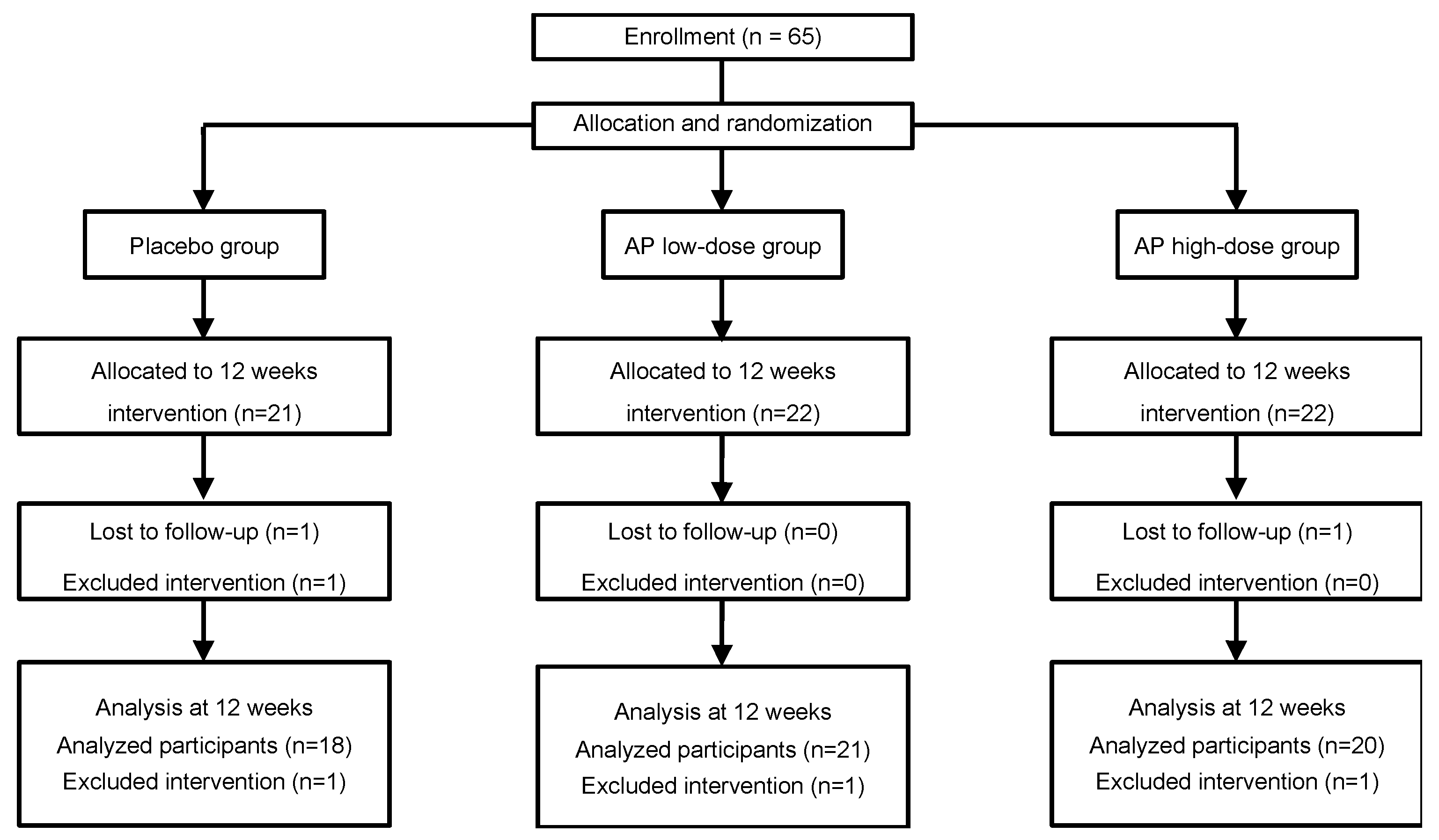
2.1. Study Participants
2.2. Dietary Supplements
2.3. Experimental Design
2.4. Ethics Statement
2.5. Determination of SOD-like Activity of Apple Procyanidins
2.6. Statistical Analysis
3. Results
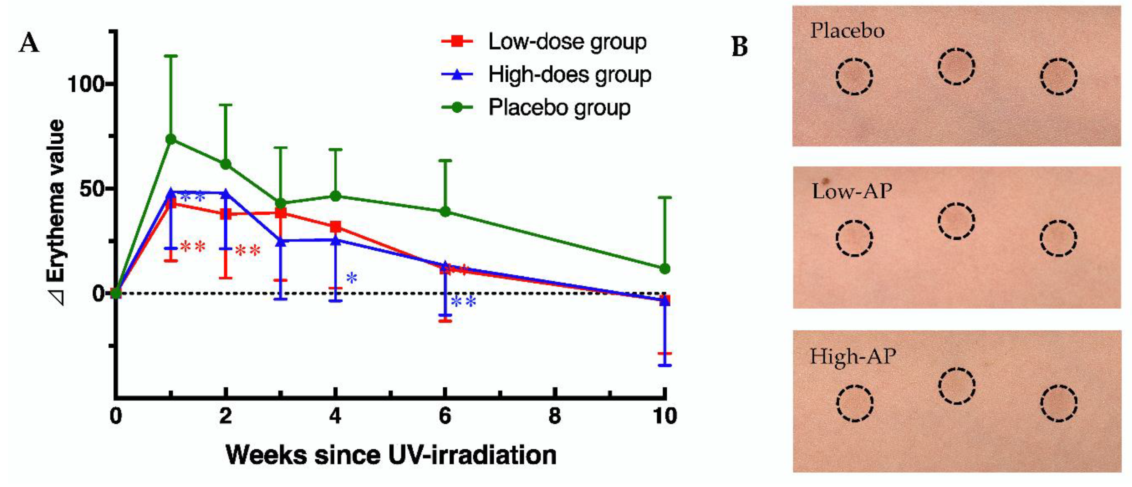
3.1. Effects of AP on UV Irradiation Induced Erythema Value
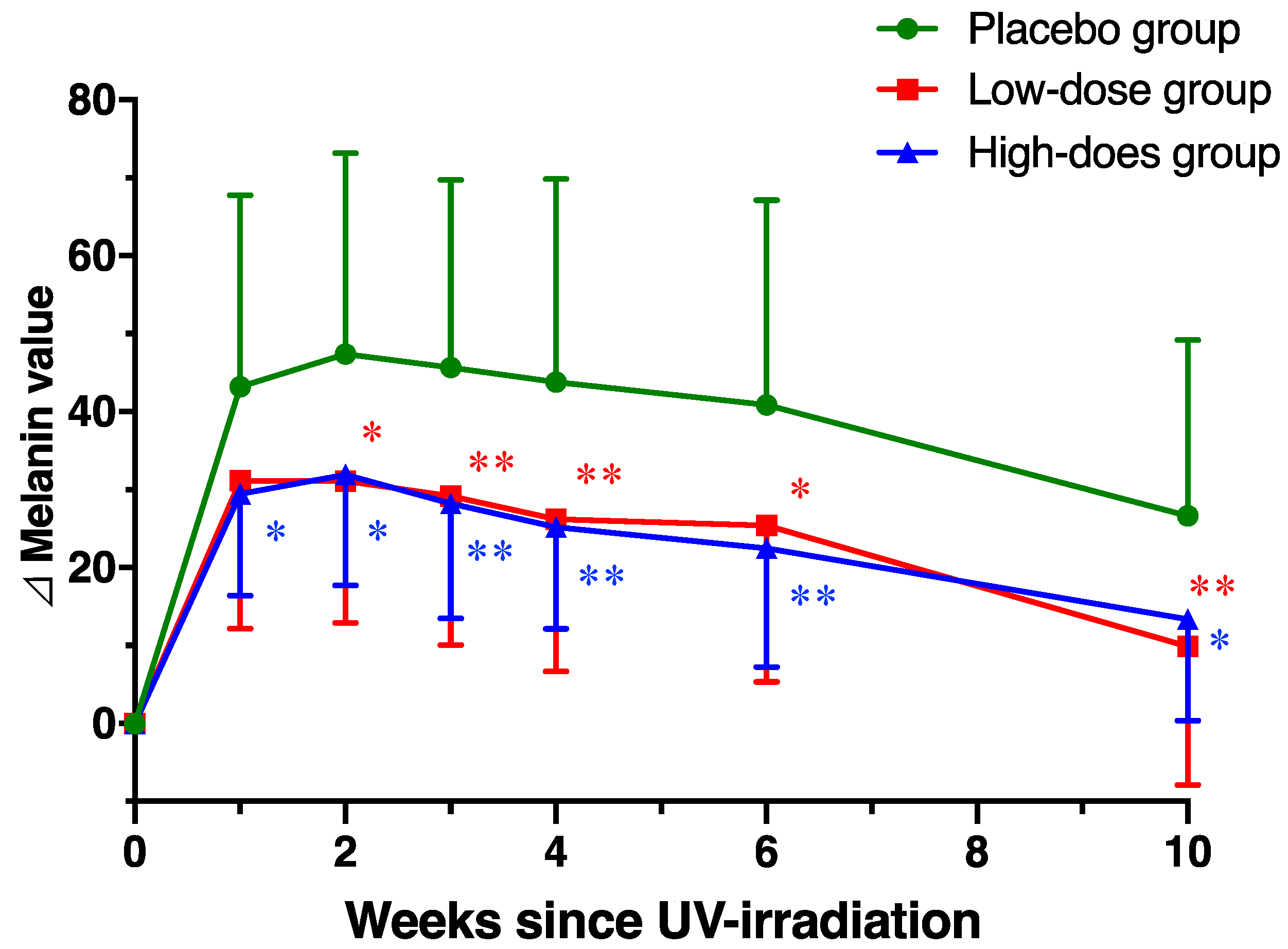
3.2. Effects of AP on UV Irradiation Induced Melanin Value
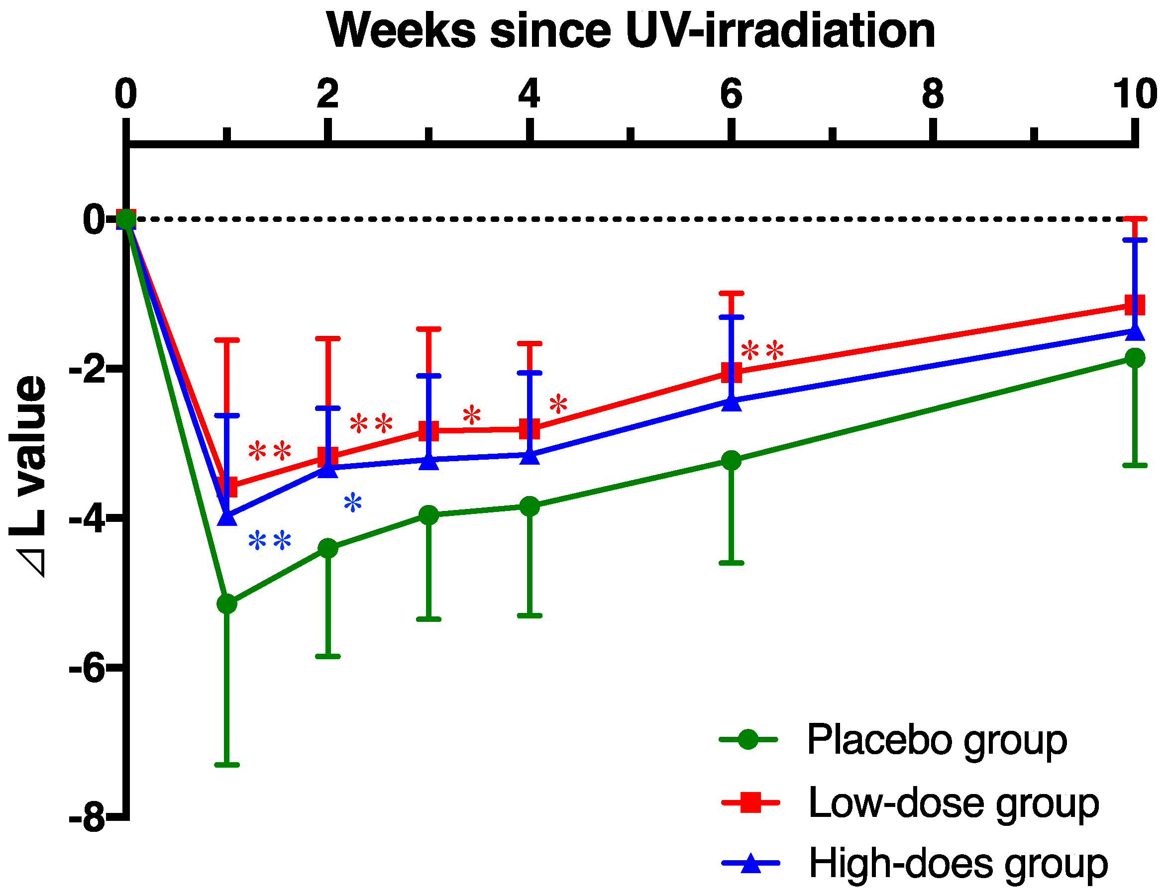
3.3. Effects of AP on UV Irradiation Induced L Value
3.4. Effect of AP on Facial Skin Condition
3.5. Effect of Apple Procyanidins on SOD-Like Activity
4. Discussion
Supplementary Materials
Author Contributions
Funding
Acknowledgments
Conflicts of Interest
References
- Perez-Sanchez, A.; Barrajon-Catalan, E.; Herranz-Lopez, M.; Micol, V. Nutraceuticals for skin care: A comprehensive review of human clinical studies. Nutrients 2018, 10. [Google Scholar] [CrossRef] [PubMed]
- Madison, K.C. Barrier function of the skin: "La raison d'etre" of the epidermis. J. Investig. Dermatol. 2003, 121, 231–241. [Google Scholar] [CrossRef] [PubMed]
- Katiyar, S.; Elmets, C.A.; Katiyar, S.K. Green tea and skin cancer: Photoimmunology, angiogenesis and DNA repair. J. Nutr. Biochem. 2007, 18, 287–296. [Google Scholar] [CrossRef] [PubMed]
- Bickers, D.R.; Athar, M. Oxidative stress in the pathogenesis of skin disease. J. Investig. Dermatol. 2006, 126, 2565–2575. [Google Scholar] [CrossRef] [PubMed]
- D’Orazio, J.; Jarrett, S.; Amaro-Ortiz, A.; Scott, T. UV radiation and the skin. Int. J. Mol. Sci. 2013, 14, 12222–12248. [Google Scholar] [CrossRef]
- Dzialo, M.; Mierziak, J.; Korzun, U.; Preisner, M.; Szopa, J.; Kulma, A. The Potential of Plant Phenolics in Prevention and Therapy of Skin Disorders. Int. J. Mol. Sci. 2016, 17, 160. [Google Scholar] [CrossRef]
- Orimo, H.; Tokura, Y.; Hino, R.; Kasai, H. Formation of 8-hydroxy-2'-deoxyguanosine in the DNA of cultured human keratinocytes by clinically used doses of narrowband and broadband ultraviolet B and psoralen plus ultraviolet A. Cancer Sci. 2006, 97, 99–105. [Google Scholar] [CrossRef]
- Premi, S.; Wallisch, S.; Mano, C.M.; Weiner, A.B.; Bacchiocchi, A.; Wakamatsu, K.; Bechara, E.J.; Halaban, R.; Douki, T.; Brash, D.E. Photochemistry. Chemiexcitation of melanin derivatives induces DNA photoproducts long after UV exposure. Science 2015, 347, 842–847. [Google Scholar] [CrossRef]
- Karg, E.; Odh, G.; Wittbjer, A.; Rosengren, E.; Rorsman, H. Hydrogen peroxide as an inducer of elevated tyrosinase level in melanoma cells. J. Investig. Dermatol. 1993, 100, 209S–213S. [Google Scholar] [CrossRef]
- Eller, M.S.; Yaar, M.; Gilchrest, B.A. DNA damage and melanogenesis. Nature 1994, 372, 413–414. [Google Scholar] [CrossRef]
- F'Guyer, S.; Afaq, F.; Mukhtar, H. Photochemoprevention of skin cancer by botanical agents. Photodermatol. Photoimmunol. Photomed. 2003, 19, 56–72. [Google Scholar] [CrossRef] [PubMed]
- Solano, F. Photoprotection and Skin Pigmentation: Melanin-Related Molecules and Some Other New Agents Obtained from Natural Sources. Molecules 2020, 25. [Google Scholar] [CrossRef]
- Grether-Beck, S.; Marini, A.; Jaenicke, T.; Stahl, W.; Krutmann, J. Molecular evidence that oral supplementation with lycopene or lutein protects human skin against ultraviolet radiation: Results from a double-blinded, placebo-controlled, crossover study. Br. J. Dermatol. 2017, 176, 1231–1240. [Google Scholar] [CrossRef] [PubMed]
- Ito, N.; Seki, S.; Ueda, F. The protective role of astaxanthin for UV-induced skin deterioration in healthy people - a randomized, double-blind, placebo-controlled trial. Nutrients 2018, 10. [Google Scholar] [CrossRef] [PubMed]
- Baswan, S.M.; Marini, A.; Klosner, A.E.; Jaenicke, T.; Leverett, J.; Murray, M.; Gellenbeck, K.W.; Krutmann, J. Orally administered mixed carotenoids protect human skin against ultraviolet A-induced skin pigmentation: A double-blind, placebo-controlled, randomized clinical trial. Photodermatol. Photoimmunol. Photomed. 2020. [Google Scholar] [CrossRef] [PubMed]
- Nichols, J.A.; Katiyar, S.K. Skin photoprotection by natural polyphenols: Anti-inflammatory, antioxidant and DNA repair mechanisms. Arch. Dermatol. Res. 2010, 302, 71–83. [Google Scholar] [CrossRef] [PubMed]
- Gu, L.; Kelm, M.A.; Hammerstone, J.F.; Beecher, G.; Holden, J.; Haytowitz, D.; Gebhardt, S.; Prior, R.L. Concentrations of proanthocyanidins in common foods and estimations of normal consumption. J. Nutr. 2004, 134, 613–617. [Google Scholar] [CrossRef]
- Koga, T.; Moro, K.; Nakamori, K.; Yamakoshi, J.; Hosoyama, H.; Kataoka, S.; Ariga, T. Increase of antioxidative potential of rat plasma by oral administration of proanthocyanidin-rich extract from grape seeds. J. Agric. Food Chem. 1999, 47, 1892–1897. [Google Scholar] [CrossRef]
- Lotito, S.B.; Actis-Goretta, L.; Renart, M.L.; Caligiuri, M.; Rein, D.; Schmitz, H.H.; Steinberg, F.M.; Keen, C.L.; Fraga, C.G. Influence of oligomer chain length on the antioxidant activity of procyanidins. Biochem Biophys Res Commun 2000, 276, 945–951. [Google Scholar] [CrossRef]
- Yamakoshi, J.; Kataoka, S.; Koga, T.; Ariga, T. Proanthocyanidin-rich extract from grape seeds attenuates the development of aortic atherosclerosis in cholesterol-fed rabbits. Atherosclerosis 1999, 142, 139–149. [Google Scholar] [CrossRef]
- Guo, Q.; Zhao, B.; Packer, L. Electron spin resonance study of free radicals formed from a procyanidin-rich pine (Pinus maritima) bark extract, pycnogenol. Free Radic. Biol. Med. 1999, 27, 1308–1312. [Google Scholar] [CrossRef]
- Akiyama, H.; Sato, Y.; Watanabe, T.; Nagaoka, M.H.; Yoshioka, Y.; Shoji, T.; Kanda, T.; Yamada, K.; Totsuka, M.; Teshima, R.; et al. Dietary unripe apple polyphenol inhibits the development of food allergies in murine models. FEBS Lett. 2005, 579, 4485–4491. [Google Scholar] [CrossRef] [PubMed]
- Sugiyama, H.; Akazome, Y.; Shoji, T.; Yamaguchi, A.; Yasue, M.; Kanda, T.; Ohtake, Y. Oligomeric procyanidins in apple polyphenol are main active components for inhibition of pancreatic lipase and triglyceride absorption. J. Agric. Food Chem. 2007, 55, 4604–4609. [Google Scholar] [CrossRef] [PubMed]
- Miura, T.; Chiba, M.; Kasai, K.; Nozaka, H.; Nakamura, T.; Shoji, T.; Kanda, T.; Ohtake, Y.; Sato, T. Apple procyanidins induce tumor cell apoptosis through mitochondrial pathway activation of caspase-3. Carcinogenesis 2008, 29, 585–593. [Google Scholar] [CrossRef]
- Obara, M.; Masumoto, S.; Ono, Y.; Ozaki, Y.; Shoji, T. Procyanidin Concentrations and H-ORAC of Apples Cultivated in Japan. Food Sci. Technol. Res. 2016, 22, 563–568. [Google Scholar] [CrossRef]
- Shoji, T.; Masumoto, S.; Moriichi, N.; Kobori, M.; Kanda, T.; Shinmoto, H.; Tsushida, T. Procyanidin trimers to pentamers fractionated from apple inhibit melanogenesis in B16 mouse melanoma cells. J. Agric. Food Chem. 2005, 53, 6105–6111. [Google Scholar] [CrossRef]
- Shoji, T.; Akazome, Y.; Kanda, T.; Ikeda, M. The toxicology and safety of apple polyphenol extract. Food Chem. Toxicol. 2004, 42, 959–967. [Google Scholar] [CrossRef]
- Shoji, T.; Masumoto, S.; Moriichi, N.; Kanda, T.; Ohtake, Y. Apple (Malus pumila) procyanidins fractionated according to the degree of polymerization using normal-phase chromatography and characterized by HPLC-ESI/MS and MALDI-TOF/MS. J. Chromatogr. A 2006, 1102, 206–213. [Google Scholar] [CrossRef]
- Ottaviani, J.I.; Heiss, C.; Spencer, J.P.E.; Kelm, M.; Schroeter, H. Recommending flavanols and procyanidins for cardiovascular health: Revisited. Mol. Aspects Med. 2018, 61, 63–75. [Google Scholar] [CrossRef]
- Yamakoshi, J.; Otsuka, F.; Sano, A.; Tokutake, S.; Saito, M.; Kikuchi, M.; Kubota, Y. Lightening effect on ultraviolet-induced pigmentation of guinea pig skin by oral administration of a proanthocyanidin-rich extract from grape seeds. Pigment Cell Res. 2003, 16, 629–638. [Google Scholar] [CrossRef]
- Eller, M.S.; Ostrom, K.; Gilchrest, B.A. DNA damage enhances melanogenesis. Proc. Natl. Acad. Sci. USA 1996, 93, 1087–1092. [Google Scholar] [CrossRef] [PubMed]
- Yamaguchi, F.; Yoshimura, Y.; Nakazawa, H.; Ariga, T. Free radical scavenging activity of grape seed extract and antioxidants by electron spin resonance spectrometry in an H(2)O(2)/NaOH/DMSO system. J. Agric. Food Chem. 1999, 47, 2544–2548. [Google Scholar] [CrossRef] [PubMed]
- Rohdewald, P. A review of the French maritime pine bark extract (Pycnogenol), a herbal medication with a diverse clinical pharmacology. Int. J. Clin. Pharmacol. Ther. 2002, 40, 158–168. [Google Scholar] [CrossRef] [PubMed]
- Heinrich, U.; Neukam, K.; Tronnier, H.; Sies, H.; Stahl, W. Long-term ingestion of high flavanol cocoa provides photoprotection against UV-induced erythema and improves skin condition in women. J. Nutr. 2006, 136, 1565–1569. [Google Scholar] [CrossRef] [PubMed]
- Saliou, C.; Rimbach, G.; Moini, H.; McLaughlin, L.; Hosseini, S.; Lee, J.; Watson, R.R.; Packer, L. Solar ultraviolet-induced erythema in human skin and nuclear factor-kappa-B-dependent gene expression in keratinocytes are modulated by a French maritime pine bark extract. Free Radic. Biol. Med. 2001, 30, 154–160. [Google Scholar] [CrossRef]
- Furumura, M.; Sato, N.; Kusaba, N.; Takagaki, K.; Nakayama, J. Oral administration of French maritime pine bark extract (Flavangenol(®)) improves clinical symptoms in photoaged facial skin. Clin. Interv. Aging 2012, 7, 275–286. [Google Scholar] [CrossRef]
- Ni, Z.; Mu, Y.; Gulati, O. Treatment of melasma with Pycnogenol. Phytother. Res. 2002, 16, 567–571. [Google Scholar] [CrossRef]
- Holt, R.R.; Lazarus, S.A.; Sullards, M.C.; Zhu, Q.Y.; Schramm, D.D.; Hammerstone, J.F.; Fraga, C.G.; Schmitz, H.H.; Keen, C.L. Procyanidin dimer B2 [epicatechin-(4beta-8)-epicatechin] in human plasma after the consumption of a flavanol-rich cocoa. Am. J. Clin. Nutr. 2002, 76, 798–804. [Google Scholar] [CrossRef]
- Sano, A.; Yamakoshi, J.; Tokutake, S.; Tobe, K.; Kubota, Y.; Kikuchi, M. Procyanidin B1 is detected in human serum after intake of proanthocyanidin-rich grape seed extract. Biosci. Biotechnol. Biochem. 2003, 67, 1140–1143. [Google Scholar] [CrossRef]
- Shoji, T.; Masumoto, S.; Moriichi, N.; Akiyama, H.; Kanda, T.; Ohtake, Y.; Goda, Y. Apple procyanidin oligomers absorption in rats after oral administration: Analysis of procyanidins in plasma using the porter method and high-performance liquid chromatography/tandem mass spectrometry. J. Agric. Food Chem. 2006, 54, 884–892. [Google Scholar] [CrossRef]
- Masumoto, S.; Aoki, S.; Miura, T.; Shoji, T. Flavan-3-ol/Procyanidin Metabolomics in Rat Urine Using HPLC-Quadrupole TOF/MS. Mol. Nutr. Food Res. 2018, 62. [Google Scholar] [CrossRef] [PubMed]
- Masumoto, S.; Terao, A.; Yamamoto, Y.; Mukai, T.; Miura, T.; Shoji, T. Non-absorbable apple procyanidins prevent obesity associated with gut microbial and metabolomic changes. Sci. Rep. 2016, 6, 31208. [Google Scholar] [CrossRef] [PubMed]
- Anhe, F.F.; Roy, D.; Pilon, G.; Dudonne, S.; Matamoros, S.; Varin, T.V.; Garofalo, C.; Moine, Q.; Desjardins, Y.; Levy, E.; et al. A polyphenol-rich cranberry extract protects from diet-induced obesity, insulin resistance and intestinal inflammation in association with increased Akkermansia spp. population in the gut microbiota of mice. Gut 2015, 64, 872–883. [Google Scholar] [CrossRef]
- Roopchand, D.E.; Carmody, R.N.; Kuhn, P.; Moskal, K.; Rojas-Silva, P.; Turnbaugh, P.J.; Raskin, I. Dietary Polyphenols Promote Growth of the Gut Bacterium Akkermansia muciniphila and Attenuate High-Fat Diet-Induced Metabolic Syndrome. Diabetes 2015, 64, 2847–2858. [Google Scholar] [CrossRef] [PubMed]
- Salem, I.; Ramser, A.; Isham, N.; Ghannoum, M.A. The Gut Microbiome as a Major Regulator of the Gut-Skin Axis. Front. Microbiol. 2018, 9, 1459. [Google Scholar] [CrossRef] [PubMed]
- Sharma, S.D.; Meeran, S.M.; Katiyar, S.K. Dietary grape seed proanthocyanidins inhibit UVB-induced oxidative stress and activation of mitogen-activated protein kinases and nuclear factor-kappaB signaling in in vivo SKH-1 hairless mice. Mol. Cancer Ther. 2007, 6, 995–1005. [Google Scholar] [CrossRef]


| Intervention Period (Weeks) | 0 | 2 | 3 | 4 | 5 | 6 | 8 | 12 |
| (Weeks since UV Irradiation) | (0) | (1) | (2) | (3) | (4) | (6) | (10) | |
| Precheck | ● | |||||||
| Visit to facility | ● | ● | ● | ● | ● | ● | ● | ● |
| Informed consent | ● | |||||||
| MED evaluation | ● | |||||||
| Evaluation of pigmentation | ||||||||
| UV-irradiation (1.5 MED) | ● | |||||||
| Erythema, melanin, L value | ● | ● | ● | ● | ● | ● | ● | |
| Evaluation of facial skin condition | ||||||||
| Water content, TEWL | ● | ● | ● | ● | ||||
| Ingestion of test sample |  | |||||||
| Recording in diary | | |||||||
| Apple Polyphenol Groups | Placebo Group | ||
|---|---|---|---|
| Low-Dose Group (300 mg/day) | High-Dose Group (600 mg/day) | ||
| N | 21 | 20 | 18 |
| Age (y) | 34.19 ± 4.29 | 35.20 ± 3.89 | 34.06 ± 4.08 |
| MED (mJ/cm2) | 5323.7 ± 478.0 | 5471.1 ± 499.7 | 5247.0 ± 531.7 |
| Water content (AU) | 88.56 ± 18.62 | 80.94 ± 14.33 | 85.32 ± 20.59 |
| TEWL (g/h m2) | 15.80 ± 5.85 | 16.12 ± 5.58 | 14.92 ± 5.10 |
| Parameters | Groups | Intervention Period (Weeks since UV-Irradiation) | ||||||
|---|---|---|---|---|---|---|---|---|
| 0 | 1 | 2 | 3 | 4 | 6 | 10 | ||
| Erythema value | Placebo | 175.6 ± 34.4 | 252.0 ± 40.3 ** | 237.3 ± 36.9 ** | 219.5 ± 34.7 ** | 222.5 ± 38.5 ** | 212.8 ± 31.9 * | 187.6 ± 35.4 |
| Low AP | 182.4 ± 32.0 | 225.5 ± 34.4 ** | 222.5 ± 31.9 ** | 224.0 ± 40.0 ** | 215.9 ± 41.5 * | 196.6 ± 31.8 | 184.0 ± 26.6 | |
| High AP | 179.5 ± 51.0 | 228.7 ± 43.5 ** | 225.0 ± 52.5 ** | 204.6 ± 40.0 | 206.1 ± 36.6 | 193.4 ± 42.8 | 176.2 ± 40.3 | |
| Melanin value | Placebo | 149.0 ± 33.3 | 192.2 ± 36.5 ** | 196.4 ± 38.4 ** | 194.7 ± 37.4 ** | 192.9 ± 36.8 ** | 189.9 ± 35.8 ** | 175.7 ± 32.1 |
| Low AP | 163.8 ± 29.5 | 194.9 ± 27.3 * | 194.9 ± 28.2 * | 193.0 ± 27.2 * | 190.0 ± 28.5 * | 189.2 ± 28.7 | 174.5 ± 23.3 | |
| High AP | 159.1 ± 36.6 | 188.6 ± 33.6 * | 191.1 ± 33.7 ** | 187.4 ± 31.7 | 184.3 ± 33.5 | 181.6 ± 33.2 | 172.5 ± 31.0 | |
| L value | Placebo | 64.4 ± 2.4 | 59.3 ± 2.7 ** | 60.0 ± 2.1 ** | 60.4 ± 2.0 ** | 60.6 ± 2.5 ** | 61.2 ± 2.1 ** | 62.5 ± 2.1 |
| Low AP | 64.4 ± 1.9 | 60.8 ± 2.4 ** | 61.2 ± 2.1 ** | 61.5 ± 1.8 ** | 61.6 ± 1.8 ** | 62.3 ± 1.6 * | 63.2 ± 1.9 | |
| High AP | 64.4 ± 2.7 | 60.4 ± 2.0 ** | 61.0 ± 2.5 ** | 61.1 ± 2.1 ** | 61.2 ± 2.0 ** | 61.9 ± 2.2 ** | 62.9 ± 2.2 ** | |
| Parameters | Groups | Intervention Period (Weeks) | |||
|---|---|---|---|---|---|
| 0 | 4 | 8 | 12 | ||
| Water content (AU) | Placebo | 85.3 ± 20.6 | 89.6 ± 16.6 | 88.0 ± 16.9 | 92.0 ± 16.8 |
| Low AP | 88.6 ± 18.6 | 92.8 ± 16.6 | 91.8 ± 22.5 | 92.2 ± 15.8 | |
| High AP | 80.9 ± 14.3 | 86.7 ± 14.6 | 88.3 ± 13.9 | 91.3 ± 12.3 | |
| TEWL (g/h m2) | Placebo | 14.9 ± 5.1 | 14.6 ± 5.3 | 15.1 ± 6.2 | 13.6 ± 3.7 |
| Low AP | 15.8 ± 5.9 | 17.0 ± 6.9 | 15.9 ± 7.5 | 15.4 ± 7.6 | |
| High AP | 16.1 ± 5.6 | 16.2 ± 4.1 | 14.5 ± 4.3 | 14.8 ± 6.4 | |
© 2020 by the authors. Licensee MDPI, Basel, Switzerland. This article is an open access article distributed under the terms and conditions of the Creative Commons Attribution (CC BY) license (http://creativecommons.org/licenses/by/4.0/).
Share and Cite
Shoji, T.; Masumoto, S.; Moriichi, N.; Ohtake, Y.; Kanda, T. Administration of Apple Polyphenol Supplements for Skin Conditions in Healthy Women: A Randomized, Double-Blind, Placebo-Controlled Clinical Trial. Nutrients 2020, 12, 1071. https://doi.org/10.3390/nu12041071
Shoji T, Masumoto S, Moriichi N, Ohtake Y, Kanda T. Administration of Apple Polyphenol Supplements for Skin Conditions in Healthy Women: A Randomized, Double-Blind, Placebo-Controlled Clinical Trial. Nutrients. 2020; 12(4):1071. https://doi.org/10.3390/nu12041071
Chicago/Turabian StyleShoji, Toshihiko, Saeko Masumoto, Nina Moriichi, Yasuyuki Ohtake, and Tomomasa Kanda. 2020. "Administration of Apple Polyphenol Supplements for Skin Conditions in Healthy Women: A Randomized, Double-Blind, Placebo-Controlled Clinical Trial" Nutrients 12, no. 4: 1071. https://doi.org/10.3390/nu12041071
APA StyleShoji, T., Masumoto, S., Moriichi, N., Ohtake, Y., & Kanda, T. (2020). Administration of Apple Polyphenol Supplements for Skin Conditions in Healthy Women: A Randomized, Double-Blind, Placebo-Controlled Clinical Trial. Nutrients, 12(4), 1071. https://doi.org/10.3390/nu12041071