Nutrition in Necrotizing Enterocolitis and Following Intestinal Resection

Abstract
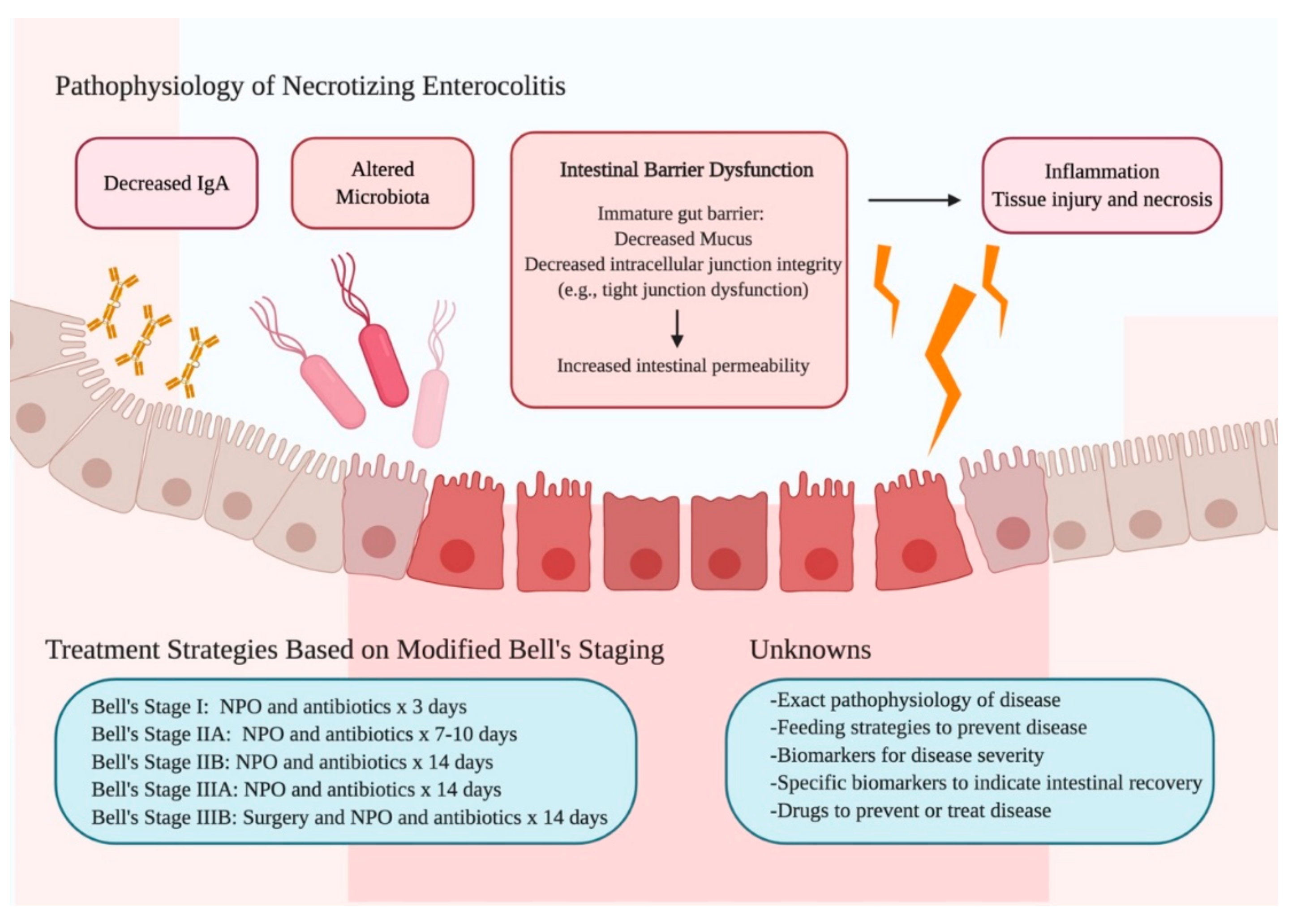
1. Introduction
2. NEC Prevention
2.1. Delivery of Feeds
2.1.1. Initiation of Feeds
2.1.2. Feeding Advancement
2.1.3. Bolus and Continuous Feeding
2.2. Composition of Feeds
2.2.1. Osmolality
2.2.2. Breast Milk
2.2.3. Donor Breast Milk
2.2.4. Cow’s Milk Formula
3. Medical NEC
3.1. NPO Duration
3.2. Parenteral Nutrition
4. Surgical NEC
4.1. Enteral Feeding
4.2. Anatomical Considerations
4.2.1. Intestinal Length
4.2.2. Segment Functionality
4.3. Ostomy Replacement
4.4. Macronutrients
4.4.1. Fat
4.4.2. Protein
4.4.3. Oligosaccharides
4.5. Parenteral Nutrition
PN Lipid Source
5. Conclusions
Author Contributions
Funding
Conflicts of Interest
References
- Neu, J.; Walker, W.A. Necrotizing enterocolitis. N. Engl. J. Med. 2011, 364, 255–264. [Google Scholar] [CrossRef]
- Bell, M.J.; Ternberg, J.L.; Feigin, R.D.; Keating, J.P.; Marshall, R.; Barton, L.; Brotherton, T. Neonatal necrotizing enterocolitis. Therapeutic decisions based upon clinical staging. Ann. Surg. 1978, 187, 1–7. [Google Scholar] [CrossRef]
- Walsh, M.C.; Kliegman, R.M. Necrotizing Enterocolitis: Treatment Based on Staging Criteria. Pediatr. Clin. North Am. 1986, 33, 179–201. [Google Scholar] [CrossRef]
- Schnabl, K.L.; E Van Aerde, J.; Thomson, A.B.; Clandinin, M.T. Necrotizing enterocolitis: A multifactorial disease with no cure. World J. Gastroenterol. 2008, 14, 2142–2161. [Google Scholar] [CrossRef]
- Warner, B.W. The Pathogenesis of Resection-Associated Intestinal Adaptation. Cell. Mol. Gastroenterol. Hepatol. 2016, 2, 429–438. [Google Scholar] [CrossRef]
- Morgan, J.; Bombell, S.; McGuire, W. Early trophic feeding versus enteral fasting for very preterm or very low birth weight infants. Cochrane Database Syst. Rev. 2013, 2013, CD000504. [Google Scholar] [CrossRef]
- AlShaikh, B.; Dharel, D.; Yusuf, K.; Singhal, N. Early total enteral feeding in stable preterm infants: a systematic review and meta-analysis. J. Matern. Neonatal Med. 2019, 1–8. [Google Scholar] [CrossRef]
- Nangia, S.; Vadivel, V.; Thukral, A.; Saili, A. Early Total Enteral Feeding versus Conventional Enteral Feeding in Stable Very-Low-Birth-Weight Infants: A Randomised Controlled Trial. Neonatol. 2019, 115, 256–262. [Google Scholar] [CrossRef]
- Dorling, J.; Abbott, J.; Berrington, J.; Bosiak, B.; Bowler, U.; Boyle, E.; Embleton, N.; Hewer, O.; Johnson, S.; Juszczak, E.; et al. Controlled Trial of Two Incremental Milk-Feeding Rates in Preterm Infants. New Engl. J. Med. 2019, 381, 1434–1443. [Google Scholar] [CrossRef]
- Olieman, J.F.; Penning, C.; Ijsselstijn, H.; Escher, J.C.; Joosten, K.F.; Hulst, J.M.; Tibboel, D. Enteral Nutrition in Children with Short-Bowel Syndrome: Current Evidence and Recommendations for the Clinician. J. Am. Diet. Assoc. 2010, 110, 420–426. [Google Scholar] [CrossRef]
- Davis, T.A.; Fiorotto, M.L.; Suryawan, A. Bolus vs. continuous feeding to optimize anabolism in neonates. Curr. Opin. Clin. Nutr. Metab. Care 2015, 18, 102–108. [Google Scholar] [CrossRef]
- Wang, Y.; Zhu, W.; Luo, B.-R. Continuous feeding versus intermittent bolus feeding for premature infants with low birth weight: a meta-analysis of randomized controlled trials. Eur. J. Clin. Nutr. 2019, 1–9. [Google Scholar] [CrossRef]
- Pearson, F.; Johnson, M.J.; Leaf, A.A. Leaf, Milk osmolality: does it matter? Arch. Dis. Child. Fetal Neonatal. Ed. 2013, 98, F166–F169. [Google Scholar] [CrossRef]
- Willis, D.M.; Chabot, J.; Radde, I.C.; Chance, G.W. Unsuspected hyperosmolality of oral solutions contributing to necrotizing enterocolitis in very-low-birth-weight infants. Pediatrics 1977, 60, 535–538. [Google Scholar]
- Sántulli, T.V.; Schullinger, J.N.; Heird, W.C.; Gongaware, R.D.; Wigger, J.; Barlow, B.; Blanc, W.A.; Berdon, W.E. Acute necrotizing enterocolitis in infancy: a review of 64 cases. Pediatrics 1975, 55, 376–387. [Google Scholar]
- Miyake, H.; Chen, Y.; Koike, Y.; Hock, A.; Lee, C.; Zani, A.; Pierro, A.; Li, B. Osmolality of enteral formula and severity of experimental necrotizing enterocolitis. Pediatr. Surg. Int. 2016, 32, 1153–1156. [Google Scholar] [CrossRef]
- Brown, J.V.E.; Embleton, N.; Harding, J.E.; McGuire, W. Multi-nutrient fortification of human milk for preterm infants. Cochrane Database Syst. Rev. 2016, 2016, CD000343. [Google Scholar] [CrossRef]
- Meinzen-Derr, J.; Poindexter, B.; Wrage, L.; Morrow, A.L.; Stoll, B.; Donovan, E.F. Role of human milk in extremely low birth weight infants’ risk of necrotizing enterocolitis or death. J. Perinatol. 2009, 29, 57–62. [Google Scholar] [CrossRef]
- Sisk, P.M.; A Lovelady, C.; Dillard, R.G.; Gruber, K.; O’Shea, T.M. Early human milk feeding is associated with a lower risk of necrotizing enterocolitis in very low birth weight infants. J. Perinatol. 2007, 27, 428–433. [Google Scholar] [CrossRef]
- Lucas, A.; Cole, T. Breast milk and neonatal necrotising enterocolitis. Lancet 1990, 336, 1519–1523. [Google Scholar] [CrossRef]
- Hanson, L.Å.; Strömbäck, L.; Erling, V.; Zaman, S.; Olcén, P.; Telemo, E. The immunological role of breast feeding. Pediatr. Allergy Immunol. 2001, 12, 15–19. [Google Scholar] [CrossRef]
- Walsh, V.; McGuire, W. Immunonutrition for Preterm Infants. Neonatol. 2019, 115, 398–405. [Google Scholar] [CrossRef]
- Gopalakrishna, K.P.; Macadangdang, B.R.; Rogers, M.B.; Tometich, J.T.; Firek, B.A.; Baker, R.; Ji, J.; Burr, A.H.P.; Ma, C.; Good, M.; et al. Maternal IgA protects against the development of necrotizing enterocolitis in preterm infants. Nat. Med. 2019, 25, 1110–1115. [Google Scholar] [CrossRef]
- Good, M.; Sodhi, C.P.; Egan, C.E.; Afrazi, A.; Jia, H.; Yamaguchi, Y.; Lu, P.; Branca, M.F.; Ma, C.; Prindle, T.; et al. Breast milk protects against the development of necrotizing enterocolitis through inhibition of Toll-like receptor 4 in the intestinal epithelium via activation of the epidermal growth factor receptor. Mucosal Immunol. 2015, 8, 1166–1179. [Google Scholar] [CrossRef]
- Sullivan, S.; Schanler, R.J.; Kim, J.H.; Patel, A.; Trawöger, R.; Kiechl-Kohlendorfer, U.; Chan, G.M.; Blanco, C.L.; Abrams, S.A.; Cotten, C.M.; et al. An Exclusively Human Milk-Based Diet Is Associated with a Lower Rate of Necrotizing Enterocolitis than a Diet of Human Milk and Bovine Milk-Based Products. J. Pediatr. 2010, 156, 562–567. [Google Scholar] [CrossRef]
- Schanler, R.J.; Lau, C.; Hurst, N.M.; Smith, E.O. Randomized Trial of Donor Human Milk Versus Preterm Formula as Substitutes for Mothers’ Own Milk in the Feeding of Extremely Premature Infants. Pediatrics 2005, 116, 400–406. [Google Scholar] [CrossRef]
- Cristofalo, E.A.; Schanler, R.J.; Blanco, C.L.; Sullivan, S.; Trawoeger, R.; Kiechl-Kohlendorfer, U.; Dudell, G.; Rechtman, D.J.; Lee, M.L.; Lucas, A.; et al. Randomized Trial of Exclusive Human Milk versus Preterm Formula Diets in Extremely Premature Infants. J. Pediatr. 2013, 163, 1592–1595. [Google Scholar] [CrossRef]
- Boyd, C.A.; Quigley, M.A.; Brocklehurst, P. Donor breast milk versus infant formula for preterm infants: systematic review and meta-analysis. Arch. Dis. Child. Fetal Neonatal Ed. 2007, 92, F169–F175. [Google Scholar] [CrossRef]
- Quigley, M.; Embleton, N.D.; McGuire, W. Formula versus donor breast milk for feeding preterm or low birth weight infants. Cochrane Database Syst. Rev. 2019, 7, CD002971. [Google Scholar] [CrossRef]
- Chowning, R.; Radmacher, P.; Lewis, S.; Serke, L.; Pettit, N.; Adamkin, D.H. A retrospective analysis of the effect of human milk on prevention of necrotizing enterocolitis and postnatal growth. J. Perinatol. 2016, 36, 221–224. [Google Scholar] [CrossRef]
- Chuang, S.-L.; Hayes, P.J.; Ogundipe, E.; Haddad, M.; Macdonald, T.T.; Fell, J.M. Cow’s milk protein-specific T-helper type I/II cytokine responses in infants with necrotizing enterocolitis. Pediatr. Allergy Immunol. 2009, 20, 45–52. [Google Scholar] [CrossRef] [PubMed]
- Li, B.; Hock, A.; Wu, R.Y.; Minich, A.; Botts, S.; Lee, C.; Antounians, L.; Miyake, H.; Koike, Y.; Chen, Y.; et al. Bovine milk-derived exosomes enhance goblet cell activity and prevent the development of experimental necrotizing enterocolitis. PLOS ONE 2019, 14, e0211431. [Google Scholar] [CrossRef] [PubMed]
- Valpacos, M.; Arni, D.; Keir, A.; Aspirot, A.; Wilde, J.C.; Beasley, S.; De Luca, D.; Pfister, R.E.; Karam, O. Diagnosis and Management of Necrotizing Enterocolitis: An International Survey of Neonatologists and Pediatric Surgeons. Neonatology 2018, 113, 170–176. [Google Scholar] [CrossRef] [PubMed]
- Hock, A.M.; Chen, Y.; Miyake, H.; Koike, Y.; Seo, S.; Pierro, A. Initiation of Enteral Feeding After Necrotizing Enterocolitis. Eur. J. Pediatr. Surg. 2018, 28, 44–50. [Google Scholar]
- Bohnhorst, B.; Müller, S.; Dördelmann, M.; Peter, C.S.; Petersen, C.; Poets, C.F. Early feeding after necrotizing enterocolitis in preterm infants. J. Pediatr. 2003, 143, 484–487. [Google Scholar] [CrossRef]
- Kuik, S.J.; Kalteren, W.S.; Mebius, M.J.; Bos, A.F.; Hulscher, J.B.F.; Kooi, E.M.W. Predicting intestinal recovery after necrotizing enterocolitis in preterm infants. Pediatr. Res. 2019, 1–9. [Google Scholar] [CrossRef]
- Heath, M.; Buckley, R.; Gerber, Z.; Davis, P.; Linneman, L.; Gong, Q.; Barkemeyer, B.; Fang, Z.; Good, M.; Penn, D.; et al. Association of Intestinal Alkaline Phosphatase With Necrotizing Enterocolitis Among Premature Infants. JAMA Netw. Open 2019, 2, e1914996. [Google Scholar] [CrossRef]
- Neu, J. Neonatal necrotizing enterocolitis: An update. Acta Paediatr. 2005, 94, 100–105. [Google Scholar] [CrossRef]
- Ibrahim, H.; Jeroudi, M.A.; Baier, R.J.; Dhanireddy, R.; Krouskop, R.W. Aggressive Early Total Parental Nutrition in Low-Birth-Weight Infants. J. Perinatol. 2004, 24, 482–486. [Google Scholar] [CrossRef]
- Can, E.; Bulbul, A.; Uslu, S.; Cömert, S.; Bolat, F.; Nuhoğlu, A.; Nuhoǧlu, A. Effects of aggressive parenteral nutrition on growth and clinical outcome in preterm infants. Pediatr. Int. 2012, 54, 869–874. [Google Scholar] [CrossRef]
- Akinkuotu, A.C.; Nuthakki, S.; Sheikh, F.; Cruz, S.M.; Welty, S.E.; Olutoye, O.O. The effect of supplemental parenteral nutrition on outcomes of necrotizing enterocolitis in premature, low birth weight neonates. Am. J. Surg. 2015, 210, 1045–1050. [Google Scholar] [CrossRef]
- Christian, V.; Polzin, E.; Welak, S.R. Nutrition Management of Necrotizing Enterocolitis. Nutr. Clin. Pr. 2018, 33, 476–482. [Google Scholar] [CrossRef]
- Goulet, O.; Ruemmele, F. Causes and Management of Intestinal Failure in Children. Gastroenterol. 2006, 130, S16–S28. [Google Scholar] [CrossRef]
- Buchman, A.L.; Scolapio, J.; Fryer, J. AGA technical review on short bowel syndrome and intestinal transplantation. Gastroenterology 2003, 124, 1111–1134. [Google Scholar] [CrossRef]
- Welters, C.F.M.; DeJong, C.H.C.; Deutz, N.E.; Heineman, E. Intestinal adaptation in short bowel syndrome. ANZ J. Surg. 2002, 72, 229–236. [Google Scholar] [CrossRef]
- Ksiazyk, J.; Piena, M.; Kierkus, J.; Lyszkowska, M. Hydrolyzed versus nonhydrolyzed protein diet in short bowel syndrome in children. J. Pediatr. Gastroenterol. Nutr. 2002, 35, 615–618. [Google Scholar] [CrossRef]
- Grabinger, T.; Garzon, J.F.G.; Hausmann, M.; Geirnaert, A.; Lacroix, C.; Hennet, T. Alleviation of Intestinal Inflammation by Oral Supplementation With 2-Fucosyllactose in Mice. Front. Microbiol. 2019, 10, 1385. [Google Scholar] [CrossRef]
- Bartholome, A.; Albin, D.; Baker, D.; Holst, J.J.; Tappenden, K. Supplementation of total parenteral nutrition with butyrate acutely increases structural aspects of intestinal adaptation after an 80% jejunoileal resection in neonatal piglets. J. Parenter. Enter. Nutr. 2004, 28, 210–222. [Google Scholar] [CrossRef]
- Barclay, A.R.; Paxton, C.E.; Gillett, P.; Hoole, D.; Livingstone, J.; Young, D.; Wilson, D.C.; Menon, G.; Munro, F. Regionally acquired intestinal failure data suggest an underestimate in national service requirements. Arch. Dis. Child. 2009, 94, 938–943. [Google Scholar] [CrossRef]
- Quirós-Tejeira, R.E.; Ament, M.E.; Reyen, L.; Herzog, F.; Merjanian, M.; Olivares-Serrano, N.; Vargas, J.H. Long-term parenteral nutritional support and intestinal adaptation in children with short bowel syndrome: A 25-year experience. J. Pediatr. 2004, 145, 157–163. [Google Scholar] [CrossRef]
- Goulet, O.; Baglin-Gobet, S.; Talbotec, C.; Fourcade, L.; Colomb, V.; Sauvat, F.; Jais, J.-P.; Michel, J.-L.; Jan, D.; Ricour, C. Outcome and Long-Term Growth After Extensive Small Bowel Resection in the Neonatal Period: A Survey of 87 Children. Eur. J. Pediatr. Surg. 2005, 15, 95–101. [Google Scholar] [CrossRef]
- Spencer, A.U.; Neaga, A.; West, B.; Safran, J.; Brown, P.; Btaiche, I.; Kuzma-O’Reilly, B.; Teitelbaum, D.H. Pediatric short bowel syndrome: redefining predictors of success. Ann. Surg. 2005, 242, 403. [Google Scholar] [CrossRef]
- Squires, R.H.; Duggan, C.; Teitelbaum, D.H.; Wales, P.W.; Balint, J.; Venick, R.; Rhee, S.; Sudan, D.; Mercer, D.; Martinez, J.A.; et al. Natural history of pediatric intestinal failure: initial report from the Pediatric Intestinal Failure Consortium. J. Pediatr. 2012, 161, 723–728. [Google Scholar] [CrossRef]
- Khan, F.A.; Squires, R.H.; Litman, H.J.; Balint, J.; Carter, B.A.; Fisher, J.G.; Horslen, S.; Kocoshis, S.; Martinez, J.A.; Mercer, D.; et al. Predictors of Enteral Autonomy in Children with Intestinal Failure: A Multicenter Cohort Study. J. Pediatr. 2015, 167, 29–34. [Google Scholar] [CrossRef]
- Tappenden, K.A. Pathophysiology of short bowel syndrome: considerations of resected and residual anatomy. JPEN J. Parenter. Enteral Nutr. 2014, 38, 14S–22S. [Google Scholar] [CrossRef]
- Amin, S.C.; Pappas, C.; Iyengar, H.; Maheshwari, A. Short bowel syndrome in the NICU. Clin. Perinatol. 2013, 40, 53–68. [Google Scholar] [CrossRef]
- Serrano, M.-S.; Schmidt-Sommerfeld, E. Nutrition support of infants with short bowel syndrome. Nutr. 2002, 18, 966–970. [Google Scholar] [CrossRef]
- Kocoshis, S.A. Medical management of pediatric intestinal failure. Semin. Pediatr. Surg. 2010, 19, 20–26. [Google Scholar] [CrossRef]
- Batra, A.; Beattie, R. Management of short bowel syndrome in infancy. Early Hum. Dev. 2013, 89, 899–904. [Google Scholar] [CrossRef]
- Tappenden, K.A. Intestinal Adaptation Following Resection. J. Parenter. Enter. Nutr. 2014, 38, 23S–31S. [Google Scholar] [CrossRef]
- Choi, P.M.; Sun, R.C.; Guo, J.; Erwin, C.R.; Warner, B.W. High-fat diet enhances villus growth during the adaptation response to massive proximal small bowel resection. J. Gastrointest. Surg. 2014, 18, 286–294. [Google Scholar] [CrossRef] [PubMed]
- Sukhotnik, I.; Shiloni, E.; Krausz, M.M.; Yakirevich, E.; Sabo, E.; Mogilner, J.; Coran, A.G.; Harmon, C.M. Low-fat diet impairs postresection intestinal adaptation in a rat model of short bowel syndrome. J. Pediatr. Surg. 2003, 38, 1182–1187. [Google Scholar] [CrossRef]
- Chen, W.-J.; Yang, C.-L.; Lai, H.-S.; Chen, K.-M. Effects of Lipids on Intestinal Adaptation Following 60% Resection in Rats. J. Surg. Res. 1995, 58, 253–259. [Google Scholar] [CrossRef] [PubMed]
- Vanderhoof, J.A.; Park, J.H.; Herrington, M.K.; Adrian, T.E. Effects of dietary menhaden oil on mucosal adaptation after small bowel resection in rats. Gastroenterol. 1994, 106, 94–99. [Google Scholar] [CrossRef]
- Kollman, K.A.; Lien, E.L.; Vanderhoof, J.A. Dietary lipids influence intestinal adaptation after massive bowel resection. J. Pediatr. Gastroenterol. Nutr. 1999, 28, 41–45. [Google Scholar] [CrossRef]
- Choi, P.M.; Sun, R.C.; Sommovilla, J.; Diaz-Miron, J.; Khil, J.; Erwin, C.R.; Guo, J.; Warner, B.W. The role of enteral fat as a modulator of body composition after small bowel resection. Surg. 2014, 156, 412–418. [Google Scholar] [CrossRef]
- Weale, A.R.; Edwards, A.G.; Bailey, M.; Lear, P. Intestinal adaptation after massive intestinal resection. Postgrad. Med J. 2005, 81, 178–184. [Google Scholar] [CrossRef] [PubMed]
- Welters, C.F.; DeJong, C.H.; Deutz, N.E.; Heineman, E. Intestinal function and metabolism in the early adaptive phase after massive small bowel resection in the rat. J. Pediatr. Surg. 2001, 36, 1746–1751. [Google Scholar] [CrossRef]
- Bines, J.; Francis, D.; Hill, D. Reducing parenteral requirement in children with short bowel syndrome: impact of an amino acid-based complete infant formula. J. Pediatr. Gastroenterol. Nutr. 1998, 26, 123–128. [Google Scholar] [CrossRef]
- Schaart, M.W.; De Bruijn, A.C.J.M.; Tibboel, D.; Renes, I.B.; Van Goudoever, J.B. Dietary Protein Absorption of the Small Intestine in Human Neonates. J. Parenter. Enter. Nutr. 2007, 31, 482–486. [Google Scholar] [CrossRef]
- Kim, M.-H.; Kim, H. The Roles of Glutamine in the Intestine and Its Implication in Intestinal Diseases. Int. J. Mol. Sci. 2017, 18, 1051. [Google Scholar] [CrossRef] [PubMed]
- Scolapio, J.S.; McGreevy, K.; Tennyson, G.; Burnett, O. Effect of glutamine in short-bowel syndrome. Clin. Nutr. 2001, 20, 319–323. [Google Scholar] [CrossRef] [PubMed]
- Koruda, M.J.; Rolandelli, R.H.; Settle, R.G.; Saul, S.H.; Rombeau, J.L. The Effect of a Pectin-Supplemented Elemental Diet on Intestinal Adaptation to Massive Small Bowel Resection. J. Parenter. Enter. Nutr. 1986, 10, 343–350. [Google Scholar] [CrossRef] [PubMed]
- Allin, B.S.R.; Long, A.M.; Gupta, A.; Lakhoo, K.; Knight, M. One-year outcomes following surgery for necrotising enterocolitis: a UK-wide cohort study. Arch. Dis. Child. Fetal Neonatal. Ed. 2018, 103, F461–F466. [Google Scholar] [CrossRef] [PubMed]
- Abi Nader, E.; Lambe, C.; Talbotec, C.; Dong, L.; Pigneur, B.; Goulet, O.A. New Concept to Achieve Optimal Weight Gain in Malnourished Infants on Total Parenteral Nutrition. JPEN J. Parenter. Enteral Nutr. 2018, 42, 78–86. [Google Scholar] [PubMed]
- Engelstad, H.J.; Barron, L.; Moen, J.; Wylie, T.N.; Wylie, K.; Rubin, D.C.; Davidson, N.; Cade, W.T.; Warner, B.B.; Warner, B.W. Remnant Small Bowel Length in Pediatric Short Bowel Syndrome and the Correlation with Intestinal Dysbiosis and Linear Growth. J. Am. Coll. Surg. 2018, 227, 439–449. [Google Scholar] [CrossRef]
- Andorsky, D.J.; Lund, D.P.; Lillehei, C.W.; Jaksic, T.; DiCanzio, J.; Richardson, D.S.; Collier, S.B.; Lo, C.; Duggan, C. Nutritional and other postoperative management of neonates with short bowel syndrome correlates with clinical outcomes. J. Pediatr. 2001, 139, 27–33. [Google Scholar] [CrossRef]
- Belza, C.; Fitzgerald, K.; de Silva, N.; Avitzur, Y.; Steinberg, K.; Courtney-Martin, G.; Wales, P.W. Predicting Intestinal Adaptation in Pediatric Intestinal Failure: A Retrospective Cohort Study. Ann. Surg. 2019, 269, 988–993. [Google Scholar] [CrossRef]
- Linseisen, J.; Hoffmann, J.; Lienhard, S.; Jauch, K.W.; Wolfram, G. Antioxidant status of surgical patients receiving TPN with an omega-3-fatty acid-containing lipid emulsion supplemented with alpha-tocopherol. Clin. Nutr. 2000, 19, 177–184. [Google Scholar] [CrossRef]
- Cotogni, P.; Muzio, G.; Trombetta, A.; Ranieri, V.M.; Canuto, R.A. Impact of the omega-3 to omega-6 polyunsaturated fatty acid ratio on cytokine release in human alveolar cells. JPEN J. Parenter. Enteral Nutr. 2011, 35, 114–121. [Google Scholar] [CrossRef]
- Wang, Y.; Feng, Y.; Lu, L.-N.; Wang, W.-P.; He, Z.-J.; Xie, L.-J.; Hong, L.; Tang, Q.-Y.; Cai, W.; Information, P.E.K.F.C. The effects of different lipid emulsions on the lipid profile, fatty acid composition, and antioxidant capacity of preterm infants: A double-blind, randomized clinical trial. Clin. Nutr. 2016, 35, 1023–1031. [Google Scholar] [CrossRef] [PubMed]
- Hukkinen, M.; Mutanen, A.; Nissinen, M.; Merras-Salmio, L.; Gylling, H.; Pakarinen, M.P. Parenteral Plant Sterols Accumulate in the Liver Reflecting Their Increased Serum Levels and Portal Inflammation in Children With Intestinal Failure. JPEN J. Parenter. Enteral Nutr. 2017, 41, 1014–1022. [Google Scholar] [CrossRef] [PubMed]
- Kurvinen, A.; Nissinen, M.J.; Andersson, S.; Korhonen, P.; Ruuska, T.; Taimisto, M.; Kalliomäki, M.; Lehtonen, L.; Sankilampi, U.; Arikoski, P.; et al. Parenteral Plant Sterols and Intestinal Failure–associated Liver Disease in Neonates. J. Pediatr. Gastroenterol. Nutr. 2012, 54, 803–811. [Google Scholar] [CrossRef] [PubMed]
- El Kasmi, K.C.; Anderson, A.L.; Devereaux, M.W.; Vue, P.M.; Zhang, W.; Setchell, K.D.R.; Karpen, S.J.; Sokol, R.J. Phytosterols Promote Liver Injury and Kupffer Cell Activation in Parenteral Nutrition-Associated Liver Disease. Sci. Transl. Med. 2013, 5, 206ra137. [Google Scholar] [CrossRef] [PubMed]
- Cober, M.P.; Killu, G.; Brattain, A.; Welch, K.B.; Kunisaki, S.M.; Teitelbaum, D.H. Intravenous Fat Emulsions Reduction for Patients with Parenteral Nutrition–Associated Liver Disease. J. Pediatr. 2012, 160, 421–427. [Google Scholar] [CrossRef]
- Nehra, D.; Fallon, E.M.; Carlson, S.J.; Potemkin, A.K.; Hevelone, N.D.; Mitchell, P.D.; Gura PharmD, K.M.; Puder, M. Provision of a soy-based intravenous lipid emulsion at 1 g/kg/d does not prevent cholestasis in neonates. JPEN J. Parenter. Enteral Nutr. 2013, 37, 498–505. [Google Scholar] [CrossRef]
- Kalish, B.T.; Le, H.D.; Fitzgerald, J.M.; Wang, S.; Seamon, K.; Gura, K.M.; Gronert, K.; Puder, M. Intravenous fish oil lipid emulsion promotes a shift toward anti-inflammatory proresolving lipid mediators. Am. J. Physiol. Liver Physiol. 2013, 305, G818–G828. [Google Scholar] [CrossRef]
- Wanten, G.; Beunk, J.; Naber, A.; Swinkels, D. Tocopherol isoforms in parenteral lipid emulsions and neutrophil activation. Clin. Nutr. 2002, 21, 417–422. [Google Scholar] [CrossRef]
- Soden, J.S.; Lovell, M.A.; Brown, K.; Partrick, D.A.; Sokol, R.J. Failure of Resolution of Portal Fibrosis during Omega-3 Fatty Acid Lipid Emulsion Therapy in Two Patients with Irreversible Intestinal Failure. J. Pediatr. 2010, 156, 327–331. [Google Scholar] [CrossRef]
- Mercer, D.F.; Hobson, B.D.; Fischer, R.T.; Talmon, G.A.; Perry, D.A.; Gerhardt, B.K.; Grant, W.J.; Botha, J.F.; Langnas, A.N.; Quirós-Tejeira, R.E. Hepatic Fibrosis Persists and Progresses Despite Biochemical Improvement in Children Treated With Intravenous Fish Oil Emulsion. J. Pediatr. Gastroenterol. Nutr. 2013, 56, 364–369. [Google Scholar] [CrossRef]
- Calkins, K.L.; Dunn, J.C.; Shew, S.B.; Reyen, L.; Farmer, D.G.; Devaskar, S.U.; Venick, R.S. Pediatric intestinal failure-associated liver disease is reversed with 6 months of intravenous fish oil. JPEN J. Parenter. Enteral Nutr. 2014, 38, 682–692. [Google Scholar] [CrossRef] [PubMed]
- Nandivada, P.; Fell, G.L.; Mitchell, P.D.; Potemkin, A.K.; O’Loughlin, A.A.; Gura, K.M.; Puder, M. Long-Term Fish Oil Lipid Emulsion Use in Children With Intestinal Failure–Associated Liver Disease. J. Parenter. Enter. Nutr. 2016, 41, 930–937. [Google Scholar] [CrossRef] [PubMed]
- Najm, S.; Löfqvist, C.; Hellgren, G.; Engström, E.; Lundgren, P.; Hård, A.-L.; Lapillonne, A.; Savman, K.; Nilsson, A.K.; Andersson, M.X.; et al. Effects of a lipid emulsion containing fish oil on polyunsaturated fatty acid profiles, growth and morbidities in extremely premature infants: A randomized controlled trial. Clin. Nutr. ESPEN 2017, 20, 17–23. [Google Scholar] [CrossRef] [PubMed]
- Muhammed, R.; Bremner, R.; Protheroe, S.; Johnson, T.; Holden, C.; Murphy, M.S. Resolution of Parenteral Nutrition–associated Jaundice on Changing From a Soybean Oil Emulsion to a Complex Mixed-Lipid Emulsion. J. Pediatr. Gastroenterol. Nutr. 2012, 54, 797–802. [Google Scholar] [CrossRef] [PubMed]
- Mundi, M.S.; Martindale, R.G.; Hurt, R.T. Emergence of Mixed-Oil Fat Emulsions for Use in Parenteral Nutrition. J. Parenter. Enter. Nutr. 2017, 41, 3S–13S. [Google Scholar] [CrossRef]

© 2020 by the authors. Licensee MDPI, Basel, Switzerland. This article is an open access article distributed under the terms and conditions of the Creative Commons Attribution (CC BY) license (http://creativecommons.org/licenses/by/4.0/).
Share and Cite
Ou, J.; Courtney, C.M.; Steinberger, A.E.; Tecos, M.E.; Warner, B.W. Nutrition in Necrotizing Enterocolitis and Following Intestinal Resection. Nutrients 2020, 12, 520. https://doi.org/10.3390/nu12020520
Ou J, Courtney CM, Steinberger AE, Tecos ME, Warner BW. Nutrition in Necrotizing Enterocolitis and Following Intestinal Resection. Nutrients. 2020; 12(2):520. https://doi.org/10.3390/nu12020520
Chicago/Turabian StyleOu, Jocelyn, Cathleen M. Courtney, Allie E. Steinberger, Maria E. Tecos, and Brad W. Warner. 2020. "Nutrition in Necrotizing Enterocolitis and Following Intestinal Resection" Nutrients 12, no. 2: 520. https://doi.org/10.3390/nu12020520
APA StyleOu, J., Courtney, C. M., Steinberger, A. E., Tecos, M. E., & Warner, B. W. (2020). Nutrition in Necrotizing Enterocolitis and Following Intestinal Resection. Nutrients, 12(2), 520. https://doi.org/10.3390/nu12020520