Transcriptomic Analysis of MAPK Signaling in NSC-34 Motor Neurons Treated with Vitamin E

 ,
,  ,
,
Abstract
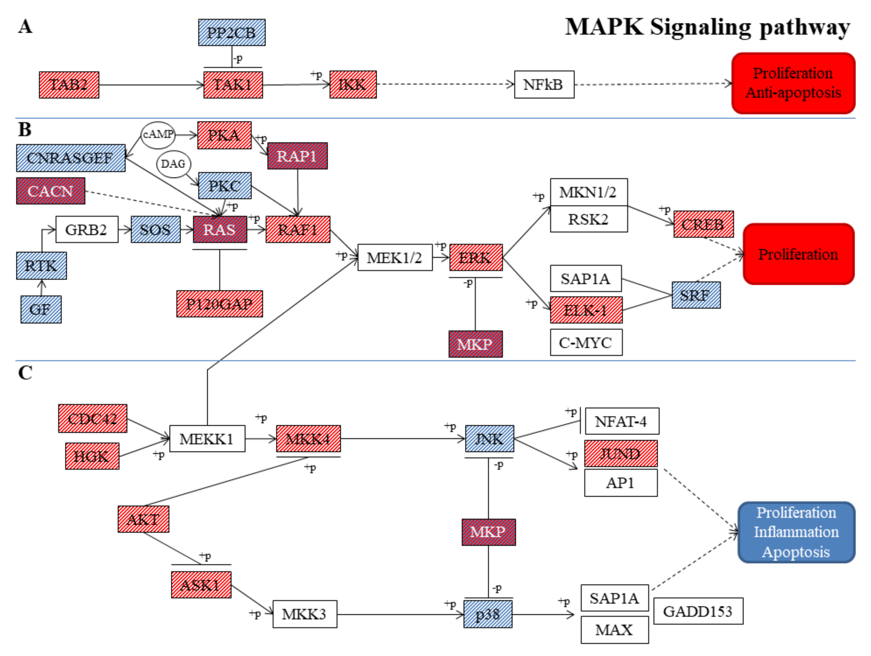
:1. Introduction
2. Materials and Methods
2.1. Cell Culture and Vitamin E Treatment
2.2. Thiazolyl Blue Tetrazolium Bromide (MTT) Assay
2.3. Statistical Validation of Cell Viability
2.4. Extraction of Total RNA and cDNA Library Preparation
2.5. RNA-Seq Data Analysis and Gene Evaluation
3. Results
3.1. Cell Viability
3.2. Transcriptome Analysis and Gene Inspection
4. Discussion
5. Conclusions
Author Contributions
Funding
Acknowledgments
Conflicts of Interest
References
- Shahidi, F.; de Camargo, A.C. Tocopherols and Tocotrienols in Common and Emerging Dietary Sources: Occurrence, Applications, and Health Benefits. Int. J. Mol. Sci. 2016, 17, 1745. [Google Scholar] [CrossRef]
- Galli, F.; Azzi, A.; Birringer, M.; Cook-Mills, J.M.; Eggersdorfer, M.; Frank, J.; Cruciani, G.; Lorkowski, S.; Ozer, N.K. Vitamin E: Emerging aspects and new directions. Free Radic. Biol. Med. 2017, 102, 16–36. [Google Scholar] [CrossRef] [PubMed]
- Zingg, J.M. Vitamin E: Regulatory Role on Signal Transduction. IUBMB Life 2019, 71, 456–478. [Google Scholar] [CrossRef] [PubMed]
- Birringer, M.; Lorkowski, S. Vitamin E: Regulatory role of metabolites. IUBMB Life 2019, 71, 479–486. [Google Scholar] [CrossRef] [PubMed]
- Ulatowski, L.; Parker, R.; Warrier, G.; Sultana, R.; Butterfield, D.A.; Manor, D. Vitamin E is essential for Purkinje neuron integrity. Neuroscience 2014, 260, 120–129. [Google Scholar] [CrossRef] [PubMed]
- Yokota, T.; Igarashi, K.; Uchihara, T.; Jishage, K.; Tomita, H.; Inaba, A.; Li, Y.; Arita, M.; Suzuki, H.; Mizusawa, H.; et al. Delayed-onset ataxia in mice lacking alpha -tocopherol transfer protein: Model for neuronal degeneration caused by chronic oxidative stress. Proc. Natl. Acad. Sci. USA 2001, 98, 15185–15190. [Google Scholar] [CrossRef] [PubMed]
- Mohammed, H.O.; Starkey, S.R.; Stipetic, K.; Divers, T.J.; Summers, B.A.; de Lahunta, A. The role of dietary antioxidant insufficiency on the permeability of the blood-brain barrier. J. Neuropathol. Exp. Neurol. 2008, 67, 1187–1193. [Google Scholar] [CrossRef] [PubMed]
- Liu, Z.; Zhou, T.; Ziegler, A.C.; Dimitrion, P.; Zuo, L. Oxidative Stress in Neurodegenerative Diseases: From Molecular Mechanisms to Clinical Applications. Oxidative Med. Cell. Longev. 2017, 2017, 2525967. [Google Scholar] [CrossRef]
- Cobley, J.N.; Fiorello, M.L.; Bailey, D.M. 13 reasons why the brain is susceptible to oxidative stress. Redox Biol. 2018, 15, 490–503. [Google Scholar] [CrossRef] [PubMed]
- Chang, K.H.; Cheng, M.L.; Chiang, M.C.; Chen, C.M. Lipophilic antioxidants in neurodegenerative diseases. Clin. Chim. Acta Int. J. Clin. Chem. 2018, 485, 79–87. [Google Scholar] [CrossRef] [PubMed]
- Gugliandolo, A.; Bramanti, P.; Mazzon, E. Role of Vitamin E in the Treatment of Alzheimer’s Disease: Evidence from Animal Models. Int. J. Mol. Sci. 2017, 18. [Google Scholar] [CrossRef] [PubMed]
- Schirinzi, T.; Martella, G.; Imbriani, P.; Di Lazzaro, G.; Franco, D.; Colona, V.L.; Alwardat, M.; Sinibaldi Salimei, P.; Mercuri, N.B.; Pierantozzi, M.; et al. Dietary Vitamin E as a Protective Factor for Parkinson’s Disease: Clinical and Experimental Evidence. Front. Neurol. 2019, 10, 148. [Google Scholar] [CrossRef] [PubMed]
- Gurney, M.E.; Cutting, F.B.; Zhai, P.; Doble, A.; Taylor, C.P.; Andrus, P.K.; Hall, E.D. Benefit of vitamin E, riluzole, and gabapentin in a transgenic model of familial amyotrophic lateral sclerosis. Ann. Neurol. 1996, 39, 147–157. [Google Scholar] [CrossRef] [PubMed]
- Chico, L.; Modena, M.; Lo Gerfo, A.; Ricci, G.; Caldarazzo Ienco, E.; Ryskalin, L.; Fornai, F.; Siciliano, G. Cross-talk between pathogenic mechanisms in neurodegeneration: The role of oxidative stress in Amyotrophic Lateral Sclerosis. Arch. Ital. Biol. 2017, 155, 131–141. [Google Scholar] [CrossRef]
- Michal Freedman, D.; Kuncl, R.W.; Weinstein, S.J.; Malila, N.; Virtamo, J.; Albanes, D. Vitamin E serum levels and controlled supplementation and risk of amyotrophic lateral sclerosis. Amyotroph. Lateral Scler. Front. Degener. 2013, 14, 246–251. [Google Scholar] [CrossRef]
- Wang, H.; O’Reilly, E.J.; Weisskopf, M.G.; Logroscino, G.; McCullough, M.L.; Schatzkin, A.; Kolonel, L.N.; Ascherio, A. Vitamin E intake and risk of amyotrophic lateral sclerosis: A pooled analysis of data from 5 prospective cohort studies. Am. J. Epidemiol. 2011, 173, 595–602. [Google Scholar] [CrossRef] [PubMed]
- Desnuelle, C.; Dib, M.; Garrel, C.; Favier, A. A double-blind, placebo-controlled randomized clinical trial of alpha-tocopherol (vitamin E) in the treatment of amyotrophic lateral sclerosis. ALS riluzole-tocopherol Study Group. Amyotroph. Lateral Scler. Other Motor Neuron Disord. 2001, 2, 9–18. [Google Scholar] [CrossRef]
- Galbussera, A.; Tremolizzo, L.; Brighina, L.; Testa, D.; Lovati, R.; Ferrarese, C.; Cavaletti, G.; Filippini, G. Vitamin E intake and quality of life in amyotrophic lateral sclerosis patients: A follow-up case series study. Neurol. Sci. 2006, 27, 190–193. [Google Scholar] [CrossRef]
- Veyrat-Durebex, C.; Corcia, P.; Dangoumau, A.; Laumonnier, F.; Piver, E.; Gordon, P.H.; Andres, C.R.; Vourc’h, P.; Blasco, H. Advances in cellular models to explore the pathophysiology of amyotrophic lateral sclerosis. Mol. Neurobiol. 2014, 49, 966–983. [Google Scholar] [CrossRef]
- Yan, X.; Liu, Y.; Xie, T.; Liu, F. alpha-Tocopherol protected against cobalt nanoparticles and cocl2 induced cytotoxicity and inflammation in Balb/3T3 cells. Immunopharmacol. Immunotoxicol. 2018, 40, 179–185. [Google Scholar] [CrossRef]
- Zappe, K.; Pointner, A.; Switzeny, O.J.; Magnet, U.; Tomeva, E.; Heller, J.; Mare, G.; Wagner, K.H.; Knasmueller, S.; Haslberger, A.G. Counteraction of Oxidative Stress by Vitamin E Affects Epigenetic Regulation by Increasing Global Methylation and Gene Expression of MLH1 and DNMT1 Dose Dependently in Caco-2 Cells. Oxidative Med. Cell. Longev. 2018, 2018, 3734250. [Google Scholar] [CrossRef] [PubMed]
- Chiricosta, L.; Diomede, F.; Trubiani, O.; Bramanti, P.; Mazzon, E. Physiological Expression of Ion Channel Receptors in Human Periodontal Ligament Stem Cells. Cells 2019, 8. [Google Scholar] [CrossRef] [PubMed]
- Conesa, A.; Madrigal, P.; Tarazona, S.; Gomez-Cabrero, D.; Cervera, A.; McPherson, A.; Szczesniak, M.W.; Gaffney, D.J.; Elo, L.L.; Zhang, X.; et al. A survey of best practices for RNA-seq data analysis. Genome Biol. 2016, 17, 13. [Google Scholar] [CrossRef]
- Kanehisa, M.; Goto, S. KEGG: Kyoto encyclopedia of genes and genomes. Nucleic Acids Res. 2000, 28, 27–30. [Google Scholar] [CrossRef] [PubMed]
- Kim, E.K.; Choi, E.J. Pathological roles of MAPK signaling pathways in human diseases. Biochim. Biophys. Acta 2010, 1802, 396–405. [Google Scholar] [CrossRef] [PubMed]
- Stankiewicz, T.R.; Linseman, D.A. Rho family GTPases: Key players in neuronal development, neuronal survival, and neurodegeneration. Front. Cell. Neurosci. 2014, 8, 314. [Google Scholar] [CrossRef] [PubMed]
- Sun, J.; Nan, G. The extracellular signal-regulated kinase 1/2 pathway in neurological diseases: A potential therapeutic target (Review). Int. J. Mol. Med. 2017, 39, 1338–1346. [Google Scholar] [CrossRef]
- Kwon, J.E.; Kim, E.K.; Choi, E.J. Stabilization of the survival motor neuron protein by ASK1. Febs Lett. 2011, 585, 1287–1292. [Google Scholar] [CrossRef] [PubMed]
- Peviani, M.; Tortarolo, M.; Battaglia, E.; Piva, R.; Bendotti, C. Specific induction of Akt3 in spinal cord motor neurons is neuroprotective in a mouse model of familial amyotrophic lateral sclerosis. Mol. Neurobiol. 2014, 49, 136–148. [Google Scholar] [CrossRef]
- Hernandez, J.M.; Floyd, D.H.; Weilbaecher, K.N.; Green, P.L.; Boris-Lawrie, K. Multiple facets of junD gene expression are atypical among AP-1 family members. Oncogene 2008, 27, 4757–4767. [Google Scholar] [CrossRef] [PubMed]
- Wang, T.H.; Wang, S.Y.; Wang, X.D.; Jiang, H.Q.; Yang, Y.Q.; Wang, Y.; Cheng, J.L.; Zhang, C.T.; Liang, W.W.; Feng, H.L. Fisetin Exerts Antioxidant and Neuroprotective Effects in Multiple Mutant hSOD1 Models of Amyotrophic Lateral Sclerosis by Activating ERK. Neuroscience 2018, 379, 152–166. [Google Scholar] [CrossRef] [PubMed]
- Zhong, J. RAS and downstream RAF-MEK and PI3K-AKT signaling in neuronal development, function and dysfunction. Biol. Chem. 2016, 397, 215–222. [Google Scholar] [CrossRef] [PubMed]
- Cullen, P.J.; Lockyer, P.J. Integration of calcium and Ras signalling. Nat. Rev. Mol. Cell Biol. 2002, 3, 339–348. [Google Scholar] [CrossRef] [PubMed]
- Glaser, T.; Arnaud Sampaio, V.F.; Lameu, C.; Ulrich, H. Calcium signalling: A common target in neurological disorders and neurogenesis. Semin. Cell Dev. Biol. 2018. [Google Scholar] [CrossRef] [PubMed]
- Patai, R.; Nogradi, B.; Engelhardt, J.I.; Siklos, L. Calcium in the pathomechanism of amyotrophic lateral sclerosis—Taking center stage? Biochem. Biophys. Res. Commun. 2017, 483, 1031–1039. [Google Scholar] [CrossRef] [PubMed]
- Chang, Q.; Martin, L.J. Voltage-gated calcium channels are abnormal in cultured spinal motoneurons in the G93A-SOD1 transgenic mouse model of ALS. Neurobiol. Dis. 2016, 93, 78–95. [Google Scholar] [CrossRef] [PubMed]
- Joshi, D.C.; Singh, M.; Krishnamurthy, K.; Joshi, P.G.; Joshi, N.B. AMPA induced Ca2+ influx in motor neurons occurs through voltage gated Ca2+ channel and Ca2+ permeable AMPA receptor. Neurochem. Int. 2011, 59, 913–921. [Google Scholar] [CrossRef] [PubMed]
- Numakawa, Y.; Numakawa, T.; Matsumoto, T.; Yagasaki, Y.; Kumamaru, E.; Kunugi, H.; Taguchi, T.; Niki, E. Vitamin E protected cultured cortical neurons from oxidative stress-induced cell death through the activation of mitogen-activated protein kinase and phosphatidylinositol 3-kinase. J. Neurochem. 2006, 97, 1191–1202. [Google Scholar] [CrossRef]
- Amer, Y.O.; Hebert-Chatelain, E. Mitochondrial cAMP-PKA signaling: What do we really know? BBA Bioenergy 2018, 1859, 868–877. [Google Scholar] [CrossRef] [PubMed]
- Salinthone, S.; Kerns, A.R.; Tsang, V.; Carr, D.W. alpha-Tocopherol (vitamin E) stimulates cyclic AMP production in human peripheral mononuclear cells and alters immune function. Mol. Immunol. 2013, 53, 173–178. [Google Scholar] [CrossRef] [PubMed]
- Krieger, C.; Lanius, R.A.; Pelech, S.L.; Shaw, C.A. Amyotrophic lateral sclerosis: The involvement of intracellular Ca2+ and protein kinase C. Trends Pharmacol. Sci. 1996, 17, 114–120. [Google Scholar] [CrossRef]
- Besnard, A.; Galan-Rodriguez, B.; Vanhoutte, P.; Caboche, J. Elk-1 a transcription factor with multiple facets in the brain. Front. Neurosci. 2011, 5, 35. [Google Scholar] [CrossRef] [PubMed]
- Hai, T.; Hartman, M.G. The molecular biology and nomenclature of the activating transcription factor/cAMP responsive element binding family of transcription factors: Activating transcription factor proteins and homeostasis. Gene 2001, 273, 1–11. [Google Scholar] [CrossRef]
- Pasini, S.; Corona, C.; Liu, J.; Greene, L.A.; Shelanski, M.L. Specific downregulation of hippocampal ATF4 reveals a necessary role in synaptic plasticity and memory. Cell Rep. 2015, 11, 183–191. [Google Scholar] [CrossRef]
- Reed, D.K.; Hall, S.; Arany, I. α-Tocopherol protects renal cells from nicotine- or oleic acid-provoked oxidative stress via inducing heme oxygenase-1. J. Physiol. Biochem. 2015, 71, 1–7. [Google Scholar] [CrossRef] [PubMed]
- Caunt, C.J.; Keyse, S.M. Dual-specificity MAP kinase phosphatases (MKPs): Shaping the outcome of MAP kinase signalling. FEBS J. 2013, 280, 489–504. [Google Scholar] [CrossRef] [PubMed]
- Collins, L.M.; O’Keeffe, G.W.; Long-Smith, C.M.; Wyatt, S.L.; Sullivan, A.M.; Toulouse, A.; Nolan, Y.M. Mitogen-activated protein kinase phosphatase (MKP)-1 as a neuroprotective agent: Promotion of the morphological development of midbrain dopaminergic neurons. Neuromol. Med. 2013, 15, 435–446. [Google Scholar] [CrossRef] [PubMed]
- Maor-Nof, M.; Romi, E.; Sar Shalom, H.; Ulisse, V.; Raanan, C.; Nof, A.; Leshkowitz, D.; Lang, R.; Yaron, A. Axonal Degeneration Is Regulated by a Transcriptional Program that Coordinates Expression of Pro- and Anti-degenerative Factors. Neuron 2016, 92, 991–1006. [Google Scholar] [CrossRef]
- Wooten, M.W. Function for NF-kB in neuronal survival: Regulation by atypical protein kinase C. J. Neurosci. Res. 1999, 58, 607–611. [Google Scholar] [CrossRef]
- Aashaq, S.; Batool, A.; Andrabi, K.I. TAK1 mediates convergence of cellular signals for death and survival. Apoptosis 2019, 24, 3–20. [Google Scholar] [CrossRef]
- Wu, H.; Wei, H.; Sehgal, S.A.; Liu, L.; Chen, Q. Mitophagy receptors sense stress signals and couple mitochondrial dynamic machinery for mitochondrial quality control. Free Radic. Biol. Med. 2016, 100, 199–209. [Google Scholar] [CrossRef] [PubMed]
- Shefa, U.; Jeong, N.Y.; Song, I.O.; Chung, H.J.; Kim, D.; Jung, J.; Huh, Y. Mitophagy links oxidative stress conditions and neurodegenerative diseases. Neural Regen. Res. 2019, 14, 749–756. [Google Scholar] [CrossRef]
- Schaaf, M.B.; Keulers, T.G.; Vooijs, M.A.; Rouschop, K.M. LC3/GABARAP family proteins: Autophagy-(un)related functions. FASEB J. 2016, 30, 3961–3978. [Google Scholar] [CrossRef]
- Cornelissen, T.; Haddad, D.; Wauters, F.; Van Humbeeck, C.; Mandemakers, W.; Koentjoro, B.; Sue, C.; Gevaert, K.; De Strooper, B.; Verstreken, P.; et al. The deubiquitinase USP15 antagonizes Parkin-mediated mitochondrial ubiquitination and mitophagy. Hum. Mol. Genet. 2014, 23, 5227–5242. [Google Scholar] [CrossRef] [PubMed]
- Corona, C.; Pasini, S.; Liu, J.; Amar, F.; Greene, L.A.; Shelanski, M.L. Activating Transcription Factor 4 (ATF4) Regulates Neuronal Activity by Controlling GABABR Trafficking. J. Neurosci. 2018, 38, 6102–6113. [Google Scholar] [CrossRef]
- Calvo-Garrido, J.; Maffezzini, C.; Schober, F.A.; Clemente, P.; Uhlin, E.; Kele, M.; Stranneheim, H.; Lesko, N.; Bruhn, H.; Svenningsson, P.; et al. SQSTM1/p62-Directed Metabolic Reprogramming Is Essential for Normal Neurodifferentiation. Stem Cell Rep. 2019, 12, 696–711. [Google Scholar] [CrossRef] [PubMed]
- Hadano, S.; Mitsui, S.; Pan, L.; Otomo, A.; Kubo, M.; Sato, K.; Ono, S.; Onodera, W.; Abe, K.; Chen, X.; et al. Functional links between SQSTM1 and ALS2 in the pathogenesis of ALS: Cumulative impact on the protection against mutant SOD1-mediated motor dysfunction in mice. Hum. Mol. Genet. 2016, 25, 3321–3340. [Google Scholar] [CrossRef]
- Le Grand, J.N.; Bon, K.; Fraichard, A.; Zhang, J.; Jouvenot, M.; Risold, P.Y.; Boyer-Guittaut, M.; Delage-Mourroux, R. Specific distribution of the autophagic protein GABARAPL1/GEC1 in the developing and adult mouse brain and identification of neuronal populations expressing GABARAPL1/GEC1. PLoS ONE 2013, 8, e63133. [Google Scholar] [CrossRef] [PubMed]

| Experimental Group | Mean (%) | Standard Deviation | p Value |
|---|---|---|---|
| Control | 100.00 | 10.10 | 0.12 |
| 1 µM Vit. E | 98.30 | 3.62 | 0.18 |
| 5 µM Vit. E | 100.10 | 6.12 | 0.67 |
| 10 µM Vit. E | 99.14 | 7.88 | 0.31 |
| 15 µM Vit. E | 97.87 | 9.86 | 0.24 |
| 20 µM Vit. E | 98.84 | 8.53 | 0.66 |
| Gene | Expression Level CTR-NSC-34 | Expression Level VitE-NSC-34 | Fold Change | q Value | KEGG |
|---|---|---|---|---|---|
| CACNA1H | 19.33 | 8.34 | −1.21 | 6.06 × 10⁻³ | CACN |
| CACNG3 | 29.49 | 46.23 | 0.65 | 9.75 × 10⁻⁴ | CACN |
| CACNG7 | 9.53 | 15.87 | 0.74 | 3.69 × 10⁻⁴ | CACN |
| CACNA1C | 27.86 | 23.16 | −0.27 | 3.69 × 10⁻⁴ | CACN |
| CACNA2D1 | 24.54 | 20.98 | −0.23 | 2.34 × 10⁻² | CACN |
| CACNA2D2 | 4.75 | 2.11 | −1.17 | 1.98 × 10⁻² | CACN |
| CACNB1 | 27.88 | 15.59 | -0.84 | 3.69 × 10⁻⁴ | CACN |
| CACNG2 | 8.68 | 5.00 | −0.80 | 3.69 × 10⁻⁴ | CACN |
| BDNF | 5.44 | 2.03 | −1.42 | 3.69 × 10⁻⁴ | GF |
| VEGFA | 519.96 | 446.13 | −0.22 | 3.69 × 10⁻⁴ | GF |
| ERBB3 | 4.51 | 2.49 | −0.86 | 3.69 × 10⁻⁴ | RTK |
| DUSP1 | 7.06 | 12.31 | 0.80 | 1.13 × 10⁻² | MKP |
| DUSP4 | 5.89 | 4.15 | −0.50 | 4.86 × 10⁻² | MKP |
| DUSP16 | 3.60 | 8.88 | 1.30 | 3.69 × 10⁻⁴ | MKP |
| RASA1 | 236.29 | 281.47 | 0.25 | 3.69 × 10⁻⁴ | P120GAP |
| SOS1 | 59.25 | 56.23 | −0.08 | 4.87 × 10⁻² | SOS |
| SOS2 | 45.24 | 40.65 | −0.15 | 3.89 × 10⁻² | SOS |
| HRAS | 70.84 | 52.08 | −0.44 | 3.69 × 10⁻⁴ | RAS |
| NRAS | 26.96 | 31.96 | 0.25 | 2.74 × 10⁻³ | RAS |
| KRAS | 74.05 | 92.48 | 0.32 | 4.08 × 10⁻³ | RAS |
| RAPGEF2 | 29.70 | 17.79 | −0.74 | 2.49 × 10⁻² | CNRASGEF |
| PRKCB | 6.42 | 2.70 | −1.25 | 4.09 × 10⁻² | PCK |
| PRKACB | 12.03 | 19.53 | 0.70 | 2.74 × 10⁻³ | PKA |
| RAP1B | 297.73 | 313.97 | 0.08 | 3.61 × 10⁻² | RAP1 |
| RAP1A | 41.91 | 31.19 | −0.43 | 3.69 × 10⁻⁴ | RAP1 |
| RAF1 | 23.14 | 33.12 | 0.52 | 2.02 × 10⁻³ | RAF1 |
| MAPK1 | 107.60 | 132.85 | 0.30 | 3.69 × 10⁻⁴ | ERK |
| ELK1 | 1.40 | 3.27 | 1.22 | 2.02 × 10⁻³ | ELK-1 |
| ATF4 | 179.33 | 197.24 | 0.14 | 6.66 × 10⁻³ | CREB |
| SRF | 20.37 | 14.62 | −0.48 | 3.69 × 10⁻⁴ | SRF |
| TAB2 | 1011.99 | 1152.61 | 0.19 | 3.69 × 10⁻⁴ | TAB2 |
| PPM1B | 23.61 | 16.21 | −0.54 | 3.69 × 10⁻⁴ | PP2CB |
| MAP3K7 | 199.57 | 262.58 | 0.40 | 3.69 × 10⁻⁴ | TAK1 |
| IKBKB | 4.45 | 9.20 | 1.05 | 6.91 × 10⁻⁴ | IKK |
| Gene | Expression Level CTR-NSC-34 | Expression Level VitE-NSC-34 | Fold Change | q Value | KEGG |
|---|---|---|---|---|---|
| JUND | 3.41 | 5.15 | 0.59 | 2.69 × 10⁻² | JUND |
| MAPK9 | 118.96 | 106.30 | −0.16 | 3.44 × 10⁻² | JNK |
| MAP2K4 | 47.27 | 77.30 | 0.71 | 3.69 × 10⁻⁴ | MKK4 |
| MAP4K4 | 164.30 | 174.25 | 0.09 | 1.78 × 10⁻³ | HGK |
| CDC42 | 323.84 | 394.04 | 0.28 | 3.69 × 10⁻⁴ | CDC42 |
| MAP3K5 | 8.91 | 16.09 | 0.85 | 9.75 × 10⁻⁴ | ASK1 |
| MAPK11 | 3.34 | 1.83 | −0.87 | 3.24 × 10⁻² | P38 |
| MAPK14 | 40.49 | 25.46 | −0.67 | 1.11 × 10⁻² | P38 |
| AKT1 | 110.17 | 136.09 | 0.31 | 3.69 × 10⁻⁴ | AKT |
| AKT3 | 288.28 | 314.60 | 0.13 | 2.25 × 10⁻³ | AKT |
| DUSP4 | 5.89 | 4.15 | −0.50 | 4.86 × 10⁻² | MKP |
| DUSP16 | 3.60 | 8.88 | 1.30 | 3.69 × 10⁻⁴ | MKP |
| Pathway | Total Number of Genes | Upregulated Genes | Downregulated Genes |
|---|---|---|---|
| Classical MAPK pathway | 34 (74%) | 16 (47%) | 18 (53%) |
| JNK and p38 MAPK pathway | 12 (26%) | 8 (67%) | 4 (33%) |
| Gene | Expression Level CTR-NSC-34 | Expression Level VitE-NSC-34 | Fold Change | q Value | KEGG |
|---|---|---|---|---|---|
| MAPK9 | 118.96 | 106.30 | −0.16 | 3.44 × 10⁻² | JNK |
| ATF4 | 179.33 | 197.24 | 0.14 | 6.66 × 10⁻³ | ATF4 |
| MFN2 | 74.11 | 54.96 | −0.43 | 2.02 × 10⁻³ | MFN2 |
| UBB | 6430.17 | 4355.22 | −0.56 | 3.69 × 10⁻⁴ | UB |
| RHOT2 | 6.52 | 1.25 | −2.38 | 1.52 × 10⁻³ | MIRO |
| BECN1 | 248.55 | 208.86 | −0.25 | 5.27 × 10⁻³ | BECLIN1 |
| USP15 | 58.66 | 69.67 | 0.25 | 3.69 × 10⁻⁴ | USP15 |
| ATG5 | 59.74 | 52.90 | −0.18 | 2.14 × 10⁻² | ATG5 |
| TAX1BP1 | 62.39 | 31.39 | −0.99 | 3.69 × 10⁻⁴ | TAX1BP1 |
| SQSTM1 | 103.55 | 160.22 | 0.63 | 3.69 × 10⁻⁴ | P62 |
| NBR1 | 52.32 | 34.50 | −0.60 | 9.60 × 10⁻³ | NBR1 |
| GABARAPL1 | 28.21 | 34.05 | 0.27 | 2.71 × 10⁻² | LC3 |
| RAB7 | 258.35 | 220.73 | −0.23 | 3.69 × 10⁻⁴ | RAB7 |
© 2019 by the authors. Licensee MDPI, Basel, Switzerland. This article is an open access article distributed under the terms and conditions of the Creative Commons Attribution (CC BY) license (http://creativecommons.org/licenses/by/4.0/).
Share and Cite
Chiricosta, L.; Gugliandolo, A.; Tardiolo, G.; Bramanti, P.; Mazzon, E. Transcriptomic Analysis of MAPK Signaling in NSC-34 Motor Neurons Treated with Vitamin E. Nutrients 2019, 11, 1081. https://doi.org/10.3390/nu11051081
Chiricosta L, Gugliandolo A, Tardiolo G, Bramanti P, Mazzon E. Transcriptomic Analysis of MAPK Signaling in NSC-34 Motor Neurons Treated with Vitamin E. Nutrients. 2019; 11(5):1081. https://doi.org/10.3390/nu11051081
Chicago/Turabian StyleChiricosta, Luigi, Agnese Gugliandolo, Giuseppe Tardiolo, Placido Bramanti, and Emanuela Mazzon. 2019. "Transcriptomic Analysis of MAPK Signaling in NSC-34 Motor Neurons Treated with Vitamin E" Nutrients 11, no. 5: 1081. https://doi.org/10.3390/nu11051081
APA StyleChiricosta, L., Gugliandolo, A., Tardiolo, G., Bramanti, P., & Mazzon, E. (2019). Transcriptomic Analysis of MAPK Signaling in NSC-34 Motor Neurons Treated with Vitamin E. Nutrients, 11(5), 1081. https://doi.org/10.3390/nu11051081