Article

Evaluation of Metal-Doped ZIF-8-Hyaluronic Acid Nanocomposites for Disruption of Salmonella Typhimurium and Escherichia coli on Food Contact (Stainless Steel) Surfaces


College of Agriculture and Life Sciences, Texas A&M University, College Station, TX 77843-2117, USA
*
Author to whom correspondence should be addressed.
Sustainability 2026, 18(6), 2792; https://doi.org/10.3390/su18062792
Submission received: 14 February 2026 / Revised: 6 March 2026 / Accepted: 9 March 2026 / Published: 12 March 2026
(This article belongs to the Section Sustainable Food)

Abstract

This study developed and evaluated multifunctional Cu-doped Zeolitic Imidazolate Framework-8 nanoparticles coated with hyaluronic acid (Cu-ZIF-8@HA) for antimicrobial application on stainless-steel food-contact surfaces. Structural characterization through SEM, TEM, and elemental mapping confirmed the successful synthesis, uniform Cu incorporation, and HA coating without compromising the crystalline ZIF-8 framework. Cu doping reduced particle size (~130 nm) and enhanced redox activity, while HA encapsulation improved colloidal stability and biocompatibility by shifting zeta potential from positive (+22.1 mV) to negative (−18.7 mV). Cytotoxicity assays demonstrated that HA significantly mitigated metal-induced toxicity, maintaining >70% cell viability at ≤1000 µg/mL. Antibacterial assessments revealed potent activity against Salmonella Typhimurium ATCC 14028 and Escherichia coli O157:H7, with Cu-ZIF-8@HA exhibiting the largest inhibition zones (18.15–20.33 mm), lowest MIC/MBC values (500/2000 µg/mL and 1000/2500 µg/mL), and over 6-log reductions in bacterial adhesion on stainless steel. Enhanced wettability (contact angle 11.77°) and surface energy (64.42 mN/m) further facilitated antimicrobial contact. These results confirm that Cu-ZIF-8@HA integrates the oxidative potency of Cu, the structural stability of ZIF-8, and the biocompatibility of HA, offering a promising and safe nanomaterial platform for controlling bacterial contamination and biofilm formation in food-processing environments.

1. Introduction

Foodborne pathogens like Salmonella enterica (serovar Typhimurium) and pathogenic Escherichia coli (e.g., O157:H7) are leading causes of illness and have been increasingly linked to contamination of fresh produce, especially leafy greens [1,2]. For instance, roughly 8% of around 1200 U.S. Salmonella outbreaks (1990–2015) were attributed to leafy vegetables [3]. These hardy microbes can persist on food and contact surfaces, and Salmonella can even survive on produce under low-humidity storage, whereas E. coli populations decline under similar dry conditions [4,5]. Such resilience enables Salmonella and E. coli to attach and form biofilms on stainless steel food-contact surfaces, even after routine cleaning and sanitization.
Given the significant public health risks posed by foodborne pathogens and the limitations associated with conventional sanitization methods, there is an urgent need to develop novel antimicrobial strategies capable of effectively disinfecting food-processing surfaces and minimizing cross-contamination. One promising approach involves the application of nanoscale metal–organic frameworks (MOFs) as antimicrobial agents. Among these materials, zeolitic imidazolate framework-8 (ZIF-8), a zinc-based MOF, has attracted considerable attention due to its high surface area, tunable porosity, structural stability, and ability to release Zn2+ ions in a controlled manner. The antimicrobial activity of ZIF-8 arises from multiple bactericidal mechanisms. Released Zn2+ ions can disrupt microbial enzymatic activity and compromise membrane integrity, while the framework itself may facilitate the generation of reactive oxygen species (ROS) that induce oxidative damage to cellular components. In addition, the imidazolate linker within the ZIF-8 structure contributes inherent antimicrobial activity [6]. Recent studies have demonstrated that incorporating secondary metal dopants into the ZIF-8 framework can further enhance its antibacterial performance [7]. Copper (Cu) has been identified as an effective dopant compared with other metals such as Mg, Mn, Co, and Fe, resulting in Cu-doped ZIF-8 materials with significantly improved bactericidal activity [8,9]. Copper is widely recognized as a broad-spectrum antimicrobial agent capable of rapidly inactivating bacteria, yeasts, and viruses through contact-mediated killing, often achieving reductions greater than 7 log within one hour [10]. Compared with silver, another commonly used antimicrobial metal, copper offers several practical advantages, including lower cost, status as an essential micronutrient with relatively lower toxicity risk, and sustained antimicrobial efficacy even under low-moisture conditions [11].
To further improve the functionality of these nano-antimicrobials, a biocompatible polymer coating can be applied. In this work, we incorporate hyaluronic acid (HA), a natural, food-safe polysaccharide, onto the surface of ZIF-8 and Cu-ZIF-8 particles, forming ZIF-8@HA and Cu-ZIF-8@HA nanocomposites. HA has been shown to synergize with ZIF-8 by enhancing stability and enabling pH-responsive release of antimicrobial agents in targeted microenvironments [12]. Moreover, embedding MOFs in a polymer matrix, such as HA, can improve particle dispersion and adhesion, enabling the nanocomposite to coat surfaces better and interact with bacterial cells. Polymer functionalization can help anchor MOF nanoparticles to bacterial membranes, promoting direct contact-mediated disruption of the cells [13].
By coating Cu-doped ZIF-8 with HA, we aim to develop a durable antimicrobial film that can be applied to stainless steel and remain effective over time. In summary, this study assesses four nanomaterials (ZIF-8, Cu-ZIF-8, ZIF-8@HA, and Cu-ZIF-8@HA) for their ability to inactivate Salmonella Typhimurium and E. coli on stainless steel surfaces. The results of this research could pave the way for an innovative nanocoating method for food-contact surfaces, combining the advantages of metal-doped MOFs and biopolymer matrices to enhance the disruption of bacterial contaminants and improve the safety in fresh produce handling environments [14].

2. Materials and Methods

2.1. Nanoparticle Synthesis

2.1.1. ZIF-8 and Cu-ZIF-8

ZIF-8 nanoparticles were synthesized following [15]. Zinc nitrate hexahydrate (2.8 mmol) and 2-methylimidazole (64.4 mmol) were separately dissolved in methanol (1.4 × 10³ mmol; Fisher Scientific, Waltham, MA, USA). The 2-methylimidazole solution was poured into the zinc nitrate solution and stirred for 24 h at 21 °C. Precipitates were collected by centrifugation (6000 rpm, 10 min), washed three times with methanol, and vacuum-dried using a Welch 1376 DuoSeal Pump (Thomas Industries Inc., Wabasha, MN, USA). The resulting ZIF-8 powders were stored in a desiccator at 21 °C until further use.
Cu-ZIF-8 was synthesized following [8]. Zinc nitrate hexahydrate (1.96 mmol), copper nitrate trihydrate (0.84 mmol), and 2-methylimidazole (64.4 mmol) were dissolved in ethanol (1.4 × 10³ mmol; Supelco®, Bellefonte, PA, USA) within Erlenmeyer flasks (Fisher Scientific, Waltham, MA, USA). The 2-methylimidazole solution was added to the mixed metal nitrates, and the mixture was stirred at 21 °C for 24 h. The resulting precipitates were collected by centrifugation (Allegra™ 25R, Beckman Coulter, Brea, CA, USA; 8000 rpm, 10 min), washed three times with ethanol, and vacuum-dried using a Welch 1376 DuoSeal pump (Thomas Industries, Wabasha, MN, USA). The dried Cu-ZIF-8 powders were stored in a desiccator at 21 °C until further use.

2.1.2. ZIF-8@HA and Cu-ZIF-8@HA

ZIF-8@HA was prepared following [8]. ZIF-8 powder (Section 2.1.1) was dispersed in 0.05 mg/mL hyaluronic acid (HA; Sigma-Aldrich, Darmstadt, Germany) at a 1:2 ratio (mg ZIF-8: mL HA solution) and vortexed for 2 min using a Vortex-Genie 2 (Scientific Industries, Bohemia, NY, USA). The suspension was stirred for 30 min to promote HA adsorption, sonicated for 1 min for uniform dispersion, and centrifuged at 8000 rpm for 10 min. The pellet was washed three times with deionized water and vacuum-dried for 4 h using a Welch 1376 DuoSeal Pump (Thomas Industries, Wabasha, MN, USA). The resulting ZIF-8–HA nanoparticle powders were stored at room temperature. Cu-ZIF-8@HA was synthesized using the same procedure, substituting Cu-ZIF-8 for ZIF-8.

2.2. Nanoparticle Characterization

2.2.1. Scanning Electron Microscopy (SEM)

Morphological characterization of ZIF-8, Cu-ZIF-8, ZIF-8@HA, and Cu-ZIF-8@HA, along with surface coupons, was performed at the University’s Microscopy and Imaging Center. Samples were sputter-coated with platinum using a High-Resolution Coater 208 HR (Cressington Scientific Instruments, Watford, UK) and imaged with a Quanta 600 FEG SEM (FEI Company, Hillsboro, OR, USA) at 20 kV, a working distance of 10 mm, and 1000–5000× magnification. Elemental composition and spatial distribution were analyzed using SEM–EDS mapping, which identifies element-specific X-ray emissions to visualize and quantify elemental distribution across the surface [8].

2.2.2. Transmission Electron Microscopy (TEM)

An amount of 1 mg of nanoparticle powder was dispersed in 2 mL of ethanol, sonicated for 5 min, and 5 µL of the suspension was drop-cast onto a carbon-coated copper grid (200 mesh; Ted Pella Inc., Redding, CA, USA). After air-drying (21 °C, 30 min) and optional staining with 2% uranyl acetate, samples were imaged using a JEOL 1200 EX TEM (JEOL Ltd., Tokyo, Japan) at 200 kV. Images were captured with a 3k × 3k CCD camera (PSI Solutions, Twinsburg, OH, USA) and SIA Micrograph MaxIm DL 5.0 software.

2.2.3. Zeta Potential (ζ, ZP)

Surface charge was determined using a Malvern Zetasizer Nano ZS (Malvern Panalytical, Westborough, MA, USA) with a DTS1070 folded capillary cell (Malvern Panalytical, Westborough, MA, USA). Nanoparticles (2 mg/mL) were dispersed in ethanol and sonicated for 5 min before measurement [16].

2.2.4. Particle Size (PS) Measurement

Particle size was measured at 21 °C using the same instrument and cell setup. Dispersions (2 mg/mL, refractive index 1.380) were sonicated for 2 min, and measurements were performed in triplicate [16].

2.2.5. Fourier Transform Infrared Spectroscopy (FTIR)

All powdered nanoparticle samples were analyzed using Fourier Transform Infrared Spectroscopy (FTIR) to determine characteristic functional groups and confirm the structural integrity of the synthesized materials. The spectra were obtained with an IR Prestige-21 spectrometer (Shimadzu, Kyoto, Japan) equipped with an attenuated total reflectance (ATR) accessory. Each spectrum was recorded over 400–4000 cm−1 to capture both fingerprint and functional group regions, enabling detailed verification of chemical bonding and surface modifications.

2.2.6. Cytotoxicity Test

The cytotoxicity of ZIF-8, Cu-ZIF-8, ZIF-8@HA, and Cu-ZIF-8@HA dispersions was evaluated using a Cell Counting Kit-8 (CCK-8; Signalway Antibody, College Park, MD, USA). Cells (3 × 10⁴ per well) were seeded in 96-well plates and incubated for 24 h at 37 °C. After adding 10 µL nanoparticle dispersion (in 0.5% DMSO medium), incubation continued for 48 h, followed by the addition of 10 µL CCK-8 reagent and incubation for 4 h. Cell viability was quantified via absorbance at 450 nm [17].

2.3. Antibacterial Activity of Nanoparticles

2.3.1. Bacterial Culture Preparation

Salmonella enterica serovar Typhimurium ATCC 14028 and Escherichia coli O157:H7 were selected because they are major Gram-negative foodborne pathogens frequently implicated in outbreaks associated with fresh produce and ready-to-eat foods. Both organisms can persist in food-processing environments and form biofilms on stainless-steel surfaces, thereby increasing the risk of cross-contamination. The use of well-characterized ATCC reference strains ensures reproducibility and comparability with prior antimicrobial surface-disinfection studies. Frozen stocks (−80 °C) were sourced from the Texas A&M University Microbiology Laboratory culture collection. Each strain was revived by inoculating a loopful into tryptic soy broth (TSB; Difco, Franklin Lakes, NJ, USA) and incubating at 35 °C for 24 h, followed by streaking on tryptic soy agar (TSA; Difco) to obtain single colonies. Working cultures were stored at 5 °C and used within three months [18].
For multi-strain inoculum preparation, overnight cultures were adjusted to 108 CFU/mL, and equal 1 mL aliquots of each strain were combined in sterile TSB, vortexed for 2 min to ensure homogeneity, and used immediately for all assays to simulate mixed-strain contamination.

2.3.2. Disk Diffusion Test

The antimicrobial efficacy of nanoparticles was assessed using the disk diffusion method to determine the optimal composition for subsequent analyses. The bacteria (Section 2.3.1) were grown in tryptic soy broth (TSB; Difco, NJ, USA) at 37 °C to ~108 CFU/mL, and 100 µL of the suspension was evenly spread on tryptic soy agar (TSA) plates.
Nanoparticle powders were dispersed in deionized water (1500 µg/mL) [8]. Sterile 0.5 cm Whatman filter disks (Sigma-Aldrich, St. Louis, MO, USA) were loaded with 0.2 mL of each nanoparticle suspension and placed onto inoculated plates. After 24 h incubation at 37 °C, inhibition zone diameters were measured [19]. The nanoparticle formulation producing the largest inhibition zone was selected for further antibacterial testing and physicochemical characterization. It should be noted that disk diffusion is a diffusion-dependent qualitative screening assay, and inhibition zone formation may occur at concentrations below broth-determined MIC values due to localized high-concentration gradients surrounding the disk.

2.3.3. Minimum Inhibitory Concentration (MIC)

The MIC of each nanoparticle formulation against S. Typhimurium and E. coli was determined by broth microdilution [8,20]. The bacterial culture (Section 2.3.1) was grown in tryptic soy broth (TSB; Difco, NJ, USA) at 37 °C for 24 h and diluted to ~108 CFU/mL in 9 mL double-strength TSB (2×TSB). Enumeration was confirmed by serial dilution and plating on TSA after 24 h incubation.
Controls included: (i) 100 µL sterile water + 100 µL 2×TSB (negative), (ii) 100 µL inoculum + 100 µL 2×TSB (positive), and (iii) 100 µL nanoparticle suspension + 100 µL 2×TSB (color correction). Nanoparticle concentrations (250–2000 µg/mL) were tested in 96-well plates (Thermo Scientific, Waltham, MA, USA), with 100 µL bacterial inoculum added per well. Optical density (OD630) was measured hourly for 24 h using a Gen5 Microplate Reader (BioTek, Winooski, VT, USA). Wells with OD630 ≤ that of the negative control were considered inhibitory, indicating complete growth suppression.

2.3.4. Minimum Bactericidal Concentration (MBC)

The minimum bactericidal concentration (MBC) of each nanoparticle formulation was determined based on the results of the minimum inhibitory concentration (MIC) assay, with minor modifications to confirm bacterial viability after treatment. Following incubation of the MIC microplates at 37 °C for 24 h, wells that exhibited no visible bacterial growth were selected for further analysis. From each of these wells, a 100 µL aliquot was aseptically withdrawn and spread onto tryptic soy agar (TSA; Difco, Franklin Lakes, NJ, USA) plates to assess the presence of surviving bacterial cells. The inoculated TSA plates were incubated aerobically at 37 °C for an additional 24 h to allow the recovery and growth of any viable bacteria that remained after nanoparticle exposure. After incubation, bacterial survival was evaluated by enumerating colony-forming units (CFU) on the agar plates using standard plate-counting procedures [8].
The MBC was defined as the lowest nanoparticle concentration resulting in ≥99.9% bacterial reduction (≥3-log decrease) relative to the initial inoculum. All assays were performed in triplicate, with untreated inoculum and media-only wells serving as positive and negative controls, respectively.

2.3.5. Evaluation of Antibiofilm Activity on Stainless-Steel Food-Contact Surfaces

Stainless-Steel Coupon Preparation
Food-grade stainless-steel sheets were cut into 1 × 1 cm coupons and polished to remove surface debris. Coupons were washed sequentially with laboratory detergent, rinsed thoroughly with deionized water, and sonicated for 10 min to eliminate residual contaminants. Cleaned coupons were air-dried and sterilized by autoclaving at 121 °C for 15 min before use. Sterile coupons were handled aseptically throughout all experiments.
Contact Angle
The surface wettability of the coupons was determined using a Pendant Drop Goniometer (OCA 11, DataPhysics Instruments, Stuttgart, Germany). A 6 µL droplet of either water or nanoparticle suspension (1000 µg/mL) was placed on the surface, and images were immediately captured [21]. Static contact angles were analyzed with ImageJ software version 1.5x (NIH, Bethesda, MD, USA). Measurements were conducted at 21 °C, and mean values were calculated from three independent replicates [8].
Surface Tension
Surface tension of nanoparticle dispersions was determined using a dynamic contact analyzer (DCA-315, Cahn Scientific, Irvine, CA, USA). Samples were prepared in deionized water at concentrations of 250, 500, 1000, and 2000 µg/mL and gently mixed to ensure uniformity. The force on a platinum plate immersed at a constant rate was recorded, and equilibrium surface tension (mN/m) was calculated from the force–wetting geometry [8]. Each measurement was performed in triplicate per formulation.
Biofilm Formation
Salmonella enterica serovar Typhimurium ATCC 14028 and Escherichia coli O157:H7 were obtained and combined to prepare a mixed bacterial culture. Stock cultures were stored at −80 °C in tryptic soy broth (TSB) supplemented with 20% glycerol (Thermo Scientific, MA, USA). Before experimentation, frozen stocks were revived in TSB (Difco™, BD, Franklin Lakes, NJ, USA) and incubated aerobically at 37 °C for 24 h (Symphony Incubator, VWR, West Chester, PA, USA). Single colonies were isolated by T-streaking onto tryptic soy agar (TSA; Difco™, BD), transferred to TSA slants, incubated for 24 h at 37 °C, and maintained at 4 °C as working cultures. All microbiological procedures were conducted under biosafety level 2 (BSL-2) conditions using a Class II biological safety cabinet (Purifier®, Labconco, Kansas, MO, USA). Media, glassware, and instruments were sterilized using a high-pressure steam autoclave (MLS-3781L, Sanyo Electric Co., Osaka, Japan).
For inoculum preparation, a loopful of cells from the working culture was transferred into 9 mL of TSB and incubated at 37 °C for 24 h. Bacterial cells were harvested by centrifugation (6000 rpm, 10 min, 21 °C), washed three times with sterile 0.1% peptone water, and resuspended in fresh TSB. The bacterial suspension was adjusted to approximately 108 CFU/mL based on an OD600 calibration curve and confirmed by serial dilution and plating on TSA. Sterile stainless-steel coupons (1 × 1 cm) were immersed in 30 mL of the bacterial suspension (108 CFU/mL) in sterile Petri dishes and incubated statically at 37 °C for 72 h to allow biofilm formation. To maintain nutrient availability while minimizing disturbance of the developing biofilm, 50% of the culture medium was aseptically replaced daily with fresh sterile TSB [22].
Effect of Nanoparticle Solutions on the Biofilm Formation on Stainless-Steel Surfaces
The bacterial inoculum, prepared as described in Section Biofilm Formation, was exposed to Cu-ZIF-8-HA nanoparticles, which were found to be the most antibacterial in this research, during dispersion in the growth medium. Nanoparticles were suspended in PBS with 5% MeOH and adjusted to working concentrations equivalent to the MIC, MBC, and 2×MBC. Sterile coupons were immersed in these solutions and incubated at 37 °C for 72 h.
After incubation, coupons were aseptically retrieved, rinsed three times with 3 mL sterile water to remove non-adherent cells, and blotted dry. Each coupon was placed into a sterile centrifuge tube containing 1 g sterile glass beads (500 µm; BioSpec Products, Bartlesville, OK, USA) and 5 mL of 0.1% peptone water, then vortexed for 1 min (Vortex-Genie 2, Scientific Industries, Bohemia, NY, USA) to detach biofilm-associated bacteria. Aliquots (1 mL) were serially diluted in 9 mL of sterile buffered peptone water (Difco™, NJ, USA), and 0.1 mL from each dilution was plated on TSA. Plates were incubated aerobically at 37 °C for 72 h for colony enumeration [23]. All treatments were conducted in triplicate with two controls: PBS + bacteria and PBS + 5% MeOH.
Statistical Analysis
All experiments were performed in independent replicates, and the outcomes are presented as means accompanied by standard deviations (SDs). Statistical analyses were conducted using JMP Pro 17 software (SAS Institute, Cary, NC, USA). Differences among treatments were assessed through one-way analysis of variance (ANOVA), followed by pairwise comparisons using Student’s t-test, with a significance level set at p < 0.05.

3. Results and Discussion

3.1. Nanoparticle Characterization

3.1.1. Scanning Electron Microscopy (SEM) with Elemental Mapping (MAP)

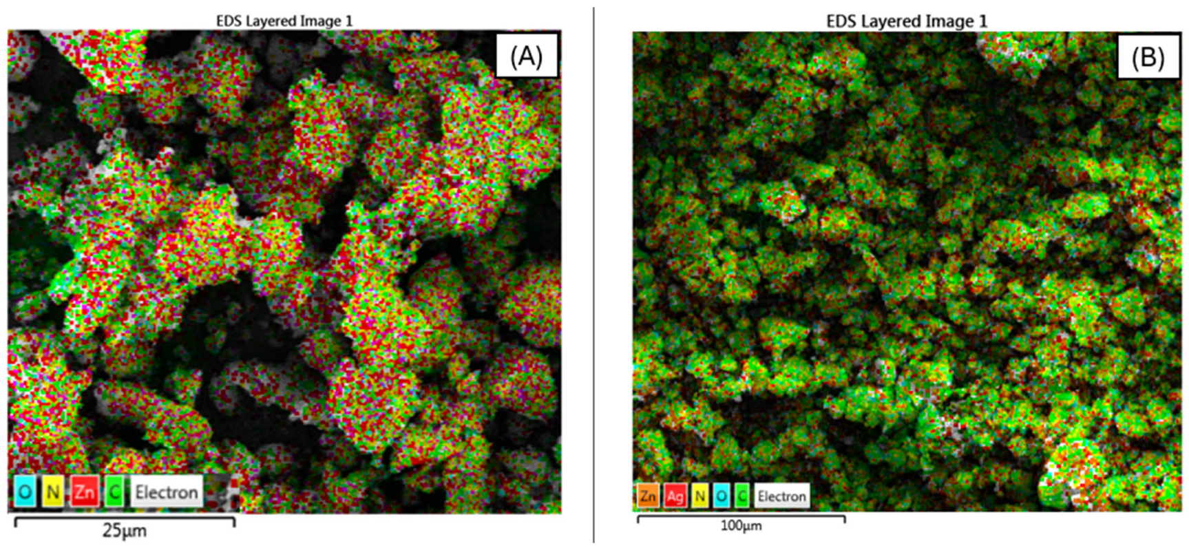
SEM and elemental mapping analyses of ZIF-8, Cu-ZIF-8, ZIF-8@HA, and Cu-ZIF-8@HA nanoparticles reveal distinct morphological and compositional features that confirm successful synthesis and surface functionalization (Figure 1, Figure 2 and Figure 3).
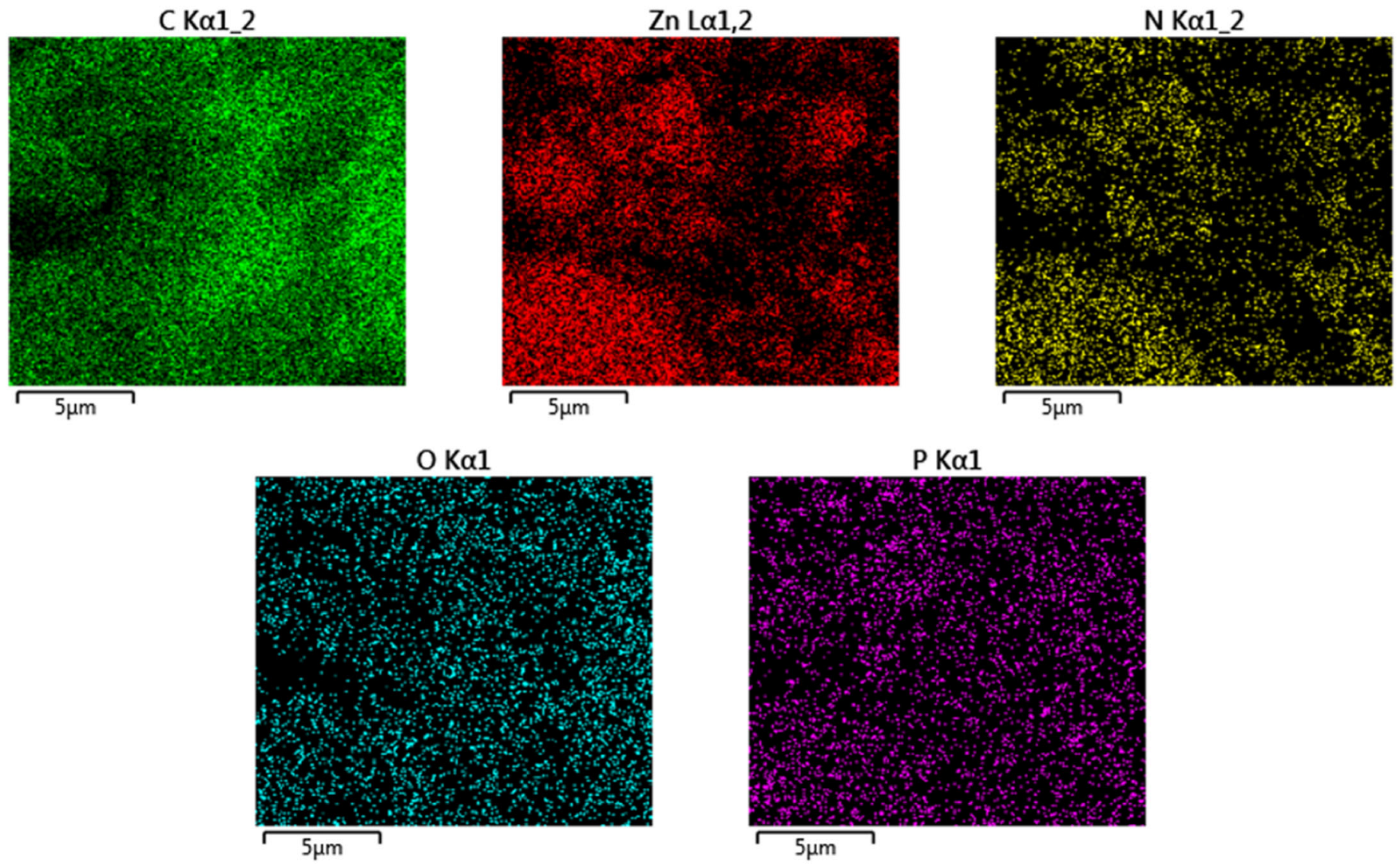
Pristine ZIF-8 displays uniform rhombic dodecahedral crystals with smooth, well-defined edges, characteristic of high crystallinity and homogeneous nucleation. Upon copper incorporation, Cu-ZIF-8 retains the general ZIF-8 morphology but exhibits slightly rougher surfaces and smaller particle sizes, indicating that Cu2+ ions partially substitute Zn2+ within the ZIF-8 framework, modulating growth kinetics and surface energy [8,24]. This morphological evolution suggests successful Cu doping rather than surface adsorption, as confirmed by the uniform distribution of Cu and Zn in Cu-ZIF-8@HA (Figure 2B). Elemental mapping shows homogeneous signals for Zn, Cu, C, N, and O, demonstrating that Cu species are evenly dispersed within the ZIF-8 lattice, preserving the integrity of the metal–organic framework and avoiding phase segregation. A minor phosphorus signal was detected in the EDS spectrum; however, phosphorus is not part of the ZIF-8 or hyaluronic acid chemical structure and is therefore attributed to trace background contamination or residual phosphate species introduced during sample preparation or analysis.
The subsequent coating of hyaluronic acid (HA) onto the nanoparticle surfaces introduces significant surface modifications. SEM images of ZIF-8@HA and Cu-ZIF-8@HA reveal smoother, less angular morphologies, and in some regions, a thin amorphous layer consistent with HA coverage. HA, a negatively charged polysaccharide, forms a uniform biopolymer shell around the ZIF-8 core through electrostatic interactions between the carboxyl groups of HAs and the positively charged metal nodes of the framework. This coating not only enhances colloidal stability and dispersibility but also improves biocompatibility and potential cellular interactions in antimicrobial or biomedical applications [25]. Elemental mapping results further support successful surface modification, with strong signals from C, N, and O dominating across the entire particle surface, confirming the presence of organic HA, while Zn and Cu remain uniformly distributed within the core region. The co-localization of C, O, and N with Zn and Cu indicates intimate interaction between HA chains and the ZIF-8 framework.
The combined SEM and EDS findings confirm that both Cu doping and HA coating were effectively achieved without compromising the characteristic ZIF-8 structure. The homogeneous distribution of constituent elements (Zn, Cu, C, N, O) reflects compositional uniformity and structural stability, which are critical for maintaining the antimicrobial and functional performance of Cu-ZIF-8@HA nanocomposites.

3.1.2. Transmission Electron Microscopy (TEM)

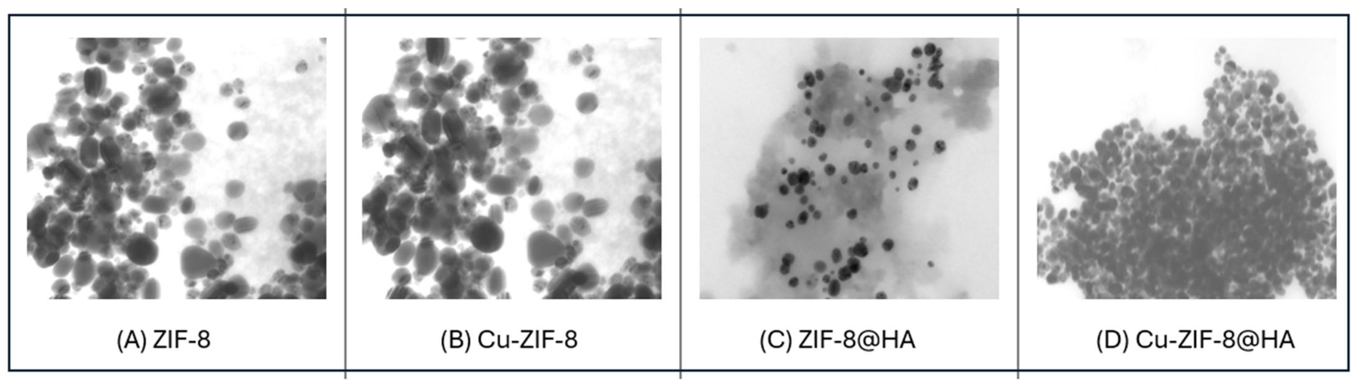
The transmission electron microscopy (TEM) images (Figure 4) provide detailed insight into the morphology and nanoscale structure of ZIF-8, Cu-ZIF-8, ZIF-8@HA, and Cu-ZIF-8@HA nanoparticles. Pristine ZIF-8 particles appear as well-defined polyhedral nanocrystals with smooth surfaces and uniform dispersion, which is characteristic of the highly crystalline ZIF-8 framework formed through the coordination of Zn2+ ions with 2-methylimidazole ligands. Following copper incorporation, the Cu-ZIF-8 nanoparticles retain a similar polyhedral morphology but exhibit slightly reduced particle size and increased image contrast. This contrast enhancement is commonly observed when heavier metal ions are incorporated into the framework and suggests partial substitution of Zn2+ nodes with Cu2+ within the ZIF-8 lattice, accompanied by minor changes in particle density and crystal growth behavior [26].
In contrast, the TEM images of ZIF-8@HA reveal a thin, less electron-dense layer surrounding the darker ZIF-8 cores, which is consistent with the presence of an amorphous hyaluronic acid coating on the nanoparticle surface. This polymeric shell appears as an irregular, lighter halo around the crystalline core, indicating successful surface encapsulation of ZIF-8 by HA. A similar core–shell morphology is observed for Cu-ZIF-8@HA; however, these particles appear more compact and occasionally form nanoscale clusters, suggesting stronger interfacial interactions between the Cu-doped ZIF-8 framework and the HA polymer chains. The enhanced aggregation behavior may arise from increased coordination interactions between Cu2+ sites and the carboxyl groups of HA.
Overall, the TEM analysis confirms that both copper doping and HA surface functionalization were successfully achieved while preserving the structural integrity of the ZIF-8 framework. The observed core–shell-like morphology and uniform nanoparticle distribution further support the successful fabrication of multifunctional Cu-ZIF-8@HA nanocomposites with controlled nanoscale architecture.

3.1.3. Zeta Potential (ZP)

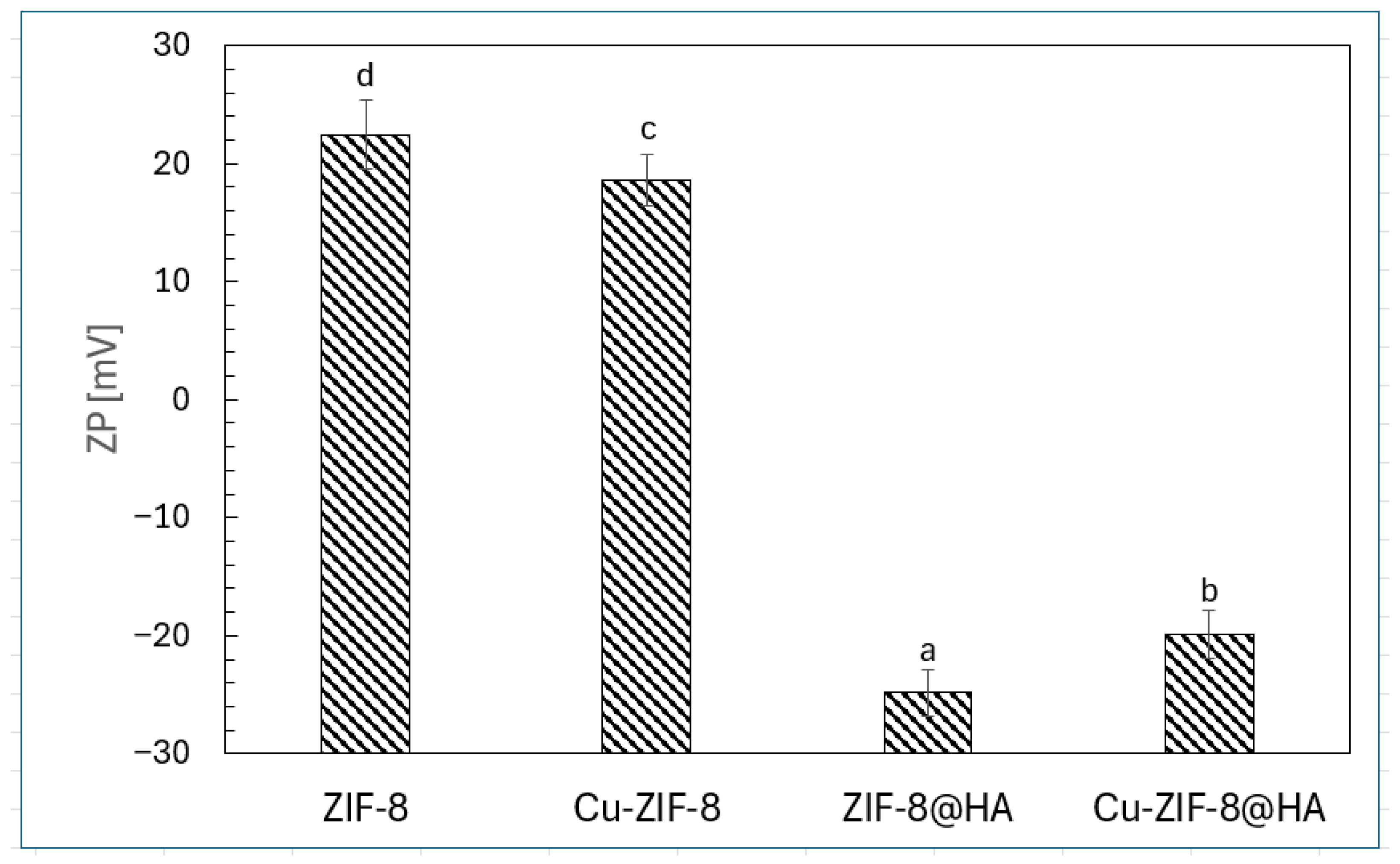
The zeta potential (ζ) values of ZIF-8, Cu-ZIF-8, ZIF-8@HA, and Cu-ZIF-8@HA nanoparticles reveal the profound effects of metal doping and hyaluronic acid (HA) surface modification on colloidal stability and surface charge behavior (Figure 5).
As shown in Figure 5, pristine ZIF-8 and Cu-ZIF-8 exhibited positive ζ values (+22.1 mV and +18.4 mV, respectively), indicating that their surfaces are dominated by metal-linked imidazolate moieties and residual Zn2+/Cu2+ ions, which impart a net positive charge that enhances electrostatic repulsion and dispersion stability in aqueous media [27]. Upon HA coating, a pronounced shift toward negative potentials was observed, with ZIF-8@HA (−26.3 mV) and Cu-ZIF-8@HA (−18.7 mV) reflecting the contribution of deprotonated carboxyl (–COO) and hydroxyl groups from HA on the nanoparticle surface [8,28]. This surface inversion confirms the successful coating and indicates the introduction of steric and electrostatic stabilization through HA’s hydrophilic polymeric shell. The slightly less negative potential of Cu-ZIF-8@HA compared with ZIF-8@HA suggests partial charge screening by Cu2+ coordination with HA carboxyl groups, which may improve particle–cell membrane interactions and antibacterial efficacy [29]. Overall, the transition from highly positive to moderately negative ζ potentials demonstrates the synergistic interplay between metal doping and HA modification, balancing colloidal stability with biofunctional interaction potential for enhanced antimicrobial applications.

3.1.4. Particle Size (PS)

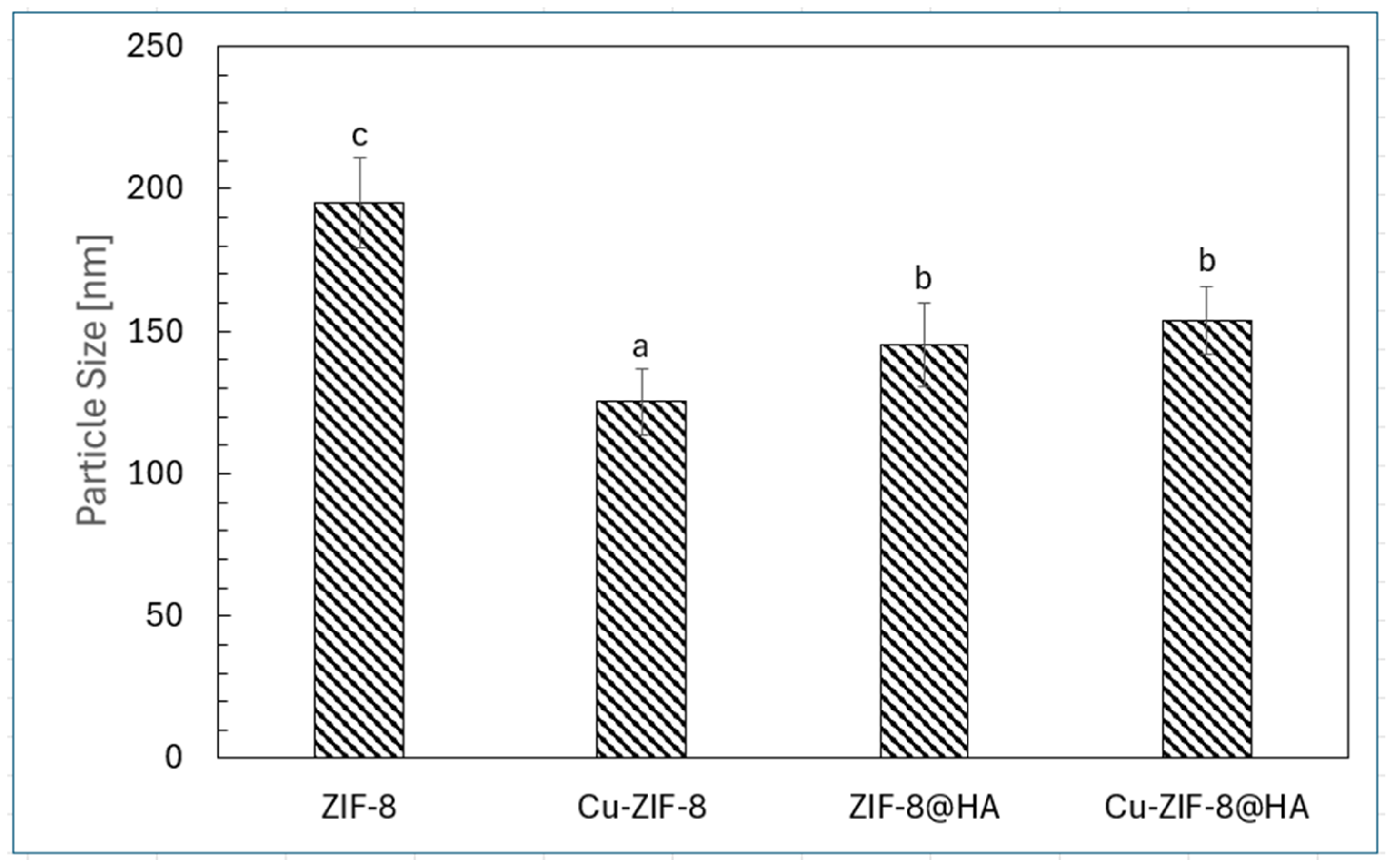
The particle size distributions of ZIF-8, Cu-ZIF-8, ZIF-8@HA, and Cu-ZIF-8@HA nanoparticles (Figure 6) illustrate the effects of metal-ion substitution and hyaluronic acid (HA) surface functionalization on nanostructural assembly and colloidal stability.
Pristine ZIF-8 exhibited the largest mean particle size (~195 nm), consistent with the typical crystalline growth of ZIF-8 through the self-assembly of Zn2+ ions with 2-methylimidazole ligands [30]. Partial substitution of Zn2+ with Cu2+ resulted in a notable reduction in particle size (~130 nm), suggesting that Cu2+ incorporation disrupts the regular coordination environment of Zn2+ nodes, thereby limiting crystal growth and promoting the formation of smaller, more compact nanoparticles. Surface functionalization with HA produced a moderate increase in hydrodynamic diameter for both ZIF-8 and Cu-ZIF-8 (~150–160 nm). This increase is attributed to the formation of a hydrated polymeric corona surrounding the nanoparticle surface, as well as the presence of extended carboxyl and hydroxyl groups that expand the hydrodynamic volume detected by dynamic light scattering [31]. The similar particle sizes observed for ZIF-8@HA and Cu-ZIF-8@HA suggest that HA coating occurred uniformly regardless of the metal composition of the framework. Control over particle size is an important factor influencing antimicrobial performance, as smaller Cu-doped nanoparticles can enhance diffusion and interactions with microbial cells, while the HA coating improves colloidal stability and biocompatibility. Overall, these findings demonstrate that Cu doping and HA encapsulation act synergistically to tune both the physicochemical properties and functional performance of ZIF-8-based nanocomposites for antimicrobial applications.

3.1.5. FTIR

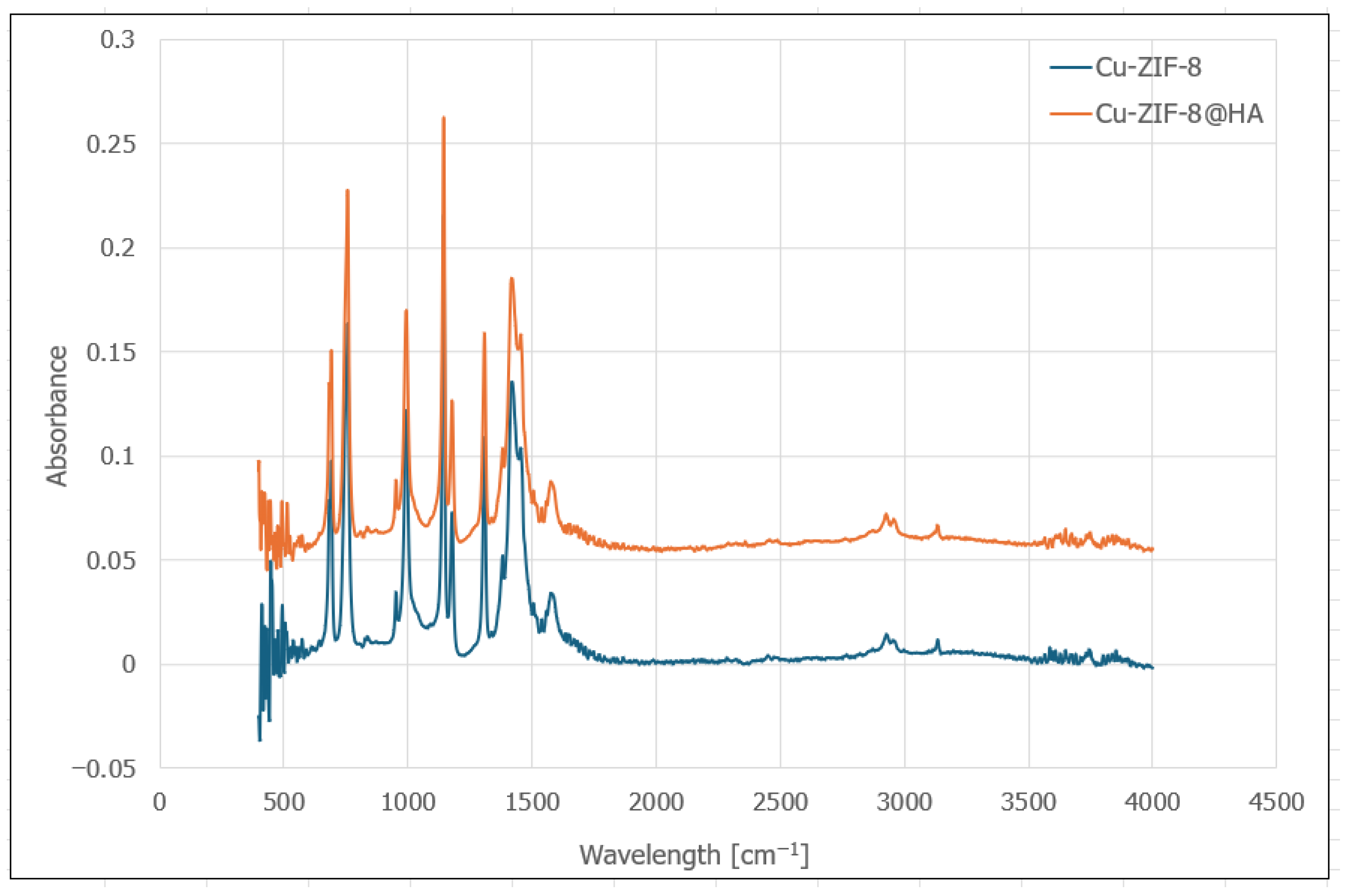
The FTIR spectra of Cu-ZIF-8 and Cu-ZIF-8@HA nanoparticles (Figure 7) exhibit characteristic vibrational bands confirming the successful synthesis and surface functionalization of the composites.
For Cu-ZIF-8, distinct peaks observed between 900–1500 cm−1 correspond to the stretching vibrations of the imidazole ring (C–N and C=N) and aromatic C–H bending, confirming the ZIF-8 framework integrity. The weak absorption below 800 cm−1 represents Zn–N and Cu–N coordination bonds within the metal–organic framework [32]. In Cu-ZIF-8@HA, additional broad peaks near 3300–3400 cm−1 and around 1650 cm−1 are attributed to O–H/N–H stretching and amide I (C=O) vibrations from hyaluronic acid, indicating successful surface coating [33]. The intensity enhancement across fingerprint regions suggests increased hydrogen bonding and surface interactions between HA and the Cu-ZIF-8 matrix, validating the formation of a stable Cu-ZIF-8@HA nanocomposite suitable for antibacterial applications.

3.1.6. Cytotoxicity Test

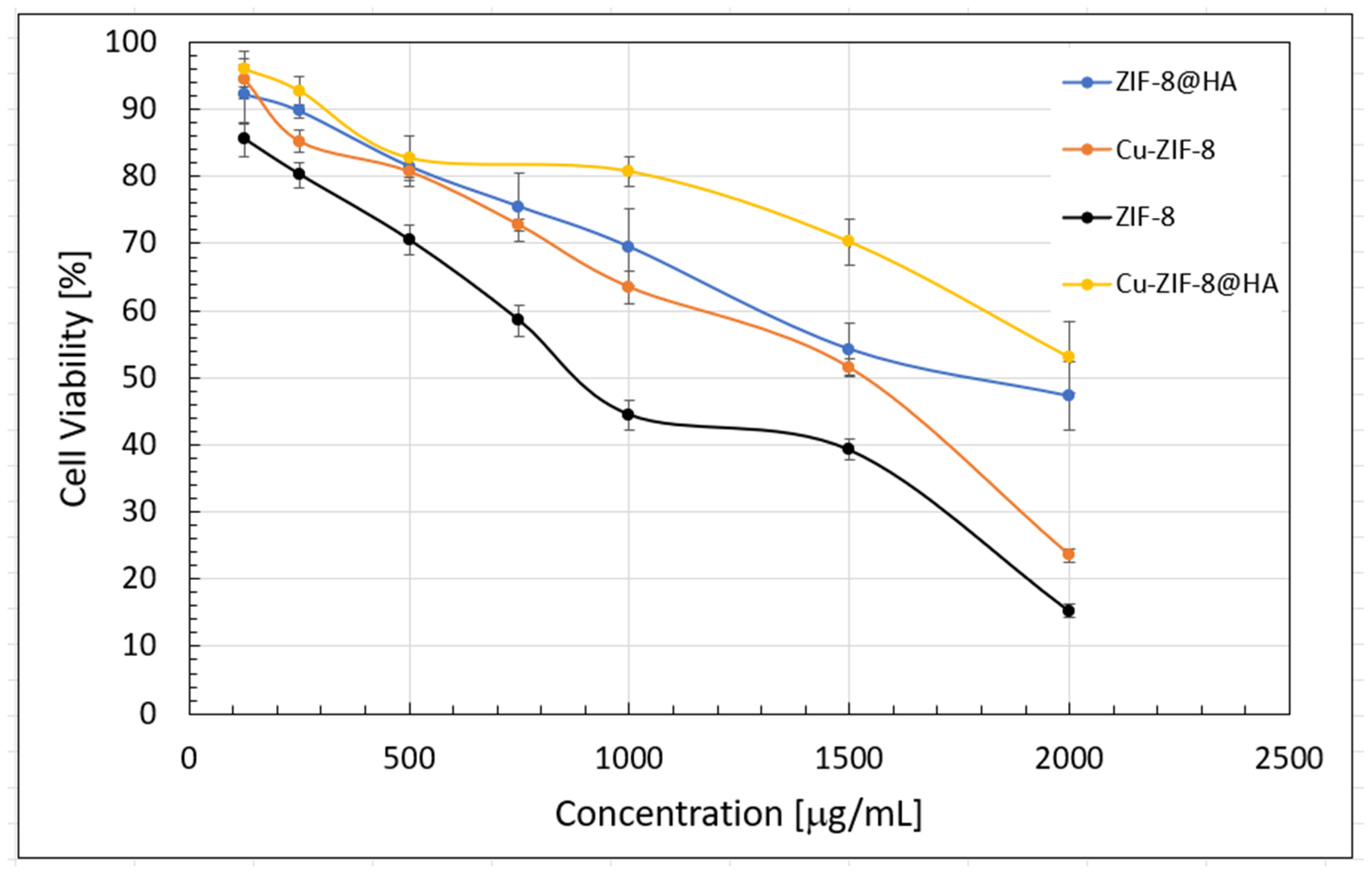
The cytotoxicity profiles of ZIF-8, Cu-ZIF-8, ZIF-8@HA, and Cu-ZIF-8@HA nanoparticles demonstrate the strong influence of metal doping and hyaluronic acid (HA) surface functionalization on biocompatibility and concentration-dependent cellular response (Figure 8).
ZIF-8 exhibited the highest cytotoxicity, with cell viability decreasing sharply to ~15% at 2000 µg/mL, indicating potential Zn2+ ion leakage and reactive oxygen species (ROS) generation that compromise membrane integrity and mitochondrial function [34]. The partial substitution of Zn2+ with Cu2+ in Cu-ZIF-8 slightly moderated cytotoxicity at lower concentrations but reduced cell viability more rapidly at higher levels, likely due to the pro-oxidative nature of Cu2+ ions and their capacity to catalyze Fenton-like reactions [8].
In contrast, HA coating markedly improved biocompatibility for both ZIF-8@HA and Cu-ZIF-8@HA, maintaining cell viability above 70% at ≤1000 µg/mL and above 50% even at the highest tested concentration. This enhancement can be attributed to HA’s hydrophilic and negatively charged surface, which reduces direct metal–cell contact, stabilizes nanoparticle dispersions, and limits the burst release of metal ions [28,35]. Among all formulations, Cu-ZIF-8@HA exhibited the best cytocompatibility, suggesting that HA functionalization effectively counterbalances Cu-induced oxidative stress and promotes favorable cell–material interactions. These results collectively demonstrate that HA encapsulation is an essential surface engineering strategy to mitigate cytotoxicity while preserving the antimicrobial efficacy of metal-doped ZIF-8 nanocomposites.

3.2. Assessment of Antibacterial Activity

3.2.1. Disk Diffusion Test

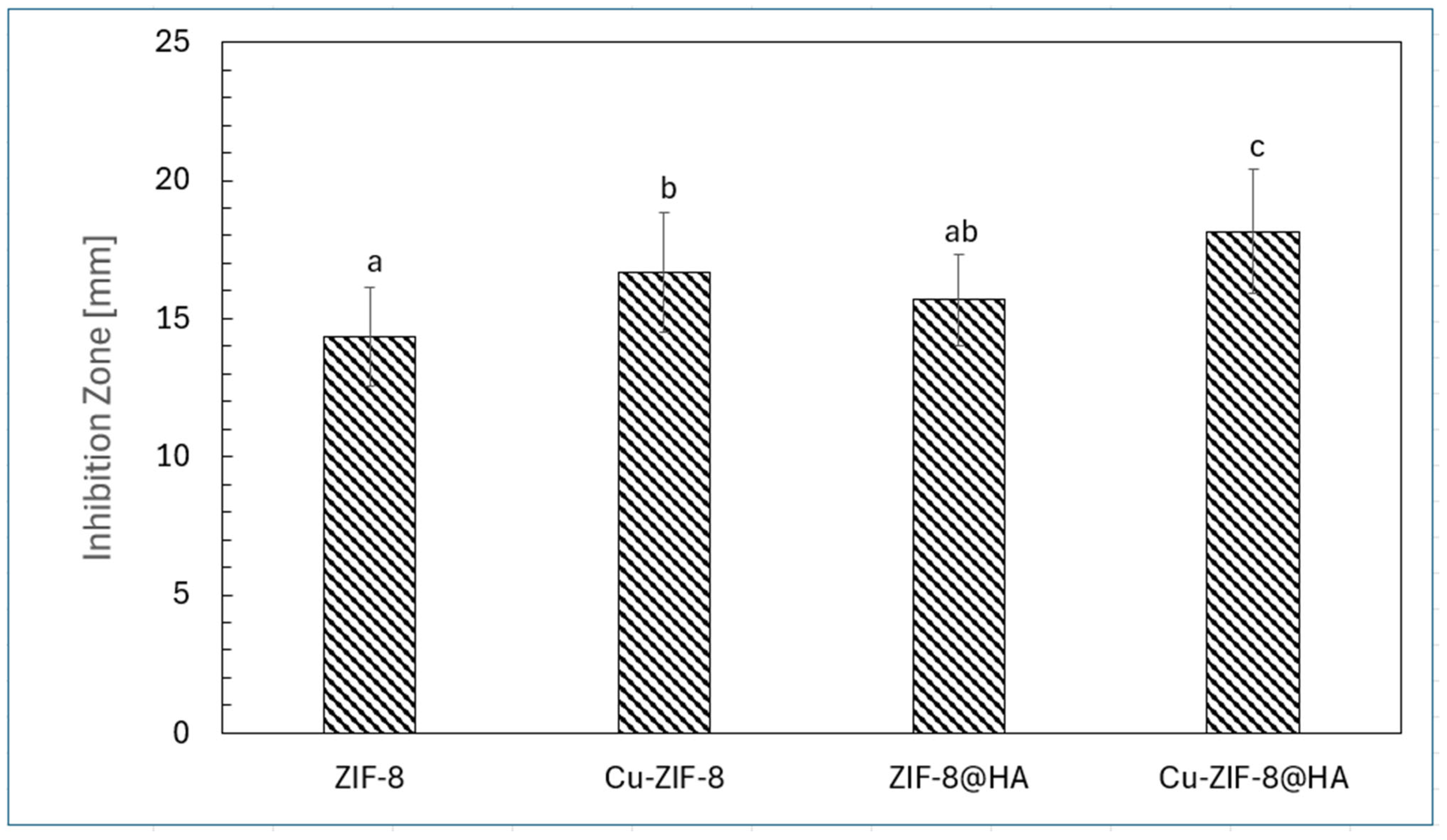
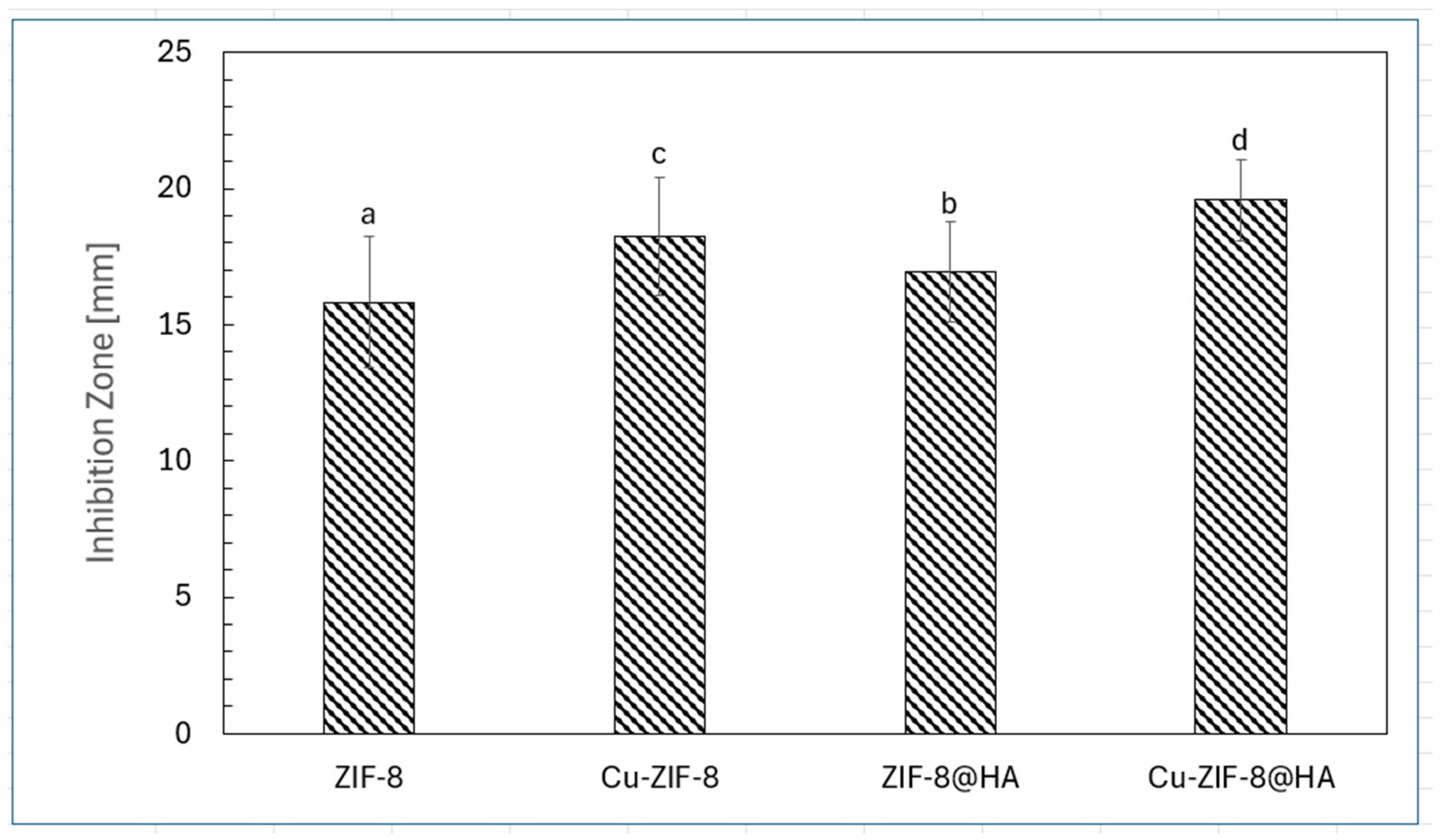
The disk diffusion assay demonstrated that both metal doping and hyaluronic acid (HA) surface functionalization significantly influence the antibacterial activity of ZIF-8–based nanocomposites against Gram-negative foodborne pathogens. As shown in Figure 9 and Figure 10, all nanoparticle formulations inhibited Salmonella enterica serovar Typhimurium ATCC 14028 and Escherichia coli O157:H7, although inhibition zones were consistently larger for E. coli, indicating greater susceptibility.
Among the tested materials, Cu-ZIF-8@HA exhibited the strongest antibacterial activity (p < 0.05), followed by Cu-ZIF-8, ZIF-8@HA, and pristine ZIF-8. The Cu-ZIF-8@HA nanocomposite produced mean inhibition zones of 18.15 ± 2.25 mm against S. Typhimurium and 20.33 ± 2.20 mm against E. coli, representing a 28–35% increase compared with pristine ZIF-8. The enhanced antibacterial performance of Cu-doped materials is likely associated with the synergistic generation of reactive oxygen species (ROS) and the sustained release of Cu2+ and Zn2+ ions from the ZIF-8 framework, which induce oxidative stress, lipid peroxidation, and membrane disruption [7]. In addition, HA functionalization improves nanoparticle dispersion and promotes interactions with bacterial and biofilm surfaces, thereby enhancing contact-mediated antimicrobial activity and stabilizing metal-ion release [36].
The observed differences in inhibition zones between E. coli and S. Typhimurium may be attributed to structural variations in their outer membranes. Although both organisms are Gram-negative, E. coli typically exhibits higher outer-membrane porin density and reduced lipopolysaccharide cross-linking, facilitating ion transport and nanoparticle diffusion [37]. In contrast, S. Typhimurium possesses stronger permeability barriers and more active efflux systems, which can increase resistance to metal-based antimicrobials [38]. These structural differences likely contribute to the greater susceptibility of E. coli observed in this study. Similar trends have been reported previously, with Cu-ZIF-8 nanoparticles producing 20–25% larger inhibition zones against E. coli compared with Salmonella species. Collectively, these findings demonstrate that the combined effects of copper doping and HA surface modification significantly enhance the antibacterial performance of ZIF-8 nanocomposites. This multifunctional design highlights the potential of Cu-ZIF-8@HA as an effective antimicrobial material for food-contact surface coatings or sanitizer formulations [39] aimed at reducing biofilm-associated contamination.

3.2.2. MIC and MBC

The Minimum Inhibitory Concentration (MIC) and Minimum Bactericidal Concentration (MBC) values obtained for ZIF-8, Cu-ZIF-8, ZIF-8@HA, and Cu-ZIF-8@HA against Salmonella Typhimurium ATCC 14028 and Escherichia coli O157:H7 demonstrate distinct differences in antibacterial performance that reflect the synergistic effects of metal doping and hyaluronic acid (HA) surface modification. As shown in Table 1 and Table 2, all formulations displayed bactericidal activity, with MBC/MIC ratios ranging from 2.0 to 4.0.
Among the tested nanoparticles, the Cu-doped and HA-coated hybrids (Cu-ZIF-8@HA) exhibited the lowest MIC and MBC values for both pathogens (500/2000 µg/mL for S. Typhimurium and 1000/2500 µg/mL for E. coli), indicating the highest antimicrobial potency. These results highlight how both copper incorporation and HA functionalization substantially enhance antibacterial efficacy compared with pristine ZIF-8.
Cu-ZIF-8@HA demonstrated a 2–3-fold reduction in MIC relative to pristine ZIF-8, indicating synergistic enhancement from copper incorporation and HA functionalization. The concentration-dependent decrease in viable counts suggests a dual-action mechanism involving sustained metal-ion release and localized surface interaction. Although ROS levels were not directly measured, the magnitude of bactericidal improvement is consistent with previously reported redox-active Cu-doped MOF systems. Importantly, the HA coating appears to balance antimicrobial potency with reduced cytotoxicity, expanding the therapeutic window for surface-contact applications.
Copper doping notably improved the antimicrobial capacity of the parent ZIF-8 framework, halving the MIC from 1500 to 750 µg/mL for S. Typhimurium and reducing it from 2000 to 1500 µg/mL for E. coli. This enhancement is consistent with previous reports demonstrating that the introduction of Cu2+ ions into ZIF-8 frameworks facilitates redox cycling between Cu2+/Cu+ species, which promotes the generation of reactive oxygen species (ROS) and oxidative stress within bacterial cells [40]. Additionally, copper ions disrupt bacterial membranes by binding to thiol and amino groups in proteins and nucleic acids, interfering with enzymatic function and cell respiration [41]. The higher antibacterial activity of Cu-ZIF-8 against both Gram-negative pathogens aligned with findings that Cu-based metal–organic frameworks (MOFs) outperform Zn-based analogs due to the higher redox reactivity and metal-ion toxicity of copper species.
The inclusion of hyaluronic acid (HA) further enhanced nanoparticle performance, as reflected in the lower MIC and MBC values of ZIF-8@HA (1250/4500 µg/mL for S. Typhimurium; 1250/3000 µg/mL for E. coli) relative to unmodified ZIF-8. HA is a biocompatible, negatively charged polysaccharide that improves aqueous dispersibility, colloidal stability, and surface charge uniformity of nanoparticles [42]. Its hydrophilic carboxyl and hydroxyl groups promote electrostatic attraction and hydrogen bonding with bacterial cell walls, facilitating stronger adhesion and localized antimicrobial action [43]. Furthermore, HA’s biopolymeric shell can modulate the diffusion and control the release rate of metal ions from the MOF core, maintaining sustained bactericidal concentrations while minimizing aggregation or premature degradation [44]. These properties contribute to the superior antimicrobial efficacy of HA-modified composites, particularly when combined with metal doping.
The dual modification in Cu-ZIF-8@HA produced the most significant improvement across both bacterial species. Its MIC values were 3 times lower than those of ZIF-8 for S. Typhimurium (500 vs. 1500 µg/mL) and 2 times lower for E. coli (1000 vs. 2000 µg/mL). The MBC/MIC ratios of 4.0 and 2.5 confirm its bactericidal nature, demonstrating that this hybrid structure efficiently kills rather than merely inhibits bacterial cells. It arises from copper’s oxidative and membrane-disruptive effects combined with HA’s stabilizing and surface-interactive roles. HA encapsulation also increases biocompatibility, mitigating potential cytotoxicity associated with free metal ions while improving material wettability and adherence to biological or food-contact surfaces [45].
The species-dependent differences observed between S. Typhimurium and E. coli likely reflect inherent variations in their outer membrane composition, lipopolysaccharide density, and efflux pump activity [46]. E. coli generally exhibits slightly higher MIC and MBC values, suggesting greater resilience to oxidative and ionic stress. However, this disparity diminishes in the presence of Cu and HA modifications, emphasizing the broad-spectrum potential of Cu-ZIF-8@HA. Previous studies confirm that Gram-negative bacteria, despite possessing outer membrane barriers, remain highly susceptible to nanoparticle-induced ROS generation and ion leakage when the particle surface is engineered for adhesion and diffusion [47].
Mechanistically, the enhanced antibacterial action of Cu-ZIF-8@HA is attributed to multiple synergistic pathways: (i) ROS production causing lipid peroxidation and DNA damage; (ii) release of Zn2+ and Cu2+ disrupting enzymatic functions; (iii) electrostatic adhesion of HA-modified particles to the bacterial envelope, facilitating localized attack; and (iv) possible catalytic decomposition of hydrogen peroxide through Cu redox cycling [48,49]. The combined outcome is rapid membrane rupture, leakage of cellular contents, and bacterial death. From an application standpoint, these findings indicate that Cu-ZIF-8@HA could serve as an efficient, biocompatible antimicrobial coating for food-contact surfaces, offering sustained bactericidal action with minimal toxicity.
In summary, the MIC/MBC data confirm that copper doping and HA functionalization significantly amplify the antibacterial capacity of ZIF-8 frameworks against both S. Typhimurium ATCC14028 and E. coli O157:H7. Cu-ZIF-8@HA demonstrates the most favorable bactericidal ratio and the lowest inhibitory concentrations, highlighting the synergistic roles of Cu ion release, ROS generation, and HA-mediated stability. These findings are consistent with contemporary studies on multifunctional MOF-based nanocomposites, underscoring their promise as next-generation antimicrobial agents for food safety and biomedical applications [44].

3.2.3. Nanoparticle Effectiveness on Bacterial Biofilms

Stainless-Steel Surface Characterization
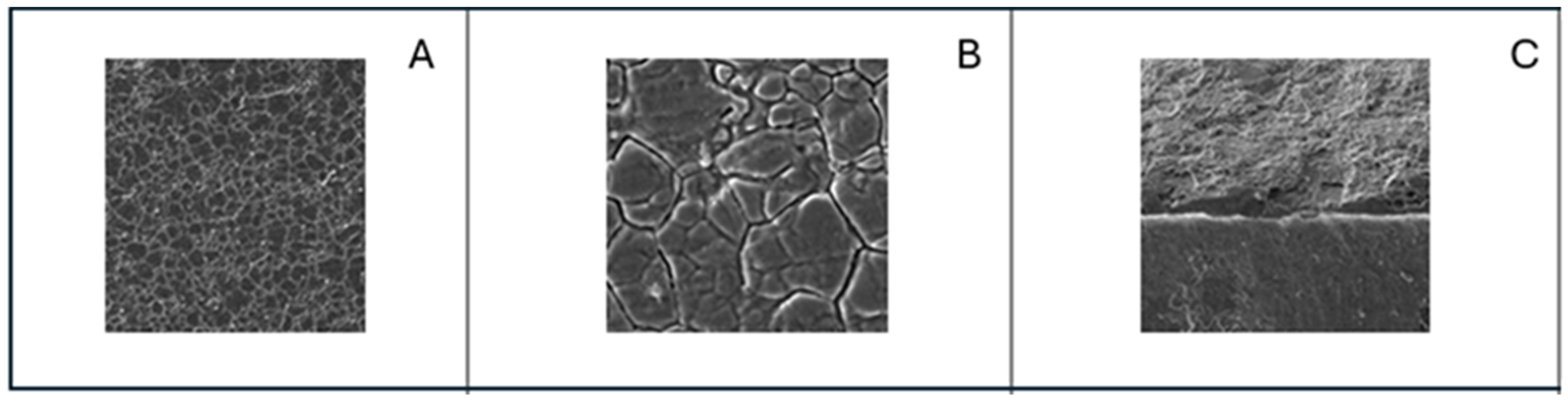
SEM micrographs (Figure 11) of the stainless-steel surface reveal the characteristic topography, with a relatively smooth matrix, fine polishing marks, and occasional microscopic irregularities typical of food-grade stainless steel.
These surface features, including minor pits and grooves, are potential sites for bacterial adhesion and biofilm formation, emphasizing the importance of surface integrity in food-contact applications. The high-magnification images highlight the uniform distribution of austenitic grains and the presence of oxide layers that contribute to corrosion resistance. However, these passive films can be disrupted by repeated cleaning or mechanical abrasion, increasing surface roughness. SEM observations, therefore, confirm that even minimal morphological imperfections can significantly influence microbial attachment and subsequent contamination risks in food processing environments.
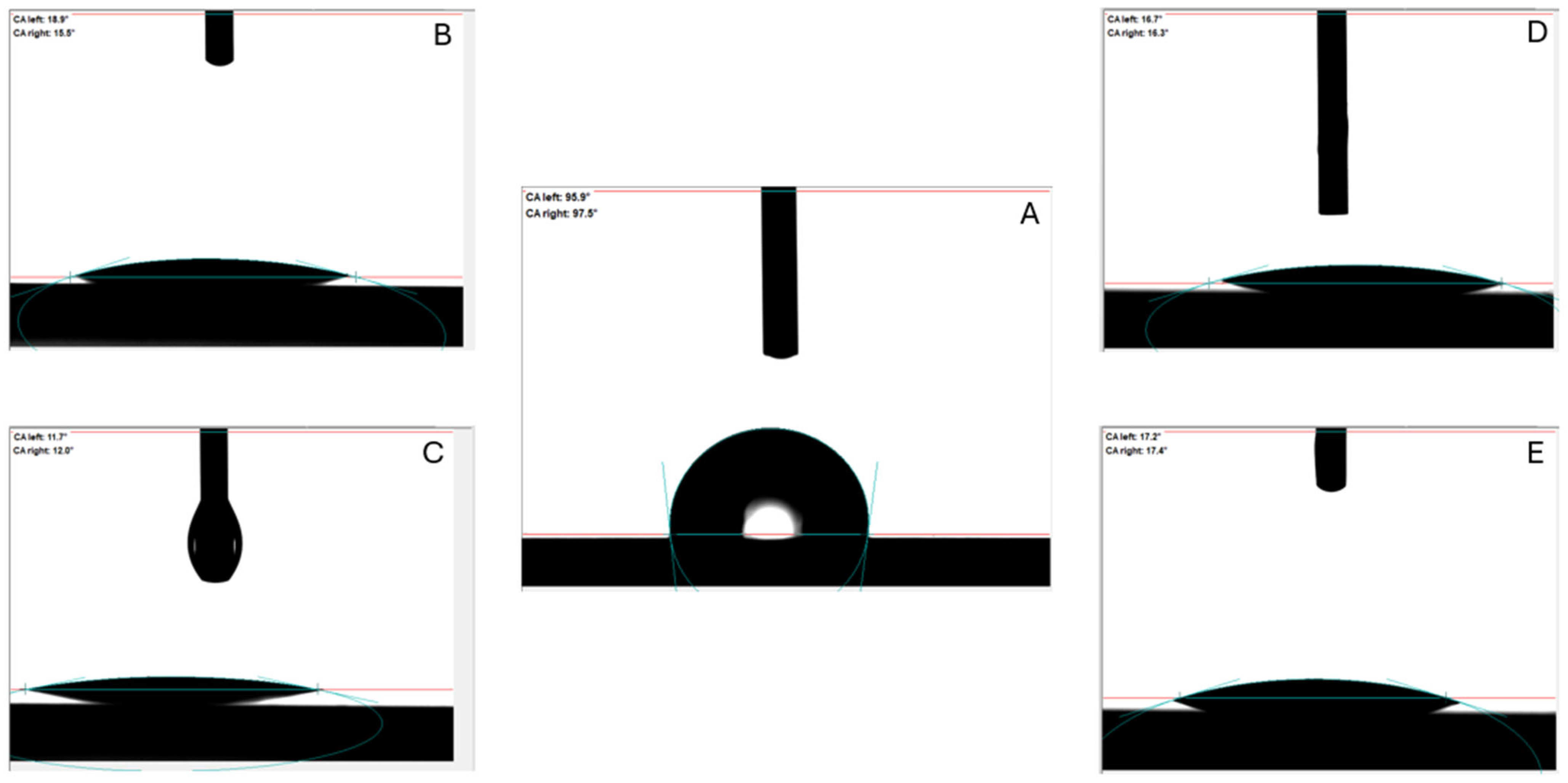
The contact angle results (Figure 12, Table 3) reveal that coating stainless-steel surfaces with ZIF-8-based nanocomposites significantly enhances hydrophilicity compared to untreated water control.
The high contact angle of water (95.80 ± 2.33°) indicates a hydrophobic surface typical of bare stainless steel, where limited polar interactions hinder wetting. In contrast, all nanoparticles (ZIF-8, Cu-ZIF-8, ZIF-8@HA, and Cu-ZIF-8@HA) produced markedly lower contact angles (11.77–18.57°; p < 0.05), suggesting that incorporating metal–organic frameworks and biopolymer coatings improves surface wettability. These findings align with reports that ZIF-8 nanostructures can modify surface chemistry and increase the concentration of polar functional groups, thereby reducing water contact angles and improving surface energy [50]. Among the nanoparticle treatments, Cu-ZIF-8 exhibited the lowest contact angle (11.77 ± 1.42°), indicating the highest hydrophilicity. Copper doping is known to alter the surface charge density and electronic distribution of ZIF-8 frameworks, introducing more oxygen-containing and hydroxyl groups that interact strongly with water molecules. This enhanced wettability can facilitate uniform film formation and improved adhesion on metallic substrates, which are critical for antimicrobial or antibiofouling applications. The slightly higher contact angles observed for Cu-ZIF-8@HA and ZIF-8@HA suggest that the hyaluronic acid layer, while biocompatible and hydrophilic, may partially screen the active polar sites of the metal centers, producing a smoother, moderately hydrophilic surface [51]. The dramatic reduction in contact angle following nanoparticle application demonstrates that ZIF-8-based nanocomposites effectively transform stainless-steel surfaces from hydrophobic to hydrophilic. Such enhanced wettability promotes better liquid spreading, which can improve the efficiency of antimicrobial contact, nutrient transport, and cleaning in food-processing environments, thereby contributing to safer and more hygienic surfaces [52].
The total surface free energy (SFE) values of stainless-steel surfaces treated with different nanoparticle solutions (Table 4) demonstrated a progressive increase from water (46.33 ± 1.27 mN/m) to Cu-ZIF-8@HA (64.42 ± 3.03 mN/m), indicating a significant (p < 0.05) enhancement in surface wettability and polarity upon nanoparticle modification. The untreated surface or water-treated condition exhibited relatively low SFE, consistent with limited hydrophilicity and higher bacterial adhesion potential typical of bare stainless steel [53]. Coating with ZIF-8 (52.18 ± 2.39 mN/m) moderately improved surface energy due to the presence of zinc nodes and imidazolate linkers that introduce polar functional groups. The incorporation of copper (Cu-ZIF-8, 56.25 ± 1.85 mN/m) further increased SFE, reflecting improved surface activation and potential for enhanced antimicrobial interaction via catalytic and redox activity. The addition of hyaluronic acid (ZIF-8@HA, 59.50 ± 2.77 mN/m) significantly increased total SFE owing to the abundance of hydrophilic hydroxyl and carboxyl groups that enhance hydrogen bonding with water molecules. The Cu-ZIF-8@HA nanocomposite exhibited the highest SFE, suggesting synergistic effects between copper doping and HA coating that yield superior wettability, improved nanoparticle dispersion, and stronger interaction with aqueous environments. Such high-energy surfaces are advantageous for reducing bacterial adhesion and facilitating antimicrobial contact on stainless-steel food-contact surfaces.
Effect of Nanoparticles on Escherichia coli and Salmonella Typhimurium on Stainless-Steel Surfaces
The adhesion and growth inhibition results summarized in Table 5 and Table 6 reveal the potent antimicrobial efficacy of Cu-ZIF-8@HA nanoparticles against both Escherichia coli O157:H7 and Salmonella Typhimurium ATCC 14028 on stainless-steel surfaces. In the untreated control groups, high bacterial adhesion levels were observed (6.86 ± 0.35 log CFU/cm2 for E. coli and 7.02 ± 0.41 log CFU/cm2 for S. Typhimurium), confirming that both species readily colonize and persist on stainless steel, a standard food-contact material, through biofilm formation and surface conditioning [54,55]. The control and PBS + 5% MeOH treatments showed no significant difference, indicating that the solvent system alone had a negligible antibacterial impact. However, the introduction of Cu-ZIF-8@HA nanoparticles led to pronounced, concentration-dependent reductions in viable surface-attached cells.
At the MIC, bacterial counts decreased markedly (p < 0.05) to 4.53 ± 0.37 log CFU/cm2 for E. coli and 5.02 ± 0.44 log CFU/cm2 for S. Typhimurium, corresponding to approximately 2–2.5 log reductions relative to the control. This partial inhibition reflects suppression of cell division and biofilm growth rather than complete eradication, consistent with the MIC’s definition as bacteriostatic [56]. Increasing the dose to the MBC produced a sharper decline, to 3.15 ± 0.42 log CFU/cm2 for E. coli and 3.64 ± 0.38 log CFU/cm2 for S. Typhimurium, indicating the onset of bactericidal action. The most striking results were observed at twice the MBC level, where bacterial adhesion nearly disappeared (0.22 ± 0.09 log CFU/cm2 for E. coli and 0.35 ± 0.11 log CFU/cm2 for S. Typhimurium), signifying near-total elimination of viable cells and disruption of the biofilm matrix. These concentration-dependent effects underscore the dual inhibitory and killing capacity of Cu-ZIF-8@HA on both planktonic and surface-attached populations.
Comparatively, E. coli O157:H7 exhibited slightly greater susceptibility than S. Typhimurium across all treatments. At MIC and MBC levels, E. coli counts were consistently about 0.3–0.5 log lower than those of S. Typhimurium, indicating that S. Typhimurium possesses stronger resistance mechanisms and adhesion capability. This difference aligns with previous findings that S. Typhimurium produces thicker extracellular polymeric substances (EPSs) and more hydrophobic cell surfaces, enabling stronger attachment to stainless steel and increased tolerance to antimicrobials [57,58]. Nonetheless, the near-complete bacterial inactivation at 2×MBC confirms that Cu-ZIF-8@HA is highly effective against both pathogens, achieving >6-log reductions, a threshold commonly required for sanitation efficacy in food-contact applications [59].
The enhanced antibacterial activity of Cu-ZIF-8@HA arises from its composite structure, which integrates metal-ion release, oxidative stress generation, and biopolymer-mediated stabilization. The ZIF-8 framework provides high surface area and controlled Zn2+ release, which interferes with bacterial enzyme systems and destabilizes cell membranes [60]. Copper doping introduces redox-active Cu2+ centers capable of generating reactive oxygen species (ROS), such as hydroxyl radicals and superoxide anions, via Fenton-like reactions, leading to lipid peroxidation and DNA fragmentation [61]. Meanwhile, the hyaluronic acid (HA) coating, composed of hydrophilic carboxyl and hydroxyl groups, enhances nanoparticle dispersibility and facilitates electrostatic adhesion to negatively charged bacterial surfaces [62]. This close contact improves the efficiency of ion transfer and ROS attack at the cell interface, while HA’s biocompatibility moderates the cytotoxicity and supports colloidal stability in aqueous systems.
The combined mechanisms act synergistically, including Cu and Zn ions that disrupt membrane integrity and enzyme function, ROS that oxidize cellular components, and HAs that promote sustained contact and controlled ion release. This multi-target approach explains the steep decline in microbial growth observed between MIC and MBC levels. Significantly, the coating’s hydrophilic nature likely enhances nanoparticle interaction with the stainless-steel surface, forming a thin film that maintains localized antimicrobial activity over time, an advantageous feature for industrial sanitation applications.
The small but measurable differences in survival between E. coli and S. Typhimurium can also be attributed to variations in cell wall architecture. E. coli’s outer membrane contains abundant porins that facilitate ion penetration [63], whereas S. Typhimurium’s more cross-linked lipopolysaccharide layer restricts diffusion of metal ions and reactive intermediates. Nevertheless, Cu-ZIF-8@HA overcame these barriers at higher concentrations, demonstrating broad-spectrum antibacterial action. Overall, the data from both tables confirm that Cu-ZIF-8@HA nanoparticles possess potent, dose-dependent inhibitory and bactericidal effects against major Gram-negative foodborne pathogens. The composite achieved significant log reductions on stainless steel, with the >6-log reduction achieved at 2×MBC meeting or exceeding the microbial reduction thresholds typically required for food-contact surface sanitation, suggesting practical applicability beyond laboratory conditions. By combining metal-ion toxicity, ROS generation, and HA-enhanced stability, Cu-ZIF-8@HA provides a practical and biocompatible approach to surface disinfection. These results strongly support its potential integration into antimicrobial coatings, cleaning formulations, or innovative sanitation systems to reduce cross-contamination and ensure food safety.

4. Conclusions

The comprehensive physicochemical and biological evaluations demonstrated that Cu-ZIF-8@HA nanocomposites possess exceptional potential as next-generation antimicrobial agents for food-contact surfaces. Copper doping effectively enhanced antibacterial potency by promoting reactive oxygen species generation and controlled metal-ion release, while HA coating improved particle dispersibility, reduced cytotoxicity, and increased surface adhesion. The synergistic modification resulted in smaller, stable nanoparticles with optimized zeta potential and vigorous bactericidal activity against both E. coli and S. Typhimurium. Application on stainless steel significantly increased surface hydrophilicity and total surface free energy, leading to improved antimicrobial film formation and substantial reductions in bacterial adhesion (>6-log CFU/cm2). These findings underscore Cu-ZIF-8@HA’s dual advantages of high antimicrobial activity and low cytotoxicity, suggesting its suitability for incorporation into coatings, sanitizers, or smart packaging materials. Beyond the current formulation, an additional promising strategy involves encapsulating natural essential oils within the Cu-ZIF-8@HA framework to further enhance antimicrobial efficacy. Essential oils could provide rapid membrane-disruptive action immediately upon contact, while the metal-doped ZIF-8 structure enables controlled and sustained release of active compounds over time. Such a hybrid system would integrate fast-acting antimicrobial activity with prolonged protection, thereby extending surface hygiene durability and reducing the risk of biofilm re-establishment. Overall, the study validates Cu-ZIF-8@HA as a robust, biocompatible nanostructure capable of mitigating biofilm-associated contamination and advancing hygienic safety standards in food-processing and biomedical applications.

Author Contributions

Conceptualization, H.L.N.; methodology, H.L.N. and T.B.N.N.; software, T.B.N.N.; validation, H.L.N. and T.B.N.N.; formal analysis, H.L.N. and T.B.N.N.; investigation, H.L.N.; resources, H.L.N.; data curation, H.L.N. and T.B.N.N.; writing—original draft preparation, H.L.N. and T.B.N.N.; writing—review and editing, H.L.N. and T.B.N.N.; visualization, H.L.N. and T.B.N.N.; supervision, H.L.N. All authors have read and agreed to the published version of the manuscript.

Funding

This research received no external funding.

Institutional Review Board Statement

Not applicable.

Informed Consent Statement

Not applicable.

Data Availability Statement

The original contributions presented in this study are included in the article. Further inquiries can be directed at the corresponding author.

Acknowledgments

The authors thank Hong Minh Xuan Nguyen for allowing us to use their facilities and instrumentation and for constructive feedback.

Conflicts of Interest

The authors declare no conflicts of interest.

References

  1. Delbeke, S.; Ceuppens, S.; Jacxsens, L.; Uyttendaele, M. Survival of Salmonella and Escherichia coli O157:H7 on Strawberries, Basil, and Other Leafy Greens During Storage. J. Food Prot. 2015, 78, 652–660. [Google Scholar] [CrossRef]
  2. Esmael, A.; Al-Hindi, R.R.; Albiheyri, R.S.; Alharbi, M.G.; Filimban, A.A.R.; Alseghayer, M.S.; Almaneea, A.M.; Alhadlaq, M.A.; Ayubu, J.; Teklemariam, A.D. Fresh Produce as a Potential Vector and Reservoir for Human Bacterial Pathogens: Revealing the Ambiguity of Interaction and Transmission. Microorganisms 2023, 11, 753. [Google Scholar] [CrossRef]
  3. Nemis Technology. Salmonella Contamination in Vegetables: Causes and Prevention; Nemis Technology: Zurich, Switzerland, 2025. [Google Scholar]
  4. Xie, Y.; Long, X.; Kim, Y.; Harris, L.J.; Nitin, N. Survival of Salmonella enterica and Enterococcus faecium on Abiotic Surfaces During Storage at Low Relative Humidity. J. Food Prot. 2024, 87, 100292. [Google Scholar] [CrossRef]
  5. Donkor, E.S.; Sosah, F.K.; Odoom, A.; Odai, B.T.; Kunadu, A.P.H. How Long Do Microorganisms Survive and Persist in Food? A Systematic Review. Microorganisms 2025, 13, 901. [Google Scholar] [CrossRef]
  6. Subhadarshini, A.; Nanda, B. Zeolitic imidazolium framework (ZIF-8) and their derivative-based material for antibacterial study: A comprehensive review. Discov. Mater. 2025, 5, 50. [Google Scholar] [CrossRef]
  7. Nguyen, H.L.; Moreira, R.G.; Castell-Perez, M.E. Antibacterial Effectiveness of Zeolitic Imidazolate Framework-8 (ZIF-8) Nanoparticle Solutions and Its Derivatives Against Salmonella Typhimurium ATCC13311 on Loose-Leaf Lettuce (Lactuca sativa). J. Food Saf. 2025, 45, e70031. [Google Scholar] [CrossRef]
  8. Nguyen, H.L. Design and Application of Metal-Doped Zeolitic Imidazolate Framework-8 Nanoparticles Coated with Hyaluronic Acid and Encapsulated with Oregano Essential Oil for The Control of Foodborne Pathogens. Doctoral Dissertation, Texas A&M University, College Station, TX, USA, 2025. [Google Scholar]
  9. Wang, X.; Wang, H.; Zhang, J.; Ma, W.; Zhang, D.; Chen, Y. Mechanism and different roles of metal-N sites on ZIF-8 for efficient antibacterial. J. Environ. Sci. 2025, 156, 68–78. [Google Scholar] [CrossRef]
  10. Grass, G.; Rensing, C.; Solioz, M. Metallic Copper as an Antimicrobial Surface. Appl. Environ. Microbiol. 2010, 77, 1541–1547. [Google Scholar] [CrossRef]
  11. Cupron. Copper vs. Silver. 2025. Available online: https://cupron.com/technology/copper-vs-silver-which-is-the-best-antimicrobial (accessed on 12 February 2026).
  12. Hsu, C.-Y.; Jasim, S.A.; Saleh, E.A.M.; Firoz, K.H.; Jyothi, S.R.; Shit, D.; Nayak, P.P.; Chauhan, A.S.; Obaida, D.S.; Hashemzadeh, A. Hyaluronic acid and ZIF-8 nanocomposites for wound care. Int. J. Biol. Macromol. 2025, 315, 144475. [Google Scholar] [CrossRef]
  13. Alipoor, R.; Ayan, M.; Hamblin, M.R.; Ranjbar, R.; Rashki, S. Hyaluronic Acid-Based Nanomaterials as a New Approach to the Treatment and Prevention of Bacterial Infections. Front. Bioeng. Biotechnol. 2022, 10, 913912. [Google Scholar] [CrossRef]
  14. Omar, W.H.W.; Mahyudin, N.A.; Azmi, N.N.; Ab Rashid, N.-K.M.; Ismail, R.; Yusoff, M.H.Y.M.; Mokhtar, N.F.K.; Sharples, G.J. Effect of natural antibacterial clays against single biofilm formation by Staphylococcus aureus and Salmonella Typhimurium bacteria on a stainless-steel surface. Int. J. Food Microbiol. 2023, 394, 110184. [Google Scholar] [CrossRef]
  15. Thanh, M.T.; Thien, T.V.; Du, P.D.; Hung, N.P.; Khieu, D.Q. Iron doped zeolitic imidazolate framework (Fe-ZIF-8): Synthesis and photocatalytic degradation of RDB dye in Fe-ZIF-8. J. Porous Mater. 2018, 25, 857–869. [Google Scholar] [CrossRef]
  16. Jongert, T.K.; Slowinski, I.A.; Dao, B.; Cortez, V.H.; Gredig, T.; Plascencia, N.D.; Tian, F. Zeta potential and size analysis of zeolitic imidazolate framework-8 nanocrystals prepared by surfactant-assisted synthesis. Langmuir 2024, 40, 6138–6148. [Google Scholar] [CrossRef]
  17. Bai, Y.; Huang, Y.; Li, Y.; Zhang, B.; Xiao, C.; Hou, X.; Yu, L. The Murine Reg3a Stimulated by Lactobacillus casei Promoted Intestinal Cell Proliferation and Inhibits the Multiplication of Porcine Diarrhea Causative Agent in vitro. Front. Microbiol. 2021, 12, 675263. [Google Scholar] [CrossRef]
  18. Yehia, H.M.; Ibraheim, S.M.; Hassanein, W.A. Prevalence of Listeria species in some foods and their rapid identification. Trop. J. Pharm. Res. 2016, 15, 1047–1052. [Google Scholar] [CrossRef]
  19. Hara, Y.; Castell-Perez, M.E.; Moreira, R.G. Antimicrobial properties of poly (vinyl alcohol) films with zeolitic imidazolate framework (ZIF-8) nanoparticles for food packaging. J. Food Sci. 2023, 88, 2512–2522. [Google Scholar] [CrossRef]
  20. Yang, X.; Castell-Perez, M.E.; Moreira, R.G.; Sevimli-Yurttas, Z. trans-Cinnamaldehyde-encapsulated zeolitic imidazolate framework-8 nanoparticle complex solutions to inactivate Escherichia coli O157:H7 on fresh spinach leaves. J. Food Sci. 2022, 87, 4649–4664. [Google Scholar] [CrossRef]
  21. Arcot, Y.; Liu, S.; Ulugun, B.; DeFlorio, W.; Bae, M.; Salazar, K.S.; Taylor, T.M.; Castillo, A.; Cisneros-Zevallos, L.; Scholar, E.M.A. Fabrication of Robust Superhydrophobic Coatings onto High-Density Polyethylene Food Contact Surfaces for Enhanced Microbiological Food Safety. ACS Food Sci. Technol. 2021, 1, 1180–1189. [Google Scholar] [CrossRef]
  22. Harvey, J.; Keenan, K.; Gilmour, A. Assessing biofilm formation by Listeria monocytogenes strains. Food Microbiol. 2007, 24, 380–392. [Google Scholar] [CrossRef]
  23. Choi, N.-Y.; Baek, S.-Y.; Yoon, J.-H.; Choi, M.-R.; Kang, D.-H.; Lee, S.-Y. Efficacy of aerosolized hydrogen peroxide-based sanitizer on the reduction of pathogenic bacteria on a stainless steel surface. Food Control 2012, 27, 57–63. [Google Scholar] [CrossRef]
  24. Doraghi, Z.; Alinezhad, H.; Ahmadi, A. A copper-doped ZIF-8: A high-performance catalyst for eco-friendly Ullmann and Biginelli reactions. Sci. Rep. 2025, 15, 24432. [Google Scholar] [CrossRef] [PubMed]
  25. Romanò, C.; De Vecchi, E.; Bortolin, M.; Morelli, I.; Drago, L. Hyaluronic Acid and Its Composites as a Local Antimicrobial/Antiadhesive Barrier. J. Bone Jt. Infect. 2017, 2, 63–72. [Google Scholar] [CrossRef] [PubMed]
  26. Velisoju, V.K.; Cerrillo, J.L.; Ahmad, R.; Mohamed, H.O.; Attada, Y.; Cheng, Q.; Yao, X.; Zheng, L.; Shekhah, O.; Telalovic, S.; et al. Copper nanoparticles encapsulated in zeolitic imidazolate framework-8 as a stable and selective CO2 hydrogenation catalyst. Nat. Commun. 2024, 15, 2045. [Google Scholar] [CrossRef]
  27. Mittal, A.; Gandhi, S.; Roy, I. Mechanistic interaction studies of synthesized ZIF-8 nanoparticles with bovine serum albumin using spectroscopic and molecular docking approaches. Sci. Rep. 2022, 12, 10331. [Google Scholar] [CrossRef]
  28. Gumustas, M.; Sengel-Turk, C.T.; Gumustas, A.; Ozkan, S.A.; Uslu, B. Effect of Polymer-Based Nanoparticles on the Assay of Antimicrobial Drug Delivery Systems. In Multifunctional Systems for Combined Delivery, Biosensing and Diagnostics; Elsevier: Amsterdam, The Netherlands, 2017; pp. 67–108. [Google Scholar] [CrossRef]
  29. Honary, S.; Zahir, F. Effect of Zeta Potential on the Properties of Nano-Drug Delivery Systems—A Review (Part 1). Tropical Trop. J. Pharm. Res. 2013, 12, 255–264. [Google Scholar] [CrossRef]
  30. Tsai, C.-W.; Langner, E.H. The effect of synthesis temperature on the particle size of nano-ZIF-8. Microporous Mesoporous Mater. 2016, 221, 8–13. [Google Scholar] [CrossRef]
  31. Li, J.; Mooney, D.J. Designing hydrogels for controlled drug delivery. Nat. Rev. Mater. 2016, 1, 16071. [Google Scholar] [CrossRef] [PubMed]
  32. Pei, Z.; Li, Y.; Fan, G.; Guo, Y.; Luan, D.; Gu, X.; Lou, X.W. Low-Coordinated Conductive ZnCu Metal-Organic Frameworks for Highly Selective H2O2 Electrosynthesis. Small 2024, 20, e2403808. [Google Scholar] [CrossRef]
  33. Aumnate, C.; Soatthiyanon, N.; Makmoon, T.; Potiyaraj, P. Polylactic acid/kenaf cellulose biocomposite filaments for melt extrusion based-3D printing. Cellulose 2021, 28, 8509–8525. [Google Scholar] [CrossRef]
  34. Liu, H.Y.; Gale, J.R.; Reynolds, I.J.; Weiss, J.H.; Aizenman, E. The Multifaceted Roles of Zinc in Neuronal Mitochondrial Dysfunction. Biomedicines 2021, 9, 489. [Google Scholar] [CrossRef]
  35. Myint, S.S.; Laomeephol, C.; Thamnium, S.; Chamni, S.; Luckanagul, J.A. Hyaluronic Acid Nanogels: A Promising Platform for Therapeutic and Theranostic Applications. Pharmaceutics 2023, 15, 2671. [Google Scholar] [CrossRef] [PubMed]
  36. Mohammed, N.J.; Zylstra, G.J.; Mutter, T.Y. Green synthesis of hyaluronic acid coated cadmium sulfide nanoparticles with enhanced antibacterial activity. Discov. Mater. 2025, 5, 225. [Google Scholar] [CrossRef]
  37. Sun, J.; Rutherford, S.T.; Silhavy, T.J.; Huang, K.C. Physical properties of the bacterial outer membrane. Nat. Rev. Microbiol. 2021, 20, 236–248. [Google Scholar] [CrossRef]
  38. Aleksandrowicz, A.; Carolak, E.; Dutkiewicz, A.; Błachut, A.; Waszczuk, W.; Grzymajlo, K. Better together—Salmonella biofilm-associated antibiotic resistance. Gut Microbes 2023, 15, 2229937. [Google Scholar] [CrossRef]
  39. Li, L.; Zhu, Y.; Li, X.; Li, D.; Jia, K.; Liu, Y. Photo-Activated Cu-ZIF-8 Integrated Sprayable Hydrogels for Accelerated Wound Healing. ACS Appl. Bio Mater. 2025, 8, 8197–8217. [Google Scholar] [CrossRef]
  40. Wang, X.; Wang, H.; Cheng, J.; Li, H.; Wu, X.; Zhang, D.; Shi, X.; Zhang, J.; Han, N.; Chen, Y. Initiative ROS Generation of Cu-doped ZIF-8 for Excellent Antibacterial Performance. Chem. Eng. J. 2023, 466, 143201. [Google Scholar] [CrossRef]
  41. Cvijan, B.B.; Jačić, J.K.; Bajčetić, M. The Impact of Copper Ions on the Activity of Antibiotic Drugs. Molecules 2023, 28, 5133. [Google Scholar] [CrossRef]
  42. Di Mola, A.; Landi, M.R.; Massa, A.; D’amora, U.; Guarino, V. Hyaluronic Acid in Biomedical Fields: New Trends from Chemistry to Biomaterial Applications. Int. J. Mol. Sci. 2022, 23, 14372. [Google Scholar] [CrossRef] [PubMed]
  43. Yang, X.; Wu, K.; Liu, Y.; Liu, Y.; Fu, Y.; Lin, W.; Mao, Z.; Wang, X.; Guo, H. Versatile Hyaluronic Acid Hydrogels via pH-Induced Gelation for Multifaceted Cutaneous Wound Healing. Adv. Healthc. Mater. 2025, 15, e03120. [Google Scholar] [CrossRef]
  44. Nguyen, H.L.; Moreira, R.G.; Castell-Perez, M.E. Multifunctional OEO-ZIF-8-HA Nanoparticles for Antibacterial Control on Latex Surfaces and Baby Arugula (Eruca sativa) Leaves. J. Food Sci. 2026, 91, e70896. [Google Scholar] [CrossRef] [PubMed]
  45. Karakocak, B.B.; Liang, J.; Biswas, P.; Ravi, N. Hyaluronate coating enhances the delivery and biocompatibility of gold nanoparticles. Carbohydr. Polym. 2018, 186, 243–251. [Google Scholar] [CrossRef] [PubMed]
  46. Meysman, P.; Sánchez-Rodríguez, A.; Fu, Q.; Marchal, K.; Engelen, K. Expression divergence between Escherichia coli and Salmonella enterica serovar Typhimurium reflects their lifestyles. Mol. Biol. Evol. 2013, 30, 1302–1314. [Google Scholar] [CrossRef]
  47. Canales, C.S.C.; Cazorla, J.I.M.; Cazorla, R.M.M.; Sábio, R.M.; Santos, H.A.; Pavan, F.R. Combating Gram-negative infections: The role of antimicrobial peptides and nanotechnology in overcoming antibiotic resistance. Mater. Today Bio 2025, 35, 102381. [Google Scholar] [CrossRef]
  48. He, A.; Li, Q.; Zhang, Y.; Dang, M.; Li, X.; Zheng, J.; Xiu, W.; Mou, Y.; Dong, H. Nanocuproptosis for precision tumor immunotherapy. Nano TransMed 2025, 100099. [CrossRef]
  49. Wang, L.; Zhou, F.; Xie, W. Advances in hyaluronic acid-based biomaterials: Applications in cancer therapy, wound healing, and disease management. J. Mater. Sci. Mater. Med. 2025, 36, 91. [Google Scholar] [CrossRef]
  50. Xiong, Y.; Shu, Y.; Deng, N.; Luo, X.; Liu, S.; Wu, X. A Novel Modified ZIF-8 Nanoparticle with Enhanced Interfacial Compatibility and Pervaporation Performance in a Mixed Matrix Membrane for De-Alcoholization in Low-Concentration Solutions. Molecules 2024, 29, 4465. [Google Scholar] [CrossRef] [PubMed]
  51. Abednejad, A.; Ghaee, A.; Nourmohammadi, J.; Mehrizi, A.A. Hyaluronic acid/carboxylated Zeolitic Imidazolate Framework film with improved mechanical and antibacterial properties. Carbohydr. Polym. 2019, 222, 115033. [Google Scholar] [CrossRef] [PubMed]
  52. Di Cerbo, A.; Mescola, A.; Rosace, G.; Stocchi, R.; Rossi, G.; Alessandrini, A.; Preziuso, S.; Scarano, A.; Rea, S.; Loschi, A.R.; et al. Antibacterial Effect of Stainless Steel Surfaces Treated with a Nanotechnological Coating Approved for Food Contact. Microorganisms 2021, 9, 248. [Google Scholar] [CrossRef]
  53. Zheng, S.; Bawazir, M.; Dhall, A.; Kim, H.-E.; He, L.; Heo, J.; Hwang, G. Implication of Surface Properties, Bacterial Motility, and Hydrodynamic Conditions on Bacterial Surface Sensing and Their Initial Adhesion. Front. Bioeng. Biotechnol. 2021, 9, 643722. [Google Scholar] [CrossRef] [PubMed]
  54. Skowron, K.; Hulisz, K.; Gryń, G.; Olszewska, H.; Wiktorczyk, N.; Paluszak, Z. Comparison of Selected Disinfectants Efficiency against Listeria monocytogenes Biofilm Formed on Various Surfaces. Int. Microbiol. 2018, 21, 23–33. [Google Scholar] [CrossRef]
  55. Olanbiwoninu, A.A.; Popoola, B.M. Biofilms and their impact on the food industry. Saudi J. Biol. Sci. 2023, 30, 103523. [Google Scholar] [CrossRef]
  56. Pankey, G.A.; Sabath, L.D. Clinical relevance of bacteriostatic versus bactericidal mechanisms of action in the treatment of Gram-positive bacterial infections. Clin. Infect. Dis. 2004, 38, 864–870. [Google Scholar] [CrossRef]
  57. Mazaheri, T.; Cervantes-Huamán, B.; Turitich, L.; Ripolles-Avila, C.; Rodríguez-Jerez, J.J. Removal of Listeria monocytogenes biofilms on stainless steel surfaces through conventional and alternative cleaning solutions. Int. J. Food Microbiol. 2022, 381, 109888. [Google Scholar] [CrossRef]
  58. Hua, Z.; Zhu, M.-J. Unlocking the Hidden Threat: Impacts of Surface Defects on the Efficacy of Sanitizers Against Listeria monocytogenes Biofilms on Food-contact Surfaces in Tree Fruit Packing Facilities. J. Food Prot. 2024, 87, 100213. [Google Scholar] [CrossRef]
  59. Aryal, M.; Muriana, P.M. Efficacy of Commercial Sanitizers Used in Food Processing Facilities for Inactivation of Listeria monocytogenes, E. Coli O157:H7, and Salmonella Biofilms. Foods 2019, 8, 639. [Google Scholar] [CrossRef]
  60. Li, Z.; Shao, Y.; Yang, Y.; Zan, J. Zeolitic imidazolate framework-8: A versatile nanoplatform for tissue regeneration. Front. Bioeng. Biotechnol. 2024, 12, 1386534. [Google Scholar] [CrossRef] [PubMed]
  61. Jomova, K.; Cvik, M.; Lauro, P.; Valko, M.; Cizmar, E.; Alomar, S.Y.; Alwasel, S.H.; Oleksak, P.; Chrienova, Z.; Nepovimova, E.; et al. The role of redox active copper(II) on antioxidant properties of the flavonoid baicalein: DNA protection under Cu(II)-Fenton reaction and Cu(II)-ascorbate system conditions. J. Inorg. Biochem. 2023, 245, 112244. [Google Scholar] [CrossRef]
  62. Zamboni, F.; Wong, C.K.; Collins, M.N. Hyaluronic acid association with bacterial, fungal and viral infections: Can hyaluronic acid be used as an antimicrobial polymer for biomedical and pharmaceutical applications? Bioact. Mater. 2022, 19, 458–473. [Google Scholar] [CrossRef] [PubMed]
  63. Kumar, S.; Inns, P.G.; Ward, S.; Lagage, V.; Wang, J.; Kaminska, R.; Booth, M.J.; Uphoff, S.; Cohen, E.A.K.; Mamou, G.; et al. Immobile lipopolysaccharides and outer membrane proteins differentially segregate in growing Escherichia coli. Proc. Natl. Acad. Sci. USA 2025, 122, e2414725122. [Google Scholar] [CrossRef] [PubMed]
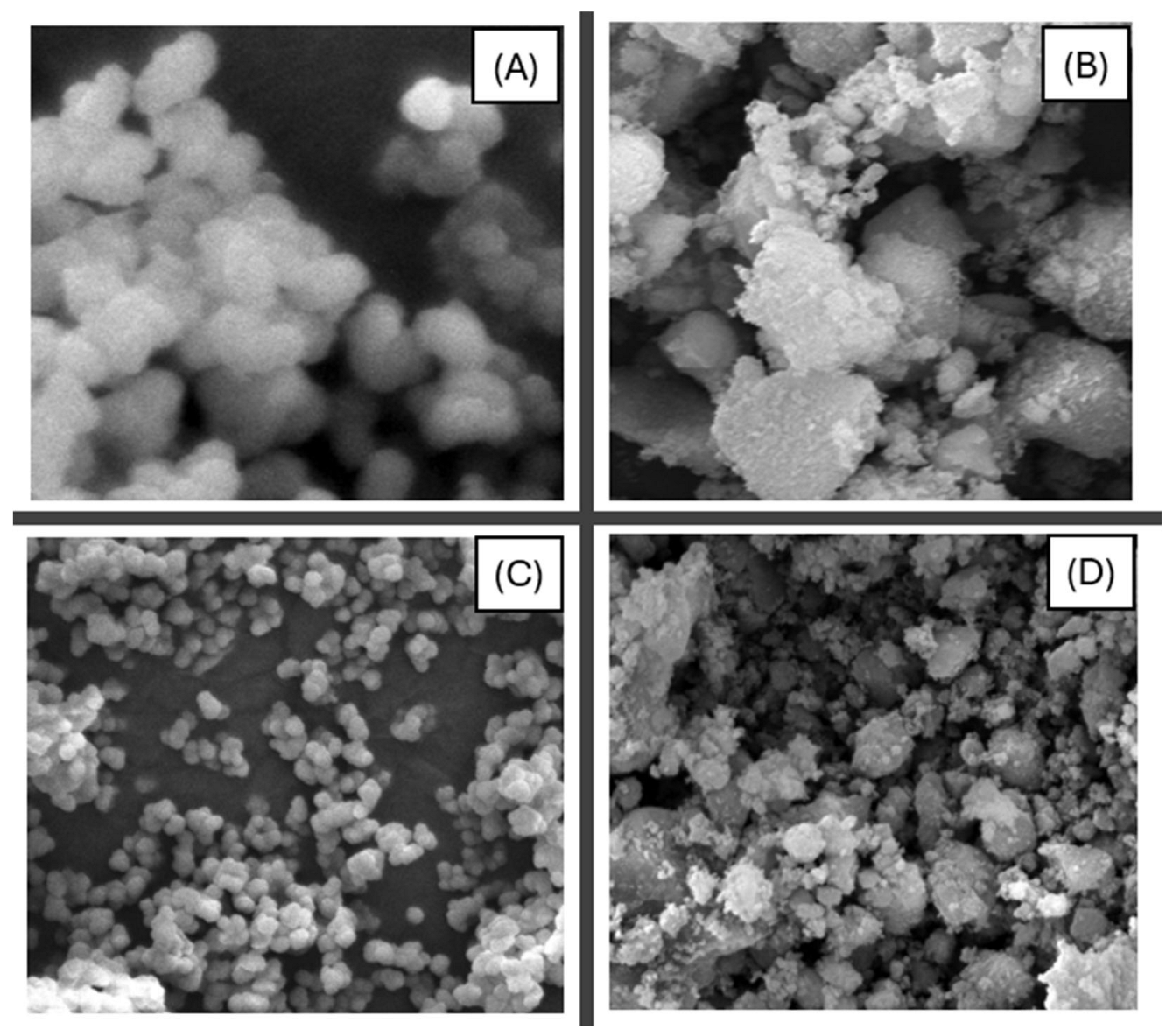
Figure 1. SEM pictures of (A) ZIF-8, (B) Cu-ZIF-8, (C) ZIF-8@HA, and (D) Cu-ZIF-8@HA nanoparticles at magnification from 1000–5000× and the scale from 10–50 μm.
Sustainability 18 02792 g001
Figure 2. Elemental Mapping of (A) ZIF-8@HA and (B) Cu-ZIF-8@HA nanoparticles at 25 and 100 μm, respectively.
Sustainability 18 02792 g002
Figure 3. Elemental Mapping of ZIF-8@HA nanoparticle with uniform spatial distribution of different elements across the surface area.
Sustainability 18 02792 g003
Figure 4. TEM images of (A) ZIF-8, (B) Cu-ZIF-8, (C) ZIF-8@HA, and (D) Cu-ZIF-8@HA nanoparticles with scale calibrations ranging from 100–200 nm.
Sustainability 18 02792 g004
Figure 5. Zeta Potential (ζ, ZP) (mV) of ZIF-8, Cu-ZIF-8, ZIF-8@HA, and Cu-ZIF-8@HA nanoparticles. a,b,c,d Means without a common subscript in the same group are significantly different (p < 0.05) from the others. Values are the average of three replications.
Sustainability 18 02792 g005
Figure 6. Particle Size (nm) of ZIF-8, Cu-ZIF-8, ZIF-8@HA, and Cu-ZIF-8@HA nanoparticles. a,b,c Means without a common subscript in the same group are significantly different (p < 0.05) from the others. Values are the average of three replications.
Sustainability 18 02792 g006
Figure 7. FTIR spectra of Cu-ZIF-8 and Cu-ZIF-8@HA nanoparticles.
Sustainability 18 02792 g007
Figure 8. Cytotoxicity test results of ZIF-8, Cu-ZIF-8, ZIF-8@HA, and Cu-ZIF-8@HA nanoparticles at different concentrations. Values are the means of three replications.
Sustainability 18 02792 g008
Figure 9. Disk diffusion test results with inhibition zone (mm) of ZIF-8, Cu-ZIF-8, ZIF-8@HA, and Cu-ZIF-8@HA against Salmonella Typhimurium ATCC14028. Values are the means of three replications. a,b,c Means within a column, which are not followed by a common superscript letter, are significantly different (p < 0.05).
Sustainability 18 02792 g009
Figure 10. Disk diffusion test results with inhibition zone (mm) of ZIF-8, Cu-ZIF-8, ZIF-8@HA, and Cu-ZIF-8@HA against Escherichia coli O157:H7. Values are the means of three replications. a,b,c,d Means within a column, which are not followed by a common superscript letter, are significantly different (p < 0.05).
Sustainability 18 02792 g010
Figure 11. SEM images of stainless-steel surfaces with (A,B) top view of the surface, and (C) side view of the coupons.
Sustainability 18 02792 g011
Figure 12. Digital images of (A) water, (B) ZIF-8, (C) Cu-ZIF-8, (D) ZIF-8-HA, and (E) Cu-ZIF-8@HA nanoparticle solutions on stainless-steel surfaces.
Sustainability 18 02792 g012
Table 1. Minimum Inhibitory Concentration (MIC) and Minimum Bactericidal Concentration (MBC) of tested nanoparticle solutions against Salmonella Typhimurium ATCC14028 after 24 h exposure.
Nanoparticle SolutionMIC Value (μg/mL)MBC Value (μg/mL)MBC/MIC Ratio
ZIF-8150060004
Cu-ZIF-875022503
ZIF-8@HA125045003.6
Cu-ZIF-8@HA50020004
Table 2. Minimum Inhibitory Concentration (MIC) and Minimum Bactericidal Concentration (MBC) of tested nanoparticle solutions against Escherichia coli O157:H7 after 24 h exposure.
Nanoparticle SolutionMIC Value (μg/mL)MBC Value (μg/mL)MBC/MIC Ratio
ZIF-8200070003.5
Cu-ZIF-8150030002.0
ZIF-8@HA125030002.4
Cu-ZIF-8@HA100025002.5
Table 3. Contact Angle of Water, ZIF-8, Cu-ZIF-8, ZIF-8@HA, and Cu-ZIF-8@HA nanoparticle solutions on stainless-steel surfaces. Values are the means of three replications. a,b,c Means within a column that are not followed by a common superscript letter are significantly different (p < 0.05).
TreatmentContact Angle (°)
Water95.80 ± 2.33 c
ZIF-815.33 ± 2.68 ab
Cu-ZIF-811.77 ± 1.42 a
ZIF-8@HA16.93 ± 1.65 ab
Cu-ZIF-8@HA18.57 ± 1.55 b
Table 4. Total Surface Free Energy [mN/m] of Water, ZIF-8, Cu-ZIF-8, ZIF-8@HA, and Cu-ZIF-8@HA nanoparticle solutions on stainless-steel surfaces. Values are the means of three replications. a,b,c,d Means within a column, which are not followed by a common superscript letter, are significantly different (p < 0.05).
TreatmentTotal Surface Free Energy [mN/m]
Water46.33 ± 1.27 a
ZIF-852.18 ± 2.39 b
Cu-ZIF-856.25 ± 1.85 bc
ZIF-8@HA59.50 ± 2.77 c
Cu-ZIF-8@HA64.42 ± 3.03 d
Table 5. Adhesion and growth inhibition of Escherichia coli O157:H7 on stainless-steel surfaces in the presence of Cu-ZIF-8@HA nanoparticles. Values are the means of three replications. a,b,c Means within a column that are not followed by a common superscript letter are significantly different (p < 0.05).
TreatmentLog CFU/cm2
Control6.86 ± 0.35 c
PBS + 5% MeOH6.14 ± 0.78 c
MIC of Cu-ZIF-8@HA4.53 ± 0.37 bc
MBC of Cu-ZIF-8@HA3.15 ± 0.42 b
2×MBC of Cu-ZIF-8@HA0.22 ± 0.09 a
Table 6. Adhesion and growth inhibition of Salmonella Typhimurium ATCC14028 on stainless-steel surfaces in the presence of Cu-ZIF-8@HA nanoparticles. Values are the means of three replications. a,b,c,d Means within a column that are not followed by a common superscript letter are significantly different (p < 0.05).
TreatmentLog CFU/cm2
Control7.02 ± 0.41 d
PBS + 5% MeOH6.45 ± 0.52 cd
MIC of Cu-ZIF-8@HA5.02 ± 0.44 c
MBC of Cu-ZIF-8@HA3.64 ± 0.38 b
2×MBC of Cu-ZIF-8@HA0.35 ± 0.11 a
Disclaimer/Publisher’s Note: The statements, opinions and data contained in all publications are solely those of the individual author(s) and contributor(s) and not of MDPI and/or the editor(s). MDPI and/or the editor(s) disclaim responsibility for any injury to people or property resulting from any ideas, methods, instructions or products referred to in the content.