Article

Repurposing Hazelnut Waste Products for a Sustainable Economy: A Metabolomic Analysis of Cuticles and Shells to Highlight Their Antioxidant Potential and Inhibitory Activity against Verocytotoxic Escherichia coli

1
Department of Veterinary Medicine and Animal Sciences—DIVAS, Università degli Studi di Milano, 26900 Lodi, Italy
2
Department of Chemical Engineering Materials & Environment, Sapienza University of Rome, Via Euossiana 18, 00184 Rome, Italy
3
Proteomics and Metabolomics Facility (ProMeFa), IRCCS San Raffaele Scientific Institute, Via Olgettina 60, 20132 Milan, Italy
*
Author to whom correspondence should be addressed.
Sustainability 2023, 15(4), 3268; https://doi.org/10.3390/su15043268
Submission received: 22 December 2022 / Revised: 7 February 2023 / Accepted: 8 February 2023 / Published: 10 February 2023

Abstract

:
This study evaluated the functional components of hazelnut waste products (cuticles and shells) for potential use in the circular economy. HPLC-HRMS and ABTS assay were used to determine the molecules of waste products with functional properties and antioxidant capacity, respectively. The antioxidant capacity of hazelnut cuticles and shells was tested using two different methods of extraction (EtOH 50/50 and acetone 40/70) by ABTS radical cation decolorization assay. The growth inhibition effect of different extracts against porcine O138 E. coli F18+ was evaluated by the microdilution bacterial growth method and confirmed with the determination of minimal inhibitory concentration (MIC). The results showed that acetone extraction produced a higher yield of metabolites compared to ethanol extraction, and the cuticles exhibited higher antioxidant and antimicrobial potential. The acetone/water extraction led to the identification of 738 signals compared to 453 detected after EtOH/water extraction. Through metabolomic analysis, it was found that the cuticles presented a much higher quantitative and qualitative polyphenolic profile than did the shells. In addition, the ABTS assay revealed that the cuticles had a higher antioxidant capacity than did the shells. Thus, we believe that hazelnut-derived waste products show a high potential for use in a circular and sustainable economy.

1. Introduction

The increasing global population has led to the need to maximize natural resources. Reducing waste and using food by-products have therefore become key aspects of the circular economy. The circular economy is defined as “a model of economics that is restorative and regenerative by design, and aims to maintain products, components, and materials at their maximum utility and value at all times, distinguishing between technical and biological cycles.” It is seen as a new business model that offers tools to improve and optimize sustainability within the Western food system [1].
The agribusiness sector offers many opportunities, and all stages of production possess the great potential to improve the sustainability and efficiency of the supply chain by minimizing upstream inputs and circulating residues/by-products downstream, where possible, using technological pathways that maximize the use of natural resources [2]. The recycling of food processing by-products, in particular, into animal feed, has long been performed to increase the sustainability of production systems. Many studies have focused on using agri-food leftovers as functional ingredients in animal diets [3,4] in order to promote the health status of animals, reduce the use of antibiotics, and reduce the environmental impact of livestock production [5,6,7]. In addition, food by-products can also be used as natural additives to increase the shelf life of food products. In fact, these by-products contain several antimicrobial compounds that can potentially be used as preservatives, ranging from enzymes, bacteriocins, fungicides, and salts to essential oils and other components [8]. The potential use of antioxidant compounds as food preservatives has also been explored, thus reducing the use of food additives such as nitrates and nitrites in the meat industry. The use of by-products can play a pivotal role in enhancing the competitiveness of the food industry for sustainable purposes, as well as from a circular economy perspective [9,10,11].
Among the possible agri-food products, hazelnuts exhibit great potential, as they are abundant and cheap, and high-added value ingredients can be obtained from their by-products. The hazelnut (Corylus avellana L.), which belongs to the Betulaceae family, is one of the most popular tree nuts consumed worldwide, ranking second in tree nut production after almonds. Hazelnuts contain several nutrients (macro and micro) with a high nutritional and functional profile. Hazelnuts are composed of seeds—the main component used in the food industry—as well as skins, or cuticles, and shells—the main waste products resulting from the hazelnut industry [12]. Hazelnut skin, which is the residual biomass of roasting, is usually used in animal feed and fertilizer or as raw material for biogas plants, while the hazelnut shell is used as a heating source during combustion. However, in recent years, from a circular economy perspective and considering that the shell represents more than 50% of the total weight of the nut, whereas the skin makes up only around 2%, these by-products have become the subject of several studies [13,14,15]. Hazelnut skin contains about 7.5% moisture, 8% protein, 14.5% fat, 1.7% ash, and 67.7% dietary fiber, which is its main constituent [16]. However, the (poly)phenolic content of the skin has gained increasing interest, especially due to its potential use in different sectors, with high-value returns.
The hazelnut shell, on the other hand, is known to contain, on average, 30.4% hemicelluloses, 26.8% cellulose, 42.9% lignin, and 3.3% other extractive matter [17]. The use of hazelnuts in agri-food industries produces tons of by-products, which given the properties just described, are far from being considered waste products. In fact, the hazelnut husk is rich in monomeric and oligomeric flavanols [18], which are a class of (poly)phenols recognized for their beneficial effects on cardiovascular and cognitive functions, which could therefore, make them a valuable dietary supplement for the nutraceutical market.
Given the current need to find food sources that are functionally rich, as well as the divided public opinion concerning the use of waste products from the food industry, the characterization of hazelnut by-products could shed light on the possible reuse of this waste product. The objective of the present study was to carry out a comprehensive chemical and functional characterization of hazelnut cuticles and shells in order to identify the metabolites they contain and to determine their feasible use in feed and food chains.

2. Materials and Methods

2.1. Sample Preparation

The extraction procedure was conducted as outlined by Yuan et al. [19], with slight modifications. Briefly, 1 g of hazelnut shells or cuticles were separately suspended in 100 mL of (1) water/acetone mixture (40/60), and (2) water/ethanol mixture (50/50). The samples were homogenized in an ultrasound bath for 2 h. After ultrasound treatment, the extracts were centrifuged for 5 min at 5000× g and 4 °C. The supernatants were collected and an aliquot was filtered and diluted (1:10) in 1 mL of initial HPLC mobile phase (95% water acidified with 0.1% HCOOH and 5% methanol). The remaining part was filtered through a 0.45 µm syringe filter and stored at −20 °C until the analysis. All samples were processed in two technical replicates.

2.2. HPLC-High Resolution Mass Spectrometry (HPLC-HRMS)

HPLC-high resolution mass spectrometry was carried out, according to the methods in a previous study [20], setting the instrumental conditions to achieve the best separation of the most important polyphenolic compounds. Briefly, a Vanquish HPLC instrument (Thermo Fisher Scientific, San Jose, CA, USA) was used for the chromatographical separation using an Restek RP column, with a programed gradient flow of 0.1% HCOOH, in water and methanol. The Exploris HRMS-Orbitrap (Thermo Scientific, San Jose, CA, USA) device was operated simultaneously in both positive mode and negative mode, using predetermined acquisition parameters for each mode. The full scan (FS) with a resolving power of 120,000 (two scan ranges of m/z 70–800 and 800–2500) was used for the screening and statistical evaluation of the chromatographic profiles. Full scan data-dependent acquisition (FS-dd-MS2) with resolving power 60,000 and 17,500 for FS and dd-MS2, respectively, was employed for the fragmentation of pseudo-molecular ions detected in the FS mode. The fragmentation of precursors was executed with stepped, normalized collision energy (NCE) set at 20, 30, and 40 eV.

2.3. HRMS Workflow Untargeted Metabolomics Approach

The detailed untargeted metabolomic workflow applied in this study is described in our recent publication [21]. Briefly, the Exploris Orbitrap raw data were submitted to Compound Discoverer (CD) 3.3 software (Thermo Fisher, Waltham, MA, USA), which enabled programmed compound identification and statistical evaluation. The criteria for the putative identification of the metabolites identified by the CD workflow were chosen as a combination of different assets, including an mzCloud match score higher than 70% and the same identification proposed by at least one external web database: the Human Metabolome platform HMDB (https://hmdb.ca/, accessed on 25 November 2022), the Kyoto Encyclopedia of Genes and Genomes (KEGG), (https://www.genome.jp/kegg, accessed on 25 November 2022), Pubchem (www.pubchem.com, accessed on 25 November 2022), the Small Molecule Pathway Database (SMPDB) (http://smpdb.ca, accessed on 28 November 2022), and Lipid Maps (https://www.lipidmaps.org, accessed on 28 November 2022). If the mass fragmentation pattern did not correspond to any of the databases, the fragmentation pattern was manually verified using ChemDrow software or by comparing it with recent publications [22]. Differential analysis was performed as part of the CD workflow, and the statistical evaluation was expressed in hierarchical cluster processing and a volcano plot graph.

2.4. Evaluation of Antioxidant Properties (ABTS Assay)

The antioxidant activity was tested using an ABTS assay, according to the methods of Dell’Anno et al. [23]. Briefly, 10 µL of the diluted sample were added to 1 mL of ABTS•+ working solution. The absorbance was recorded after 6 min of incubation in the dark, and all determinations were performed in triplicate.
In order to obtain different dilutions for testing, the extracts were diluted in their solvent, the water/acetone mixture (40/60), or the water/ethanol mixture (50/50), respectively. Concerning the shell extracts, the samples were tested, i.e., as is, 1:2, 1:4, and 1:8. On the other hand, for the cuticle extracts, the samples were tested in the following ratios: 1:16, 1:32, 1:64, and 1:100.
The total antioxidant capacity was expressed as the percentage of the inhibition of radical scavenging activity (PI%), according to the following equation:
PI   % = ( AbsABTS + Abs   Sample / AbsABTS + ) 100
AbsABTS•+ denotes the initial absorbance of diluted ABTS•+, and the Abs sample denotes the absorbance of the sample after 6 min of reaction. All assays were performed in technical triplicate and with two biological replicates aimed at verifying the replicability of the experiment using the same procedures, which included repeating the experiment starting with the sample extraction and repeating the test on different days.

2.5. Growth Inhibition Assay against Escherichia coli

A growth inhibition assay was performed against the O138 E. coli strain. Briefly, extracts of hazelnut shells and hazelnut cuticles were filtered with a 0.22 µm syringe filter and stored at −20 °C until the analysis. According to the methods of Reggi et al. [24], a liquid culture-based growth inhibition assay against E. coli O138 was performed to evaluate the ability of hazelnut shell and hazelnut cuticle extracts to inhibit bacterial growth. Briefly, samples were diluted in Luria–Bertani (LB) broth in order to obtain four different concentrations (1:4; 1:8; 1:16; 1:32). A total of 100 µL of the diluted extracts was added in co-culture with 30 µL of E. coli inoculum. The negative controls were used to correct the background color. All samples were then incubated at 37 °C in a shaking incubator for 6 h. The growth rate of E. coli was estimated every hour for 6 h by measuring the absorbance with a microplate reader spectrophotometer (ScanReady P-800, Life Real, Hangzhou, China) at an optical density (OD) of 620 nm. The measured OD was converted into log10 of the number of cells/mL, considering 1 OD = 1 × 10⁹ cells/mL [25]. All assays were performed in technical quadruplicate and with three biological replicates.

2.6. Determination of Minimal Inhibitory Concentration (MICs)

Minimum inhibitory concentrations (MICs) were determined through broth microdilution method. Briefly, different concentrations (35 mg/mL, 25 mg/mL, 10 mg/mL, 5 mg/mL, 1 mg/mL, 0.5 mg/mL, 0.1 mg/mL, and 0 mg/mL) of hazelnut cuticle and shell extracts were prepared by diluting the dried extracts with LB broth. A total of 100 µL of the different concentrations were plated in a 96-well microplate, and 10 µL of an overnight culture of E. coli O138 in Luria–Bertani (LB) broth (approximately 106 CFU/mL) was inoculated in each well of the plate, except for the blank and negative control (CTRL-) wells, and incubated at 37 °C for 20 h. Bacterial growth was determined by the change in absorbance after reading the microplates at 620 nm in a microplate reader spectrophotometer (ScanReady P-800, Life Real, Hangzhou, China). The inhibition rate was estimated by the following formula:
I n h i b i t i o n   R a t e   % = 100 O D C T R L O D s a m p l e O D C T R L O D B l a n k
The MIC was defined as the lowest extract concentration that did not produce turbidity when compared with positive control (0 mg of extract/mL) [26]. The experiment was performed in technical triplicate and with two biological replicates.

2.7. Statistical Analysis

All the data were analyzed using GraphPad Prism software (Version 9.0.0). Statistical analysis was performed after the evaluation of the normal distribution of data through Shapiro–Wilk and D’Agostino–Pearson tests. Concerning data obtained from the assays of antioxidant activity and bacterial growth inhibition, a two-way analysis of variance (ANOVA) was performed, evaluating the effect of treatment, time, and their interaction with each other. Post hoc pairwise comparisons were performed using the Bonferroni–Sidak test. Data were reported as mean ± standard error, and differences were considered statistically significant for p < 0.05.

3. Results and Discussion

Given the current importance of the use of agro-industrial by-products in economic terms, enabling industries to reduce the disposal costs of waste biomass, and livestock farmers to mitigate animal feed costs, this study focused on the metabolomic characterization and in vitro evaluation of the functional properties of hazelnut shells and cuticles in order to establish their further use as functional additives.

3.1. Evaluation of Molecules with Antioxidant Properties

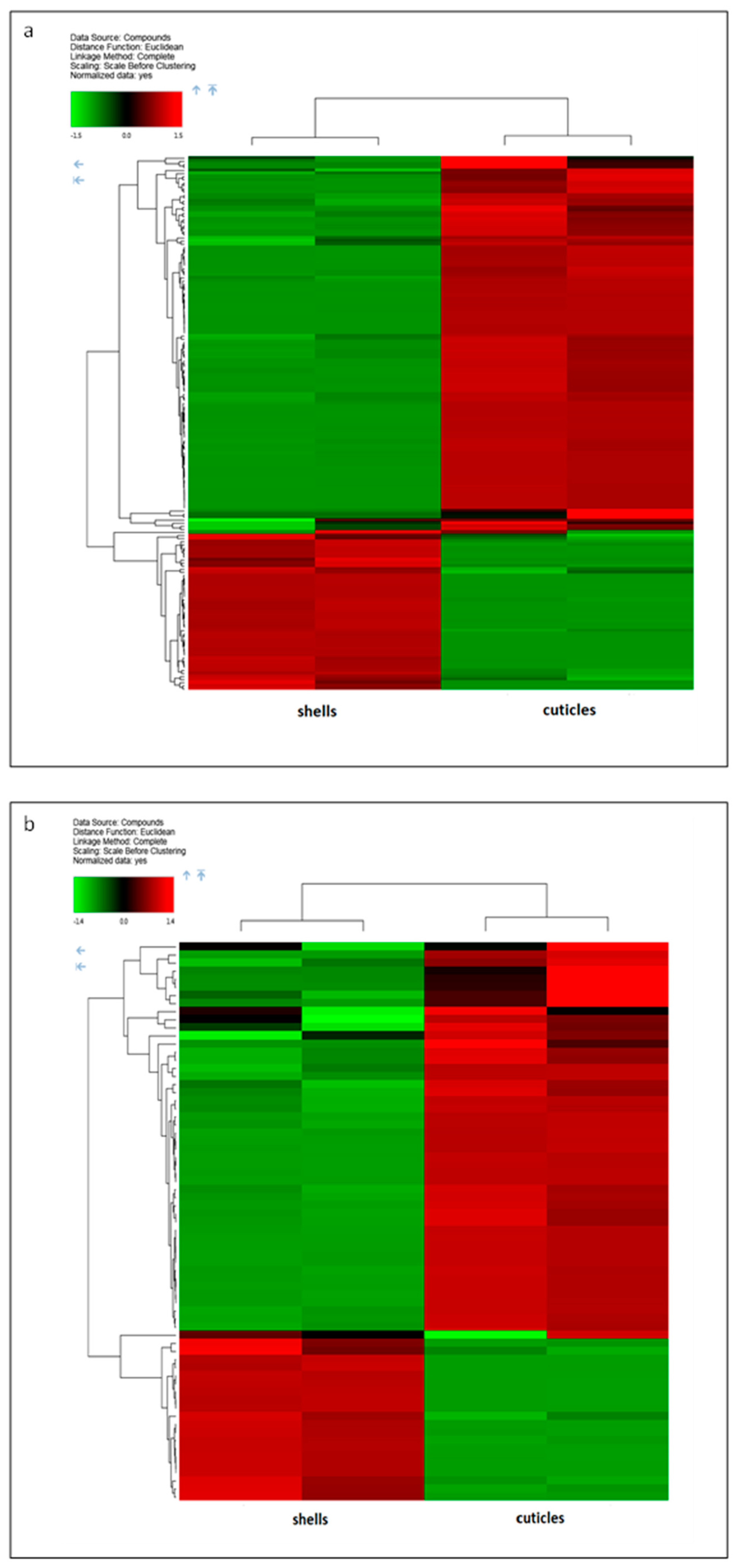
In this study, an HRMS-based metabolomics investigation was conducted for the first time in both polarization modalities (positive and negative) synchronously, within the same analytical run. A reliable, sensitive, and double-confirmed identification of key polyphenolic metabolites in complex matrices of hazelnut shells and cuticles was thus achieved. For the acetone/water extract, a total of 738 signals were obtained (Figure 1a). The most abundant polyphenolic/antioxidant/natural products (165 structures), detected with a high level of confirmation certainty, are presented in Figure 2a and Table 1. On the other hand, the ethanol/water extract showed a reduced metabolomic profile, with 453 signals (Figure 1b), of which only 67 were considered as important differentiators (Figure 2b and Table 2).
The metabolomic profiles of the hazelnut shells and cuticles were significantly different in both extracts. The profile of the acetone/water extract was much more varied than the profile obtained from the ethanol/water solvent. The acetone/water hazelnut shell extract showed a high amount of specific flavonoids, polyphenolic acids, and sesquiterpenes: 5,6,7,3’,4’-pentamethoxyflavanone, coniferyl ferulate, scopoletin acetate, brosimacutin H and A, 5,6,7-Trimethoxy-2H-chromen-2-one, chalconaringenin 2’-xyloside, Hesperetin, 4,2’,6’-Trihydroxy-4’-methoxy-3’,5’-dimethyldihydrochalcone, cnicin, spicatin, etc. On the other hand, the cuticles were characterized by higher levels of amino acids, upregulated compounds from the chalcone group, higher amounts of most hydrolyzable tannins, and a higher relative quantity of the entire proanthocyanidins class that belongs to the group of condensed tannins [27].
The ethanol/water treatment was not as qualitatively efficient as the acetone/water extraction, although the metabolomic profiles were similar. Some important compounds identified in the acetone/water extract (listed above) were also found in the ethanol/water extract. The particular characteristic of ethanol extract was the appearance of oxidated fatty acids (9,12,13-Trihydroxy-15-octadecenoic acid and 13-Hydroperoxylinoleic acid). Moreover, the very intensive signal of didodecyl-3,3-thiodipropionate (DLTDP) found exclusively in the ethanol/water extract of both the shells and cuticles requires further clarification.
The aqueous acetone solution extracted significantly higher amounts of phenolics, with a higher antioxidant capacity than aqueous ethanol. This may be due to the fact that the leftover hazelnut material exhibits a particular matrix due to its rigid and compact inner structure, which slowed down the solvent penetration. Acetone has a lower viscosity than ethanol, which evidently encourages the diffusion of the solvent into the shell cavitation or interior cuticles. This process notably increases the yield of the extracted polyphenolic material.
The cuticles generally presented a better qualitative and quantitative polyphenolic profile than did the shells. Special attention should therefore be paid to these hazelnut by-products in terms of the possible accumulation of antioxidant compounds in the cuticle membranes, which to date, has not been studied in detail.

3.2. Evaluation of Functional Properties

The results of the functional activity assays of the hazelnut cuticles and shells revealed the antioxidant and antimicrobial properties of both by-products. These activities are likely to be related to the high amounts of secondary compounds, especially phenols, condensed tannins, and vitamins [28,29]. Given their nutritional and functional value, by-products of the hazelnut industry could be successfully used as supplements in the field of animal nutrition. In fact, as reported in other studies, the biologically active compounds in these products can also be used to enrich animal diets, thereby improving the nutritional value and quality of meat and milk [30,31].

3.2.1. Antioxidant Activity

The antioxidant properties were evaluated after 6 min of reaction between ABTS•+ and the shell or cuticle extracts, based on the timing defined for the radical cation decolorization assay [32,33]. This study was performed using two different types of extraction solvents: water/acetone mixture (40/60) and water/ethanol mixture (50/50).
For both skins (Figure 3a) and shells (Figure 3b), significant differences (p < 0.05) were highlighted between the two extraction solvents for each dilution, excluding the first one. For both matrices, the extraction with acetone (40/60) was more effective, leading to an approximately 10% higher extraction of antioxidant compounds, than that obtained from the water/ethanol extraction (Supplementary Table S1). This result also been shown in previous studies, in which the acetone extraction led to a better extraction of both polyphenolic and phytosterol compounds from the plant matrix [34,35,36]. This is because the extraction of phenolic compounds, which are the main factors responsible for antioxidant activity, is closely related to both the solvent used and the temperature chosen for extraction [36]. In fact, extraction is the main step in the recovery and isolation of bioactive compounds in the sample under analysis. The extraction process is influenced by the nature of the compounds themselves, the methodology used, and the particle size of the sample, as well as the presence of interfering substances [37]. The solvent used plays a key role during the extraction, as it is strongly related to the solubility of the phenolic compounds [12]. For a solvent to be optimal for phenol extraction, it needs to be able to extract the maximum number of phenols and the minimum number of foreign substances [36]. Among the different solvates used for the extraction of phenolic compounds, several studies [35,36] have shown that acetone yields the best results for hazelnut shell and husk waste. This could also be partly explained by the fact that acetone has a low viscosity (0.306 cP at 25 °C), which would increase the concentration gradient, making the extraction more effective [38].
In any case, for both solvent mixtures, a dose-dependent effect was observed. In fact, at the highest concentration, a 90% inhibition of radical scavenging activity was found, which decreased as the concentration decreased, reaching values of around 25% at the lowest concentration tested (Figure 3).

3.2.2. Evaluation of the Growth Inhibitory Activity against Verocytotoxic Escherichia coli and Determination of Minimal Inhibitory Concentrations (MICs)

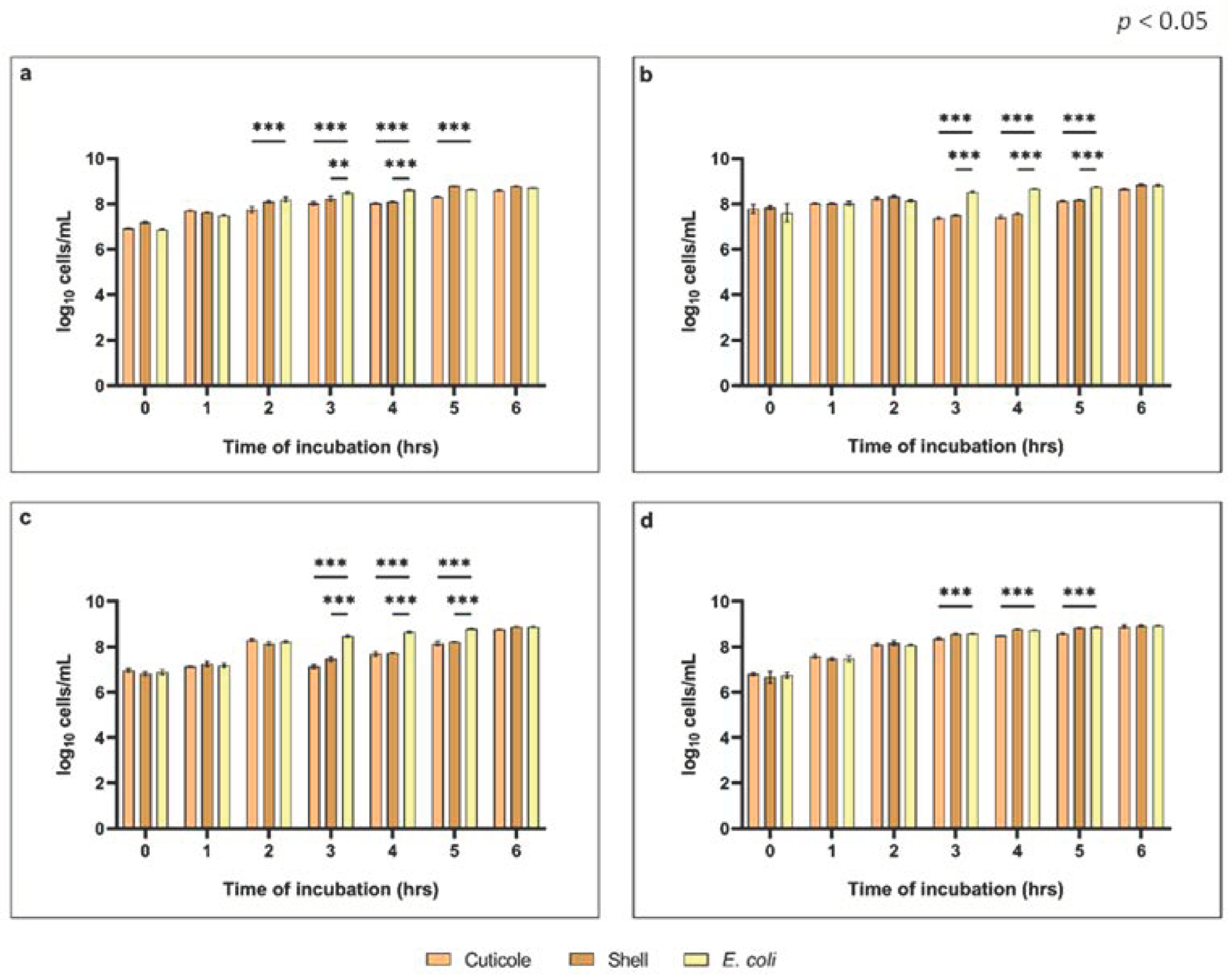
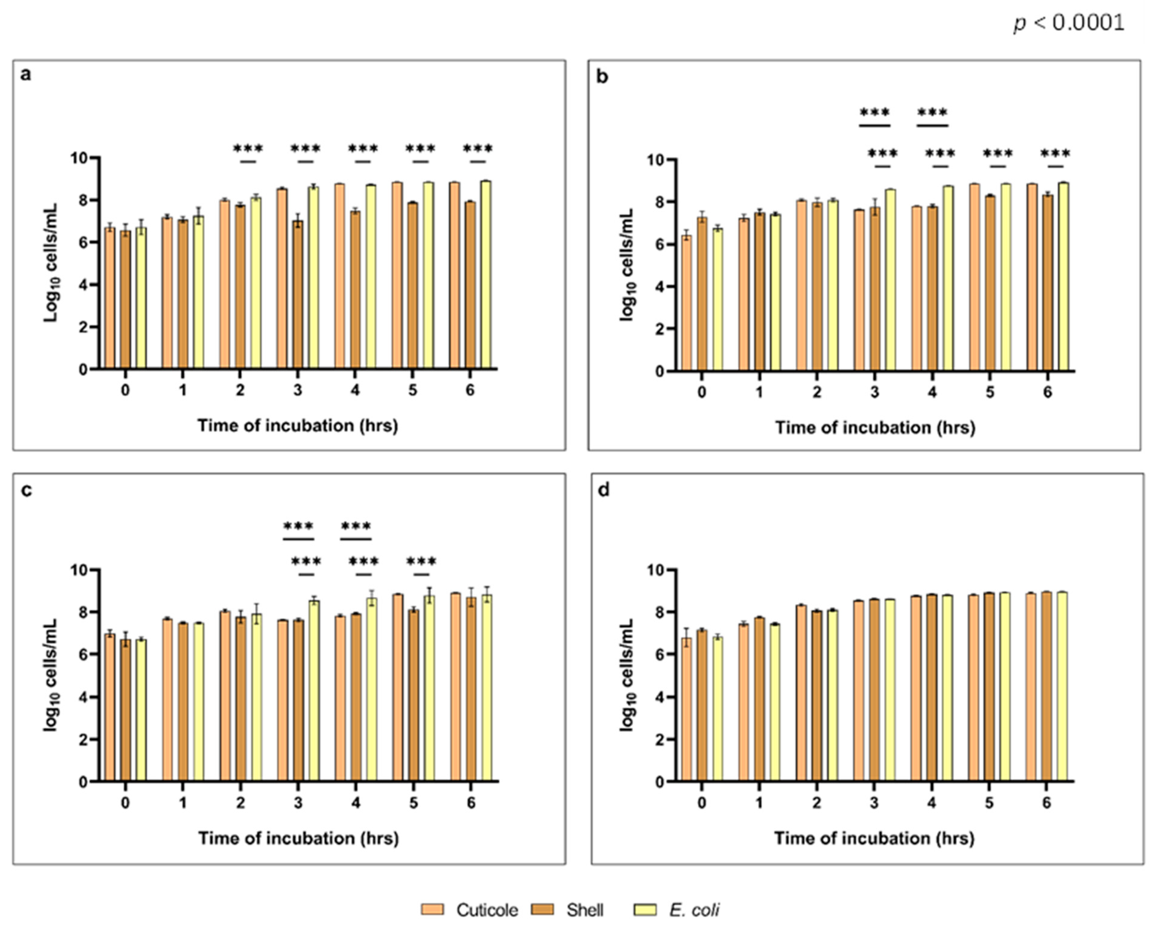
Due to the growing problem of antibiotic resistance, there has been increasing interest in natural antimicrobial compounds. Several studies on hazelnuts and their derivatives have been conducted aimed at evaluating their antimicrobial power [39]. In this study, antimicrobial properties were evaluated considering the inhibitory activity against an Escherichia coli strain from our strain library (O138 E. coli F18+). The growth inhibition activity against O138 E. coli F18+ was evaluated for each extract, considering different dilutions (1:4; 1:8; 1:16; 1:32). Both solvents led to the extraction of bioactive compounds showing antimicrobial activity at dilutions of 1:4, 1:8, and 1:16 (Figure 4 and Figure 5). At a dilution of 1:4, the use of EtOH (50/50) as an extraction solvent highlighted the growth inhibition of E. coli for both cuticle and shell matrices (Figure 4a), while at the same dilution, the acetone (40/60) only showed the antimicrobial capacity of the shell extract (Figure 5a). For the next two dilutions, the evaluation of E. coli growth inhibition was significant (p < 0.0001) for both the cuticle and shell extracts, regardless of whether they were produced using methanol (50/50) or acetone (40/60), particularly after 3 and 4 h of incubation (Figure 4b,c and Figure 5b,c). In any case, the data obtained from the growth inhibition assay showed that the use of acetone led to better extraction of the bioactive compounds from the cuticles of the hazelnuts, compared to the husks. On the other hand, while allowing for more growth of E. coli, the use of ethanol showed more evidence of the antimicrobial capabilities of both the cuticles and the shells (Figure 4). For the latter, in fact, extraction in ethanol led to the detection of a significantly lower cell/mL (log10) value, even for a dilution of 1:32 (Figure 4d). The results obtained from the growth inhibition assay showed that both cuticles and shells revealed an antimicrobial effect, probably related to some of their components, such as tannins and phenols [40,41]. Further studies are needed, however, to identify the correlation between the solvent used for extraction and the extracted matrix in order to more accurately identify the extractive power of different solvents.
In addition, a time-dependent effect in relation to inhibitory activity was observed. In fact, the assay revealed that in the first few hours, the inhibition abilities of the tested extracts were comparable to the positive control (E. coli). Only after at least 2 h did the difference in inhibition ability between the tested compounds and the positive control become significant, reaching a maximum inhibitory effect at 3 and 4 h of incubation time (Figure 4 and Figure 5). For the ethanol extracts, as shown in Figure 4, regardless of the dilution, after 5 h of incubation, the inhibitory activity was similar between the tested extracts and the positive control (E. coli). This suggests the exhaustion of bioactivity due to the degradation of bioactive compounds within the extract tested, or due to the development of bacterial resistance [42]. In any case, it is important to consider the transit time of the feed in the gastrointestinal tract of animals, which, for example in swine lasts for 4 h, thus guaranteeing the effect throughout the digestive tract.
Data obtained by the evaluation of the growth inhibition of E. coli were confirmed by the determination of the minimal inhibitory concentrations (MICs). As shown in Table 3, extracts in methanol are found to show an inhibitory capacity at lower concentrations than are the extracts in acetone. The lowest inhibitory concentrations determined reveal an inhibition rate of 96% for the extracts derived from hazelnut cuticles (96.56% for the extract in methanol and 95.57% for the extract in acetone), while the shell extracts show an inhibition rate of 53% (54.86% and 52.21%, respectively).

4. Conclusions

Given the growing need to maximize the use of natural resources, waste products are currently playing a prominent role. However, given the wide variety of waste products created within the food industry, it is necessary to evaluate them nutritionally and functionally in order to determine which ones can be efficiently introduced within a circular economy approach, for example, employing them as functional ingredients in both food and feed, but also employing them as useful substances for extending the shelf life of food products. Agro-food waste derived from hazelnuts has been shown to have important potential for multiple applications, given its high nutritional value. In this study metabolomic analyses highlighted the richness of the cuticles and shells in terms of biologically active compounds, thus making them a rich source of natural antioxidants and antimicrobial compounds. The cuticles presented a qualitatively and quantitatively higher polyphenolic profile than did the shells. This was also reflected in the in vitro evaluation of both their antioxidant and antimicrobial activities. In fact, although both cuticles and shells showed both antioxidant and antimicrobial capacities, these was significantly greater in the cuticles than in the shells. Special attention should thus be paid to these hazelnut by-products in relation to the possible accumulation of antioxidant compounds in the cuticle membrane. This study also highlights the role of the extraction solvent used. The aqueous acetone solution extracted significantly higher amounts of phenolics, with higher antioxidant and antimicrobial capacities, than did the aqueous ethanol. Additional in vitro studies will be required to further investigate the use of different extraction solvents. Moreover, preliminary studies should also investigate their possible in vivo use for animal and human nutrition.

Supplementary Materials

The following supporting information can be downloaded at: https://www.mdpi.com/article/10.3390/su15043268/s1, Table S1: Percentage inhibition of the radical scavenging activity of hazelnut cuticles and shells.

Author Contributions

Conceptualization, L.R. and S.P.; methodology, S.F., A.Z., S.P. and R.P.; investigation, S.F., A.Z., S.P. and R.P.; data curation, S.F. and S.P.; writing—original draft preparation, S.F. and S.P.; writing—review and editing, S.F., S.P. and L.R.; visualization, S.F. and S.P.; supervision, S.P., L.R. and C.A.S.R. All authors have read and agreed to the published version of the manuscript.

Funding

This research was funded by “Efficacy study of oral feed additive products; founding number: CTE_INT17ABALD_01”. The APC was funded by the University of Milan.

Data Availability Statement

The data presented in this study are available in article.

Acknowledgments

The authors would like to thank Simlab s.r.l., (SIMIC s.p.a. Spin-Off Research and Development Center for Industrial Innovation) as promoters of the project for the recovery and reuse of by-products of the food industry. The authors acknowledge the support of the APC central fund of the University of Milan.

Conflicts of Interest

The authors declare no conflict of interest.

References

  1. Ellen MacArthur Circular Economy. Available online: https://ellenmacarthurfoundation.org/topics/circular-economy-introduction/overview (accessed on 21 December 2022).
  2. McCarthy, B.; Kapetanaki, A.B.; Wang, P. Circular Agri-Food Approaches: Will Consumers Buy Novel Products Made from Vegetable Waste? Rural. Soc. 2019, 28, 91–107. [Google Scholar] [CrossRef]
  3. Ben-Othman, S.; Jõudu, I.; Bhat, R. Bioactives from Agri-Food Wastes: Present Insights and Future Challenges. Molecules 2020, 25, 510. [Google Scholar] [CrossRef] [PubMed]
  4. Melini, V.; Melini, F.; Luziatelli, F.; Ruzzi, M. Functional Ingredients from Agri-Food Waste: Effect of Inclusion Thereof on Phenolic Compound Content and Bioaccessibility in Bakery Products. Antioxidants 2020, 9, 1216. [Google Scholar] [CrossRef] [PubMed]
  5. Salami, S.A.; Luciano, G.; O’Grady, M.N.; Biondi, L.; Newbold, C.J.; Kerry, J.P.; Priolo, A. Sustainability of Feeding Plant By-Products: A Review of the Implications for Ruminant Meat Production. Anim. Feed Sci. Technol. 2019, 251, 37–55. [Google Scholar] [CrossRef]
  6. Caprarulo, V.; Giromini, C.; Rossi, L. Review: Chestnut and Quebracho Tannins in Pig Nutrition: The Effects on Performance and Intestinal Health. Animal 2021, 15, 100064. [Google Scholar] [CrossRef] [PubMed]
  7. Dell’Anno, M.; Reggi, S.; Caprarulo, V.; Hejna, M.; Sgoifo Rossi, C.; Callegari, M.; Baldi, A.; Rossi, L. Evaluation of Tannin Extracts, Leonardite and Tributyrin Supplementation on Diarrhoea Incidence and Gut Microbiota of Weaned Piglets. Animals 2021, 11, 1693. [Google Scholar] [CrossRef]
  8. Carocho, M.; Morales, P.; Ferreira, I.C.F.R. Natural Food Additives: Quo Vadis? Trends Food Sci. Technol. 2015, 45, 284–295. [Google Scholar] [CrossRef]
  9. Nikmaram, N.; Budaraju, S.; Barba, F.J.; Lorenzo, J.M.; Cox, R.B.; Mallikarjunan, K.; Roohinejad, S. Application of Plant Extracts to Improve the Shelf-Life, Nutritional and Health-Related Properties of Ready-to-Eat Meat Products. Meat. Sci. 2018, 145, 245–255. [Google Scholar] [CrossRef]
  10. Ribeiro, J.S.; Santos, M.J.M.C.; Silva, L.K.R.; Pereira, L.C.L.; Santos, I.A.; da Silva Lannes, S.C.; da Silva, M.V. Natural Antioxidants Used in Meat Products: A Brief Review. Meat. Sci. 2019, 148, 181–188. [Google Scholar] [CrossRef]
  11. Bellucci, E.R.B.; Bis-Souza, C.V.; Domínguez, R.; Bermúdez, R.; Barretto, A.C. da S. Addition of Natural Extracts with Antioxidant Function to Preserve the Quality of Meat Products. Biomolecules 2022, 12, 1506. [Google Scholar] [CrossRef]
  12. Shahidi, F.; Naczk, M. Phenolics in Food and Nutraceuticals; CRC Press: Boca Raton, FL, USA, 2003; ISBN 9780429211645. [Google Scholar]
  13. Lelli, V.; Molinari, R.; Merendino, N.; Timperio, A.M. Detection and Comparison of Bioactive Compounds in Different Extracts of Two Hazelnut Skin Varieties, Tonda Gentile Romana and Tonda Di Giffoni, Using a Metabolomics Approach. Metabolites 2021, 11, 296. [Google Scholar] [CrossRef]
  14. Bener, M.; Şen, F.B.; Önem, A.N.; Bekdeşer, B.; Çelik, S.E.; Lalikoglu, M.; Aşçı, Y.S.; Capanoglu, E.; Apak, R. Microwave-Assisted Extraction of Antioxidant Compounds from by-Products of Turkish Hazelnut (Corylus avellana L.) Using Natural Deep Eutectic Solvents: Modeling, Optimization and Phenolic Characterization. Food Chem. 2022, 385, 132633. [Google Scholar] [CrossRef] [PubMed]
  15. Salem, M.A.; Aborehab, N.M.; Al-Karmalawy, A.A.; Fernie, A.R.; Alseekh, S.; Ezzat, S.M. Potential Valorization of Edible Nuts By-Products: Exploring the Immune-Modulatory and Antioxidants Effects of Selected Nut Shells Extracts in Relation to Their Metabolic Profiles. Antioxidants 2022, 11, 462. [Google Scholar] [CrossRef]
  16. Özdemir, K.S.; Yılmaz, C.; Durmaz, G.; Gökmen, V. Hazelnut Skin Powder: A New Brown Colored Functional Ingredient. Food Res. Int. 2014, 65, 291–297. [Google Scholar] [CrossRef]
  17. Demirbas, A. Furfural Production from Fruit Shells by Acid-Catalyzed Hydrolysis. Energy Sources Part A Recovery Util. Environ. Eff. 2006, 28, 157–165. [Google Scholar] [CrossRef]
  18. del Rio, D.; Calani, L.; Dall’Asta, M.; Brighenti, F. Polyphenolic Composition of Hazelnut Skin. J. Agric. Food Chem. 2011, 59, 9935–9941. [Google Scholar] [CrossRef]
  19. Yuan, B.; Lu, M.; Eskridge, K.M.; Hanna, M.A. Valorization of Hazelnut Shells into Natural Antioxidants by Ultrasound-Assisted Extraction: Process Optimization and Phenolic Composition Identification. J. Food Process Eng. 2018, 41, e12692. [Google Scholar] [CrossRef]
  20. Frazzini, S.; Scaglia, E.; Dell’Anno, M.; Reggi, S.; Panseri, S.; Giromini, C.; Lanzoni, D.; Sgoifo Rossi, C.A.; Rossi, L. Antioxidant and Antimicrobial Activity of Algal and Cyanobacterial Extracts: An In Vitro Study. Antioxidants 2022, 11, 992. [Google Scholar] [CrossRef]
  21. Leoni, V.; Giupponi, L.; Pavlovic, R.; Gianoncelli, C.; Cecati, F.; Ranzato, E.; Martinotti, S.; Pedrali, D.; Giorgi, A.; Panseri, S. Multidisciplinary Analysis of Italian Alpine Wildflower Honey Reveals Criticalities, Diversity and Value. Sci. Rep. 2021, 11, 19316. [Google Scholar] [CrossRef]
  22. Cerulli, A.; Napolitano, A.; Masullo, M.; Hošek, J.; Pizza, C.; Piacente, S. Chestnut Shells (Italian Cultivar “Marrone Di Roccadaspide” PGI): Antioxidant Activity and Chemical Investigation with in Depth LC-HRMS/MSn Rationalization of Tannins. Food Res. Int. 2020, 129, 108787. [Google Scholar] [CrossRef] [PubMed]
  23. Dell’Anno, M.; Sotira, S.; Rebucci, R.; Reggi, S.; Castiglioni, B.; Rossi, L. In Vitro Evaluation of Antimicrobial and Antioxidant Activities of Algal Extracts. Ital. J. Anim. Sci. 2020, 19, 103–113. [Google Scholar] [CrossRef]
  24. Reggi, S.; Giromini, C.; Dell’Anno, M.; Baldi, A.; Rebucci, R.; Rossi, L. In Vitro Digestion of Chestnut and Quebracho Tannin Extracts: Antimicrobial Effect, Antioxidant Capacity and Cytomodulatory Activity in Swine Intestinal IPEC-J2 Cells. Animals 2020, 10, 195. [Google Scholar] [CrossRef] [PubMed]
  25. Myers, J.A.; Curtis, B.S.; Curtis, W.R. Improving Accuracy of Cell and Chromophore Concentration Measurements Using Optical Density. BMC Biophys. 2013, 6, 4. [Google Scholar] [CrossRef] [PubMed]
  26. Collins, C.H.; Lyne, P.M.; Grange, J. Collins and Lyne’s Microbiological Methods; Butterworth-Heinemann: Oxford, UK, 1995; Volume 493. [Google Scholar]
  27. Smeriglio, A.; Barreca, D.; Bellocco, E.; Trombetta, D. Proanthocyanidins and Hydrolysable Tannins: Occurrence, Dietary Intake and Pharmacological Effects. Br. J. Pharmacol. 2017, 174, 1244–1262. [Google Scholar] [CrossRef]
  28. Taş, N.G.; Gökmen, V. Bioactive Compounds in Different Hazelnut Varieties and Their Skins. J. Food Compos. Anal. 2015, 43, 203–208. [Google Scholar] [CrossRef]
  29. Masullo, M.; Cerulli, A.; Mari, A.; de Souza Santos, C.C.; Pizza, C.; Piacente, S. LC-MS Profiling Highlights Hazelnut (Nocciola Di Giffoni PGI) Shells as a Byproduct Rich in Antioxidant Phenolics. Food Res. Int. 2017, 101, 180–187. [Google Scholar] [CrossRef] [PubMed]
  30. Caccamo, M.; Valenti, B.; Luciano, G.; Priolo, A.; Rapisarda, T.; Belvedere, G.; Marino, V.M.; Esposto, S.; Taticchi, A.; Servili, M.; et al. Hazelnut as Ingredient in Dairy Sheep Diet: Effect on Sensory and Volatile Profile of Cheese. Front. Nutr. 2019, 6, 125. [Google Scholar] [CrossRef]
  31. Natalello, A.; Luciano, G.; Morbidini, L.; Valenti, B.; Pauselli, M.; Frutos, P.; Biondi, L.; Rufino-Moya, P.J.; Lanza, M.; Priolo, A. Effect of Feeding Pomegranate Byproduct on Fatty Acid Composition of Ruminal Digesta, Liver, and Muscle in Lambs. J. Agric. Food Chem. 2019, 67, 4472–4482. [Google Scholar] [CrossRef]
  32. Dong, J.-W.; Cai, L.; Xing, Y.; Yu, J.; Ding, Z.-T. Re-Evaluation of ABTS•+ Assay for Total Antioxidant Capacity of Natural Products. Nat. Prod. Commun. 2015, 10, 1934578X1501001. [Google Scholar] [CrossRef]
  33. Xie, J.; Schaich, K.M. Re-Evaluation of the 2,2-Diphenyl-1-Picrylhydrazyl Free Radical (DPPH) Assay for Antioxidant Activity. J. Agric. Food Chem. 2014, 62, 4251–4260. [Google Scholar] [CrossRef] [PubMed]
  34. Rusu, M.E.; Fizeșan, I.; Pop, A.; Gheldiu, A.-M.; Mocan, A.; Crișan, G.; Vlase, L.; Loghin, F.; Popa, D.-S.; Tomuta, I. Enhanced Recovery of Antioxidant Compounds from Hazelnut (Corylus avellana L.) Involucre Based on Extraction Optimization: Phytochemical Profile and Biological Activities. Antioxidants 2019, 8, 460. [Google Scholar] [CrossRef] [PubMed]
  35. Daniela, G.; Simona, P.; Giuseppe, Z.; Vincenzo, G. Phenolic Acid Profile and Antioxidant Capacity of Hazelnut (Corylus avellana L.) Kernels in Different Solvent Systems. J. Food Nutr. Res. 2010, 49, 195–205. [Google Scholar]
  36. Contini, M.; Baccelloni, S.; Massantini, R.; Anelli, G. Extraction of Natural Antioxidants from Hazelnut (Corylus avellana L.) Shell and Skin Wastes by Long Maceration at Room Temperature. Food Chem. 2008, 110, 659–669. [Google Scholar] [CrossRef]
  37. Stalikas, C.D. Extraction, Separation, and Detection Methods for Phenolic Acids and Flavonoids. J. Sep. Sci. 2007, 30, 3268–3295. [Google Scholar] [CrossRef]
  38. Cacace, J.E.; Mazza, G. Extraction of Anthocyanins and Other Phenolics from Black Currants with Sulfured Water. J. Agric. Food Chem. 2002, 50, 5939–5946. [Google Scholar] [CrossRef]
  39. Pereira, J.A.; Oliveira, I.; Sousa, A.; Ferreira, I.C.F.R.; Bento, A.; Estevinho, L. Bioactive Properties and Chemical Composition of Six Walnut (Juglans regia L.) Cultivars. Food Chem. Toxicol. 2008, 46, 2103–2111. [Google Scholar] [CrossRef]
  40. Fraga-Corral, M.; Otero, P.; Cassani, L.; Echave, J.; Garcia-Oliveira, P.; Carpena, M.; Chamorro, F.; Lourenço-Lopes, C.; Prieto, M.A.; Simal-Gandara, J. Traditional Applications of Tannin Rich Extracts Supported by Scientific Data: Chemical Composition, Bioavailability and Bioaccessibility. Foods 2021, 10, 251. [Google Scholar] [CrossRef]
  41. Kaczmarek, B. Tannic Acid with Antiviral and Antibacterial Activity as A Promising Component of Biomaterials—A Minireview. Materials 2020, 13, 3224. [Google Scholar] [CrossRef]
  42. Zoetendal, E.G.; Smith, A.H.; Sundset, M.A.; Mackie, R.I. The BaeSR Two-Component Regulatory System Mediates Resistance to Condensed Tannins in Escherichia coli. Appl. Environ. Microbiol. 2008, 74, 535–539. [Google Scholar] [CrossRef] [Green Version]
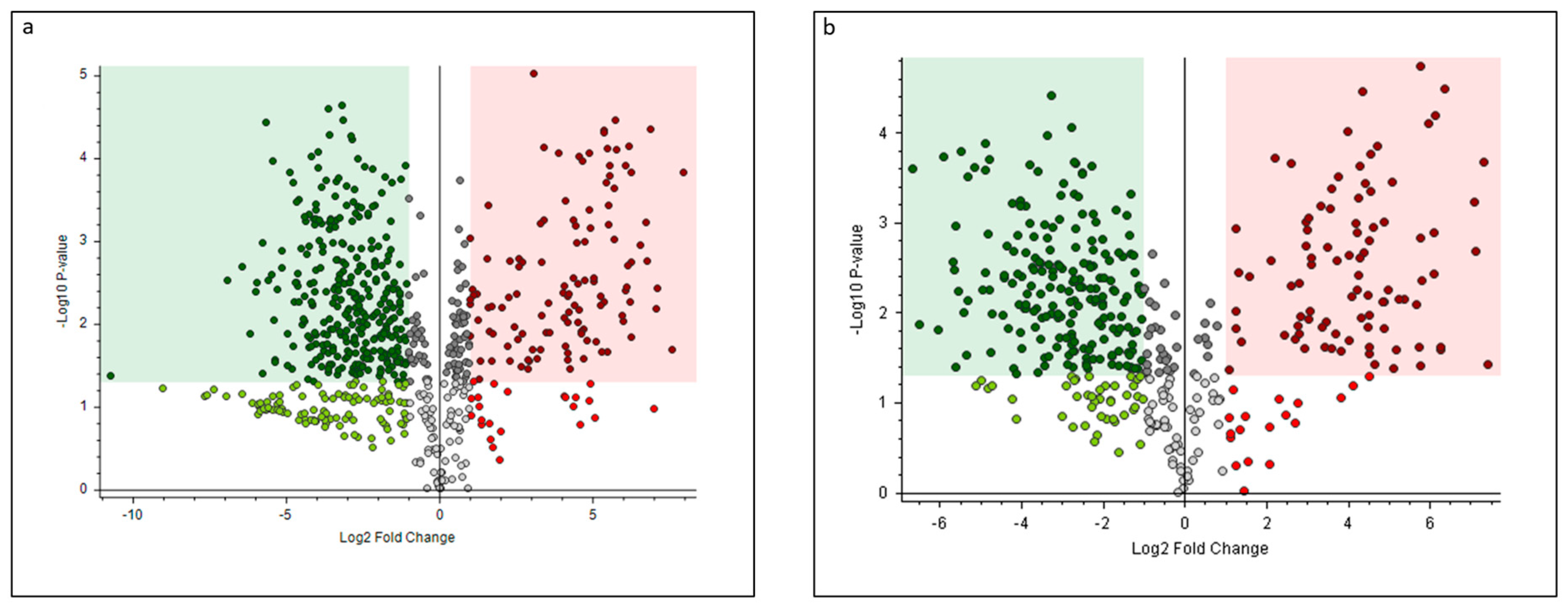
Figure 1. Volcano plot comparison between the relative intensity revealed in the hazelnut shell and cuticle extracts: (a) chromatographic peak of 738 compounds in acetone/water solvent; (b) chromatographic peak of 453 compounds in ethanol/water solvent. The right region contains an upregulated signal with significantly higher intensities obtained from the shells than from the cuticles, and these intensities were greater than those in the upper fold-change (FC) threshold. The left region includes downregulated peaks, in which the intensities from the shells were significantly lower than those from cuticles, and less than those in the lower FC threshold; p-value (PV) = 0.05.
Sustainability 15 03268 g001
Figure 2. Hierarchical cluster analysis for (a) the 165 most abundant species identified in the water/acetone extracts, (b) the 67 most abundant species identified in the water/ethanol extracts. The heatmap reflects the differences between the relative amounts through normalized chromatographic peak areas in relation to the sample type; the z-color scale indicates normalized peak area value: red and green indicate more and less abundant, respectively.
Sustainability 15 03268 g002
Figure 3. Percentage of inhibition (PI%) of the radical scavenging activity of hazelnut cuticles and shells. (a) PI% of radical scavenging activity of hazelnut cuticles at four different concentrations (1:16; 1:32; 1:64: 1:100). (b) PI% of radical scavenging activity of hazelnut shells at four different concentrations (as is; 1:2; 1;4; 1:8). Data are shown as means and standard error. Asterisk means (n = 3), with different superscripts, are significantly different; *** p < 0.0001, ** p < 0.05.
Sustainability 15 03268 g003
Figure 4. Evaluation of the growth inhibition of hazelnut cuticle and shell extracts with ethanol against E. coli F18+: (a) growth inhibition assay at a dilution of 1:4; (b) growth inhibition assay at a dilution of 1:8; (c) growth inhibition assay at a dilution of 1:16; (d) growth inhibition assay at a dilution of 1:32. Data are shown as means and standard error. * Asterisk means (n = 3), with different superscripts, are significantly different; *** p < 0.0001, ** p < 0.05.
Sustainability 15 03268 g004
Figure 5. Evaluation of growth inhibition of hazelnut cuticle and shell extracts with acetone against E. coli F18+; (a) growth inhibition assay at a dilution of 1:4; (b) growth inhibition assay at a dilution of 1:8; (c) growth inhibition assay at a dilution of 1:16; (d) growth inhibition assay at a dilution of 1:32. Data are shown as means and standard error. * Asterisk indicates statistically significant differences among tested compounds (treatment *** p < 0.0001).
Sustainability 15 03268 g005
Table 1. Compounds from hierarchical cluster analysis of acetone/H2O extracts identified as the most significant differentiators between shells and cuticles, with statistical evaluation and regulation trend.
ClassNameShells vs. Cuticles
Log2 Fold
Change
p-ValueRegulation
LignanLariciresinol 4-o-glucoside−2.760.0002down
Cycloolivil5.550.0002up
Dihydroconiferin−2.910.0225down
PyranonesDihydrokavain0.850.0032up
Amino acids and metabolitesCitrulline−3.270.0034down
Glutamic acid−2.660.0004down
Isoleucine−2.590.0020down
Tryptophan−40.0004down
Arginine−4.640.0549down
Aspartic acid−1.120.1216down
Asparagine−4.080.1099down
Alpha-ketoglutaric acid−0.30.1150ns
O-heptanoylcarnitine−4.380.1412down
Propionylcarnitine−4.560.1450down
Kynurenic acid−2.760.0398down
Anisoles2-[4-(3-hydroxypropyl)-2-methoxyphenoxy]-1,3-propanediol−0.220.1818ns
Vanillin4.210.0224up
Homovanillic acid4.220.0073up
Anthracenecarboxylic acidsCarminic acid−6.920.0030down
Aurone flavonoidsAureusidin 6-glucuronide−5.770.0010down
Bractein−2.770.0048down
2,6,3’,4’-Tetrahydroxy-2-benzylcoumaranone−4.170.0185down
Nigrescin−1.90.0392down
Castillene B2.70.0018up
Amaronol A2.310.0017up
Azaspirodecane derivativesPetasitenine0.660.0502ns
Benzoquinones5-o-methylembelin−0.750.4685ns
Carbohydrates and derivativesTrehalose−4.190.0310down
Hexose1.220.0043up
Saccharic acid−0.80.0117down
Gluconic acid−0.710.0098down
Mannitol−0.960.0133down
Xylitol−0.350.1614ns
Paeonolide−3.060.0144down
Chalcones and dihydrochalconesPseudosindorin−3.490.0007down
Lusianin2.250.0662ns
Desmosdumotin C−0.30.0740ns
Hamilcone4.730.0264up
2’,6’-Dihydroxy-4’-methoxy-3’-prenyldihydrochalcone−3.450.0057down
2’-Hydroxy-4’,6’-dimethoxy-3’-methyldihydrochalcone4.40.0029up
2’,4’,4-Trihydroxy-3’,3-methoxychalcone0.990.0303up
Derricin−3.610.0000down
Pseudosindorin−0.560.1130ns
Brosimacutin H6.80.0017up
Grandiflorone−1.380.0280down
Phloretin−2.840.0006down
Trilobatin−2.730.0064down
Kanzonol B−5.660.0000down
4,2’,6’-Trihydroxy-4’-methoxy-3’,5’-dimethyldihydrochalcone4.750.0030up
Isoneobavachalcone−1.610.0056down
2’,3’,4’,6’-Tetramethoxychalcone1.660.0125up
Okanin−2.720.0174down
Aspalathin0.250.0391up
Okanin 3,4,3’,4’-tetramethyl ether4.110.0003up
Chalconaringenin 2’-xyloside5.450.0002up
4-Hydroxy-2’,4’-dimethoxydihydrochalcone4.230.0044up
Purpuritenin B−3.080.0033down
Phloretin 3’,5’-Di-C-glucoside−1.360.0499down
ChromenesSec-o-glucosylhamaudol2.470.0108up
2-Methyl-5-acetonyl-7-hydroxychromone4.840.0161up
Eugenitin−2.250.0261down
CoumarinsScopoletin acetate6.90.0000up
5,6,7-Trimethoxy-2H-chromen-2-one6.120.0037up
Glabrocoumarone A−3.910.0010down
Diarylheptanoid1,7-bis(4-hydroxyphenyl)-3,5-heptanediol−0.960.1334ns
DipeptideLeucylproline0.340.0195up
Flavanones and conjugatesEriodictyol−1.820.0257down
Pectolinarigenin 7-rhamnoside−1.570.2553ns
Protofarrerol5.550.0006up
Naringenin−3.390.0048down
5,6,7,3’,4’-Pentamethoxyflavanone7.60.0207up
Fustin−4.010.0136down
Astilbin−3.130.0018down
Hesperetin5.370.0000up
Brosimacutin A4.880.0004up
5,4’-Dihydroxy-6-C-prenylflavanone 4’-xylosyl-(1->2)-rhamnoside2.90.0276up
Naringin dihydrochalcone−2.320.0038down
FlavanonolDihydromorin−3.380.0007down
Flavans and conjugates3,4,7-Trihydroxy-5,4’-dimethoxy-6,8-dimethylflavan4.550.0069up
Epiafzelechin 3-O-gallate−3.880.0021down
Nitenin−3.330.0251down
Vitexin 2”-O-p-coumarate−2.450.0075down
Myricitrin−2.570.0347down
Tricetin−6.410.0021down
Tangeritin4.40.0061up
Baohuoside 16.080.0001up
Luteolin−3.880.0249down
5,7,3’-Trihydroxy-6,4’,5’-trimethoxyflavanone5.70.0002up
Barbatoflavan−2.760.0153down
8-Hydroxytricetin 7-glucuronide−1.760.0002down
6-Methoxyluteolin 7-glucuronide−1.530.0212down
Flavonols and conjugatesKaempferol-3-glucoside−3.20.0031down
Quercetin−3.420.0039down
Miquelianin−4.810.0353down
Myricetin−4.320.0015down
Ampelopsin 3’-methyl ether 4’-rhamnoside−4.490.0012down
Kaempferol 3-(2”-p-coumaryl-alpha-L-arabinopyranoside)−1.890.0773down
Quercetin 3-(2”-galloylgalactoside)−2.350.0225down
Diffutin−2.220.0013down
Glycinol derivate(6αs,11αs)-4-dimethylallyl-3,6α,9-trihydroxypterocarpan5.790.0001down
Hydrolyzable tannins3,4-di-o-methylellagic acid−3.120.0023down
Gallic acid−0.470.0176down
Robinetinidol 3-O-gallate−6.160.0131down
3,4,3’-Tri-O-methylellagic acid−1.270.0157down
Propyl galiate4.090.0085up
Epigallocatechin−2.790.0165down
Ellagic acid−1.640.0746down
Quinate2.030.1993ns
Organic acid2-methylcitric acid−1.150.0145down
Malic acid−0.520.0249down
2,4-Dihydroxybenzoic acid−1.920.0356down
Citric acid−0.430.1457down
IsoflavoneGenistein−1.790.0229down
IsoprenoidAbscisic acid−1.690.0018down
Lipids and derivatesCholine−1.550.0037down
Monoolein−3.080.0268down
Oleic acid−3.590.0312down
1-Linoleoyl glycerol−2.580.0068down
9(Z),11(E)-Conjugated linoleic acid−3.810.0694down
2-methoxyestrone 3-glucosiduronic acid−2.060.0031down
Palmitoleic Acid2.290.0049down
Ethyl myristate−1.090.1308ns
Stearic acid−0.220.3352down
5-Hydroxy-2-furoic acid−0.390.0504down
2,3-Dihydroxypropyl stearate1.120.0500up
Stearidonic acid−1.150.0484down
Nucleotide/nucleosideAdenosine−4.110.0059down
Cytidine−3.450.0198down
Phenolic acid and derivates4-Methoxycinnamic acid3.310.0205up
Coniferyl ferulate7.070.0066up
2-Protocatechuoyl phloroglucinol carboxylic acid−4.370.0006down
Chlorogenic acid−5.130.0021down
N-feruloylglycine−4.940.0321down
Sinapinic acid−3.660.0134down
Scutellarioside II−4.630.0024down
Phenolic glycosidesAvenein−5.760.0393down
Piperazines2-piperazinecarboxamide−4.470.0103down
Piperidinecarboxylic acidNipecotic acid1.780.0532up
Proanthocyanidins/
tannins
Catechin−0.370.0471down
Epicatechin−0.50.0025down
Cinnamtannin A2−3.540.0117down
Cinnamtannin D2−3.830.0325down
Fisetinidol−0.980.0234down
4’-methyl-epigallocatechin-3’-glucuronide−1.580.0013down
Gallocatechin gallate−4.370.0005down
Epicatechin 5-O-beta-D-glucopyranoside-3-benzoate−2.560.0395down
PterocarpansPhaseollin−1.780.0025down
QuinazolinesFumiquinazoline D3.590.0127up
SesquiterpeneCnicin5.130.0165up
Spicatin4.180.0047up
Shikimic acid derivate4-coumaroylshikimic acid−0.910.0266down
Chorismic acid4.650.0122up
Stilbenoid4-prenyloxyresveratrol0.870.0135up
Vanillyl mandelic acid−2.890.0001down
Ampelopsin 3’-methyl ether 4’-rhamnoside−1.80.0132down
TerpenoidsGenipin1.320.0462up
Loganin−4.010.0023down
TriterpeneBruceine D1.560.0016up
Unclassified5,6,7-Trimethoxy-2-(2,3,4-trimethoxybenzylidene)indan-1-one4.40.0006up
(4E)-1,7-Bis(4-hydroxyphenyl)-4-hepten-3-one−4.110.0005down
3-[3-(beta-D-Glucopyranosyloxy)-2-methoxyphenyl]propanoic acid−3.560.0007down
2-[(5Z)-5-tetradecenyl]cyclobutanone0.870.1403ns
1,7-bis(4-hydroxyphenyl)heptan-3-one−3.740.0179down
4-(4-Hydroxyphenyl)-2-butanyl 6-O-[(4ξ)-α-L-threo-pentofuranosyl]-β-D-glucopyranoside−3.480.0192down
Table 2. Compounds from hierarchical cluster analysis of EtOH/H2O extracts identified as the most relevant differentiators between shells and cuticles, with statistical evaluation and regulation trend.
ClassNameShells vs. Cuticles
Log2 Fold
Change
p-ValueRegulation
LignanDihydroconiferin3.020.0012up
Amino acids and metabolitesTryptophan−2.070.0129down
Citrulline−2.70.0266down
Phenylalanine−1.620.0387down
AnisolesVanillin−2.090.0041down
Anthracenecarboxylic acidsCarminic acid−0.980.0054down
Aurone flavonoidsCastillene B−1.370.0276down
Carbohydrates and derivativesSaccharic acid−1.30.0507ns
Raffinose−5.120.0650ns
Trehalose−4.80.0706ns
Chalcones and dihydrochalconesPhlorizin−3.350.0015down
Brosimacutin H−3.520.0029down
Phloretin−3.320.0029down
Okanin 3’-glucoside−3.890.0081down
2’,3’,4’,6’-Tetramethoxychalcone0.50.0189up
Okanin 3,4,3’,4’-tetramethyl ether6.280.0244up
Lusianin−4.970.0561ns
4-Hydroxy-2’,4’-dimethoxydihydrochalcone−2.620.0848ns
Okanin−0.310.3281ns
CoumarinsGlabrocoumarone A5.990.0001up
Scopoletin acetate−1.840.0027down
Diarylheptanoid1,7-Bis(4-hydroxyphenyl)-3,5-heptanediol−1.430.0008down
DipeptideLeucylproline−0.430.0322down
Flavanones and conjugatesFustin−2.770.0001down
Naringin dihydrochalcone5.780.0015up
Naringenin−3.880.0090down
5,6,7,3’,4’-Pentamethoxyflavanone7.440.0382up
Flavans and conjugates5,7-Dihydroxy-3-methoxy-4’-prenyloxyflavone4.430.0004up
Myricitrin−3.230.0011down
3,4,7-Trihydroxy-5,4’-dimethoxy-6,8-dimethylflavan−2.180.0054down
6-Methoxyluteolin 7-glucuronide5.780.0387up
8-Hydroxytricetin 7-glucuronide−2.330.0518ns
4’,5,6,7-Tetramethoxyflavanone−0.080.3485ns
Tricetin0.110.7318ns
Myricetin−3.090.0029down
Quercitrin−4.060.0082down
Kaempferol-3-glucoside0.340.0199up
Hydrolyzable tannins3,4,3’-Tri-O-methylellagic acid50.0057up
Quinate2.820.0175up
Propyl galiate−1.60.0270down
Robinetinidol 3-O-gallate−4.220.0914ns
Gallic acid−0.670.1800ns
IsoflavoneGenistein0.30.0903up
IsoprenoidAbscisic acid−1.110.0051down
Lipids and derivates2-Arachidonoyl glycerol−2.570.0051down
Choline−2.190.0077down
Oleic acid−2.10.0921ns
9,12,13-Trihydroxy-15-octadecenoic acid−1.10.2899ns
13-Hydroperoxylinoleic acid−0.150.2955ns
Nucleotide/nucleosideAdenine−1.730.0150down
Adenosine−2.050.1434ns
Organic acid2,4-Dihydroxybenzoic acid−1.170.0160down
Malic acid−1.130.0855ns
Phenolic acid and derivates4-Methoxycinnamic acid7.110.0006up
Chlorogenic acid−0.70.0145down
Coniferyl ferulate0.570.0226up
2-Protocatechuoyl phloroglucinol carboxylic acid2.940.0253up
Proanthocyanidins/
tannins
Catechin−5.320.0073down
Epicatechin−0.520.0937ns
PterocarpansMedicocarpin1.580.0040up
Phaseollin0.740.0534ns
Shikimic acid derivate4-coumaroylshikimic acid−1.710.0660ns
Unclassified3-hydroxy-3,4-bis[(4-hydroxy-3-methoxyphenyl)methyl]oxolan-2-one7.320.0002up
3-[3-(beta-D-Glucopyranosyloxy)-2-methoxyphenyl]propanoic acid−3.760.0051down
(4E)-1,7-Bis(4-hydroxyphenyl)-4-hepten-3-one−3.840.0063down
5,6,7-Trimethoxy-2-(2,3,4-trimethoxybenzylidene)indan-1-one4.520.0108up
Didodecyl-3,3-thiodipropionate (DLTDP)−0.350.0455down
Table 3. Minimal inhibitory concentration (MIC) of hazelnut cuticles and shells on E. coli F18+.
Extraction SolventMIC (mg/mL)
CuticleShell
Acetone (40/60)510
Methanol (50/50)15
Disclaimer/Publisher’s Note: The statements, opinions and data contained in all publications are solely those of the individual author(s) and contributor(s) and not of MDPI and/or the editor(s). MDPI and/or the editor(s) disclaim responsibility for any injury to people or property resulting from any ideas, methods, instructions or products referred to in the content.