Article

Efficacy of Metarhizium anisopliae against the Greater Pumpkin Fly Dacus bivitattus

International Centre of Insect Physiology and Ecology (icipe), Nairobi 00100, Kenya
*
Author to whom correspondence should be addressed.
Sustainability 2023, 15(17), 13185; https://doi.org/10.3390/su151713185
Submission received: 1 July 2023 / Revised: 9 August 2023 / Accepted: 21 August 2023 / Published: 1 September 2023
(This article belongs to the Special Issue Sustainable Integrated Pest Management: Achievements and Challenges)

Abstract

:
The greater pumpkin fly Dacus bivittatus (Bigot) is a fruit fly indigenous to Africa, which causes extensive damage to Cucurbitaceae. To control this pest, farmers rely on synthetic chemicals, often organophosphates, which have negative effects on human health and the environment. However, the sustainable management of D. bivittatus may be obtained through integrated pest management (IPM) practices, with the use of biopesticides as a key component. In this study, the effect of nine isolates of the entomopathogenic fungus Metarhizium anisopliae (Metschnikoff) Sorokin (ICIPE 18, ICIPE 20, ICIPE 30, ICIPE 48, ICIPE 62, ICIPE 69, ICIPE 84, ICIPE 91 and ICIPE 94) was directly evaluated on adult D. bivittatus mortality. Adult flies were allowed to walk for 5 min on 0.3 g of dry conidia of each isolate and monitored daily for 10 days. We also evaluated the effect of sand inoculated with M. anisopliae on larval and pupal mortality and adult eclosion and mortality in three replicated experiments. Larvae were exposed to the same isolates at a concentration of 1 × 10⁷ conidia/mL in sterile sand, and adult eclosion and mortality were monitored for 15 days. The median lethal time (LT50) of adults after direct exposure was shortest for ICIPE 18, ICIPE 20, ICIPE 30 and ICIPE 69 (3.11–3.52 days). In infested sand, larval mortality was highest for ICIPE 18 and ICIPE 20 (≥42.50%), while pupal mortality was highest for ICIPE 30 (≥41.25%). The lowest eclosion was observed for ICIPE 18, ICIPE 20, ICIPE 30 and ICIPE 69 (≤40.00%). The LT50 of adults eclosed from infested sand was shortest for ICIPE 18, ICIPE 20 and ICIPE 30 (4.48–6.95 days). ICIPE 18, ICIPE 20, ICIPE 30 and ICIPE 69 are, therefore, potential isolates for subsequent field testing on D. bivittatus populations.

Graphical Abstract

1. Introduction

Cucurbits are an important dietary source of vitamins and minerals [1]. In sub-Saharan Africa, cucurbits are equally valued for their medicinal properties and their potential as a source of income for smallholder farmers [2]. In Kenya, the major species of cucurbits commonly grown by smallholder famers are butternut Cucurbita moschata Duchesne, pumpkin Cucurbita maxima Lamarck, cucumber Cucumis sativus L., courgette Cucurbita pepo L. and watermelon Citrullus lanatus (Thunberg) Matsumura & Nakai. These crops are primarily grown in Kajiado, Machakos, Makueni, Isiolo, Tharaka Nithi and Embu counties [3]. However, cucurbit production in the country is threatened by abiotic and biotic factors [3]. Tephritid fruit flies are the most important biotic constraints and include both alien species, such as the melon fly Zeugodacus cucurbitae (Coquillett), and native African species, such as the lesser pumpkin fly Dacus ciliatus Loew and the greater pumpkin fly Dacus bivittatus (Bigot) [4,5,6,7]. For example, in Kenya, based on a field study in the coastal region, 67% of the losses of bitter gourd Momordica charantia L. were largely attributed to infestation by a complex of these fruit fly species [8]. In Africa, members of the genus Dacus are the most dominant on and damaging of cucurbits, resulting in significant yield losses [8,9,10,11,12]. Sexually mature female flies oviposit on young fruits, and, once the eggs hatch, the larvae feed in the fruits [13]. Although the dominant Dacus species infesting cucurbits vary, 12 Dacus species are economically important in sub-Saharan Africa, including D. bivittatus, which is a widespread species occurring on the continent and was found to be the dominant species attacking cucurbits in Ghana [4,14,15,16].
Cucurbit production in Kenya is characterized by the heavy use of insecticides to control tephritid fruit flies [17]. However, this intensive use has negative effects on human health and the environment and may result in the development of pesticide resistance [18]. Alternatives to chemical insectides for control of tephritid fruit flies, such as the use of entomopathogenic fungi, are being explored [19,20]. Recently, Metarhizium anisopliae (Metschnikoff) Sorokin was commercialized for use against adults of the oriental fruit fly Bactrocera doralis (Hendel) on fruit trees in Africa [21]. Entomopathogenic fungi were also found to be effective against the puparia of various species of tephritid fruit flies. In Lybia, M. anisopliae isolate F52 (MET52) was found to be effective for control of puparia (90% mortality) and adults (100% mortality) of the greater melon fly Dacus frontalis Becker [22].
Although D. bivitattus is among the notorious species of fruit flies affecting cucurbits and was reported in various countries in Africa [12,13,23,24], the pathogenicity of entomopathogenic fungi for control of this pest has not yet been investigated. Therefore, the purpose of the study was to evaluate the virulence of nine African isolates of M. anisopliae against larvae, puparia and adults of D. bivitattus.

2. Materials and Methods

2.1. Insect Source and Rearing Conditions

Dacus bivittatus adults were obtained from incubated cucurbit fruits collected in Nguruman, Kenya, in December 2019 and used to establish a colony on C. pepo at the International Centre for Insect Physiology and Ecology (icipe), Nairobi, Kenya, according to Dimbi et al. [25]. The colony was maintained in the laboratory at 45 ± 2% relative humidity (RH), 27 ± 2 °C and a photoperiod of 12 h light:12 h dark. Prior to the experiments, to boost D. bivittatus populations and obtain young flies of the same age, adults were exposed to C. pepo for 24–48 h for oviposition. A plastic container (35 cm × 20 cm × 12 cm) containing sterilized sand up to a depth of 5 cm and a wire mesh placed at 15 cm was used to hold infested C. pepo. After 10 days of incubation, 3rd instar larvae emerged and dropped into the sand to pupate. Puparia were collected from the sand and placed in plastic Petri dishes (90 mm diameter) with a thin layer of sand. Petri dishes holding the puparia were then placed in acrylic glass cages (15 cm × 15 cm × 15 cm) and monitored until adult emergence. Eclosed flies were maintained on a sugar and yeast hydrolysate-based artificial diet containing enzymatic yeast hydrolysate (ICN Biomedical, Irvine, CA, USA) and sucrose (ratio 1:3) supplied in a 90 mm diameter plastic Petri dish, while a wet cotton ball was placed inside the rearing cage as a source of water.

2.2. Fungi

Nine M. anisopliae isolates (ICIPE 18, ICIPE 20, ICIPE 30, ICIPE 48, ICIPE 62, ICIPE 69, ICIPE 84, ICIPE 91 and ICIPE 4), preserved at −80 °C prior to use, were obtained from icipe. The isolates were revived by culturing them on Sabouraud dextrose agar (SDA) (Oxoid, Basingstoke, UK) at 26 ± 2 °C in complete darkness for 21 days. Conidial viability was assessed by scraping the surface of 21-day-old fungal cultures and suspending the inoculum in 10 mL of sterile (autoclaved at 121 °C for 1 h) 0.01% Triton in a 30 mL universal bottle containing four 3 mm diameter glass beads. The conidial suspension was vortexed for 3 min at 700 rpm to attain homogeneity, from which a final concentration of 3 × 10⁶ conidia/mL was prepared using an improved Neubauer hemocytometer (Sigma, Burlington, VT, USA) under a light microscope (LEICA DM 2000, Leica Microsystems, Morrisville, NC, USA) at 40× magnification. A volume of 0.1 mL of conidial suspension was then spread onto sterilized SDA in 90 mm diameter plastic Petri dishes, using three replicates per isolate. The inoculated Petri dishes were incubated at 26 °C for 16–18 h in total darkness, followed by fixing with lactophenol cotton blue (Millipore Corporation, Billerica, MA, USA) to halt fungal growth. Sterile slide cover slips (2 cm × 2 cm) were placed on top of cultures in each Petri dish, and viability was recorded using a compound microscope (LEICA DM 500). Viability was determined by counting a total number of 100 conidia per cover slip. A conidium was deemed viable if it had germinated and the length of the germ tube was at least twice the diameter of the conidium. Percentage germination per cover slip was equal to the number of germinated conidia.

2.3. Effect of M. anisopliae Sprays on Adult D. bavitattus Mortality

The effect of the nine M. anisopliae isolates on adult D. bivittatus mortality was tested in the laboratory following a completely randomized design (CRD) with five replicates per treatment (nine isolates and a control) and repeated thrice. A mass of 0.3 g dry conidia of each isolate was harvested as described in Section 2.2 and evenly spread on velvet material in a sterile contaminating device using a spatula. The contaminating device was a 9.5 cm × 4.8 cm cylindrical plastic vial with velvet material covering the inside and a white netting at the bottom. Twenty-five flies aged 5–7 days were randomly picked from the insect colony, introduced in the contaminating device and allowed to walk on the velvet material for 5 min. Five flies from each treatment were randomly selected and set aside for conidial acquisition studies. The remainder of the treated flies were subsequently transferred into 15 cm × 15 cm × 15 cm clean acrylic glass cages. The flies were provided with an artificial diet as described in Section 2.1 and 10 mL water in Falcon tube lids filled with pumice granules. Flies were maintained at the same laboratory conditions as described in Section 2.1, and mortality was recorded daily for 10 days. Dead insects were surface-sterilized in 70% ethyl alcohol and 2.5% sodium hydroxide for 2–3 min, rinsed thrice in sterile distilled water and transferred into 90 mm diameter plastic Petri dishes lined with moist sterilized Whatman filter paper to allow for mycosis. Petri dishes were kept for 4 days in an incubator (45% RH, 27 ± 2 °C and 12 h light:12 h dark), after which mycosis was confirmed from incubated cadavers by outgrowth of green-colored mycelium on the surface of the cadavers identical to M. anisopliae morphology from mother cultures. When in doubt, slides were prepared from mycelial outgrowth and conidia to confirm fungal identity.

2.4. Effect of Sand Inoculated with M. anisopliae on Larval and Pupal D. bavitattus Mortality and Adult Eclosion and Survival

The effect of the nine M. anisopliae isolates on larval and pupal D. bivittatus mortality and adult eclosion and mortality was tested in the laboratory following a CRD with four replicates per treatment (nine isolates and a control) and repeated thrice. Dacus bavitattus larvae were collected from infested C. pepo fruits as described in Section 2.1. Larvae were subsequently picked using soft forceps and placed in sterile 90 mm diameter plastic Petri dishes prior to the experiment. A fungal suspension of 1 × 10⁷ conidia/mL from the nine M. anisopliae isolates was prepared in 0.1% Triton-X 100 (Sigma-Aldrich, St. Louis, MO, USA) as described in Section 2.2. For the control, 0.1% Triton-X 100 solution was used without any conidia. Using a 500 mL hand sprayer, a 20 mL suspension was evenly sprayed on 100 g of sterile sand placed in 15 cm × 15 cm × 15 cm acrylic glass cages. The sand was thoroughly mixed to ensure homogeneity according to Ekesi et al. [26]. Fifty 3rd instar larvae were introduced into the sterile sand of each cage by individually transferring them using forceps. Larval mortality was assessed daily for 4 days. Larvae were considered dead when they turned black and were void of movement after disturbing. After 4 days, when all larvae had either died or pupated, the puparia were removed, placed in a clean 90 mm Petri dish and transferred into clean 15 cm × 15 cm × 15 cm acrylic glass cages, using a separate cage for each treatment, for 10 days to allow for eclosion. After 10 days, puparia that had not eclosed were considered dead, and death of puparia was further confirmed through dissection of all puparia that had failed to eclose. Emerged adults were provided with diet and water as described in Section 2.3, and mortality of eclosed adults was recorded daily for 10 days. Adult cadavers were removed from the acrylic glass cages, surface-sterilized and assessed for mycosis as described in Section 2.3.

2.5. Statistics

Data analyses were performed using R software version 4.1.0 [27]. To ensure normality and homoscedasticity of variance, data on conidial acquisition of adults was log10(x + 1)-transformed before subjected to a linear mixed effect model implemented in the lme4 package with the lmer function [28]. Data on percentage adult mortality were corrected by adjusting treatment mortality with control mortality using Abbott’s correction [29]. Adjusted mortality was subjected to probit regression using the ecotox package [30]. This analysis provided the estimates for lethal time-response mortality to 50% (LT50) of the population, fiducial limit (FL) and regression slopes. Differences in LT50 were assessed by comparing the LT estimates and the overlapping 95% FL at α = 0.05. The Cox mixed effect regression model implemented in the coxme package [31] was used to model survival of adults (both adults directly exposed to fungal sprays and eclosed adults from fungus-treated sand). In this model, cage membership repetition was used as a random factor. Survival curves were generated using the Kaplan–Meier estimator. Larval and pupal mortality and eclosion datasets were analyzed using a generalized linear mixed effect model, while mortality of eclosed adults was analyzed with logistic regression in the glmer function of the lme4 package. Cage membership was used as a random factor. When factors showing significant differences, means were separated using Tukey’s honestly significant difference (HSD) test with the lsmeans package [32].

3. Results

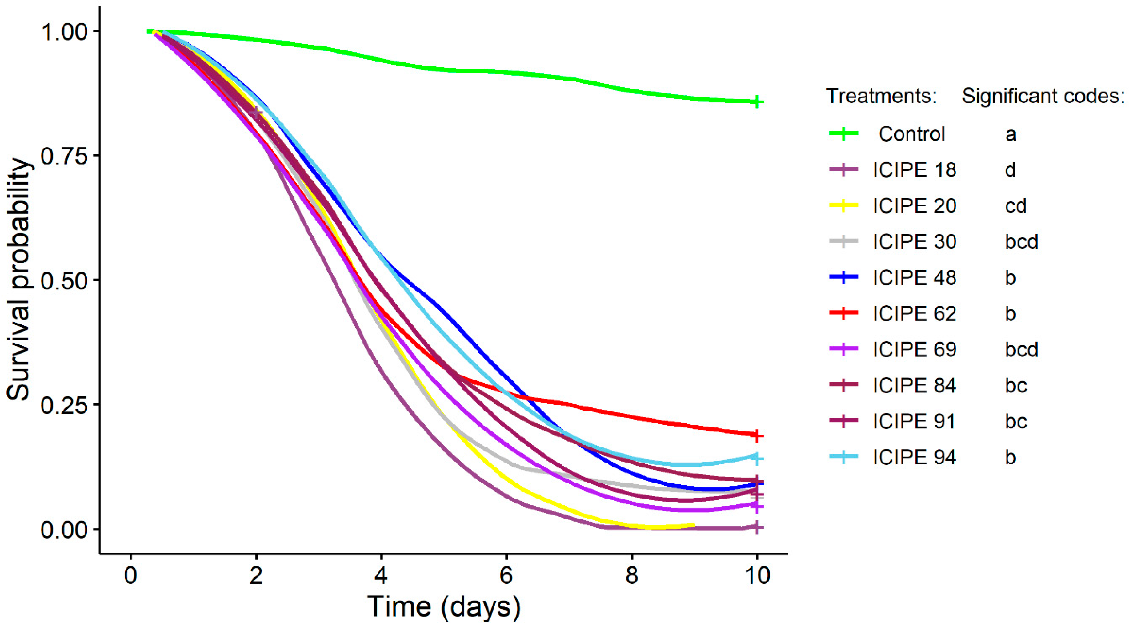
3.1. Conidial Acquisition and Adult Mortality Following Direct Exposure to M. anisopliae

Conidial germination did not vary among the fungal isolates (χ2 = 5.73; df = 8; p = 0.68) and ranged from 93.61% to 96.28%. All fungus-exposed fruit flies acquired conidia, while no conidia were observed in the control, and, therefore, controls were omitted from the analysis of the conidial acquisition. There was no significant difference in the conidial acquisition among experiments (χ2 = 4.50; df = 2; p = 0.11) nor isolates (F = 0.93; df = 8; p = 0.50). The mean number of conidia acquired by a single fruit fly ranged between 4.78 × 10⁶ to 6.54 × 10⁶ conidia/mL.
The survival of D. bivittatus adults significantly differed among the fungal isolates (χ2 = 263.46; df = 9; p < 0.0001) and was lowest for ICIPE 18, although not significantly different from that for ICIPE 20, ICIPE 30 and ICIPE 69 (Figure 1).
The mortality in M. anisopliae treatments ranged from 83.75 to 100.00%, while it was only 14.17% in the control. The mortality of D. bivittatus adults caused by the different fungal isolates was as follows, in descending order: ICIPE 20, 100.00 ± 0.00%; ICIPE 18, 99.60 ± 0.40%; ICIPE 69, 95.40 ± 1.90%; ICIPE 91, 92.90 ± 3.20%; ICIPE 30, 90.40 ± 4.30%; ICIPE 84, 89.60 ± 1.70%; ICIPE 48, 88.80 ± 3.60%; ICIPE 94, 85.80 ± 3.70%; and ICIPE 62, 83.75 ± 4.10%. The LT50 estimates for D. bivittatus adults were the shortest when exposed to ICIPE 18, ICIPE 20, ICIPE 30 and ICIPE 69 and the longest when exposed to ICIPE 48, ICIPE 62, ICIPE 84 and ICIPE 94 (Table 1).

3.2. Larval and Pupal Mortality in M. anisopliae-Treated Sand

Larval mortality significantly differed across the experiments (χ2 = 306.88; df = 2; p < 0.0001) (Table 2). Also, larval mortality significantly differed among the different fungal isolates (χ2 = 118.60; df = 8; p < 0.0001; χ2 = 77.6; df = 8; p < 0.0001; χ2 = 123.74; df = 8; p < 0.0001 for the first, second and third experiments, respectively). ICIPE 18 and ICIPE 20 consistently caused the highest mortality (≥42.50%) in all experiments, while ICIPE 91 caused the lowest mortality (<30.00%).
Likewise, pupal mortality significantly varied across the experiments (χ2 = 16.88; df = 2; p = 0.0003) and among the fungal isolates (χ2 = 99.91; df = 8; p < 0.0001; χ2 = 163.65; df = 8; p < 0.0001; χ2 = 135.28; df = 8; p < 0.0001 for the first, second and third experiments, respectively), with ICIPE 30 consistently causing the highest mortality (≥41.25%).

3.3. Adult Eclosion from M. anisopliae-Treated Sand

The eclosion of adults from all fungus-exposed puparia was lower compared to the control (≥86.25%) (Table 3). The number of eclosed adults after exposure significantly differed across the experiments (χ2 = 15.10; df = 2; p = 0.0005). Also, eclosion varied among the different fungal isolates for all the experiments (χ2 = 121.68; df = 9; p < 0.0001; χ2 = 105.78; df = 9; p < 0.0001; χ2 = 111.05; df = 9; p < 0.0001 for the first, second and third experiments, respectively). Eclosion after exposure to M. anisopliae ranged from 8.75 to 62.50%. The lowest eclosion was recorded when puparia were infected with ICIPE 18, ICIPE 20, ICIPE 30 and ICIPE 69.

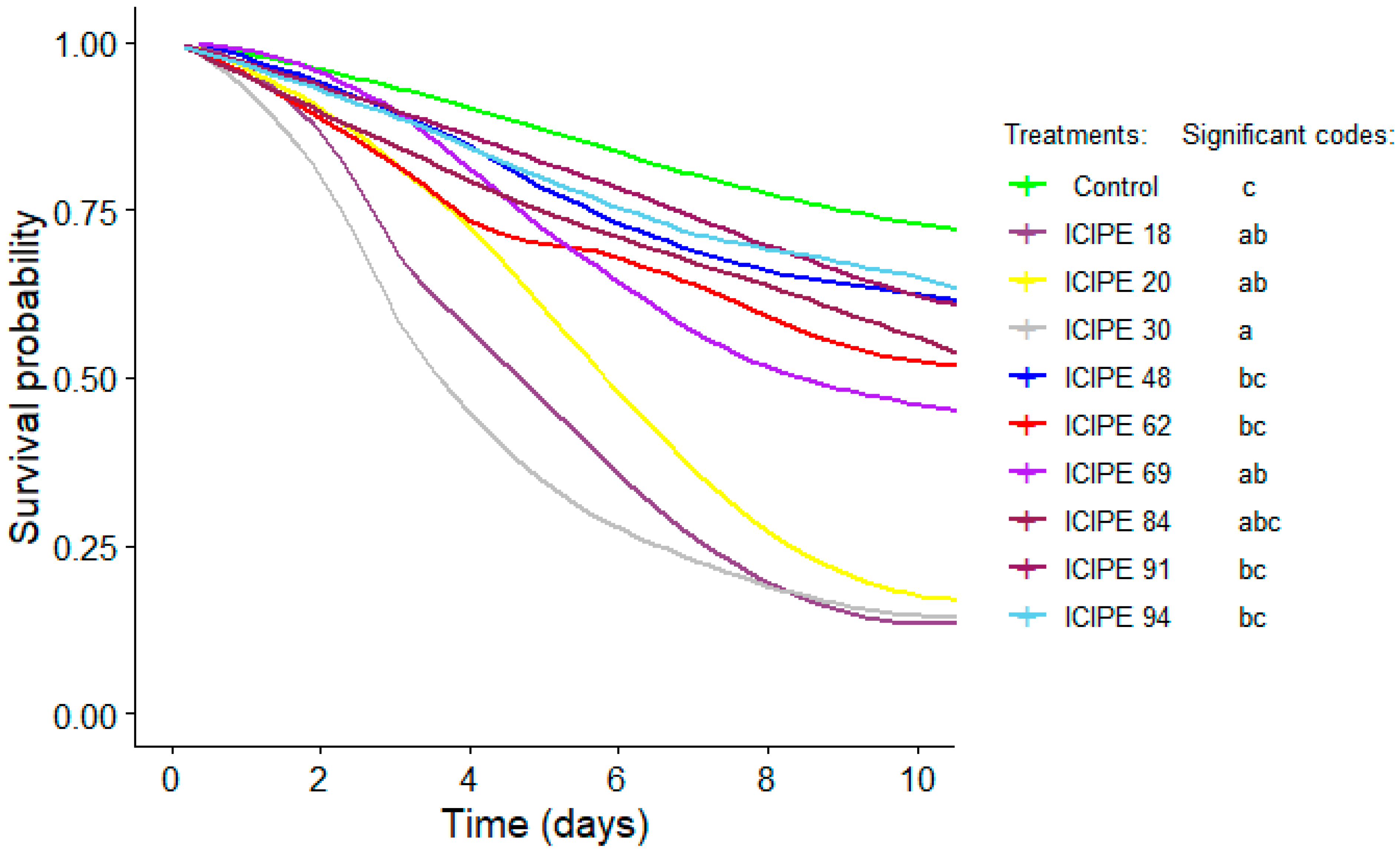
3.4. Mortality of Adults Eclosed from M. anisopliae-Treated Sand

The survival of eclosed D. bivittatus adults significantly differed among the treatments (χ2 = 55.57; df = 9; p < 0.0001) (Figure 2 and Table 4). The mortality of adults eclosed from the fungal-treated puparia varied among the isolates and equaled, in descending order, ICIPE 30, 81.10 ± 7.00%; ICIPE 18, 69.40 ± 7.00%; ICIPE 20, 66.70 ± 6.90%; ICIPE 69, 56.80 ± 6.50%; ICIPE 84, 55.10 ± 4.40%; ICIPE 62, 47.90 ± 6.30%; ICIPE 91, 47.70 ± 6.30%; ICIPE 48, 43.90 ± 2.60%; and ICIPE 94, 43.30 ± 6.50%. The LT50 estimates for D. bivittatus adults were the shortest when exposed to ICIPE 18 and ICIPE 30 and the longest when exposed to ICIPE 48, ICIPE 91 and ICIPE 94.

4. Discussion

Afrotropical pestiferous fruit flies such as D. bivitattus are polyphagous, multivoltine and largely concealed for a greater part of their lifecycle and, therefore, require a holistic IPM approach for sustainable management. Fungal-based biopesticides, being environmentally benign and with no-to-mimimal health risks to animals, humans and nontarget organisms, represent an ideal option that could complement other IPM components for the suppression of these pests. A key prerequisite for the development and commercialization of any entomopathogenic fungal product is understanding the efficacy of the entomopathogenic fungus against the target pest.
One of the major factors that determines the efficacy of an entomopathogenic fungus is its ability to adhere to the body of the target insect [33]. In the current study, the conidial acquisition by a single D. bivittatus fly was relatively high and ranged between 4.78 × 10⁶ and 6.54 × 10⁶ conidia/mL. This was higher than the conidial acquisition previously reported for the Mediterranean fruit fly Ceratitis capitata (Weidemann), the Natal fruit fly Ceratitis fasciventris Karsch and the mango fruit fly Ceratitis cosyra (Walker) across 12 isolates of Beauveria bassiana (Balsamo) Vuillemin and M. anisopliae, including some used in the present study, which ranged between 4.2 × 10⁵ and 1.0 × 10⁶ conidia/mL [34]. The increased conidial acquisition by D. bivittatus compared to Ceratitis spp. and reported in this study could be attributed to the variation in the flies’ size and the increased amount of time (5 min instead of 3 min) they were allowed to walk on the fungus-impregnated material. Among the different fungal isolates tested in this study, there was no difference in the conidial acquisition. Only one species of entomopathogenic fungus (M. anisopliae) was used in this study. Indeed, previous studies showed that many M. anisopliae isolates have similar surface characteristics such as adhesins as a surface attachment cue [35].
ICIPE 18, ICIPE 20, ICIPE 30 and ICIPE 69 caused the highest reduction in adult survival. Interestingly, ICIPE 18, ICIPE 30 and ICIPE 69 were also reported to be the most virulent isolates against adult Z. cucurbitae in laboratory bioassays [36]. ICIPE 69 is a particularly promising candidate for further investigation, as the isolate was found to be effective against a wide range of pests and was commercialized for use against fruit flies and other pests [21].
Although there was a significant difference among experiments, all the tested isolates were pathogenic to D. bivittatus larvae and puparia and significantly reduced eclosion. The concentration used to spray the sand in our study (1 × 10⁷ conidia/mL) was lower than the concentrations usually recommended for entomopathogenic fungi for commercial use in the field [37], and, therefore, we hypothesize that similar pupal and adult mortality percentages may be achieved with the tested isolates under field conditions. The differences among the experiments could be attributed to the slightly different environmental conditions within the substrate. Temperature and relative humidity were reported to affect the effectiveness of M. anisopliae [36,38]. Nevertheless, across experiments, ICIPE 18 and ICIPE 20 caused the highest adult mortality, while ICIPE 30 caused the highest pupal mortality. As a result, eclosion, which combined the effect of larval and pupal mortality, was lowest for isolates ICIPE 18, ICIPE 20, ICIPE 30 and ICIPE 69. As such, the most virulent isolates against D. bivittatus larvae and puparia were the same as those against adults.
The differential pathogenicity of M. anisopliae isolates toward D. bivittatus larvae is in concordance with reports from related fruit fly species. For example, Lezama-Gutierrez et al. [39] reported that M. anisopliae isolates differed in pathogenicity against the larvae of the Mexican fruit fly Anastrepha ludens (Loew). Likewise, Usman et al. [19], who evaluated the virulence of nine M. anisopliae isolates against different stages of the peach fruit fly Bactrocera zonata (Saunders) and the oriental fruit fly Bactrocera dorsalis (Hendel), demonstrated that larval mortality varied across the different isolates of this fungus for both fruit fly species. However, the larval mortality reported in our study for the best performing isolates (ICIPE 18 with 67.50% mortality and ICIPE 20 with 61.25% mortality in experiment 3) was lower than that reported by Lezama-Gutierrez et al. [39] and Usman et al. [19], where larval mortality reached 98.75%, 75.2% and 69.3% for A. ludens, B. zonata and B. dorsalis, respectively. This could be explained in part by the differences in the fruit fly species and isolates used, as well as the concentration of the fungal suspension and the mode of infection. Indeed, Lezama-Gutierrez et al. [39] exposed the larvae to M. anisopliae by direct immersion in a conidial suspension, while in the current study larvae were placed on sand sprayed with fungal conidia to better mimick field conditions.
A similar finding of the differential B. dorsalis pupal mortality among M. anisopliae isolates was reported by Wang et al. [40]. However, the pupal mortality of D. bivittatus in our study (reaching 48.75% for ICIPE 30) was higher than that found by Wang et al. [40] for B. dorsalis, where the best performing M. anisopliae isolate (MA04) only yielded 15% mortality. In our study, the D. bivittatus were still larvae when placed in fungus-infected sand to pupate, while Wang et al. [40] directly exposed B. dorsalis puparia to the pathogen. Dacus bivitattus used in our study likely acquired conidia as larvae before or just after pupation, before the cuticle hardened, explaining the higher pupal mortality. Indeed, higher larval susceptibility across different fungal species and isolates compared to that of puparia was well-documented for several fruit fly species. For instance, Usman et al. [19] demonstrated that the larvae of B. zonata and B. dorsalis were more susceptible to M. anisopliae and B. bassiana than their respective puparia. Likewise, Mahmoud et al. [41] also reported that B. bassiana, M. anisopliae and Lecanicillium muscarium R. Zare & W. Gams caused the greatest mortality in the adults of B. zonata, followed by the larvae and puparia.
We found different effects among M. anisopliae isolates on D. bivittatus eclosion, which is in accordance with the findings reported by Onsongo et al. [42], who tested eclosion for the related fruit fly species Z. cucurbitae using some of the same isolates from our study. Whereas, in a study by Onsongo et al. [42], ICIPE 69 was clearly found to most suppress Z. cucurbitae eclosion (compared to ICIPE 18 and ICIPE 30); in our study, ICIPE 18, ICIPE 20, ICIPE 30 and ICIPE 69 all resulted in the highest reduction in eclosion. Interestingly, the reduction in D. bivitattus eclosion caused by ICIPE 18 in the current study was higher than that reported for Z. cucurbitae by Onsongo et al. [42], using the same methodology and concentration (1 × 10⁷ conidia/mL) to treat the substrate. Both D. bivitattus and ICIPE 18 are native to Africa and, therefore, share a similar evolutionary history, while Z. cucurbitae is an alien pest of Asian origin, which could explain this discrepancy. Metarhizium anisopliae isolates were reported to caused a significant reduction in eclosion in other fruit fly species such as C. capitata, C. fasciventris, C. cosyra [26] and the blueberry maggot fly Rhagoletis mendax Curran [43]. When evaluated in cages under field conditions, a granular formulation of ICIPE 20 was found to reduce the eclosion of C. capitata, C. fasciventris and C. cosyra by 37–54%, and it would be interesting to investigate eclosion reduction in the most promising isolates against D. bivittatus under field conditions.
The survival of adults eclosed from M. anisopliae-infected sand was significantly reduced compared to that of the control for all the isolates, with ICIPE 18, ICIPE 20 and ICIPE 30 causing the highest mortality. Hypothetically, the adults acquired conidia while emerging from the inoculated sand. A similar effect was reported by Onsongo et al. [42] when studying the effect of M. anisopliae-infected sand on adult Z. cucurbitae survival. In our study, the LT50 of adults eclosing from infested sand was longer than that of adults directly exposed to sprays; this was to be expected, as emerging adults likely acquired fewer conidia through infested sand than when directly exposed. However, our results illustrated how fungal sand treatments may indirectly affect adult survival and suggested how a possible soil treatment in field conditions may negatively affect not only larvae and puparia but also adults. Onsongo et al. [42] found that ICIPE 30 was most virulent in reducing adult Z. cucurbitae survival in infested sand sprayed at lower concentrations, whereas ICIPE 69 was most virulent at higher concentrations.
Based on our findings, it was evident that all M. anisopliae isolates are virulent to D. bivitattus larvae, puparia and adults and reduce the longevity of adults when emerging from M. anisopliae-infested sand. ICIPE 18, ICIPE 20, ICIPE 30 and ICIPE 69 were consistently the most virulent against larvae, puparia and adults, while, in addition, ICIPE 18 and ICIPE 30 reduced adult survival in treated sand. We, therefore, recommended further screenhouse and field studies toward the IPM of D. bivitattus to identify the best isolate under field conditions.

Author Contributions

Conceptualization, S.K.O. and T.D.; methodology, J.A.O., S.K.O. and T.D.; analysis, S.K.O., E.R.O. and T.D.; validation, S.K.O., E.R.O. and T.D.; data curation S.K.O., J.A.O., E.R.O. and T.D.; writing—original draft preparation, S.K.O.; supervision, K.S.A., S.A.M. and T.D.; project administration, T.D.; funding acquisition, T.D. and S.A.M. All authors have read and agreed to the published version of the manuscript.

Funding

This work received financial support from the German Federal Ministry for Economic Cooperation and Development (BMZ), commissioned and administered through the Deutsche Gesellschaft für Internationale Zusammenarbeit (GIZ) Fund for International Agricultural Research (FIA), grant number 17.7860.4–001; the Norwegian Agency for Development Cooperation, the Section for Research, Innovation and Higher Education, grant number RAF-3058 KEN-18/0005; the UK’s Foreign, Commonwealth and Development Office (FCDO), grant number B2291A-FCDO-BIOPESTICIDE; the Swedish International Development Cooperation Agency (Sida); the Swiss Agency for Development and Cooperation (SDC); the Federal Democratic Republic of Ethiopia; and the Government of the Republic of Kenya. The views expressed herein do not necessarily reflect the official opinion of the donors.

Institutional Review Board Statement

Not applicable.

Informed Consent Statement

Not applicable.

Data Availability Statement

No data were deposited in an official repository.

Acknowledgments

The authors gratefully acknowledge icipe’s Arthropod Pathology Unit (APU) and African Fruit Fly Program (AFFP) teams for their technical support during the experimental setup and data collection. The authors are also grateful to Moses Ambaka, Jane Kimemia and Frida Mueni.

Conflicts of Interest

The authors declare no conflict of interest. The funders had no role in the design of the study; in the collection, analyses or interpretation of data; in the writing of the manuscript; or in the decision to publish the results.

References

  1. McCreight, J.D. Cultivation and uses of cucurbits. In Genetics and Genomics of the Cucurbitaceae; Grumet, R., Katzir, N., Garcia-Mas, J., Eds.; Springer: New York, NY, USA, 2016. [Google Scholar]
  2. Rolnik, A.; Olas, B. Vegetables from the Cucurbitaceae family and their products: Positive effect on human health. Nutrition 2020, 78, 110788. [Google Scholar] [CrossRef] [PubMed]
  3. Horticultural Crops Directorate (HCD). Horticulture Validated Report 2016–2017; Ministry of Agriculture, Livestock and Fisheries: Nairobi, Kenya, 2017. [Google Scholar]
  4. Badii, K.B.; Billah, M.K.; Afreh-Nuamah, K.; Obeng-Ofori, D. Species composition and host range of fruit-infesting flies (Diptera: Tephritidae) in northern Ghana. Int. J. Trop. Insect Sci. 2015, 35, 137–151. [Google Scholar] [CrossRef]
  5. De Meyer, M.; Delatte, H.; Mwatawala, M.; Quilici, S.; Vayssières, J.F.; Virgilio, M. A review of the current knowledge on Zeugodacus cucurbitae (Coquillett) (Diptera, Tephritidae) in Africa, with a list of species included in Zeugodacus. ZooKeys 2015, 540, 539–557. [Google Scholar] [CrossRef]
  6. Mwatawala, M.; Kudra, A.; Mkiga, A.; Godfrey, E.; Jeremiah, S.; Virgilio, M.; De Meyer, M. Preference of Zeugodacus cucurbitae (Coquillett) for three commercial fruit vegetable hosts in natural and semi natural conditions. Fruits 2015, 70, 333–339. [Google Scholar] [CrossRef]
  7. Dhillon, M.K.; Singh, R.; Naresh, J.S.; Sharma, H.C. The melon fruit fly, Bactrocera cucurbitae: A review of its biology and management. J. Insect Sci. 2005, 5, 40. [Google Scholar]
  8. Kambura, C.; Tanga, C.M.; Kilalo, D.; Muthomi, J.; Salifu, D.; Rwomushana, I.; Mohamed, S.A.; Ekesi, S. Composition, host range and host suitability of vegetable-infesting Tephritids on cucurbits cultivated in Kenya. Afr. Entomol. 2018, 26, 379–397. [Google Scholar] [CrossRef]
  9. Zida, I.; Nacro, S.; Dabiré, R.; Somda, I. Seasonal abundance and diversity of fruit flies (Diptera: Tephritidae) in three types of plant formations in Western Burkina Faso, West Africa. Ann. Entomol. Soc. Am. 2020, 113, 343–354. [Google Scholar] [CrossRef]
  10. Tuo, Y.; Kone, K.; Yapo, M.L.; Herve, K.K. Abundance and incidence of zucchini (Cucurbita pepo L.) flies in the Korhogo Department of northern Côte d’Ivoire and pest control methods used by farmers. J. Exp. Agric. 2018, 21, 1–7. [Google Scholar] [CrossRef]
  11. Mwatawala, M.W.; De Meyer, M.; Makundi, R.H.; Maerere, A.P. Host range and distribution of fruit-infesting pestiferous fruit flies (Diptera, Tephritidae) in selected areas of Central Tanzania. Bull. Entomol. Res. 2009, 99, 629–641. [Google Scholar] [CrossRef]
  12. Mwatawala, M.W.; De Meyer, M.; Makundi, R.H.; Maerere, A.P. Biodiversity of fruit flies (Diptera, Tephritidae) in orchards in different agro-ecological zones of the Morogoro region, Tanzania. Fruits 2006, 61, 321–332. [Google Scholar] [CrossRef]
  13. Mokam, D.G.; Djiéto-Lordon, C.; Bilong Bilong, C.F.; Lumaret, J.P. Host susceptibility and pest status of fruit flies (Diptera: Tephritidae) attacking cucurbits in two agroecological zones of Cameroon, Central Africa. Afr. Entomol. 2018, 26, 317–332. [Google Scholar] [CrossRef]
  14. Doorenweerd, C.; Leblanc, L.; Norrbom, A.L.; San Jose, M.; Rubinoff, D. A global checklist of the 932 fruit fly species in the tribe Dacini (Diptera, Tephritidae). ZooKeys 2018, 730, 17–54. [Google Scholar] [CrossRef] [PubMed]
  15. Layodé, B.F.R.; Onzo, A.; Karlsson, M.F. Watermelon-infesting Tephritidae fruit fly guild and parasitism by Psyttalia phaeostigma (Hymenoptera: Braconidae). Int. J. Trop. Insect Sci. 2020, 40, 157–166. [Google Scholar] [CrossRef]
  16. White, I.M.; Elson-Harris, M. Fruit Flies of Economic Significance: Their Identification and Bionomics; CAB International: Wallingford, UK, 1992. [Google Scholar]
  17. Kibira, M.; Affognon, H.; Njehia, B.; Muriithi, B.; Mohamed, S.; Ekesi, S. Economic evaluation of integrated management of fruit fly in mango production in Embu County, Kenya. Afr. J. Agric. Resour. Econ. 2015, 10, 343–353. [Google Scholar]
  18. Vontas, J.; Hernández-Crespo, P.; Margaritopoulos, J.T.; Ortego, F.; Feng, H.T.; Mathiopoulos, K.D.; Hsu, J.C. Insecticide resistance in Tephritid flies. Pestic. Biochem. Physiol. 2011, 100, 199–205. [Google Scholar] [CrossRef]
  19. Usman, M.; Gulzar, S.; Wakil, W.; Wu, S.; Piñero, J.C.; Leskey, T.C.; Nixon, L.J.; Oliveira-Hofman, C.; Toews, M.D.; Shapiro-Ilan, D. Biological and microbial control virulence of entomopathogenic fungi to Rhagoletis pomonella (Diptera: Tephritidae) and interactions with entomopathogenic nematodes. J. Econ. Entomol. 2020, 113, 2627–2633. [Google Scholar]
  20. Toledo-Hernández, R.A.; Toledo, J.; Sánchez, D. Effect of Metarhizium anisopliae (Hypocreales: Clavicipitaceae) on food consumption and mortality in the Mexican fruit fly, Anastrepha ludens (Diptera: Tephritidae). Int. J. Trop. Insect Sci. 2018, 38, 254–260. [Google Scholar] [CrossRef]
  21. Akutse, K.S.; Subramanian, S.; Maniania, N.K.; Dubois, T.; Ekesi, S. Biopesticide research and product development in Africa for sustainable agriculture and food security–experiences from the International Centre of Insect Physiology and Ecology. Front. Sustain. Food Syst. 2020, 4, 563016. [Google Scholar] [CrossRef]
  22. Elghadi, E.; Port, G.R. Use of entomopathogenic fungi for the biological control of the greater melon fly Dacus frontalis in Libya. In Area-Wide Management of Fruit Fly Pests; Perez-Staples, D., Diaz-Fleischer, F., Montoya, P., Maria Vera, M., Eds.; CRC Press: Boca Raton, FL, USA, 2019. [Google Scholar]
  23. Muriuki, C.; Guantai, M.; Namikoye Samita, E.; Mulwa, J.; Nyamai, M.; Kasina, M. Abundance and diversity of frugivorous fruit flies in Kandara, Murang’a County, Kenya. Afr. Phytosanitary J. 2020, 2, 41–50. [Google Scholar] [CrossRef]
  24. Isabirye, B.E.; Akol, A.M.; Mayamba, A.; Nankinga, C.K.; Rwomushana, I. Species composition and community structure of fruit flies (Diptera: Tephritidae) across major mango-growing regions in Uganda. Int. J. Trop. Insect Sci. 2015, 35, 69–79. [Google Scholar] [CrossRef]
  25. Dimbi, S.; Maniania, N.K.; Lux, S.A.; Ekesi, S.; Mueke, J.K. Pathogenicity of Metarhizium anisopliae (Metsch.) Sorokin and Beauveria bassiana (Balsamo) Vuillemin, to three adult fruit fly species: Ceratitis capitata (Weidemann), C. rosa var. fasciventris Karsch and C. cosyra (Walker) (Diptera: Tephritidae). Mycopathologia 2003, 156, 375–382. [Google Scholar] [CrossRef] [PubMed]
  26. Ekesi, S.; Maniania, N.K.; Lux, S.A. Mortality in three African tephritid fruit fly puparia and adults caused by the entomopathogenic fungi, Metarhizium anisopliae and Beauveria bassiana. Biocontrol Sci. Technol. 2002, 12, 7–17. [Google Scholar] [CrossRef]
  27. R Development Core Team. R: A Language and Environment for Statistical Computing; R Foundation for Statistical Computing: Vienna, Austria, 2016. [Google Scholar]
  28. Bates, D.; Mächler, M.; Bolker, B.; Walker, S. Fitting linear mixed effects models using lme4. J. Stat. Softw. 2015, 67, 1–48. [Google Scholar] [CrossRef]
  29. Abbott, W.S. A method of computing the effectiveness of an insecticide. J. Econ. Entomol. 1925, 18, 265–267. [Google Scholar] [CrossRef]
  30. Hlina, B.L. Ecotox: A Package for Analysis of Ecotoxicology. R Package Version 1.4.2. 2020. Available online: https://cran.r-project.org/package=ecotox (accessed on 24 November 2021).
  31. Therneau, T.M. A Package for Survival Analysis in R. 2020. Available online: https://cran.r-project.org/package=survival (accessed on 24 November 2021).
  32. Lenth, R.V. Least-squares means: The R Package lsmeans. J. Stat. Softw. 2016, 69, 1–33. [Google Scholar] [CrossRef]
  33. Ortiz-Urquiza, A.; Keyhani, N.O. Action on the surface: Entomopathogenic fungi versus the insect cuticle. Insects 2013, 4, 357–374. [Google Scholar] [CrossRef]
  34. Dimbi, S.; Maniania, N.K.; Ekesi, S. Effect of Metarhizium anisopliae inoculation on the mating behavior of three species of African tephritid fruit flies, Ceratitis capitata, Ceratitis cosyra and Ceratitis fasciventris. Biol. Control 2009, 50, 111–116. [Google Scholar] [CrossRef]
  35. Bidochka, M.J.; Small, C. Phylogeography of Metarhizium, an insect pathogenic fungus. In Insect-Fungal Associations; Vega, F.E., Blackwell, M., Eds.; Oxford University Press: New York, NY, USA, 2005. [Google Scholar]
  36. Onsongo, S.K.; Gichimu, B.M.; Akutse, K.S.; Dubois, T.; Mohamed, S.A. Performance of three isolates of Metarhizium anisopliae and their virulence against Zeugodacus cucurbitae under different temperature regimes, with global extrapolation of their efficiency. Insects 2019, 10, 270. [Google Scholar] [CrossRef]
  37. Quesada-Moraga, E.; Martin-Carballo, I.; Garrido-Jurado, I.; Santiago-Álvarez, C. Horizontal transmission of Metarhizium anisopliae among laboratory populations of Ceratitis capitata (Wiedemann) (Diptera: Tephritidae). Biol. Control 2008, 47, 115–124. [Google Scholar] [CrossRef]
  38. Arthurs, S.; Thomas, M.B. Effects of temperature and relative humidity on sporulation of Metarhizium anisopliae var. acridum in mycosed cadavers of Schistocerca gregaria. J. Invertebr. Pathol. 2001, 78, 59–65. [Google Scholar] [CrossRef] [PubMed]
  39. Lezama-Gutiérrez, R.; La Luz, A.T.D.; Molina-Ochoa, J.; Rebolledo-Dominguez, O.; Pescador, A.R.; López-Edwards, M.; Aluja, M. Virulence of Metarhizium anisopliae (Deuteromycotina: Hyphomycetes) on Anastrepha ludens (Diptera: Tephritidae): Laboratory and field trials. J. Econ. Entomol. 2000, 93, 1080–1084. [Google Scholar] [CrossRef] [PubMed]
  40. Wang, D.; Liang, Q.; Chen, M.; Ye, H.; Liao, Y.; Yin, J.; Lü, L.; Lei, Y.; Cai, D.; Jaleel, W.; et al. Susceptibility of oriental fruit fly, Bactrocera dorsalis (Diptera: Tephritidae) pupae to entomopathogenic fungi. Appl. Entomol. Zool. 2021, 56, 269–275. [Google Scholar] [CrossRef]
  41. Mahmoud, M.F. Susceptibility of the peach fruit fly, Bactorecera zonata (Saunders) (Diptera: Tephritidae) to three entomopathogenic fungi. Egypt. J. Biol. Pest Control 2009, 19, 169–175. [Google Scholar]
  42. Onsongo, S.K.; Mohamed, S.A.; Akutse, K.S.; Gichimu, B.M.; Dubois, T. The entomopathogenic fungi Metarhizium anisopliae and Beauveria bassiana for management of the melon fly Zeugodacus cucurbitae: Pathogenicity, horizontal transmission, and compatability with cuelure. Insects 2022, 13, 859. [Google Scholar] [CrossRef] [PubMed]
  43. Renkema, J.; Cutler, G.C.; Sproule, J.M.; Johnson, D.L. Effect of Metarhizium anisopliae (Clavicipitaceae) on Rhagoletis mendax (Diptera: Tephritidae) pupae and adults. Can. Entomol. 2020, 152, 237–248. [Google Scholar] [CrossRef]
Figure 1. Kaplan–Meier survival curves for Dacus bivittatus adults directly treated with sprays of different Metarhizium anisopliae isolates. Survival curves labeled with the same letters are not significantly different at α = 0.05 according to Tukey’s test.
Sustainability 15 13185 g001
Figure 2. Kaplan–Meier survival curves for Dacus bivittatus adults eclosed from sand inoculated with different Metarhizium anisopliae isolates. Survival curves labeled with the same letters are not significantly different at α = 0.05 according to Tukey’s test.
Sustainability 15 13185 g002
Table 1. Regression slope and median lethal time (LT50) of Dacus bivittatus adults directly treated with sprays of different Metarhizium anisopliae isolates. Means followed by the same letter are not significantly different at α = 0.05 according to Tukey’s test. a SE = standard error. b FL = fiducial limit at 95%.
IsolateSlope (Mean ± SE a)LT50 (Days) (95% FL b)
ICIPE 184.65 ± 0.033.11 (2.90–3.31) a
ICIPE 204.53 ± 0.033.46 (3.13–3.76) ab
ICIPE 303.76 ± 0.023.62 (3.25–3.98) ab
ICIPE 483.66 ± 0.024.39 (4.08–4.70) bc
ICIPE 622.64 ± 0.024.30 (3.76–4.86) bc
ICIPE 693.76 ± 0.023.52 (3.27–3.76) ab
ICIPE 843.76 ± 0.024.07 (3.84–4.30) bc
ICIPE 913.85 ± 0.023.86 (3.61–4.12) b
ICIPE 943.54 ± 0.024.47 (4.18–4.75) c
Table 2. Larval and pupal mortality of Dacus bavittatus in sand inoculated with different Metarhizium anisopliae isolates. Means within a column followed by the same letter are not significantly different at α = 0.05 according to Tukey’s test. a SE = standard error.
IsolatesLarval Mortality (%) (Mean ± SE a)Pupal Mortality (%) (Mean ± SE)
Exp 1Exp 2Exp 3Exp 1Exp 2Exp 3
ICIPE 1846.25 ± 2.39 a43.75 ± 5.15 a67.50 ± 3.23 a41.25 ± 4.73 ab35.00 ± 6.12 ab20.00 ± 4.08 c
ICIPE 2043.75 ± 3.15 ab42.50 ± 3.23 a61.25 ±5.54 a42.5 ± 4.33 ab36.25 ± 4.27 ab20.00 ± 6.45 c
ICIPE 3040.00 ± 3.54 ab31.25 ± 4.27 bc45.00 ± 2.04 b48.75 ± 2.39 a41.25 ± 2.39 a46.25 ± 3.15 a
ICIPE 4826.25 ± 3.15 c22.50 ± 3.23 cd26.25 ± 2.39 d35.00 ± 4.08 b15.00 ± 3.54 d37.50 ± 2.50 ab
ICIPE 6233.75 ± 4.73 bc31.25 ± 3.75 bc61.25 ± 2.39 a36.25 ± 5.54 b13.75 ± 4.27 d13.75 ± 3.75 c
ICIPE 6943.75 ± 2.39 ab31.25 ± 3.15 bc41.25 ± 2.39 b40.00 ± 5.77 ab28.75 ± 3.15 bc45.00 ± 2.04 a
ICIPE 8433.75 ± 3.75 bc35.00 ± 2.04 ab27.50 ± 1.44 cd37.50 ± 2.50 b15.00 ± 2.04 d36.25 ± 3.75 ab
ICIPE 9126.25 ± 3.75 c21.25 ± 2.39 d30.00 ± 2.04 cd22.50 ± 4.33 c16.25 ± 2.39 d38.75 ± 6.25 ab
ICIPE 9423.75 ± 4.27 c31.25 ± 4.27 bc37.50 ± 5.20 bc42.50 ± 3.23 ab20.00 ± 2.04 cd33.75 ± 3.15 b
Table 3. Percentage of adult Dacus bavitattus eclosion (mean ± standard error) from sand inoculated with different Metarhizium anisopliae isolates. Means within a column followed by the same letter are not significantly different at α = 0.05 according to Tukey’s test.
IsolatesExp 1Exp 2Exp 3
ICIPE 1812.50 ± 3.23 de21.25 ± 6.57 d12.50 ± 1.44 cd
ICIPE 2013.75 ± 2.39 de21.25 ± 2.39 d18.75 ± 1.25 bcd
ICIPE 3011.25 ± 2.39 e27.50 ± 2.50 cd8.75 ± 2.39 d
ICIPE 4841.25 ± 4.27 bc62.5 ± 4.79 b36.25 ± 2.39 b
ICIPE 6230.00 ± 2.04 bcde55.00 ± 6.45 b25.00 ± 2.04 bcd
ICIPE 6916.25 ± 3.75 cde40.00 ± 5.40 bcd13.75 ± 2.39 cd
ICIPE 8428.75 ± 3.15 cde50.00 ± 3.54 bc36.25 ± 3.75 b
ICIPE 9151.25 ± 4.73 b62.50 ± 4.33 b31.25 ± 4.27 bc
ICIPE 9433.75 ± 3.15 bcd48.75 ± 5.15 bc28.75 ± 4.27 bcd
Control91.25 ± 3.75 a91.25 ± 3.15 a86.25 ± 1.25 a
Table 4. Regression slope and median lethal time (LT50) of Dacus bivittatus adults eclosed from sand inoculated with different Metarhizium anisopliae isolates. Means followed by the same letter are not significantly different at α = 0.05 according to Tukey’s test. a SE = standard error. b FL = fiducial limit at 95%.
IsolatesSlope (Mean ± SE a)LT50 (Days) (95% FL b)
ICIPE 182.04 ± 0.0035.81 (5.05–6.60) ab
ICIPE 202.13 ± 0.0036.95 (6.17–7.80) b
ICIPE 301.78 ± 0.0034.48 (3.66–5.28) a
ICIPE 481.75 ± 0.00415.70 (14.50–17.20) ef
ICIPE 621.47 ± 0.00311.70 (10.30–13.90) de
ICIPE 692.10 ± 0.0038.70 (7.84–9.74) c
ICIPE 841.85 ± 0.00411.60 (10.60–12.90) d
ICIPE 911.84 ± 0.00414.10 (12.60–16.40) def
ICIPE 941.63 ± 0.00415.40 (13.10–19.30) ef
Disclaimer/Publisher’s Note: The statements, opinions and data contained in all publications are solely those of the individual author(s) and contributor(s) and not of MDPI and/or the editor(s). MDPI and/or the editor(s) disclaim responsibility for any injury to people or property resulting from any ideas, methods, instructions or products referred to in the content.

