Article

Low-Fouling Plate-and-Frame Ultrafiltration for Juice Clarification: Part 1—Membrane Preparation and Characterization

by
Yusuf Wibisono
1,*,
Dikianur Alvianto
2,
Bambang Dwi Argo
1,
Mochamad Bagus Hermanto
2,
Jatmiko Eko Witoyo
3 and
Muhammad Roil Bilad
4,*
1
Department of Bioprocess Engineering, Universitas Brawijaya, Jl. Veteran, Malang 65145, Indonesia
2
Department of Agricultural and Biosystems Engineering, Universitas Brawijaya, Jl. Veteran, Malang 65145, Indonesia
3
Department of Agro-Industrial Technology, Universitas Brawijaya, Jl. Veteran, Malang 65145, Indonesia
4
Faculty of Integrated Technologies, Universiti Brunei Darussalam, Jalan Tungku Link, Bandar Seri Begawan BE 1410, Brunei
*
Authors to whom correspondence should be addressed.
Sustainability 2023, 15(1), 806; https://doi.org/10.3390/su15010806
Submission received: 8 November 2022 / Revised: 15 December 2022 / Accepted: 16 December 2022 / Published: 2 January 2023
(This article belongs to the Special Issue Sustainable Food Waste Valorisation by Membrane Technology)

Abstract

:
Membrane technology provides advantages for separating and purifying food materials, including juice clarification. Ultrafiltration processes for fruit juices aim to remove haze components and maintain the stabilization of the juices. However, the membrane process during the clarification of fruit juices suffers from fouling, which deteriorates the process performance and products. Biofouling usually is found in the applications of the membrane in food processing. In this study, phenolic substances extracted from garlic bulbs are incorporated into a matrix of polymeric membranes to prevent fouling during juice clarification. Hydrophilic cellulose acetate was used as the polymer matrix of the membrane, and dimethylformamide was used as the solvent. The phenolic substances from garlic bulbs were incorporated into polymer solutions with different concentrations of 0%, 1%, 1.25%, and 1.5% w/v. The composite membrane was prepared using the phase inversion method, and the resulting membranes were later characterized. The results show that incorporating those phenolic compounds as the dope solution additive resulted in membranes with higher hydrophilic properties. They also modeled antibacterial properties, as shown by the reduced attachment of Bacillus subtilis of up to 91.5 ± 11.7% and Escherichia coli of up to 94.0 ± 11.9%.

1. Introduction

The membrane is a selective thin layer with the critical property of having the ability to control the permeation rate of different species [1]. One of the obstacles of membrane technology is fouling (pore closure) during the filtration operation. One of the foulant materials is made of biological components (bacteria, fungi, and algae), or biofouling. Biofouling is the accumulation of microorganisms attached to the surface, pores, or in the pores of a medium that can interfere with its performance [2], especially on membranes [3]. It accounts for >45% of total fouling [4]. Membrane fouling decreases the filtration flux, which eventually lowers the efficiency and performance of the filtration [5].
One of the materials used to make polymeric membranes is cellulose acetate (CA). It offers a few advantages, such as cost-effectiveness, high hydrophilicity, biocompatibility, and high flux potential [6], and can be obtained from waste material. The performance of CA-based membranes can be improved by adding suitable additives [7]. The main disadvantage of CA-based membranes is that the attached microorganisms can secrete cellulase enzymes that can degrade the membrane. Therefore, to reduce this potential drawback, efforts are required to control biofouling [8]. The methods that reduce biofouling include modifying the membrane by adding both organic and inorganic antibacterial additives (antibacterial agents) into the membrane matrix [9]. The antibacterial additives mostly use metallic (silver, copper, gold, iron, bismuth, cobalt, nickel), inorganic (TiO2, ZnO MgO, RuO2, MXene), carbonic (CNT, GO, carbon quantum dots), or organic materials (chitosan, 2-aminoimidazole). Table 1 summarizes the use of metallic, inorganic, carbonic, and organic additives to promote the antibacterial properties of the membranes.
While metallic, inorganic, carbonic, and organic additives promote better anti-biofouling properties of the membranes, the additives also hinder some negative impacts, such as toxicity [49,50,51,52]. On the other hand, many membranes used to separate food substances, which have no tolerance for food contaminants, include the membrane separators themselves [53,54,55]. It is, therefore, very important to research sustainable alternatives for these additives that are safe and effective for reducing biofouling. Phenolic compounds extracted from natural resources have shown great potential.
The addition of the active substance of phenolic compounds has potential because of its abundance at relatively low prices. Phenolic compounds are widely distributed in plants and have a defensive function [56]. Natural ingredients that contain phenols preventing antibacterial activity include olive leaf [57,58], Uncaria gambir [59], cocoa pod husk [60,61], honey [62], ginger [63], and garlic [64,65,66].
Garlic (Allium sativum) contains phenolic compounds, such as allicin compounds, which give a distinctive aroma [67]. It is a defense molecule from biological activity that inhibits bacterial proliferation with antibiotic, antioxidant, anti-inflammatory, and antimicrobial properties [68]. Garlic has antibacterial and antifungal properties. It contains a high enough allicin compound, and hence, it can control both Gram-negative and Gram-positive bacteria (Escherichia coli, Pasteurella multocida, Bacillus subtilis, and Staphylococcus aureus) [65], including Bacillus spp. and Streptococcus spp. [69], Salmonella typhimurium [70], and Pseudomonas syringae [71]. Phenolic compounds are found in the fresh garlic bulb [27,28] and fermented garlic [72]. While fresh garlic bulbs and black garlic are mainly used for human consumption, garlic peel waste may be a phenolic source [73]. Garlic husk waste contains phenolic compounds, such as caffeic, p-coumaric, ferulic, and di-ferulic acids [74]. The use of waste materials for membranes utilized for food processing will be environmentally and economically beneficial.
In the authors’ previous preliminary research, the phenolic compounds extracted from a garlic bulb showed antibacterial properties when used in membrane applications [66]. In this follow-up study, the phenolic compounds were extracted from garlic and added as an additive to fabricate CA-based ultrafiltration membranes. The phenolic substances were first evaluated, and then further assessment was conducted once they were incorporated into the membranes. The application of the low-fouling ultrafiltration membrane for juice clarification was then conducted.

2. Materials and Methods

2.1. Materials

The primary polymer for membrane fabrication was CA powder (39.3–40.3 wt% acetyl content), and an average molecular weight (Mn) of 30,000 g/mol was measured by gel permeation chromatography (GPC). The CA polymer was dissolved in dimethylformamide (DMF) solvent for membrane synthesis (Merck, Darmstadt, Germany). Fresh garlic bulbs were purchased from a local market in Malang City, Indonesia. Technical-grade chemicals of acetone, 96% ethanol, and formaldehyde were provided by local chemical suppliers. B. subtilis strain ATCC 6051 and E. coli strain K-12 (ATCC 10798) cultures were provided by the Medical Laboratory of Universitas Brawijaya.

2.2. Garlic Extraction

Garlic extraction was performed according to a method previously detailed elsewhere [75]. The initial garlic weight was 5040 g with an initial moisture content of 35.40%, and the final garlic weight was 1784 g with a moisture content of 7.7% The process was continued by maceration and evaporation (Rotavapor, Buchi Labortechnik AG, Flawil, Switzerland), resulting in a concentrated extract with a volume of 177 mL. The extract was dried to obtain a paste of 77.94 g with a moisture content of 1.08%. The garlic extract paste’s composition was then analyzed using an FTIR (Spirit/ATR-S, Shimadzu I.R., Kyoto, Japan). The total phenolic compound was measured using a spectrophotometer (Jasco V-730, Easton, MD, USA) and was equivalent to 271 mg GAE/g.

2.3. LC-MS/MS Analysis

The garlic extract paste sample was diluted to 10% and filtered through a 0.22 µm filter. The analysis was carried out using LCMS (Shimadzu LCMS-8060, Kyoto, Japan) as the primary tool and a vacuum pump GAST DOA P-504BN, an ultrasonic bath wise (Wiseclean), a vortex mixer (Maximix II, Thermoscientific, Waltham, MA, USA), and a micropipette (Eppendorf, Hamburg, Germany) was supporting tools. The LCMS was equipped with a Phenomenex HPLC column, Kinetex C18, 100 mmL × 2.1 mm ID, 2.6 um, 100 A, and an ESI ion source. Other analysis parameters included an injection volume of 10 uL, a sampling speed of 5 uL/s, CID gas of 270 KPa, a conversion dynode -of 10.00 kV, a time of 0–45 min, a start of 50.00 and an end of 1200 m/z, a scan speed of 2500 u/s, and an interface temperature of 300 °C. The phenolic compounds in the garlic extract were screened and identified using LC-MS/MS.

2.4. Preparation of Composite Membrane

The CA-based membrane with the addition of the garlic extract paste was prepared using DMF as the solvent, and the dope solutions were cast using a casting knife (Elcometer, Manchester, UK) according to a method described in a previous research study [66] with some modifications. The compositions of the dope solutions can be seen in Table 2.

2.5. Membrane Properties

2.5.1. Final Thickness

The membrane thickness was measured using a digital micrometer (Mitutoyo, Kawasaki, Japan) with an accuracy of 0.001 mm. The test was carried out at five measurement points: middle, top, bottom, left, and right. The samples were measured by repeating these measurements 3 times on each membrane sample.

2.5.2. Tensile Strength and Elongation

The tensile strength and elongation of the membrane were measured using a tensile strength instrument (IMADA ZP50N, Northbrook, IL, USA). Samples were cut to 7 × 1.5 cm and tested in duplicate for each membrane sample. A membrane with good mechanical strength is required to operate at a high operating pressure of around 1–5 bar for ultrafiltration [76]. The change in the maximum length of the membrane film before breaking is called elongation. The elongation test was carried out by calculating the increase in length divided by the length of the sample before the tensile test was carried out. The membrane was cut to 7 × 1.5 cm and tested in duplicate.

2.5.3. Contact Angle

The contact angle measurement was carried out to determine the membrane surface’s level of hydrophilicity/hydrophobicity. It was completed using the Contact Angle and Surface Tension Measurement Instrument (DataPhysics OCA 25, Filderstadt, Germany). The small membrane coupon was determined with the contact angle instrument by using small water droplets.

2.5.4. Membrane Permeability

Membrane clean water permeability (Pw) was measured by a method adapted from [61]. A membrane coupon with a diameter of 60 mm was placed in a filtration cell, and the transmembrane pressure was set at 0.5 bar. The permeate was collected every 2 min for 20 min, and then the mass was weighed. The permeability was calculated using Equation (1).
Pw = V/(A × Δt × Δp)
where Pw is the membrane permeability (L/m2·h.bar), Q is the volume of the permeate (liters), t is the time (hours), A is the membrane surface area (m2), and p is the transmembrane pressure (bar).

2.5.5. Membrane Morphology

Membrane morphology was observed by using a Scanning Electron Microscope (FESEM-FEI Quanta FEG 650, Hillsboro, OR, USA). The membrane sample was cut using a lancet to a size of 50 × 50 mm. Before cutting, the membrane piece was immersed in liquid nitrogen to turn it stiff and rigid. The cutting pressure did not interfere with the macro cavity structure and various cross-sectional areas of the membrane cavity. Cross-section observation was conducted by attaching the membrane sheet to the sample holder at a position of 90°. Prior to observation, a gold coating was applied to the membrane to enhance the conductivity and ease the SEM observation. The image magnifications of 1500–5000× were selected.

2.5.6. Anti-Biofouling Test

The anti-biofouling properties of the membrane were tested by using two types of bacteria, B. subtilis and E. coli, which are generally used as good representatives of Gram-positive and Gram-negative bacteria [61,77]. The membrane coupon of 5 × 5 mm was immersed in a bacterial solution for 18 h and then incubated in an incubator (Memmert IN110, Schwabach, Germany). The bacterial cells, which were approximately 1–6 μm in size, were adhered to the membrane surface depending on the anti-biofouling properties of the membrane. The exposed membranes were dried, and the bacterial cells were fixed using formaldehyde. The dried fouled membranes were then observed using SEM and obtained from 4 randomly selected spots on the membrane surface (FESEM-FEI Quanta FEG 650, Hillsboro, OR, USA). The SEM images were then processed using image processing software (ImageJ, National Institutes of Health, Bethesda, MD, USA) to quantify the number of bacterial attachments and determine the fouled membrane’s percentage area.

2.5.7. Phenolic Content in the Membranes

The phenol content (from the garlic extract) that remained on the membrane samples was measured by using a spectrophotometer (Genesys 10S UV-Vis, Thermoscientific, Waltham, MA, USA) with the sulfanilic acid (H3NC6H4SO3) reagent. Measurement of the phenol functional group in the membrane was also carried out using an FTIR (Shimadzu I.R. Prestige 21, Japan). The samples were mixed with KBr (ratio 1:10) and then placed between two optically polished KBr plates prior to FTIR measurement.

3. Results and Discussion

3.1. LC-MS/MS Analysis

A screening and identification process was carried out without a target compound using the LC-MS/MS methods to prove the presence of phenolic compounds in the ethanolic extract of garlic. Tentative compounds were proposed by comparing the retention time, MS data, and MS/MS data with the previous Allium references and searching for phytochemical compounds through the natural material database [78]. The identification results found that six phenolic compounds were identified, and the rest were two unknown compounds presented in the ethanolic extract of garlic. The list of phenolic compounds with the retention time and the LC-MS/MS data are presented in Table 3 and Figure 1.
The analysis of Peak 1, based on an m/z of 368 and a single ion fragment fraction at an m/z of 338, found that it corresponded to 3-3-Feruloylquinic acid [79,80,81]. A chlorogenic acid compound in the garlic’s ethanolic extract was tentatively identified at an m/z of 354 with an ion fragment at an m/z of 191, as revealed in Peak 2. Peak 3, with an m/z of 180 and four ion fragments at m/z ratios of 89,134, 145, 163, and 180, was identified as caffeic acid [74,78,79,80,82,83]. Furthermore, Peak 5 was tentatively identified as having three overlapping phenolic compounds. P-coumaric acid was identified tentatively based on an m/z of 164 with its three typical ion fragments at 91, 199, and 147 [80,81,83,84]. Peak 5 also depicted the presence of ferulic acid (m/z of 194), accompanied by ion fragments (m/z ratios of 134 and 169) [74,78,80,81,83,84]. At this peak, other phenolic compounds with m/z ratios of 338 and ion fragments with m/z ratios of 163 and 173 were identified as 3-p-Coumaroylquinic acid [74,78]. However, Peak 4 was not identified in the literature database.
All of the phenolic compounds identified in the LC-MS/MS measurements are phenolic acids of the subclass of hydroxycinnamic acids [78,80]. The results are in agreement with the literature [74,85], which discovered phenolic acids from the subclass of hydroxybenzoic acids (gallic acid and 4-hydroxybenzoic acid) and the subclass of hydroxycinnamic acids (caffeic acid, p-coumaric acid, trans-ferulic acid, ferulic acid, chlorogenic acid, caffeic acid-O-glucoside, coumaroylquinic acid, coumaric acid-O-glucoside, and caffeoylputrescine) from the phenolic compound extracts from garlic husk. These compounds are known to possess antioxidant properties. Moreover, hydroxycinnamic acids are a subclass of phenolic acids with antimicrobial activity based on in vitro tests on various microbial species [86,87,88,89]. These results confirm that the ethanolic extract of garlic can function as a natural antibacterial agent and has the opportunity and prospect for further application in both food and non-foods fields, such as for anti-biofouling agents in membranes.

3.2. Membrane Properties

3.2.1. Thickness

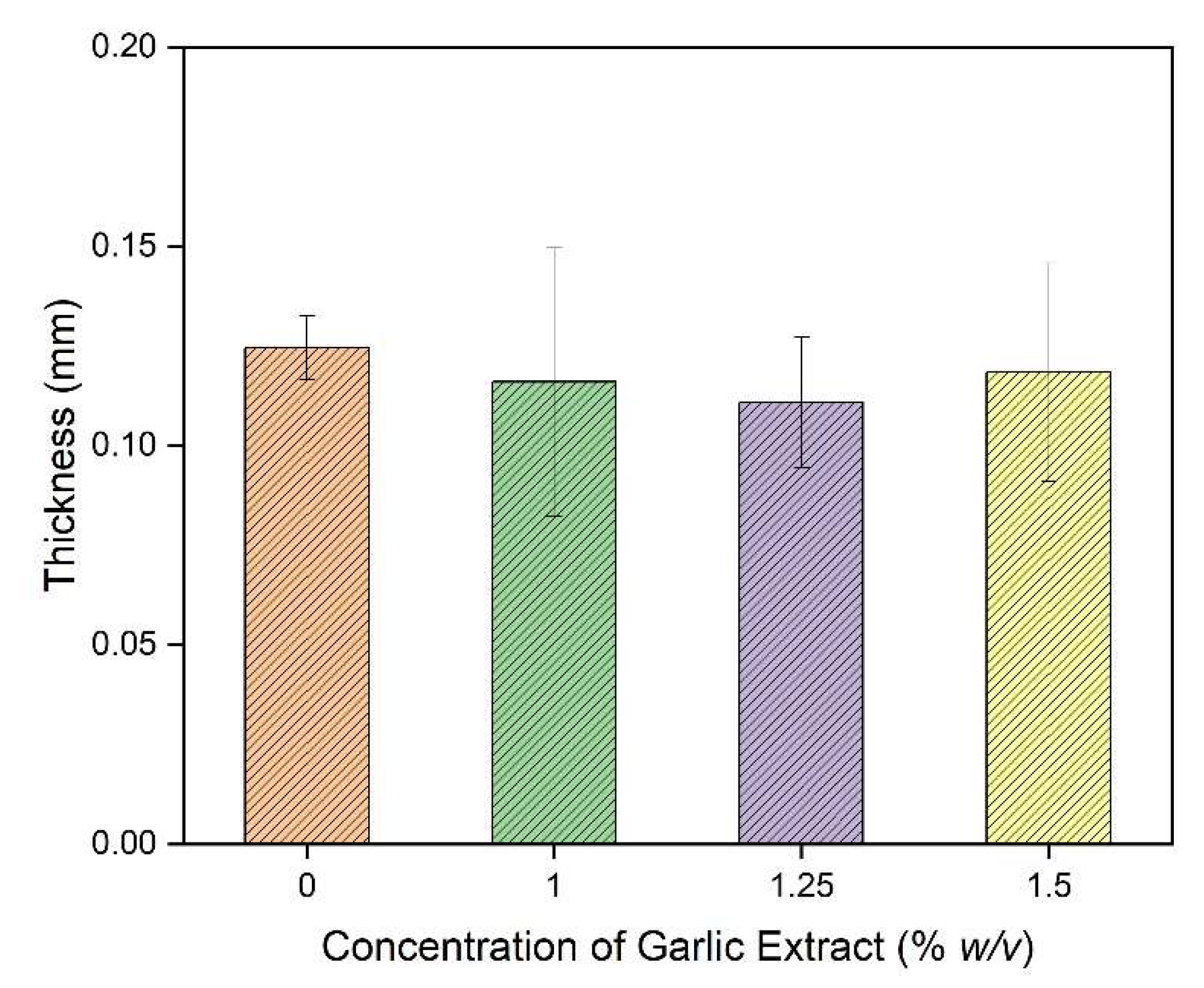
The membrane thickness measurement was conducted to ensure that the fabricated membranes have comparable thicknesses. The thickness affects the liquid mass transfer of the membranes, along with the membrane tortuosity [90,91,92].
The thicknesses of the membrane samples are shown in Figure 2. In Figure 2, the final thicknesses of all of the membrane samples were comparable, and the loading of the garlic extract into the dope solution had no effect. The addition of the garlic extract in the membrane solution did not affect the solubility of the solvent and non-solvent, which could affect the physical properties of the membrane. These findings indicate that loading garlic extract in the dope solution does not affect the inflow of water (as a non-solvent) during the phase inversion process. The membrane thickness is sensitive to the volume of water penetrating the cast film. The presence of water in the film will eventually expand the film thickness and create a void to enhance the porosity.

3.2.2. Tensile Strength and Elongation

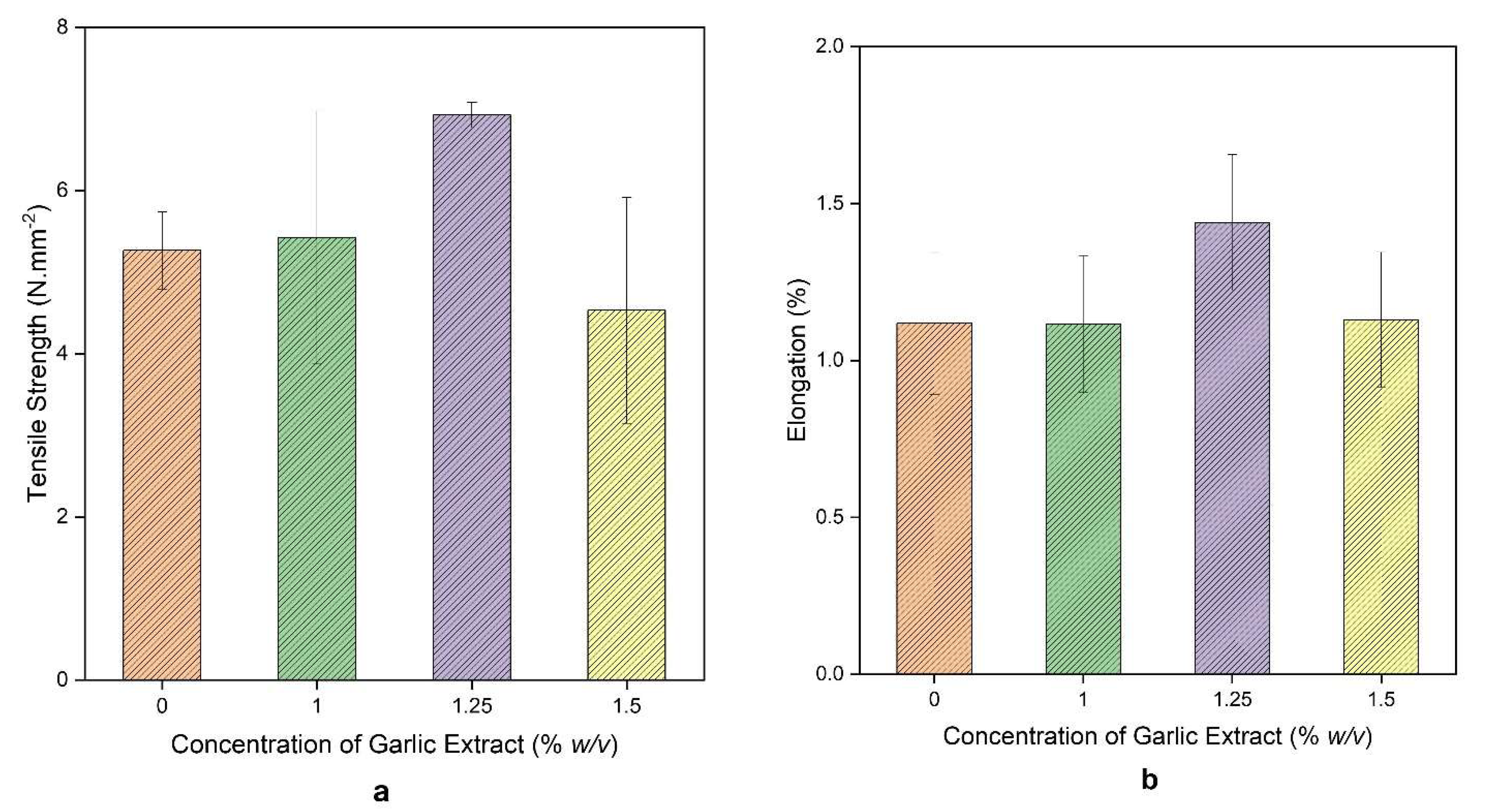
Tensile strength is a measure of a good membrane for withstanding mechanical damage during filtration. The tensile strengths and elongations of the resulting membranes are shown in Figure 3. The highest tensile strength of 6.93 N/mm2 was obtained for the membrane made from a dope solution containing 1.25% (w/v) garlic paste, and the lowest was obtained at a concentration of 1.5%. The tensile strengths of 1% and 1.5% (w/v) were comparable to the pristine membrane (without the garlic paste). The membrane thickness was similar; hence, the tensile strength was also expected to be comparable. A membrane with higher tensile strength is better at resisting mechanical damage. For instance, the addition of PDA and CNCs to the CA membrane may increase tensile strength and elongation by 76% and 35%, respectively. The CNC’s nature resulted in strong interactions with the polymers and PDA, which form hydrogen bonds with CA molecules [93].
The tensile strengths of the CA-based membranes were relatively small compared to the ones reported elsewhere [94], with a tensile strength value of 30 MPa for the CA-based membrane material, 20 MPa for the polyvinyl chloride-based membrane, 42 MPa for the polystyrene-based membrane, and 10 MPa for the LDPE-based membrane materials. Adding garlic extract may promote tensile strength reduction and change the membrane’s structure. Loading 1% Moringa powder resulted in changes to the structural and mechanical properties of the membrane [95].
The elongation of the membrane is directly proportional to the tensile strength. The higher the tensile strength, the higher the membrane’s elasticity, so that the relationship between the two is directly proportional. The highest elongation was achieved by the membrane made from a dope solution containing 1.25% (w/v) garlic extract with an elongation value of 1.44%. Loading the additives promoted polymer interactions, and the elasticity increased due to the selection of a suitable additive molecular weight. Structural changes suppressed the formation of macro voids (hydrogen bonds in the membrane pores) and increased mechanical strength [96]. The decrease in the tensile strength and elongation was associated with a phase separation from the pore structure, which decreased its mechanical properties [97].

3.2.3. Membrane Hydrophilicity

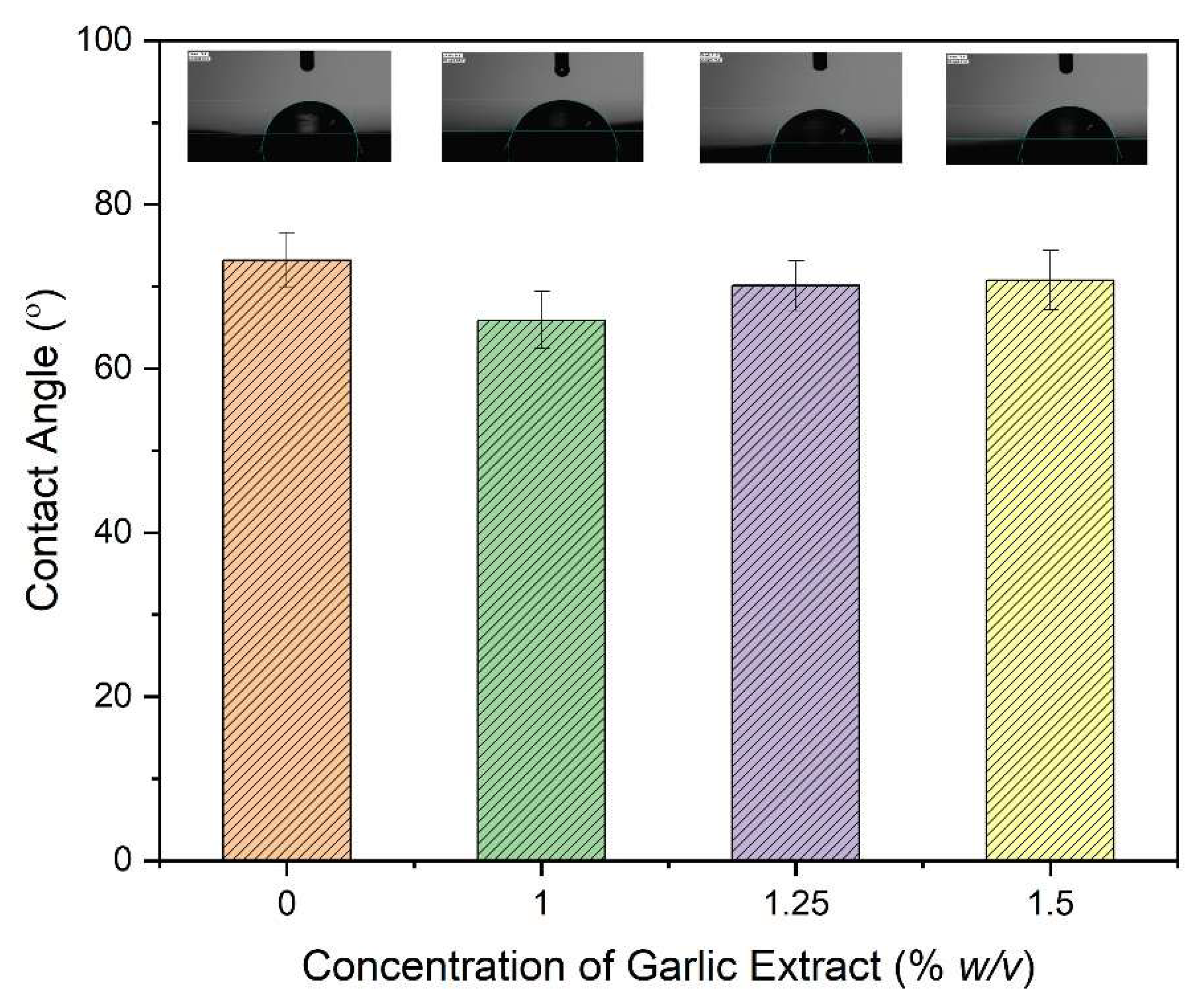
The contact angle data for the resulting membranes are shown in Figure 4. The contact angles for all samples were <90°, which means they were hydrophilic. The smallest value was for the membrane with 1% (w/v) garlic extract, with a contact angle of 64.9°, and the largest was 72.4° for the pristine Ca membrane. If a membrane has a contact angle value below 90°, it represents hydrophilic characteristics, while, if the value is more than 90°, it is hydrophobic [98]; however, if the contact angle exceeds 140°, it can be classified as superhydrophobic [99].
The contact angles for the prepared membranes were higher than those of CA-based membranes loaded with cacao pod husk extract as an additive using DMF and DMAC solvents, which had contact angle values of 40–60° [61]. Another study reported that for PES and PES/ZNO membranes with DMF as the solvent, the contact angle values were 67° and 46°, respectively. This value difference was caused by increased hydrophilicity and pore density on the membrane surface [100].
The contact angle is related to the roughness of the membrane’s outer surface [101]. The hydrophilicity measurement has almost no effect on the membrane surface, but it is essential to show the antibacterial properties of the membrane, which are a direct effect of the natural phenolic extracts—not due to the nature of the membrane surface [61]. The hydrophilicity of the membrane is influenced by several factors, namely shelf-life, fouling, efficiency, and flux, by measuring the contact angle on the membrane surface between the water droplet and the solid surface [98]. The contact angle value has an inverse relationship with the permeation rate. If the material has high hydrophilicity (a small contact angle value), the permeation rate (flux) increases [102].

3.2.4. Permeability

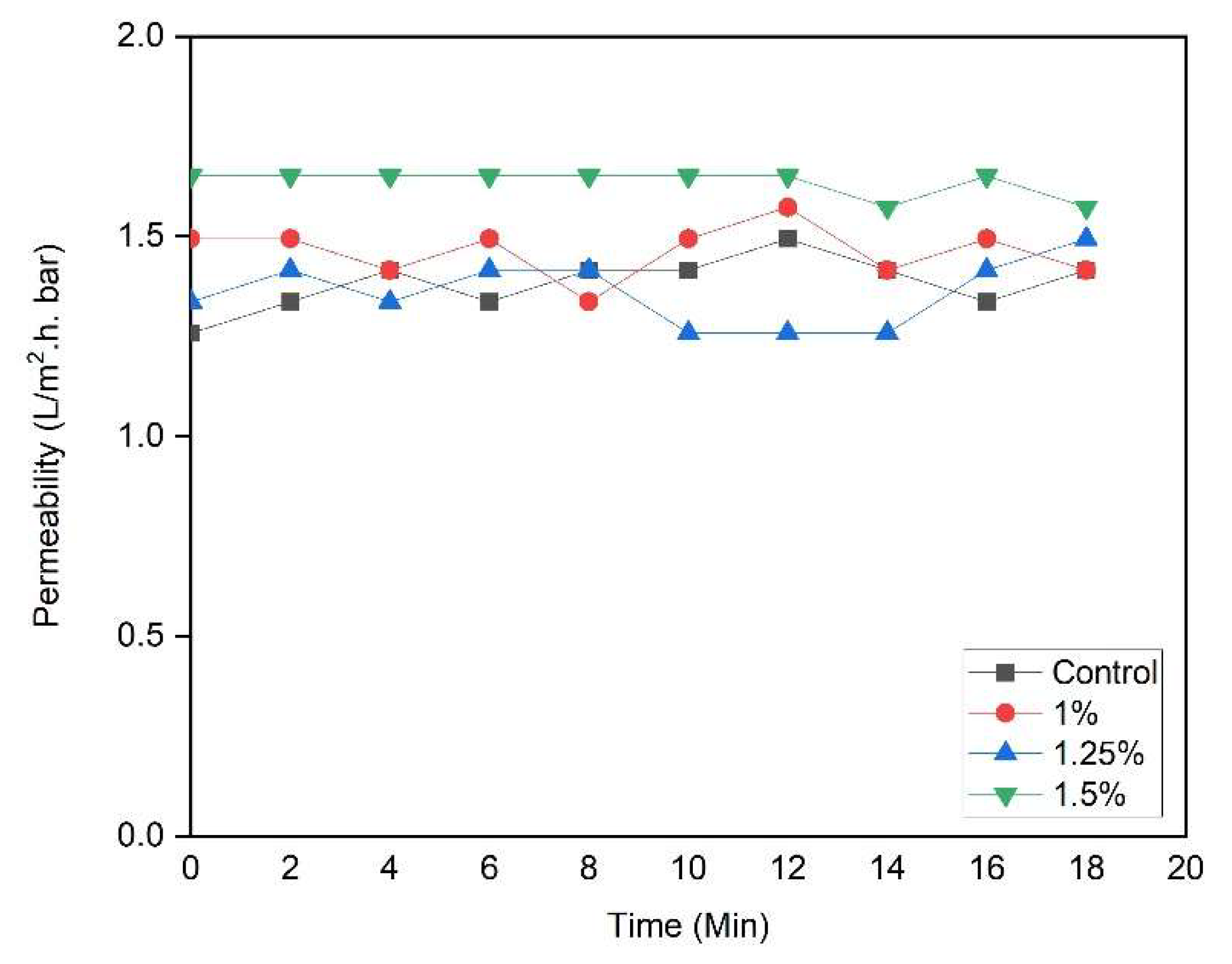
The mass transfer of the membrane can be calculated by measuring the flux of clean water passing through the membrane’s pores. The clean water permeability of the composite membrane is shown in Figure 5. Clean water permeability was used to measure water mass transport through a porous membrane. All membranes had a stable clean water permeability value. The membrane with the smallest pore produced the most considerable flux value due to higher resistance when water passes through the membrane. The mass flow rate through a membrane decreases in value due to scaling, concentration polarization, and fouling, which typically occurs when treating an actual feed.
Based on the permeability tests, it can be estimated that all membranes were of the ultrafiltration type. The estimation was based on the simplified categorization determined by [1,95]. In another study, a CA-based membrane was enriched with the PEG 600 additive, and it was found that the pure flux of water increased with the addition of the concentration of the additive because more pores were formed [7].

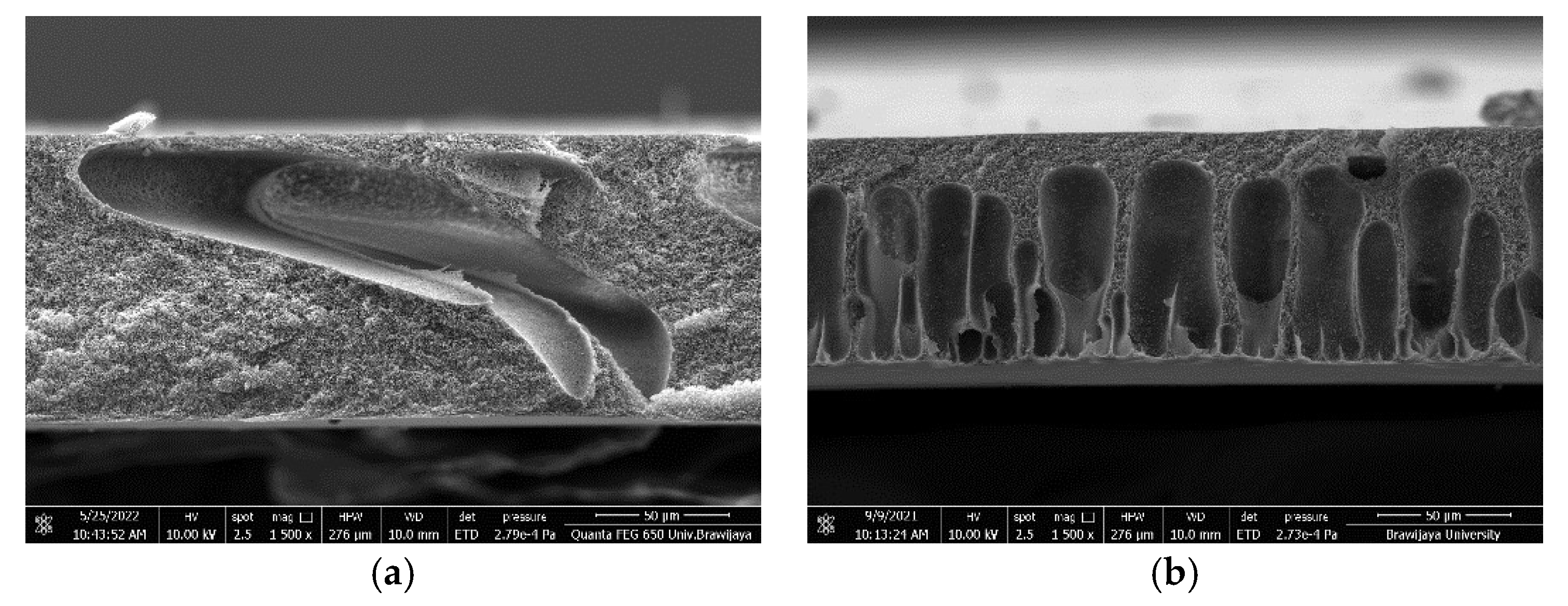
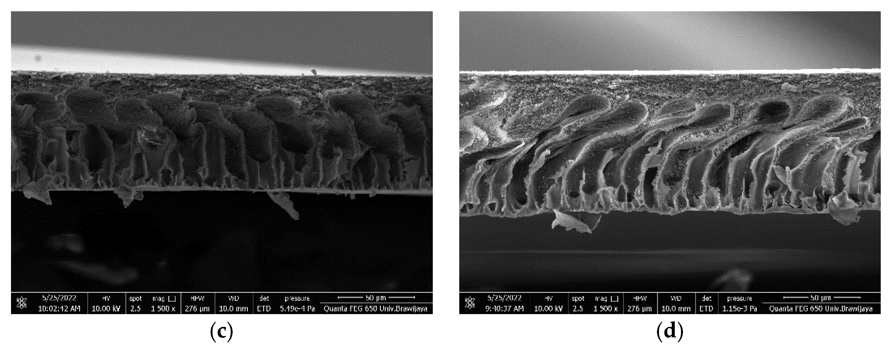
3.2.5. Membrane Morphology

A cross-section of the cellulose acetate composite membrane supported by the garlic extract paste is shown in Figure 6. The addition of garlic extracts during the fabrication of CA-based membranes affects the number and the size of micro voids. Increasing the concentration of garlic extract causes the macro void shape to become rounder, longer, and more uniform than the pristine CA-membrane. The change to the cross-section morphology can be attributed to the phase inversion process. A few large macro voids formed for the pristine CA-based membrane, while smaller, more finger-like macro voids formed on the membranes containing garlic extract. It seems that adding garlic extract hastens the phase inversion process, leading to the formation of finger-like macro voids near the top of the membrane surface.
At 1% and 1.25% (w/v) garlic extract, the macro void forms were visible and more regular than those from the 1.5% (w/v). The formation of micro voids can occur due to faster water and solvent changes during the phase inversion method, so the exchange of the solvent and non-solvent becomes an essential factor in forming membrane pores [103]. The macro voids are responsible for the formation of a greater thickness. The decrease in the membrane’s pore size is directly proportional to the decrease in the thickness of the membrane due to the lack of macro cavities [104]. Changes in the structure of the membrane can occur due to the selection of a suitable additive molecular weight to suppress the formation of macro voids, leading to an increase in mechanical properties [96]. The formation of a sponge-like structure in the entire cross-section occurs due to a decrease in the coagulation rate of PVDF-HFP with an increase in the copolymer concentration, which is suitable for slow coagulation processes [105].

3.2.6. Anti-Biofouling Tests

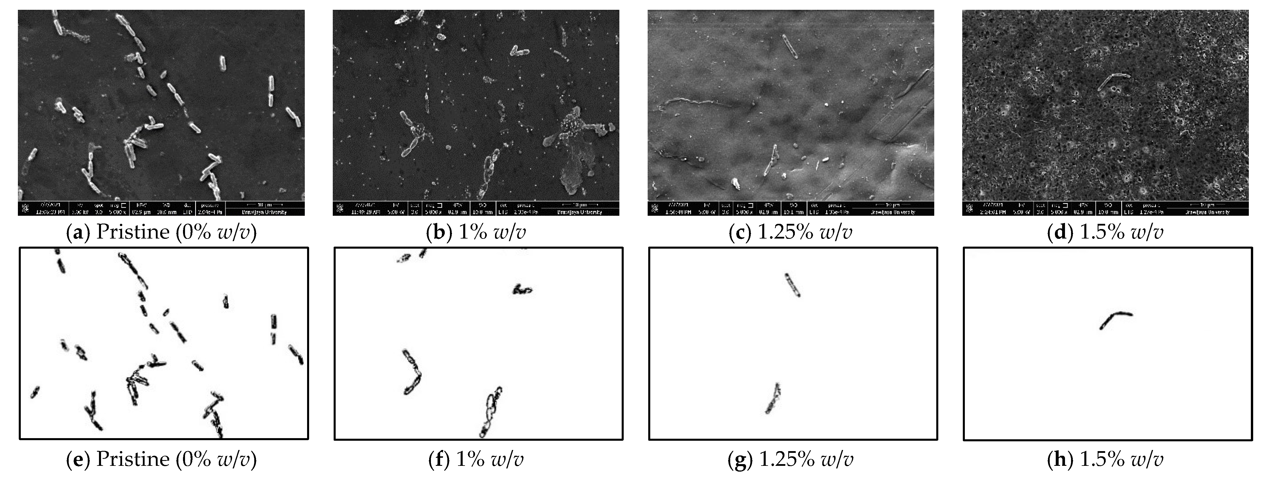
The antibacterial activity was tested by observing the attachment of bacteria to the surface of the cellulose acetate composite membrane. The bacterial attachments to the membrane surface are shown in Figure 7 and Figure 8. All membranes are adhered by the bacteria. E. coli have a length of about 2 μm with a rod shape and a diameter of 0.5 μm, live at temperatures of 20–40 °C, and belong to the Gram-negative category. B. subtilis has a rod shape measuring 0.5–2.5 × 1.2–10 m, withstands −5–75 °C, and belongs to the Gram-positive category. The phenolic compound in the membrane composites may reduce the bacterial adhesion, yet the effect was more advanced in Gram-negative bacteria due to a low-affinity, thinner cell wall, and only contains a small amount of peptidoglycan [6].
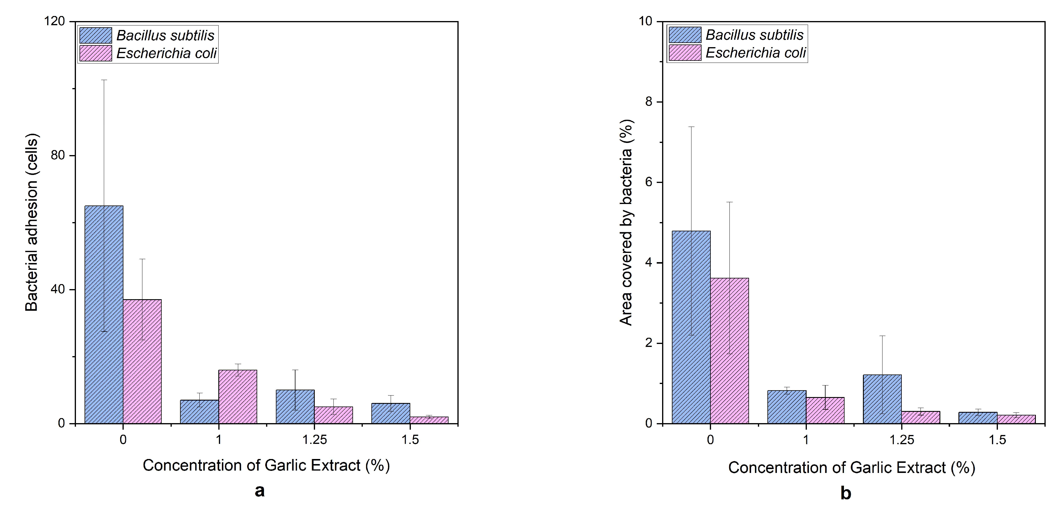
The number of bacteria and the percentage of the attachment area to the membrane can be seen in Figure 9. The bacterial adhesion on the membrane surface, both for the B. subtilis and E. coli bacteria, decreased in the presence of the garlic extract paste in the dope solution, as compared to the pristine membrane. The bacterial adhesion of B. subtilis decreased by 91.5 + 11.7%, and E. coli decreased by 94.0 ± 11.9%, the highest presence of the garlic phenolic extract. A Gram-negative bacterium has 1–3 nm peptidoglycan, while a Gram-positive bacterium has 20–50 nm peptidoglycan [106].
Garlic has natural antibacterial properties due to its content of phenolic compounds (allicin), which can inhibit bacterial activity (both Gram-positive and -negative) [68]. Adding garlic extract paste to the membrane inhibited the antibacterial activity by decreasing the number and percent of bacterial attachment areas compared to the pristine membrane. An increase in the concentration of the garlic extract paste will increase the antibacterial activity.

3.2.7. Membrane Phenolic Content

Spectroscopic methods also analyzed the phenol content of the membrane loaded with the garlic extract. The phenol content in the membrane is summarized in Table 4.
Based on Table 4, the membranes contained 0.94%, 0.81%, and 0.58% phenolic substances for 1%, 1.25%, and 1.5% (w/v) garlic extract concentrations, respectively. This indicates that, during the mixing, the phenol content of the extract was not lost and remained in the membrane to provide antibacterial activity, as proven from LC-MS/MS analysis in Section 3.1.
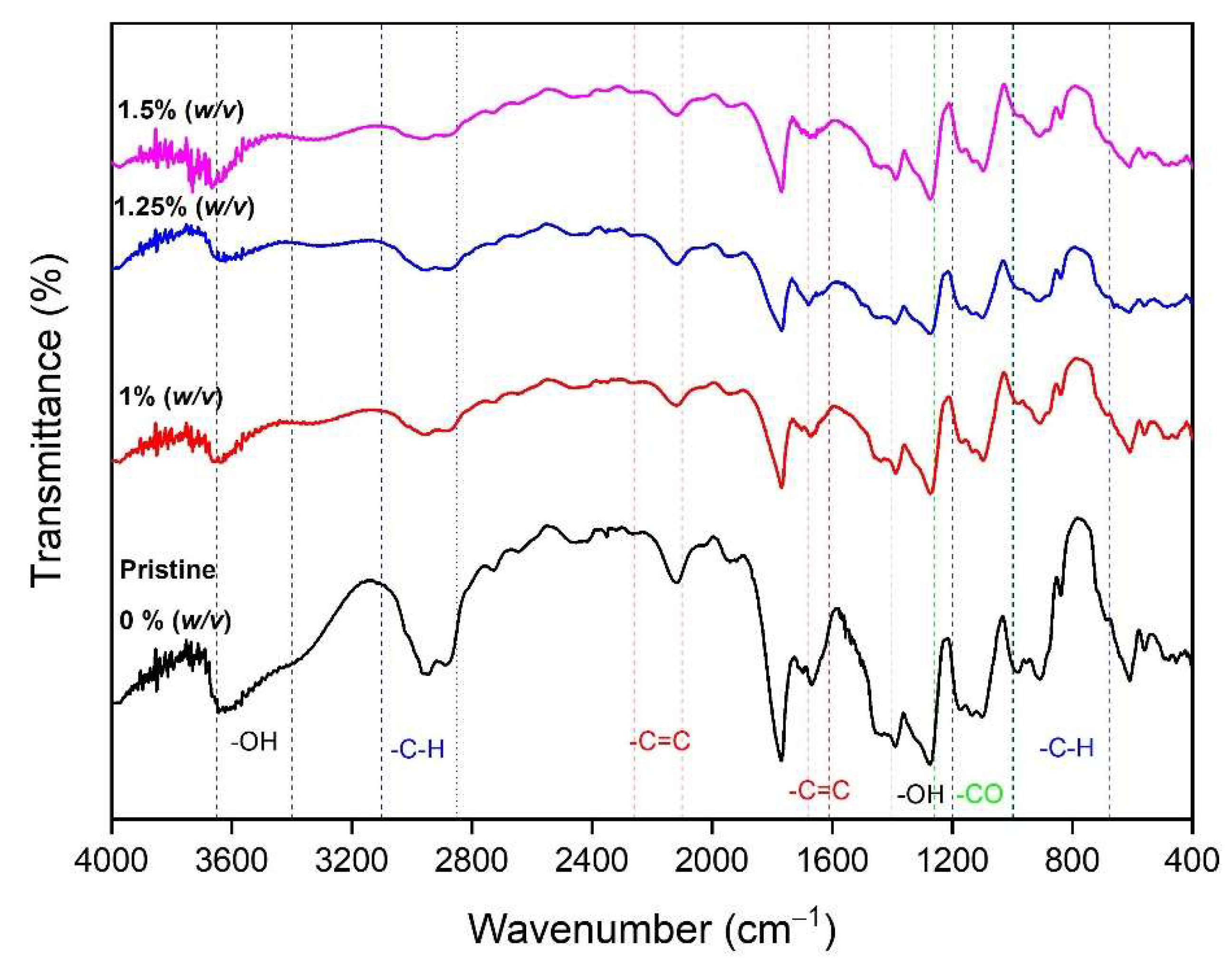
In addition, the phenolic content of the membrane was also measured using FTIR to see its functional groups (Figure 10). The measurement of the functional groups obtained broad absorption peaks on the membrane loaded with the garlic extract paste in a strong absorption area extending 3650–3400 cm−1 (O-H) and was supported by absorption in the fingerprint region of 1400–1200 cm−1 (O-H). In addition, the contents of aromatic ester groups or alcohol and phenolic compounds were detected in the 1260–1000 cm−1 (C-O) area. The phenolic compounds generally had a known aromatic ring with an aromatic C-H functional group that appears at wavelengths of 3010–3100 and 690–900 cm−1. The wave region of 2850–2970 cm−1 indicated the presence of the functional groups C-H alkanes and C-H alkenes at 675–995 cm−1. The strong absorption in this region can be used to determine the position of the substituent on the aromatic ring. Meanwhile, the aromatic ring’s C=C (alkene) vibration can be seen in the 1610–1680 cm−1 and the C=C alkyne at 2100–2260 cm−1.

4. Conclusions

Our overall findings demonstrate that adding garlic extract affected the thickness, tensile strength, elongation, clean water permeability, and attachment of B. subtilis and E. coli bacteria. The membranes loaded with garlic extract paste had hydrophilic properties suitable for water filtration. Immersion for 18 h in bacterial broths demonstrated the antibacterial activity of the membranes as shown by the suppression of the number of bacteria by 91.5 ± 11.7% on B. subtilis and 94.0 ± 11.9% on E. coli. The phenolic content was immensely detected in the membranes with up to 0.94% compositions. Antifouling additives did prevent the attachment of fouling to the membrane surface at the beginning of the process, yet they did not easily prevent further foulant adhesion. Nevertheless, based on the overall characterization results, the resulting membranes can be used for juice clarification via ultrafiltration. The membrane sheets will be used as materials for plate-and-frame membrane modules and will be a subject of future study.

Author Contributions

Conceptualization and methodology, Y.W.; investigation and data curation, D.A.; writing—original draft preparation, D.A. and Y.W.; writing—review and editing, Y.W., B.D.A., M.B.H., J.E.W., and M.R.B.; supervision, Y.W. and B.D.A.; project administration, D.A.; funding acquisition, Y.W. All authors have read and agreed to the published version of the manuscript.

Funding

This research was funded by the Ministry of Research and Technology of the Republic of Indonesia, the National Research and Innovation Agency, and Indonesia Endowment Fund for Education at Universitas Brawijaya.

Acknowledgments

The authors would like to thanks to Angky Wahyu Putranto, Fara Aulia Agustin Nurhadi, and Amelia Saraswati for fruitful collaboration and discussion.

Conflicts of Interest

The authors declare no conflict of interest.

References

  1. Mulder, M.; Mulder, J. Basic Principles of Membrane Technology; Springer: Dordrecht, The Netherlands, 1996. [Google Scholar]
  2. Dürr, S.; Thomason, J.C. Biofouling; Wiley: Hoboken, NJ, USA, 2009. [Google Scholar]
  3. Wibisono, Y.; El Obied, K.E.; Cornelissen, E.R.; Kemperman, A.J.B.; Nijmeijer, K. Biofouling removal in spiral-wound nanofiltration elements using two-phase flow cleaning. J. Membr. Sci. 2015, 475, 131–146. [Google Scholar] [CrossRef]
  4. Komlenic, R. Rethinking the causes of membrane biofouling. Filtr. Sep. 2010, 47, 26–28. [Google Scholar] [CrossRef]
  5. Wibisono, Y.; Cornelissen, E.R.; Kemperman, A.J.B.; van der Meer, W.G.J.; Nijmeijer, K. Two-phase flow in membrane processes: A technology with a future. J. Membr. Sci. 2014, 453, 566–602. [Google Scholar] [CrossRef]
  6. Wibisono, Y.; Amalia Rachmawati, S.; Septyaningrum Mylani, V.; Rosalia Dewi, S.; Wahyu Putranto, A.; Arif, C.; Shalahuddin, I.; Bagus Hermanto, M. Cellulose acetate polymer matrix loaded Olea europaea nanosolids as low fouling membrane composite. Alex. Eng. J. 2023, 64, 119–129. [Google Scholar] [CrossRef]
  7. Arthanareeswaran, G.; Thanikaivelan, P.; Srinivasn, K.; Mohan, D.; Rajendran, M. Synthesis, characterization and thermal studies on cellulose acetate membranes with additive. Eur. Polym. J. 2004, 40, 2153–2159. [Google Scholar] [CrossRef]
  8. Wibisono, Y.; Yandi, W.; Golabi, M.; Nugraha, R.; Cornelissen, E.R.; Kemperman, A.J.B.; Ederth, T.; Nijmeijer, K. Hydrogel-coated feed spacers in two-phase flow cleaning in spiral wound membrane elements: A novel platform for eco-friendly biofouling mitigation. Water Res. 2015, 71, 171–186. [Google Scholar] [CrossRef] [Green Version]
  9. Liu, C.X.; Zhang, D.R.; He, Y.; Zhao, X.S.; Bai, R. Modification of membrane surface for anti-biofouling performance: Effect of anti-adhesion and anti-bacteria approaches. J. Membr. Sci. 2010, 346, 121–130. [Google Scholar] [CrossRef]
  10. Zhang, D.Y.; Liu, J.; Shi, Y.S.; Wang, Y.; Liu, H.F.; Hu, Q.L.; Su, L.; Zhu, J. Antifouling polyimide membrane with surface-bound silver particles. J. Membr. Sci. 2016, 516, 83–93. [Google Scholar] [CrossRef]
  11. Zhang, D.Y.; Hao, Q.; Liu, J.; Shi, Y.S.; Zhu, J.; Su, L.; Wang, Y. Antifouling polyimide membrane with grafted silver nanoparticles and zwitterion. Sep. Purif. Technol. 2018, 192, 230–239. [Google Scholar] [CrossRef]
  12. Liu, Z.; Hu, Y. Sustainable Antibiofouling Properties of Thin Film Composite Forward Osmosis Membrane with Rechargeable Silver Nanoparticles Loading. ACS Appl. Mater. Interfaces 2016, 8, 21666–21673. [Google Scholar] [CrossRef]
  13. Ahmad, J.; Wen, X.; Li, F.; Wang, B. Novel triangular silver nanoparticle modified membranes for enhanced antifouling performance. RSC Adv. 2019, 9, 6733–6744. [Google Scholar] [CrossRef] [Green Version]
  14. Karkhanechi, H.; Takagi, R.; Ohmukai, Y.; Matsuyama, H. Enhancing the antibiofouling performance of RO membranes using Cu(OH)2 as an antibacterial agent. Desalination 2013, 325, 40–47. [Google Scholar] [CrossRef]
  15. Liu, C.; Wang, Z.; He, Q.; Jackson, J.; Faria, A.F.; Zhang, W.; Song, D.; Ma, J.; Sun, Z. Facile preparation of anti-biofouling reverse osmosis membrane embedded with polydopamine-nano copper functionality: Performance and mechanism. J. Membr. Sci. 2022, 658, 120721. [Google Scholar] [CrossRef]
  16. García, A.; Rodríguez, B.; Giraldo, H.; Quintero, Y.; Quezada, R.; Hassan, N.; Estay, H. Copper-modified polymeric membranes for water treatment: A comprehensive review. Membranes 2021, 11, 93. [Google Scholar] [CrossRef] [PubMed]
  17. Ngo, T.H.A.; Nguyen, T.D.; Nguyen, V.D.; Tran, D.T. Improvement in anti-biofouling property of polyamide thin-film composite membranes by using copper nanoparticles. J. Appl. Polym. Sci. 2022, 139, e52739. [Google Scholar] [CrossRef]
  18. Esfahani, M.R.; Koutahzadeh, N.; Esfahani, A.R.; Firouzjaei, M.D.; Anderson, B.; Peck, L. A novel gold nanocomposite membrane with enhanced permeation, rejection and self-cleaning ability. J. Membr. Sci. 2019, 573, 309–319. [Google Scholar] [CrossRef]
  19. Armendariz Ontiveros, M.; Quintero, Y.; Llanquilef, A.; Morel, M.; Argentel, L.; Garcia, A.; García, A. Anti-Biofouling and Desalination Properties of Thin Film Composite Reverse Osmosis Membranes Modified with Copper and Iron Nanoparticles. Materials 2019, 12, 2081. [Google Scholar] [CrossRef] [Green Version]
  20. Köse-Mutlu, B.; Türken, T.; Güçlü, S.; Güçlü, M.; Durmaz, G.; Okatan, S.; Övez, S.; Koyuncu, I. Bismuth Chelate-Doped Microfiltration Membrane and Its Anti-Biofouling Performance During a High-Flux Membrane Bioreactor Operation. CLEAN—Soil Air Water 2017, 45, 1500923. [Google Scholar] [CrossRef]
  21. Yavuz, F.; Sengur, R.; Turken, T.; Urper-Bayram, G.; Koyuncu, I. Improvement of anti-biofouling properties of hollow fiber membranes with bismuth-BAL chelates (BisBAL). Environ. Technol. 2017, 40, 1–31. [Google Scholar] [CrossRef]
  22. Gzara, L.; Ahmad Rehan, Z.; Khan, S.B.; Alamry, K.A.; Albeirutty, M.H.; El-Shahawi, M.S.; Rashid, M.I.; Figoli, A.; Drioli, E.; Asiri, A.M. Preparation and characterization of PES-cobalt nanocomposite membranes with enhanced anti-fouling properties and performances. J. Taiwan Inst. Chem. Eng. 2016, 65, 405–419. [Google Scholar] [CrossRef]
  23. Dadari, S.; Rahimi, M.; Zinadini, S. Novel antibacterial and antifouling PES nanofiltration membrane incorporated with green synthesized nickel-bentonite nanoparticles for heavy metal ions removal. Chem. Eng. J. 2022, 431, 134116. [Google Scholar] [CrossRef]
  24. Khorshidi, B.; Biswas, I.; Ghosh, T.; Thundat, T.; Sadrzadeh, M. Robust fabrication of thin film polyamide-TiO2 nanocomposite membranes with enhanced thermal stability and anti-biofouling propensity. Sci. Rep. 2018, 8, 784. [Google Scholar] [CrossRef] [PubMed] [Green Version]
  25. Anan, N.S.M.; Jaafar, J.; Sato, S.; Mohamud, R. Titanium Dioxide Incorporated Polyamide Thin Film Composite Photocatalytic Membrane for Bisphenol A Removal. IOP Conf. Ser.: Mater. Sci. Eng. 2021, 1142, 012015. [Google Scholar] [CrossRef]
  26. Samree, K.; Srithai, P.U.; Kotchaplai, P.; Thuptimdang, P.; Painmanakul, P.; Hunsom, M.; Sairiam, S. Enhancing the Antibacterial Properties of PVDF Membrane by Hydrophilic Surface Modification Using Titanium Dioxide and Silver Nanoparticles. Membranes 2020, 10, 289. [Google Scholar] [CrossRef]
  27. Kotlhao, K.; Lawal, I.A.; Moutloali, R.M.; Klink, M.J. Antifouling Properties of Silver-Zinc Oxide Polyamide Thin Film Composite Membrane and Rejection of 2-Chlorophenol and 2,4-Dichlorophenol. Membranes 2019, 9, 96. [Google Scholar] [CrossRef] [Green Version]
  28. Kamaludin, R.; Abdul Majid, L.; Othman, M.H.D.; Mansur, S.; Sheikh Abdul Kadir, S.H.; Wong, K.Y.; Khongnakorn, W.; Puteh, M.H. Polyvinylidene Difluoride (PVDF) Hollow Fiber Membrane Incorporated with Antibacterial and Anti-Fouling by Zinc Oxide for Water and Wastewater Treatment. Membranes 2022, 12, 110. [Google Scholar] [CrossRef]
  29. Cheng, H.; Guan, Q.; Villalobos, L.F.; Peinemann, K.-V.; Pain, A.; Hong, P.-Y. Understanding the antifouling mechanisms related to copper oxide and zinc oxide nanoparticles in anaerobic membrane bioreactors. Environ. Sci.: Nano 2019, 6, 3467–3479. [Google Scholar] [CrossRef] [Green Version]
  30. Abdulsalam, M.; Che Man, H.; Goh, P.S.; Yunos, K.F.; Zainal Abidin, Z.; Isma, M.I.A.; Ismail, A.F. Permeability and Antifouling Augmentation of a Hybrid PVDF-PEG Membrane Using Nano-Magnesium Oxide as a Powerful Mediator for POME Decolorization. Polymers 2020, 12, 549. [Google Scholar] [CrossRef] [Green Version]
  31. Yigit, B.; Ozay, Y.; Emen, F.; Ozsoy, E.; Ocakoglu, K.; Dizge, N. Development of ruthenium oxide modified polyethersulfone membranes for improvement of antifouling performance including decomposition kinetic of polymer. J. Polym. Environ. 2022, 1–13. [Google Scholar] [CrossRef]
  32. Zha, X.-J.; Zhao, X.; Pu, J.-H.; Tang, L.-S.; Ke, K.; Bao, R.-Y.; Bai, L.; Liu, Z.-Y.; Yang, M.-B.; Yang, W. Flexible Anti-Biofouling MXene/Cellulose Fibrous Membrane for Sustainable Solar-Driven Water Purification. ACS Appl. Mater. Interfaces 2019, 11, 36589–36597. [Google Scholar] [CrossRef]
  33. Rasool, K.; Mahmoud, K.A.; Johnson, D.J.; Helal, M.; Berdiyorov, G.R.; Gogotsi, Y. Efficient Antibacterial Membrane based on Two-Dimensional Ti3C2Tx (MXene) Nanosheets. Sci. Rep. 2017, 7, 1598. [Google Scholar] [CrossRef]
  34. Zhang, T.; Zhang, L.-Z. Development of a MXene-based membrane with excellent anti-fouling for air humidification-dehumidification type desalination. J. Membr. Sci. 2022, 641, 119907. [Google Scholar] [CrossRef]
  35. Nitodas, S.F.; Das, M.; Shah, R. Applications of Polymeric Membranes with Carbon Nanotubes: A Review. Membranes 2022, 12, 454. [Google Scholar] [CrossRef] [PubMed]
  36. Kim, Y.; Yang, E.; Park, H.; Choi, H. Anti-biofouling effect of a thin film nanocomposite membrane with a functionalized-carbon-nanotube-blended polymeric support for the pressure-retarded osmosis process. RSC Adv. 2020, 10, 5697–5703. [Google Scholar] [CrossRef] [PubMed] [Green Version]
  37. Kim, E.-S.; Hwang, G.; Gamal El-Din, M.; Liu, Y. Development of nanosilver and multi-walled carbon nanotubes thin-film nanocomposite membrane for enhanced water treatment. J. Membr. Sci. 2012, 394–395, 37–48. [Google Scholar] [CrossRef]
  38. Chae, H.-R.; Lee, J.; Lee, C.-H.; Kim, I.-C.; Park, P.-K. Graphene oxide-embedded thin-film composite reverse osmosis membrane with high flux, anti-biofouling, and chlorine resistance. J. Membr. Sci. 2015, 483, 128–135. [Google Scholar] [CrossRef]
  39. Wang, J.; Gao, X.; Yu, H.; Wang, Q.; Ma, Z.; Li, Z.; Zhang, Y.; Gao, C. Accessing of graphene oxide (GO) nanofiltration membranes for microbial and fouling resistance. Sep. Purif. Technol. 2019, 215, 91–101. [Google Scholar] [CrossRef]
  40. Mokkapati, V.R.S.S.; Koseoglu-Imer, D.Y.; Yilmaz-Deveci, N.; Mijakovic, I.; Koyuncu, I. Membrane properties and anti-bacterial/anti-biofouling activity of polysulfone–graphene oxide composite membranes phase inversed in graphene oxide non-solvent. RSC Adv. 2017, 7, 4378–4386. [Google Scholar] [CrossRef] [Green Version]
  41. Zhao, D.L.; Das, S.; Chung, T.-S. Carbon Quantum Dots Grafted Antifouling Membranes for Osmotic Power Generation via Pressure-Retarded Osmosis Process. Environ. Sci. Technol. 2017, 51, 14016–14023. [Google Scholar] [CrossRef]
  42. Mahat, N.A.; Shamsudin, S.A.; Jullok, N.; Ma’Radzi, A.H. Carbon quantum dots embedded polysulfone membranes for antibacterial performance in the process of forward osmosis. Desalination 2020, 493, 114618. [Google Scholar] [CrossRef]
  43. Zhao, D.L.; Chung, T.-S. Applications of carbon quantum dots (CQDs) in membrane technologies: A review. Water Res. 2018, 147, 43–49. [Google Scholar] [CrossRef] [PubMed]
  44. Khoerunnisa, F.; Kulsum, C.; Dara, F.; Nurhayati, M.; Nashrah, N.; Fatimah, S.; Pratiwi, A.; Hendrawan, H.; Nasir, M.; Ko, Y.G.; et al. Toughened chitosan-based composite membranes with antibiofouling and antibacterial properties via incorporation of benzalkonium chloride. RSC Adv. 2021, 11, 16814–16822. [Google Scholar] [CrossRef] [PubMed]
  45. Zhao, S.; Tao, Z.; Chen, L.; Han, M.; Zhao, B.; Tian, X.; Wang, L.; Meng, F. An antifouling catechol/chitosan-modified polyvinylidene fluoride membrane for sustainable oil-in-water emulsions separation. Front. Environ. Sci. Eng. 2020, 15, 63. [Google Scholar] [CrossRef]
  46. Zhai, X.; Chen, B.; He, Y.; An, L.; Chen, S.; Yan, X.; Zhang, Y.; Meng, J. A novel loose nanofiltration membrane with superior anti-biofouling performance prepared from zwitterion-grafted chitosan. J. Taiwan Inst. Chem. Eng. 2022, 132, 104191. [Google Scholar] [CrossRef]
  47. Atkinson, A.J.; Wang, J.; Grzebyk, K.; Zhang, Z.; Jung, D.; Zeng, D.; Pollard, A.; Gold, A.; Coronell, O. Scalable fabrication of anti-biofouling membranes through 2-aminoimidazole incorporation during polyamide casting. J. Membr. Sci. 2019, 579, 151–161. [Google Scholar] [CrossRef]
  48. Atkinson, A.J.; Armstrong, M.D.; Eskew, J.T.; Coronell, O. 2-Aminoimidazole reduces fouling and improves membrane performance. J. Membr. Sci. 2021, 629, 119262. [Google Scholar] [CrossRef]
  49. Vimbela, G.V.; Ngo, S.M.; Fraze, C.; Yang, L.; Stout, D.A. Antibacterial properties and toxicity from metallic nanomaterials. Int. J. Nanomed. 2017, 12, 3941–3965. [Google Scholar] [CrossRef] [Green Version]
  50. Chen, L.Q.; Fang, L.; Ling, J.; Ding, C.Z.; Kang, B.; Huang, C.Z. Nanotoxicity of Silver Nanoparticles to Red Blood Cells: Size Dependent Adsorption, Uptake, and Hemolytic Activity. Chem. Res. Toxicol. 2015, 28, 501–509. [Google Scholar] [CrossRef]
  51. Zhao, F.; Zhao, Y.; Liu, Y.; Chang, X.; Chen, C.; Zhao, Y. Cellular uptake, intracellular trafficking, and cytotoxicity of nanomaterials. Small 2011, 7, 1322–1337. [Google Scholar] [CrossRef]
  52. Qiu, H.; Feng, K.; Gapeeva, A.; Meurisch, K.; Kaps, S.; Li, X.; Yu, L.; Mishra, Y.K.; Adelung, R.; Baum, M. Functional polymer materials for modern marine biofouling control. Prog. Polym. Sci. 2022, 127, 101516. [Google Scholar] [CrossRef]
  53. Kotsanopoulos, K.V.; Arvanitoyannis, I.S. Membrane processing technology in the food industry: Food processing, wastewater treatment, and effects on physical, microbiological, organoleptic, and nutritional properties of foods. Crit. Rev. Food Sci. Nutr. 2015, 55, 1147–1175. [Google Scholar] [CrossRef] [PubMed]
  54. Conidi, C.; Drioli, E.; Cassano, A. Perspective of Membrane Technology in Pomegranate Juice Processing: A Review. Foods 2020, 9, 889. [Google Scholar] [CrossRef] [PubMed]
  55. Ahmad, S.; Ahmed, S.K.M. Application of Membrane Technology in Food Processing. In Food Processing: Strategies for Quality Assessment; Malik, A., Erginkaya, Z., Ahmad, S., Erten, H., Eds.; Springer: New York, NY, USA, 2014; pp. 379–394. [Google Scholar] [CrossRef]
  56. Maddox, C.E.; Laur, L.M.; Tian, L. Antibacterial activity of phenolic compounds against the phytopathogen Xylella fastidiosa. Curr Microbiol 2010, 60, 53–58. [Google Scholar] [CrossRef] [PubMed] [Green Version]
  57. Sudjana, A.N.; D’Orazio, C.; Ryan, V.; Rasool, N.; Ng, J.; Islam, N.; Riley, T.V.; Hammer, K.A. Antimicrobial activity of commercial Olea europaea (olive) leaf extract. Int. J. Antimicrob. Agents 2009, 33, 461–463. [Google Scholar] [CrossRef] [PubMed]
  58. Liu, Y.; McKeever, L.C.; Malik, N.S. Assessment of the Antimicrobial Activity of Olive Leaf Extract Against Foodborne Bacterial Pathogens. Front. Microbiol. 2017, 8, 113. [Google Scholar] [CrossRef] [PubMed] [Green Version]
  59. Melia, S.; Novia, D.; Juliyarsi, I. Antioxidant and antimicrobial activities of gambir (Uncaria gambir Roxb) extracts and their application in rendang. Pak. J. Nutr. 2015, 14, 938. [Google Scholar] [CrossRef] [Green Version]
  60. Dewi, S.R.; Stevens, L.A.; Pearson, A.E.; Ferrari, R.; Irvine, D.J.; Binner, E.R. Investigating the role of solvent type and microwave selective heating on the extraction of phenolic compounds from cacao (Theobroma cacao L.) pod husk. Food Bioprod. Processing 2022, 134, 210–222. [Google Scholar] [CrossRef]
  61. Wibisono, Y.; Diniardi, E.M.; Alvianto, D.; Argo, B.D.; Hermanto, M.B.; Dewi, S.R.; Izza, N.; Putranto, A.W.; Saiful, S. Cacao Pod Husk Extract Phenolic Nanopowder-Impregnated Cellulose Acetate Matrix for Biofouling Control in Membranes. Membranes 2021, 11, 748. [Google Scholar] [CrossRef]
  62. Leyva-Jimenez, F.J.; Lozano-Sanchez, J.; Borras-Linares, I.; Cadiz-Gurrea, M.d.l.L.; Mahmoodi-Khaledi, E. Potential antimicrobial activity of honey phenolic compounds against Gram positive and Gram negative bacteria. LWT 2019, 101, 236–245. [Google Scholar] [CrossRef]
  63. Beristain-Bauza, S.D.C.; Hernández-Carranza, P.; Cid-Pérez, T.S.; Ávila-Sosa, R.; Ruiz-López, I.I.; Ochoa-Velasco, C.E. Antimicrobial Activity of Ginger (Zingiber Officinale) and Its Application in Food Products. Food Rev. Int. 2019, 35, 407–426. [Google Scholar] [CrossRef]
  64. Magryś, A.; Olender, A.; Tchórzewska, D. Antibacterial properties of Allium sativum L. against the most emerging multidrug-resistant bacteria and its synergy with antibiotics. Arch. Microbiol. 2021, 203, 2257–2268. [Google Scholar] [CrossRef] [PubMed]
  65. Abdul Qadir, M.; Shahzadi, S.K.; Bashir, A.; Munir, A.; Shahzad, S. Evaluation of Phenolic Compounds and Antioxidant and Antimicrobial Activities of Some Common Herbs. Int. J. Anal. Chem. 2017, 2017, 3475738. [Google Scholar] [CrossRef] [PubMed] [Green Version]
  66. Wibisono, Y.; Putri, A.Y.; Dewi, S.R.; Putranto, A.W.; Izza, N. Garlic-based phenolic nanopowder as antibiofouling agent in mixed-matrix membrane. IOP Conf. Ser.: Earth Environ. Sci. 2020, 542, 012005. [Google Scholar] [CrossRef]
  67. Bayan, L.; Koulivand, P.H.; Gorji, A. Garlic: A review of potential therapeutic effects. Avicenna J. Phytomed. 2014, 4, 1–14. [Google Scholar] [PubMed]
  68. Borlinghaus, J.; Albrecht, F.; Gruhlke, M.C.; Nwachukwu, I.D.; Slusarenko, A.J. Allicin: Chemistry and biological properties. Molecules 2014, 19, 12591–12618. [Google Scholar] [CrossRef] [Green Version]
  69. Cavallito, C.J.; Bailey, J.H. Allicin, the Antibacterial Principle of Allium sativum. I. Isolation, Physical Properties and Antibacterial Action. J. Am. Chem. Soc. 1944, 66, 1950–1951. [Google Scholar] [CrossRef]
  70. Feldberg, R.S.; Chang, S.C.; Kotik, A.N.; Nadler, M.; Neuwirth, Z.; Sundstrom, D.C.; Thompson, N.H. In vitro mechanism of inhibition of bacterial cell growth by allicin. Antimicrob. Agents Chemother. 1988, 32, 1763–1768. [Google Scholar] [CrossRef] [Green Version]
  71. Curtis, H.; Noll, U.; Störmann, J.; Slusarenko, A.J. Broad-spectrum activity of the volatile phytoanticipin allicin in extracts of garlic (Allium sativum L.) against plant pathogenic bacteria, fungi and Oomycetes. Physiol. Mol. Plant Pathol. 2004, 65, 79–89. [Google Scholar] [CrossRef]
  72. Kim, J.-S.; Kang, O.-J.; Gweon, O.-C. Comparison of phenolic acids and flavonoids in black garlic at different thermal processing steps. J. Funct. Foods 2013, 5, 80–86. [Google Scholar] [CrossRef]
  73. Kim, G.-H.; Duan, Y.; Lee, S.-C.; Kim, H.-S. Assessment of Antioxidant Activity of Garlic (Allium sativum L.) Peels by Various Extraction Solvents. J. Korean Oil Chem. Soc. 2016, 33, 204–212. [Google Scholar] [CrossRef]
  74. Kallel, F.; Driss, D.; Chaari, F.; Belghith, L.; Bouaziz, F.; Ghorbel, R.; Chaabouni, S.E. Garlic (Allium sativum L.) husk waste as a potential source of phenolic compounds: Influence of extracting solvents on its antimicrobial and antioxidant properties. Ind. Crops Prod. 2014, 62, 34–41. [Google Scholar] [CrossRef]
  75. Wibisono, Y.; Izza, N.; Savitri, D.; Dewi, S.R.; Putranto, A.W. The Extraction of Phenolic Compound from Garlic (Allium sativum L.) for Anti-Biofouling Agent in Membrane. J. Ilm. Rekayasa Pertan. Dan Biosist 2020, 8, 100–109. [Google Scholar] [CrossRef] [Green Version]
  76. Wibisono, Y.; Faradilla, A.; Utoro, P.A.; Sukoyo, A.; Izza, N.; Dewi, S.R. Natural Anti-Biofoulant Moringa oleifera Impregnated Cellulose Acetate Mixed Matrix Membranes for Juice Clarification. J. Rekayasa Kim. Lingkung. 2018, 13, 100–109. [Google Scholar] [CrossRef] [Green Version]
  77. Pan, Y.; Yu, Z.; Shi, H.; Chen, Q.; Zeng, G.; Di, H.; Ren, X.; He, Y. A novel antifouling and antibacterial surface-functionalized PVDF ultrafiltration membrane via binding Ag/SiO2 nanocomposites. J. Chem. Technol. Biotechnol. 2017, 92, 562–572. [Google Scholar] [CrossRef]
  78. Farag, M.A.; Ali, S.E.; Hodaya, R.H.; El-Seedi, H.R.; Sultani, H.N.; Laub, A.; Eissa, T.F.; Abou-Zaid, F.O.F.; Wessjohann, L.A. Phytochemical Profiles and Antimicrobial Activities of Allium cepa Red cv. and A. sativum Subjected to Different Drying Methods: A Comparative MS-Based Metabolomics. Molecules 2017, 22, 761. [Google Scholar] [CrossRef] [PubMed] [Green Version]
  79. Liu, J.; Guo, W.; Yang, M.; Liu, L.; Huang, S.; Tao, L.; Zhang, F.; Liu, Y. Investigation of the dynamic changes in the chemical constituents of Chinese “Laba” garlic during traditional processing. RSC Adv. 2018, 8, 41872–41883. [Google Scholar] [CrossRef] [PubMed] [Green Version]
  80. Ali, A.; Bashmil, Y.M.; Cottrell, J.J.; Suleria, H.A.R.; Dunshea, F.R. LC-MS/MS-QTOF Screening and Identification of Phenolic Compounds from Australian Grown Herbs and Their Antioxidant Potential. Antioxidants. 2021, 10, 1770. [Google Scholar] [CrossRef]
  81. Phan, A.D.T.; Netzel, G.; Chhim, P.; Netzel, M.E.; Sultanbawa, Y. Phytochemical Characteristics and Antimicrobial Activity of Australian Grown Garlic (Allium sativum L.) Cultivars. Foods 2019, 8, 358. [Google Scholar] [CrossRef] [Green Version]
  82. Fedosov, A.I.; Kyslychenko, A.A.; Gudzenko, A.V.; Semenchenko, O.M.; Kyslychenko, V.S. The determination of phenolic compounds in garlic extracts by HPLC GC/MS technique. Der Pharma Chemica 2016, 8, 118–124. [Google Scholar]
  83. Corbu, V.M.; Gheorghe, I.; Marinaș, I.C.; Geană, E.I.; Moza, M.I.; Csutak, O.; Chifiriuc, M.C. Demonstration of Allium sativum Extract Inhibitory Effect on Biodeteriogenic Microbial Strain Growth, Biofilm Development, and Enzymatic and Organic Acid Production. Molecules 2021, 26, 7195. [Google Scholar] [CrossRef]
  84. Emir, A.; Emir, C.; Yıldırım, H. Characterization of phenolic profile by LC-ESI-MS/MS and enzyme inhibitory activities of two wild edible garlic: Allium nigrum L. and Allium subhirsutum L. J. Food Biochem. 2020, 44, e13165. [Google Scholar] [CrossRef] [PubMed]
  85. Chhouk, K.; Uemori, C.; Wahyudiono; Kanda, H.; Goto, M. Extraction of phenolic compounds and antioxidant activity from garlic husk using carbon dioxide expanded ethanol. Chem. Eng. Process. Process Intensif. 2017, 117, 113–119. [Google Scholar] [CrossRef]
  86. Gurbuzer, A. Investigation of in vitro antimicrobial activities of some hydroxybenzoic and hydroxycinnamic acids commonly found in medicinal and aromatic plants. Int. J. Plant Based Pharm. 2021, 1, 42–47. [Google Scholar]
  87. Mitani, T.; Ota, K.; Inaba, N.; Kishida, K.; Koyama, H.A. Antimicrobial Activity of the Phenolic Compounds of Prunus mume against Enterobacteria. Biol. Pharm. Bull. 2018, 41, 208–212. [Google Scholar] [CrossRef] [Green Version]
  88. Guzman, J.D. Natural cinnamic acids, synthetic derivatives and hybrids with antimicrobial activity. Molecules 2014, 19, 19292–19349. [Google Scholar] [CrossRef]
  89. Feshchenko, H.; Marchyshyn, S.; Budniak, L.; Slobodianiuk, L.; Basaraba, R. Study of antibacterial and antifungal propoerties of the lyophilized extract of fireweed (Chamaenerion angustifolium L.) herb. Pharmacologyonline 2021, 2, 1464–1472. [Google Scholar]
  90. Frallicciardi, J.; Melcr, J.; Siginou, P.; Marrink, S.J.; Poolman, B. Membrane thickness, lipid phase and sterol type are determining factors in the permeability of membranes to small solutes. Nat. Commun. 2022, 13, 1605. [Google Scholar] [CrossRef]
  91. Hwang, S.-T.; Kammermeyer, K. Effect of Thickness on Permeability. In Permeability of Plastic Films and Coatings: To Gases, Vapors, and Liquids; Hopfenberg, H.B., Ed.; Springer: Boston, MA, USA, 1974; pp. 197–205. [Google Scholar] [CrossRef]
  92. Li, W.; Cao, Z.; Zhu, X.; Yang, W. Effects of membrane thickness and structural type on the hydrogen separation performance of oxygen-permeable membrane reactors. J. Membr. Sci. 2019, 573, 370–376. [Google Scholar] [CrossRef]
  93. Yao, A.; Yan, Y.; Tan, L.; Shi, Y.; Zhou, M.; Zhang, Y.; Zhu, P.; Huang, S. Improvement of filtration and antifouling performance of cellulose acetate membrane reinforced by dopamine modified cellulose nanocrystals. J. Membr. Sci. 2021, 637, 119621. [Google Scholar] [CrossRef]
  94. Harrison, I.; Huttenhuis, P.; Heesink, A.; Enschede, P. BIOCA—Biomass Streams to Produce Cellulose Acetate; Department of Chemical Engineering, Twente University, Enschede, Procede Twente BV: Enschede, The Netherlands, 2004. [Google Scholar]
  95. Utoro, P.A.R.; Sukoyo, A.; Sandra, S.; Izza, N.; Dewi, S.R.; Wibisono, Y. High-Throughput Microfiltration Membranes with Natural Biofouling Reducer Agent for Food Processing. Processes 2019, 7, 1. [Google Scholar] [CrossRef] [Green Version]
  96. Ma, Y.; Shi, F.; Ma, J.; Wu, M.; Zhang, J.; Gao, C. Effect of PEG additive on the morphology and performance of polysulfone ultrafiltration membranes. Desalination 2011, 272, 51–58. [Google Scholar] [CrossRef]
  97. Guo, P.; Wang, F.; Duo, T.; Xiao, Z.; Xu, A.; Liu, R.; Jiang, C. Facile Fabrication of Methylcellulose/PLA Membrane with Improved Properties. Coatings 2020, 10, 499. [Google Scholar] [CrossRef]
  98. Hebbar, R.S.; Isloor, A.M.; Ismail, A.F. Chapter 12—Contact Angle Measurements. In Membrane Characterization; Hilal, N., Ismail, A.F., Matsuura, T., Oatley-Radcliffe, D., Eds.; Elsevier: Amsterdam, The Netherlands, 2017; pp. 219–255. [Google Scholar] [CrossRef]
  99. Tylkowski, B.; Tsibranska, I. Overview of main techniques used for membrane characterization. J. Chem. Technol. Metall. 2014, 50, 3–12. [Google Scholar]
  100. Zhao, S.; Yan, W.; Shi, M.; Wang, Z.; Wang, J.; Wang, S. Improving permeability and antifouling performance of polyethersulfone ultrafiltration membrane by incorporation of ZnO-DMF dispersion containing nano-ZnO and polyvinylpyrrolidone. J. Membr. Sci. 2015, 478, 105–116. [Google Scholar] [CrossRef]
  101. Feng, C.Y.; Khulbe, K.C.; Matsuura, T.; Ismail, A.F. Recent progresses in polymeric hollow fiber membrane preparation, characterization and applications. Sep. Purif. Technol. 2013, 111, 43–71. [Google Scholar] [CrossRef]
  102. Al-Amoudi, A.; Williams, P.; Al-Hobaib, A.S.; Lovitt, R.W. Cleaning results of new and fouled nanofiltration membrane characterized by contact angle, updated DSPM, flux and salts rejection. Appl. Surf. Sci. 2008, 254, 3983–3992. [Google Scholar] [CrossRef]
  103. Nasrollahi, N.; Vatanpour, V.; Aber, S.; Mahmoodi, N.M. Preparation and characterization of a novel polyethersulfone (PES) ultrafiltration membrane modified with a CuO/ZnO nanocomposite to improve permeability and antifouling properties. Sep. Purif. Technol. 2018, 192, 369–382. [Google Scholar] [CrossRef]
  104. Darvishmanesh, S.; Jansen, J.C.; Tasselli, F.; Tocci, E.; Luis, P.; Degrève, J.; Drioli, E.; Van der Bruggen, B. Novel polyphenylsulfone membrane for potential use in solvent nanofiltration. J. Membr. Sci. 2011, 379, 60–68. [Google Scholar] [CrossRef]
  105. García-Payo, M.C.; Essalhi, M.; Khayet, M. Preparation and characterization of PVDF–HFP copolymer hollow fiber membranes for membrane distillation. Desalination 2009, 245, 469–473. [Google Scholar] [CrossRef]
  106. Silhavy, T.J.; Kahne, D.; Walker, S. The bacterial cell envelope. Cold Spring Harb. Perspect. Biol. 2010, 2, a000414. [Google Scholar] [CrossRef]
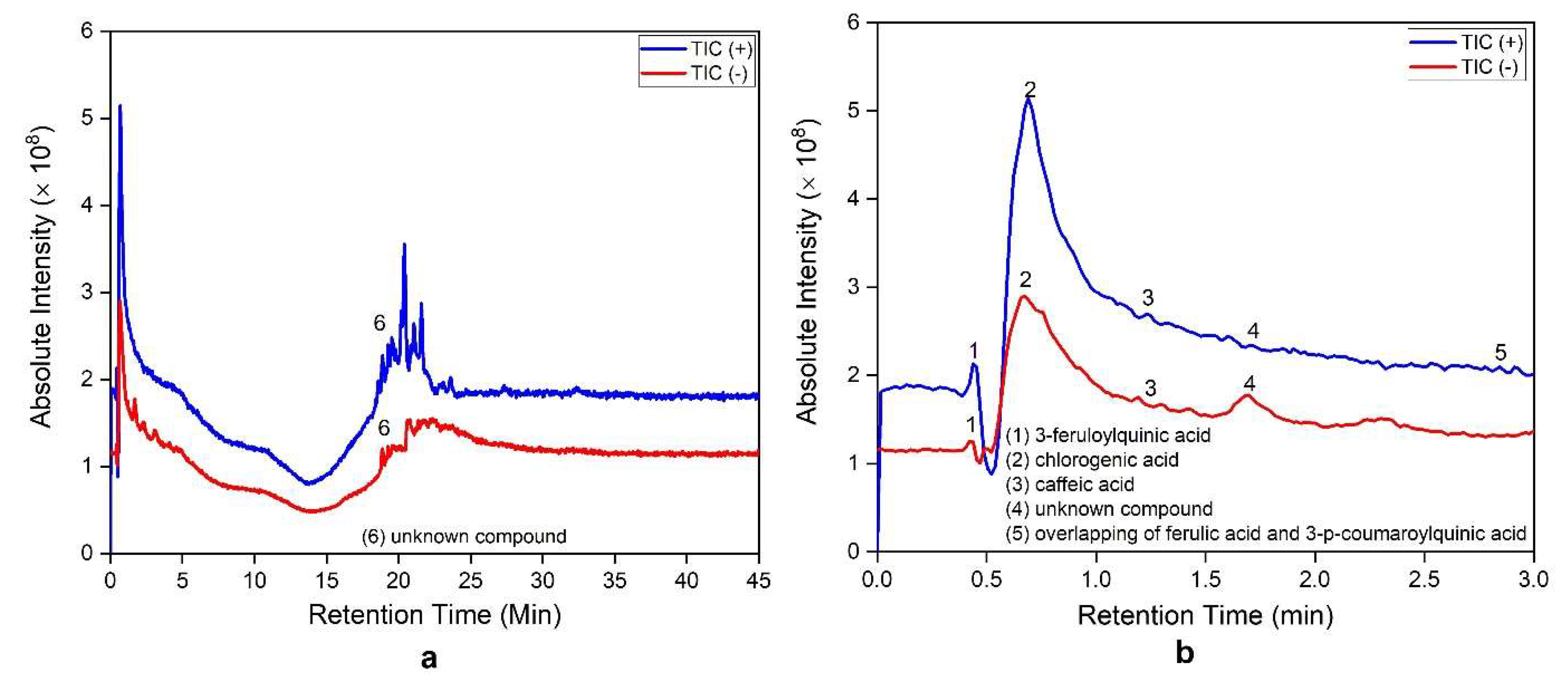
Figure 1. LC-MS/MS chromatogram of ethanolic extract of garlic at a retention time of 0–45 min (a) and the onset of a retention time of 0–3 min (b).
Sustainability 15 00806 g001
Figure 2. Thicknesses of the resulting membranes prepared from variable loadings of garlic extract.
Sustainability 15 00806 g002
Figure 3. Tensile strength (a) and elongation (b) of the resulting membranes prepared from variable loadings of garlic extract.
Sustainability 15 00806 g003
Figure 4. Contact angles for the resulting membranes prepared from variable loadings of garlic extract.
Sustainability 15 00806 g004
Figure 5. Clean water permeability of the resulting membranes prepared from variable loadings of garlic extract.
Sustainability 15 00806 g005
Figure 6. Cross-section of the resulting membranes prepared from variable loadings of garlic paste. (a) Pristine membrane; (b) 1% w/v garlic extract in the dope solution; (c) 1.25% w/v garlic extract in the dope solution; (d) 1.5% w/v garlic extract in the dope solution.
Sustainability 15 00806 g006aSustainability 15 00806 g006b
Figure 7. B. subtilis attachment to the membrane surface: (ad) SEM images; (eh) processed images by ImageJ software.
Sustainability 15 00806 g007
Figure 8. E. coli attachment to the membrane surface: (ad) SEM images; (eh) processed images by ImageJ software.
Sustainability 15 00806 g008
Figure 9. The number of bacterial cells attached to the membrane surface (a) and membrane area covered by the bacterial attachment (b).
Sustainability 15 00806 g009
Figure 10. FTIR spectra of the resulting membranes prepared from variable loadings of garlic extract.
Sustainability 15 00806 g010
Table 1. Antibacterial additives of membranes.
Antifoulant TypesCompoundsMethods of IncorporationReferences
MetallicSilvercoating, grafting, filling[10,11,12,13]
Coppercoating, grafting, filling[14,15,16,17]
Goldcoating[18]
Ironfilling[19]
Bismuthchelation, filling[20,21]
Cobaltfilling[22]
Nickelfilling[23]
InorganicTiO2coating, filling[24,25,26]
ZnOfilling[27,28,29]
MgOfilling[30]
RuO2filling[31]
MXenecoating[32,33,34]
CarbonsCarbon nanotubesfilling[35,36,37]
Graphene oxidefilling[38,39,40]
Carbon quantum dotsgrafting, filling[41,42,43]
OrganicsChitosancoating, filling[44,45,46]
2-Aminoimidazolefilling, blending[47,48]
NaturalPhenolicfillingthis study
Table 2. Composition of the dope solutions for preparation of composite membranes.
Membrane Composition
Garlic Extract (g)Garlic Extract (% w/v)DMF (mL)Cellulose Acetate (g)Initial Thickness (mm)
0 0204.000.3
0.041203.960.3
0.051.25203.950.3
0.061.5203.940.3
Table 3. A list of phenolic compounds in the ethanolic extract of garlic identified using LC-MS/MS data along with retention time (RT).
Peak aTentative Proposed CompoundsRT (min)Mode (+/−)MS b (m/z)MS/MS (m/z)References
13-Feruloylquinic acid0.444368338[79,80,81]
2Chlorogenic acid0.678354191[74,82,83]
3Caffeic acid1.23518089, 134, 145, 163, 180[74,78,79,80,82,83]
4Unknown1.702+589265, 297, 587
5p-coumaric acid2.84416491, 199, 147[80,81,83,84]
Ferulic acid194134, 149[74,78,80,81,83,84]
3-p-Coumaroylquinic acid338163, 173[74,78]
6Unknown18.897+465329, 330, 397
Note: a Peak number and retention time (RT) refers to LC-MS chromatogram and b MS: mass spectrophotometry.
Table 4. Phenolic substances of the resulting membranes prepared from variable loadings of garlic paste.
Membrane-Embedded Garlic Extract (% w/v)Phenolic Substances (%)
10.94
1.250.81
1.50.58
Disclaimer/Publisher’s Note: The statements, opinions and data contained in all publications are solely those of the individual author(s) and contributor(s) and not of MDPI and/or the editor(s). MDPI and/or the editor(s) disclaim responsibility for any injury to people or property resulting from any ideas, methods, instructions or products referred to in the content.