Article

Development of Bacterium for Crack Healing and Improving Properties of Concrete under Wet–Dry and Full-Wet Curing

by
Arunachalam Sumathi
1,
Gunasekaran Murali
1,*,
Dharmalingam Gowdhaman
2,
Mugahed Amran
3,4,
Roman Fediuk
5,
Nikolai Ivanovich Vatin
6,
Ramamurthy Deeba Laxme
1 and
Thillai Seenu Gowsika
2
1
School of Civil Engineering, SASTRA Deemed University, Thanjavur 613401, India
2
School of Chemical and Biotechnology, SASTRA Deemed University, Thanjavur 613401, India
3
Department of Civil Engineering, College of Engineering, Prince Sattam Bin Abdulaziz University, Alkharj 11942, Saudi Arabia
4
Department of Civil Engineering, Faculty of Engineering and I.T, Amran University, Quhal, Amran 9677, Yemen
5
School of Engineering, Far Eastern Federal University, 8, Sukhanova Str., 690950 Vladivostok, Russia
6
Higher School of Industrial, Civil and Road Construction, Peter the Great St. Petersburg Polytechnic University, 195251 St. Petersburg, Russia
*
Author to whom correspondence should be addressed.
Sustainability 2020, 12(24), 10346; https://doi.org/10.3390/su122410346
Submission received: 10 October 2020 / Revised: 27 October 2020 / Accepted: 28 October 2020 / Published: 11 December 2020
(This article belongs to the Special Issue Green Deal in Construction and Building Materials)

Abstract

:
Concrete cracking is inevitable, coupled with increased permeability, exacerbating the adverse impacts of atmospheric conditions and chemical attacks. Calcium carbonate precipitation resulting from certain microorganisms’ metabolism is a novel approach that can self-heal the cracks and improve concrete properties. In this study, the development and effect of bacteria Bacillus cohnii on crack healing, regained compressive strength after pre-cracking, sorptivity, water absorption, and concrete microstructures were investigated. For this purpose, a Bacillus cohnii bacterial concentration of 105 cells/mL was used as a water replacement in the concrete mixtures. Two methods subsequently cured the prepared concrete specimens: wet–dry (W-D) cycle and full-wet (F-W). In the wet–dry cycle, the cast specimens were immersed in water for 24 h and then kept at room temperature for 24 h, which was considered as one cycle; this process was repeated for 28 days. In the full-wet curing, specimens were immersed in water for 28 days. However, the curing water was changed every 24 h to facilitate the essential oxygen supply for bacterial activity to precipitate calcium carbonate. The results revealed that 90% and 88% surface healing was noticed in full-wet and full-dry pre-cracked specimens at 28 days.

1. Introduction

Concrete dominates the construction sector globally because of its low cost, durability, enhanced compressive strength, thermal mass, and versatility. Concrete is low in tensile stress and high in compressive stress, and cracks are unavoidable. When cracks form, the life span of the concrete frameworks may be reduced. To repair the cracks, various repair techniques are available, but they are extremely costly and time-consuming methods. However, a matter of grave concern is vulnerability to failure due to invariable cracks under service resulting from general loading, drying shrinkage, creep, and thermal stress [1]. These cracks offer paths for ingress for aggressive chemicals and fluids, impairing the embedded reinforcing steel or cement matrix. The formation of a crack in a structure is inevitable during its service duration; thus, crack self-healing is required. Self-healing of cracks is a process involving solid substance formation inside the cracks due to chemical or physical reaction, thus obstructing the path for migration of aggressive agents. A substitute method of repairing cracks through microbially mineralized deposition has been widespread in developing concretes due to its easy application and low maintenance cost [2].
Current widely used repairing methods in construction engineering include concrete replacement, filling, pressure grouting, and surface repair. There are some repair strategies for concrete repair in post-earthquake scenarios in case of minor cracking—the reinforced concrete member can be repaired by injecting suitable grout as follows: remove plaster and any loose material in the vicinity of crack; fix the grouting nipples in the cracks at a spacing of 6–8 inch (150–200 mm) c/c; inject water through the nipple so that the dust inside the cracks is washed off and the concrete is saturated with water; inject the grout until it comes out from the next nipple and then move to next nipple; after injection of grout through all the nipples is completed, re-plaster the surface and finish as required. These inactive repair technologies, however, can fail to fulfil the necessities of advanced constructions of concrete materials. Consequently, the necessity of creating new approaches for repairing concrete cracks is paramount [3]. With the evolution of microbiological and concrete technologies, the technique of microbial self-healing and its application to restoration of cracks in concrete has particularly attracted attention. Compared to other repairing methods, it has excellent benefits as follows [4,5,6]; (1) microbial usage before breaking, as Bacillus subtilis may be left alive in the form of spores inside concrete. Several studies indicated that bacterial spores can survive beyond 200 years of age in dry conditions; hence, they can meet the duration of concrete buildings’ service necessities. (2) Within the concrete, the bacteria are uniformly dispersed. The oxygen and water enter into the cracked concrete, which wakes up bacteria, and mineralization of microbial metabolic sediments accumulates in the cracks; hence, self-repair and self-diagnosis of concrete occur. (3) Calcium carbonate (CaCO3) is provided by a microbial mineralization reaction that is environmentally compatible and durable.
While the earlier studies have reported various methods of using bacteria to develop calcium carbonate sediment, other studies experimented with various bacteria concentrations. The earlier proposed methods are outlined as follows:
  • Bacteria addition while mixing concrete [7,8];
  • Bacteria addition into the water for concrete curing [9];
  • Treatment of the concrete surface [10,11,12,13] can be performed in different forms such as bacteria coating on a concrete surface, immersion concrete curing in liquid cultures for bacteria growth, and spraying bacteria.
  • Utilizing bacterial spores and bacterial protection in concrete [14].
De Muynck et al. [15] investigated the influence of bacteria on various mortar samples’ durability with different porosities. The surface deposition of CaCO3 crystals exhibited that the rate of chloride penetration and carbonation dropped from 10 to 40% and 25–30%, respectively, while samples showed exceptional freezing–thawing resistance under many cycles. Wang et al. [16] studied the impact of CaCO3 on the penetrability of concrete by using immobilized cells of Bacillus sphaericus. Experimental findings revealed that samples comprising polyurethane immobilized bacteria displayed a decrease in concrete penetrability of over six times that of the plain samples. Tziviloglou et al. [17] studied the influence of water immersion and wet–dry curing conditions on CaCO3 productivity for bio-mortar cracks. Findings indicated that the specimens immersed in water exhibited excellent performance compared to the wet–dry cycle curing method. National Centre for Biotechnology Information
Additionally, in the curing immersion method, the crack’s width is completely closed up to 0.36 mm, while the wet–dry cycle displayed no precipitation. Nosouhian et al. [18] studied the utilization of Bacillus sphaericus, Bacillus subtilis, and Sporosarcina pasteurii treated concrete under salt environments. They also investigated the sedimentation’s influence on volume changes, rapid chloride penetration, compressive strength, water absorption, and concrete weight. Findings revealed that bacteria-based specimens exhibited excellent performance compared to the control specimen. Furthermore, the bacterially treated specimens displayed a reduction in chloride penetration, water absorption, weight, and volume, but the compressive strength was increased by about 20%. The reduction of permeability in surface concrete’s microbial mediation is achieved by utilizing treated gel for bacteria [19]. Nosouhian et al. [20] investigated the impact of bacteria on concrete durability in a salt environment. Research was conducted by Hosseini et al. to study the impact of two strains of bacteria on the lightweight aggregate concrete produced. The results showed that the average decrease in chloride permeability and water absorption was about 20% and 10%, respectively, while the compressive intensity increased by 20% [21,22].
Tayebani and Mostofinejad [23] investigated the influence of B. subtilis and S. pasteurii type bacteria on electrical resistivity, corrosion potential, and penetrability of concrete. The specimens were prepared with two different mix designs, and 28 or 91 consecutive days of curing was adopted in urea–calcium lactate and urea–calcium chloride solution. The observed outcomes indicated reduced chloride penetrability and water absorption. However, the electrical resistance and compressive strength was significantly improved for S. pasteurii bacteria-based concrete compared to B. subtilis-based concrete [24]. Parastegari et al. investigated the improved mechanical properties of air-entrained concrete with bacteria incorporation. Conclusions indicated that a 6% increase in electrical resistivity and 28% decrease in chloride penetration were observed due to precipitation of CaCO3, which is assisted bacterially [25]. Microbially induced CaCO3 precipitation has been previously investigated and used for many applications: enhancement of concrete durability [26,27], improvement of concrete strength [14,28,29], repairing concrete cracks [29,30,31], repairing calcareous monument [30], sand consolidation [32,33,34], and improvement of soil [35].

2. Research Significance

The strength and durability of cement mortar and concrete assessment are of uttermost importance for the integrity of structures. While many investigations cited above have focused on microbially induced self-healing concrete’s efficiency under immersion curing conditions, the literature on the wet–dry cycle curing condition is limited. Furthermore, earlier works did not study the development of bacteria for crack healing. The current investigation addressed the development and utilization of Bacillus cohnii, exploring the strength and durability properties of concrete under wet–dry and full-wet curing conditions and comparing the two. Additionally, the crack healing was assessed by a portable microscope. For the chemical composition of the healing agent, scanning electron microscopy (SEM) and X-ray diffraction (XRD) were utilized.

3. Experimental Program

3.1. Preparation of Bacillus Cohnii Bacterial Solution

3.1.1. Isolation of a Bacterial Strain

Soil samples were collected from a used concrete dumping area besides Nirman Vihar, SASTRA Deemed University, Thanjavur, Tamil Nadu, India. Large particles in the soil sample were removed and sieved. Soil sample (1 g) was suspended in 100 mL of sterile saline solution, which was considered a stock solution. From this solution, 1ml was taken and serially diluted up to 10−7. Using the pour plate method, 0.1mL from dilutions 10−5 and 10−6 was plated on nutrient agar medium in basal media containing (w v-1) 0.5% peptone, 0.1% NaCl, 0.2% K2HPO4, 0.01% CaCl2, 0.01% MgSO4, 0.1% yeast extract, 1.5% bacteriological agar, and 1% glucose. Plates were kept in an incubator for 24 h at 370 °C. Morphologically different colonies were separated, and pure cultures were preserved at 40 °C [36,37].

3.1.2. Identification of Newly Isolated Bacterial Strain by Morphological and Biochemical Characterization

Pure cultures of the selected isolates were prepared and identified by morphological, biochemical, and molecular characterization. The isolates were purified to approximate purity on nutrient agar and studied for their colony characteristics. Morphological characterization was carried out, including Gram staining, negative staining, endospore staining, and motility tests. The bacterial strains were subject to various biochemical tests and then characterized using Bergey’s Manual of Determinative Bacteriology [38]. Bacteria underwent metabolic changes during incubation, demonstrated by a color shift in the media that was either visually perceived or occurred after the addition of a reagent. Indole, methyl red, Voges–Proskauer, citrate usage, triple sugar iron test, catalase, starch hydrolysis, casein hydrolysis, hydrolysis of urease, and gelatin [39,40] were the biochemical studies used.

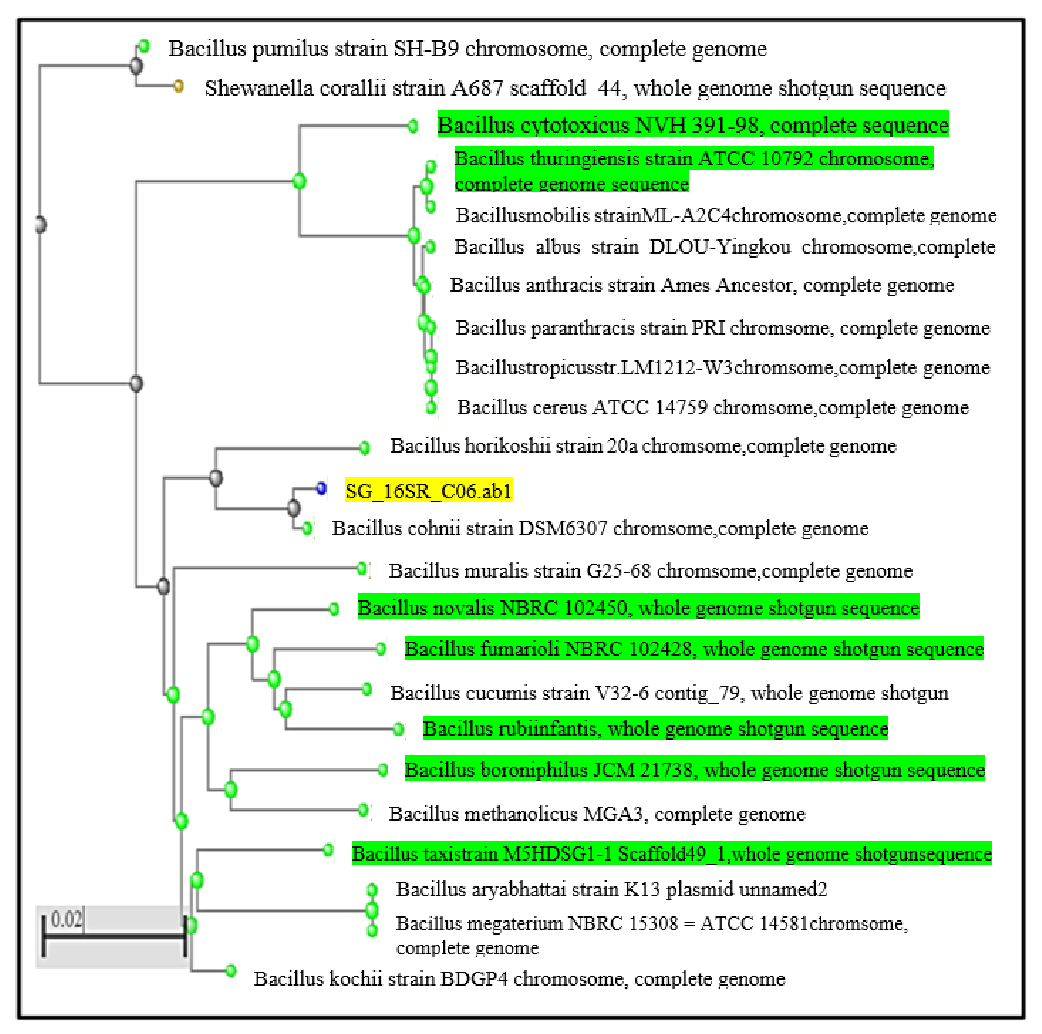
3.1.3. Molecular Characterization of Newly Isolated Bacteria

With 16S rRNA sequencing, molecular identification of the selected bacterial isolate was carried out. At Genurem Biosciences LLP, Thanjavur, Tamil Nadu, India (www.genurem.org), the isolated bacterial culture was deposited. Using a DNA extraction package, genomic DNA was extracted from that organism. The 16srRNA sequence gene was analyzed and amplified by universal primers (forward primer: 5′-GGGGGGTTTCCGCCCCTTAGTGCTGC-3′ and reverse primer: 5′-GGCTTAATGCGTTAGCTAGCTGCCAGCACTA-3′). PCR thermal cycling was performed with an initial denaturation of 94 °C for 2 min, followed by 40 denaturation cycles for 1 min at 94 °C, annealing for 1 min at 55 °C, and extension for 1 min at 72 °C [40]. Then, the PCR amplified rDNA strands were sequenced with an Applied Biosystems DNA analyzer. Applied Biosystems analysis tools were used.
Before conducting the bioinformatics analysis, the plus and minus strands were matched using DNA software analysis. In order to find the similarity of the sequence from National Centre for Biotechnology Information (NCBI), Basic Local Alignment Search Tool (BLAST) as used. The NCBI database was compared with sequences, and the most significant alignments were compared on the basis of E value and percent identity. Using the ClustalW method for multiple sequence alignment, the most similar sequence was aligned. For closely related species, a phylogenetic tree was built [41,42]. With the use of the neighbor-joining process, evolutionary history was inferred. The 16S rRNA sequences of isolates were sent to the GenBank database, NCBI, for their accession numbers.

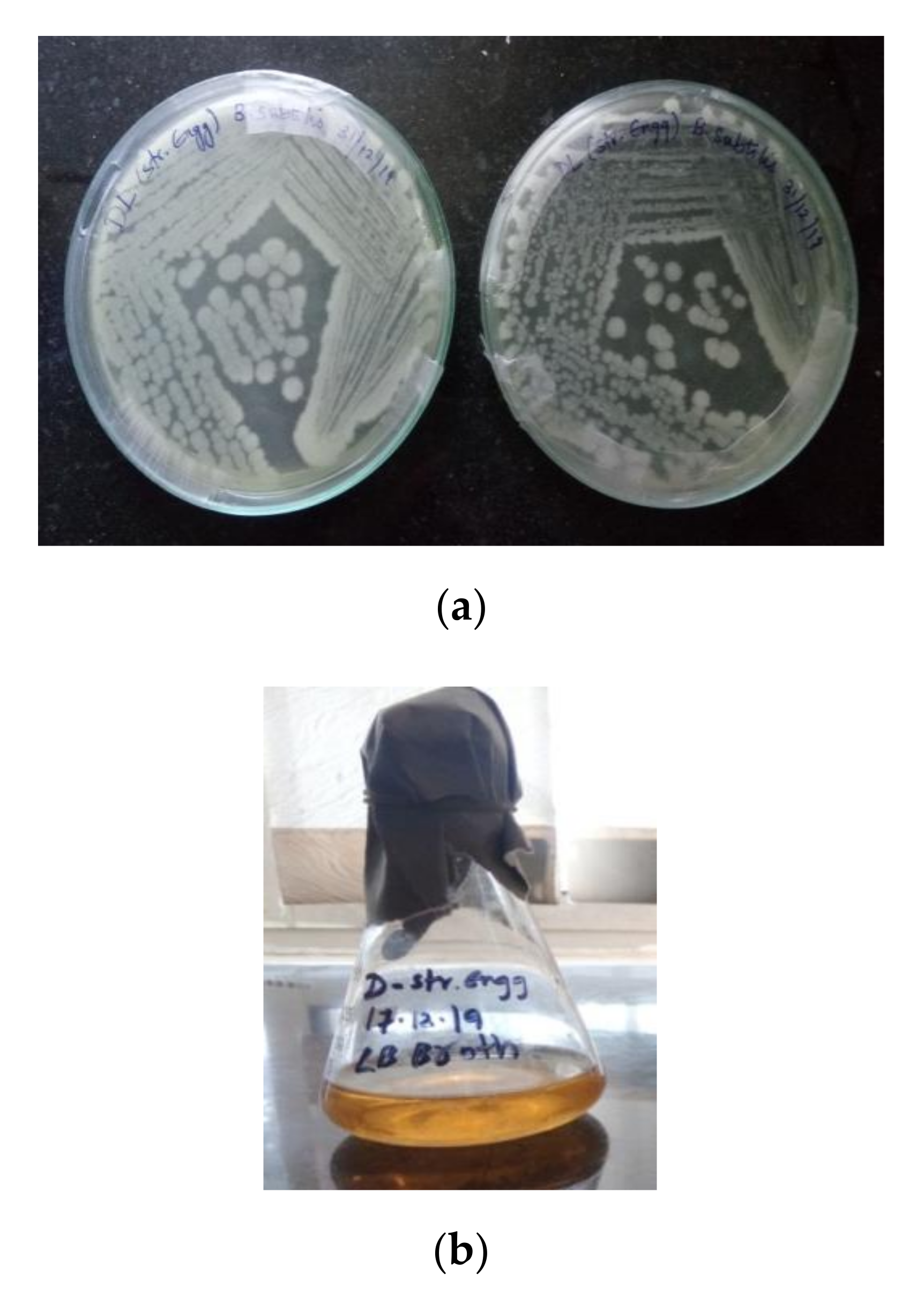
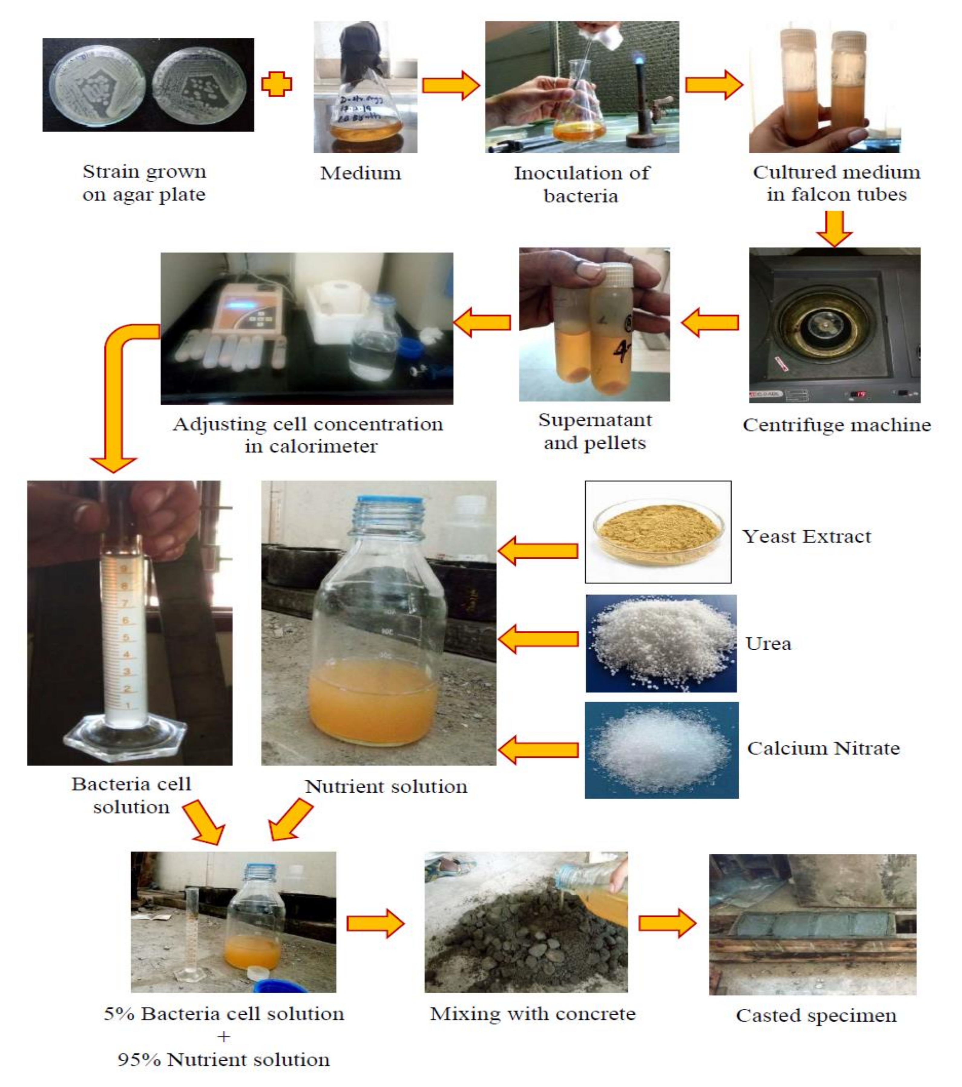
3.1.4. Preparation of Bacillus cohnii Self-Healing Agent

A loop full of newly isolated pure culture (from the plate culture) was inoculated in 50 mL of nutrient broth in a 250 mL Erlenmeyer flask and incubated at 150 rpm in an orbital shaker for 2–3 days at ambient temperature (37 ± 2 °C) [43]. The overnight Bacillus cohnii bacterial culture was transferred to 50 mL falcon tubes. Each falcon tube was centrifuged at 10,000 rpm for 10 min to separate bacterial cells, the supernatant was disposed of, and bacterial cells (pellets) were harvested for re-suspension in physiological solution (NaCl—9 g/L). The required concentration of 105 cells/mL was adjusted by checking with a calorimeter at 600nm. Then, the bacterial cells were suspended in a nutrient solution containing calcium nitrate (20 g/L), urea (20 g/L) and yeast extract (2 g/L) of cement mass. The solution was then mixed to avoid precipitation. The concentrations of calcium nitrate, urea, and yeast extract were 2%, 2%, and 0.2%, respectively [43].

3.1.5. Results and Interpretation

Bacteria are usually categorized based on studies of their morphological and biochemical characteristics. The initial screening revealed that the aerobic, Gram-positive, short rod was a motile Bacilli isolate. Positive reactions to catalase, triple sugar iron, Simmons’ citrate, and methyl red tests and adverse reactions to Voges–Proskauer, indole, and MacConkey agar tests were exhibited in biochemical tests. Table 1 shows the morphological and biochemical characteristics of Bacillus cohnii. However, these methods alone are inadequate for various morphological and biochemical characteristics exhibited by bacteria within a single bacterial genus. An alternative to this approach is the DNA sequence analysis method and its rapid, reliable, and reproducible method of identifying novel bacterial species [43]. The newly isolated bacterium’s phylogenetic relationship could be inferred using the neighbor-joining approach shown in Figure 1. With 16S rRNA sequencing analysis, the bacterial isolate was verified as Bacillus cohnii. The sequence was deposited in Gene Bank with Accession No. MT704504. Figure 2a illustrates that the isolated bacteria show discrete colonies, and Figure 2b illustrates the Gram staining of the isolated bacteria.

3.1.6. Bacterial Species: Cultivation

The bacteria used was Bacillus cohnii, and it was cultured on nutrient agar plates in streaks, as shown in Figure 3a. The Luria–Bertani (LB) broth medium was prepared and sterilized to inoculate the medium’s bacterial culture. The bacteria inoculated LB medium is shown in Figure 3b. Sterilization of the LB broth medium was carried out at 121 °C at 15 lbs for 1 h before inoculation. After inoculation, it was kept in a shaker for a minimum of 24 h to enhance the growth of bacteria.

3.1.7. Nutrient or Mineral Substrate

Incorporation of bacterial cells into concrete led to the formation of calcium carbonate precipitate. An appropriate nutrient substrate was added to the bacteria. In this study, calcium nitrate was used for the calcium source, urea for the nitrogen source, and yeast extract as growth media for the bacterial cells to undergo bacterial activity.

3.1.8. Bacterial Healing Agent Preparation

At first, the soil samples were collected from the used concrete. Large particles in the soil sample were removed and sieved. Soil sample (1 g) was suspended in 100 mL of sterile saline solution, which was considered a stock solution. The bacteria used was Bacillus cohnii, and this was cultured on nutrient agar plates in streaks. The LB broth medium was prepared and sterilized to inoculate the medium’s bacterial culture. Sterilization of the LB broth medium was carried out at 121 °C at 15 lbs for 1 h before inoculation. After inoculation, it was kept in a shaker for a minimum of 24 h to enhance the growth of bacteria. The bacterial species, Bacillus cohnii, was cultivated by adopting the same procedure as explained earlier. The bacterial cultured medium was moved into 50 mL falcon tubes to isolate bacterial cells from the medium. In order to isolate bacterial cells, each falcon tube was centrifuged at 10,000 rpm for 10 min. Figure 4 shows the falcon tubes with a bacterial cultured medium placed inside the centrifuge machine. As shown in Figure 4, the supernatant was disposed of, and bacterial cells (pellets) were harvested for re-suspension in physiological solution (NaCl, 9 g/L). The required concentration of 105 cells/mL was adjusted by checking with a calorimeter, as shown in Figure 4. An appropriate nutrient substrate was added to the bacteria. In this study, calcium nitrate was used for the calcium source, urea for the nitrogen source, and yeast extract as growth media for the bacterial cells to undergo bacterial activity. For the nutrient solution, the nutrient substrates such as calcium nitrate, urea, and yeast extract were mixed afterward to avoid precipitation. The concentrations of calcium nitrate, urea, and yeast extract were 2%, 2%, and 0.2% by cement mass, respectively. The bacterial solution (5%) and nutrient solution (95%) were mixed to create the healing agent. The prepared healing agent was added into the concrete during mixing instead of water.

3.2. Raw Material and Specimen Preparation

Ordinary Portland cement (OPC) grade 53 was used as a binder material with a specific gravity of 3.16 and a specific surface area of 245 m2/kg, in compliance with IS 12269-2013 [44]. The cement was found to have an initial and final setting time of 160 and 260 min, respectively. Locally procured river sand was used as fine aggregate (FA), free of silt, mud, and organic impurities, with a maximum size of 4.75 mm and zone II grading according to IS 383-1970 [45] and a specific gravity of 2.62, bulk density of 1696 kg/m3, water absorption of 1.37%, and a fine modulus of 3.39. Crushed granite gravel collected from a local source was used as coarse aggregate (CA) of 12.5 and 20 mm in scale, with 2.78 specific gravity, 1.56 % water absorption, 5.25 fineness modulus, and 17.18% impact value. M40 grade concrete was used to cast all specimens with a water/cement ratio of 0.45. The water content was fully replaced with a healing agent. The healing agent comprised 5% Bacillus cohnii bacterial solution and 95% nutrient solution and was directly mixed with concrete ingredients. First, for 2 min, cement and fine aggregate were combined and mixed, then coarse aggregate was added and mixed for another 2 min. Finally, to guarantee an equal distribution of bacterial cells and nutrients, the curing agent was applied and mixed for 3 min. Two different specimens were prepared; the first specimen was prepared with ordinary portable water, and the second specimen with a healing agent. The healing agents’ specimens were cured using the wet–dry cycle and full-wet method. Table 2 shows the specifics of the mixing composition used in this analysis. The 100 mm cubical specimens were used to find the compressive strength of concrete. For the durability test, cylindrical specimens of 100 mm diameter and a height of 50 mm were cast for sorptivity and porosity tests. For water absorption, 100 mm cube specimens were used.

3.3. Self-Healing

All the concrete specimens were cured for 28 days under two different curing conditions and tested against compression using a 300 T capacity compression testing machine (AIMIL, India). The compression load was applied gradually until the appearance of crack and the crack widths were measured at various specimen areas by the crack-measuring device. The measured crack width was in the range of 0.2 to 0.6 mm. After that, to assess crack healing efficiency of the Bacillus cohnii, cracked specimens were exposed to their respective curing conditions. The wet–dry cycle method of curing involves the specimens being immersed in water for 24 h, followed by being kept for 24 h at room temperature. This process was repeated for 28 days. Full-wet curing involves the specimens being immersed in water for 28 days. For crack healing quantification, the crack healing was monitored at 0, 3, 7, 14, and 28 days by measuring the crack width. The degree of crack healing is defined as the variation between the initial crack width and observed crack width at different days of healing and is expressed in Equation (1) as follows.
Degree   of   crack   healing = C i C o C o × 100
where Ci is initial width crack and Co is crack width after healing.

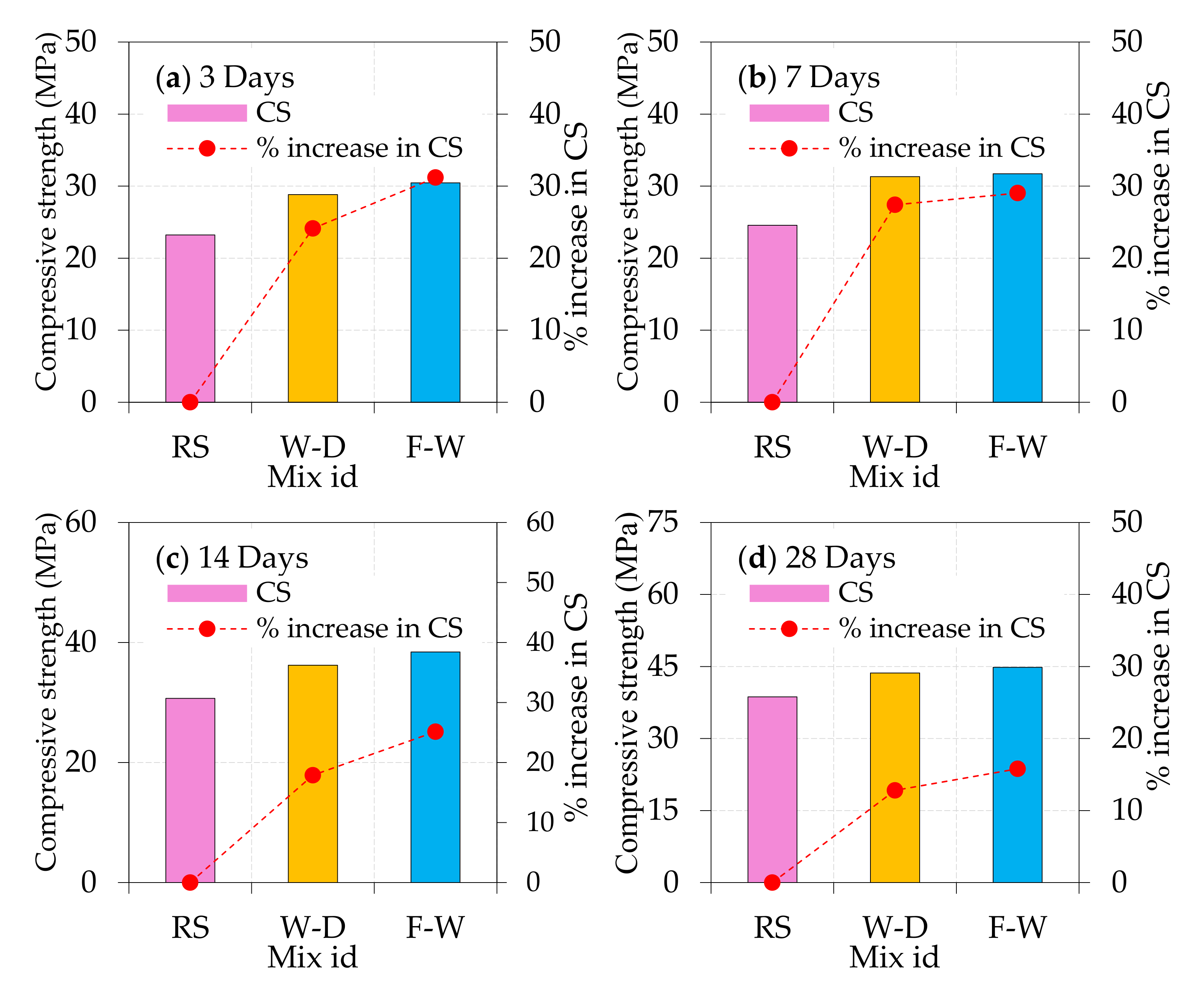
3.4. Compressive Strength

The 100 × 100 mm cubical concrete specimens were tested to assess their compressive strength, according to IS: 516-1959 [46]. The compression testing machine was used with a loading rate of 2.5 kN/s to test all the cubes at 3, 7, 14, and 28 days. The improvement in compressive strength of the bacterially treated concrete specimens with respect to the reference specimen was then determined using Equation (2).
Improvement   in   compressive   strength = f r f b f r × 100
where fr and fb are the compressive strength of reference and bacterially treated specimens, respectively.

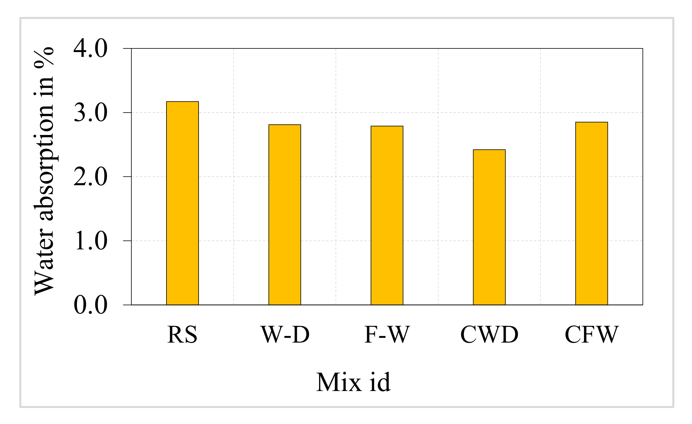
3.5. Saturated Water Absorption (SWA)
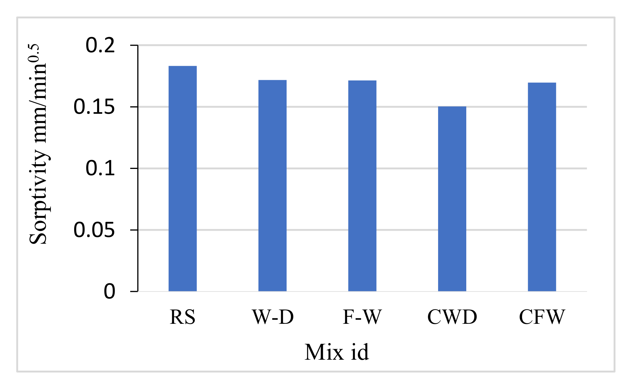
In accordance with ASTM C642 [47], saturated water absorption tests were performed on concrete mixes using 100 × 100 × 100 mm cube specimens after 28 days of curing. The mass of the cube specimens was measured prior to drying. The cubes were then dried at 105 °C in a hot air oven and continued until approximately coinciding with the mass distinction of two progressive 24 h interim figures. Next, the samples were allowed to cool under room temperature and after that submerged in water. The samples were taken out at a standard interim duration, the surfaces were dried utilizing a perfect fabric, and the samples were then weighed. This procedure was sustained until the weights were steady (completely soaked). The variation in the mass of the saturated and the oven dried specimens, at the rate of oven dry mass, provides the water absorption, which was computed.

3.6. Sorptivity

Sorptivity quantified the amount of water diffusion through the pores of the concrete by capillary suction. Although the increasing amount of water that penetrated the exposed specimen “q” unit surface area was plotted associated with the square root of exposure time, SQRT (t), and the corresponding graph may be determined by a straight line heading towards the origin. From the straight line’s slope drawn through the origin, which is called sorptivity, the rate of diffusion of water through the pores can be computed. In the current investigation, the sorptivity test was carried out on cylindrical specimens with a diameter of 100 mm and a thickness of 50 mm in accordance with ASTM C1585 by drying the specimens to an invariant mass at 105 °C in an oven and then inundating them in water after cooling the specimens to room temperature and measuring the mass increase for two hours at the normal interval. By considering the plot “p” versus “SQRT (t)” slope, the sorptivity was determined. The test was carried out as per ASTM 1585-20 [48].

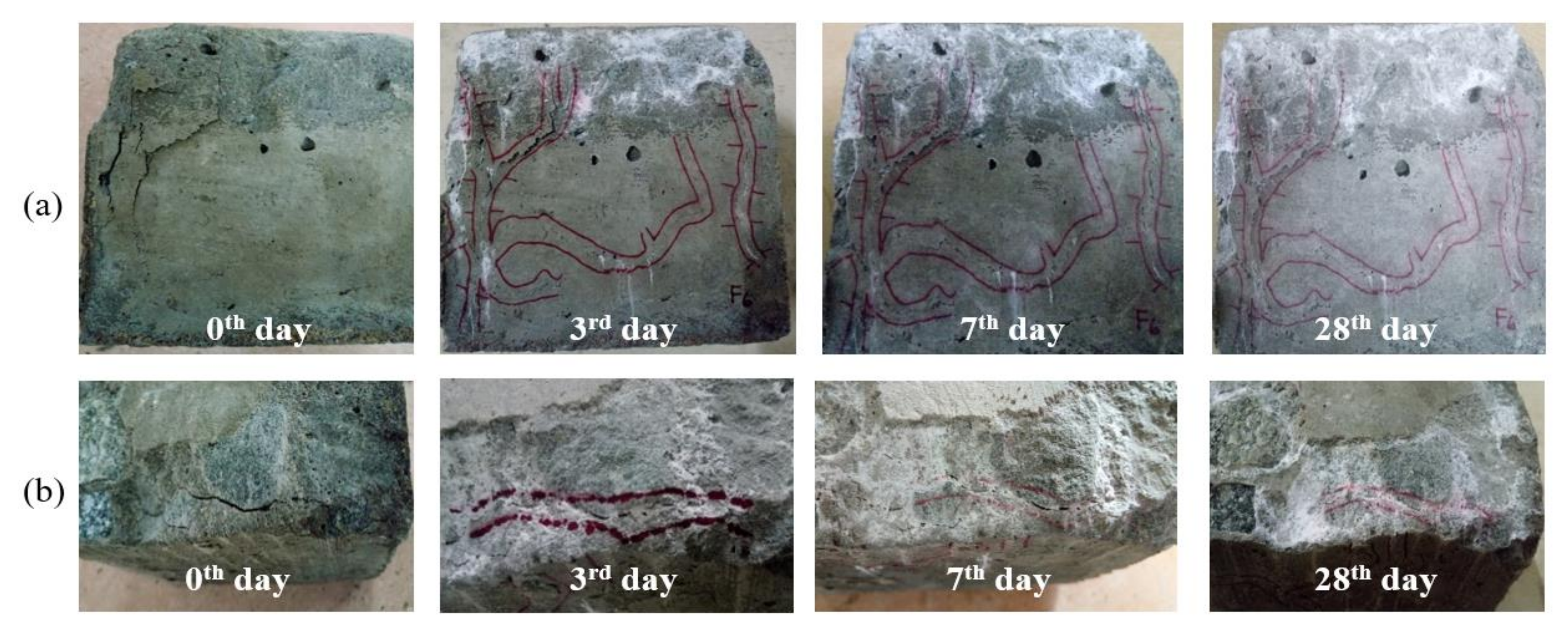
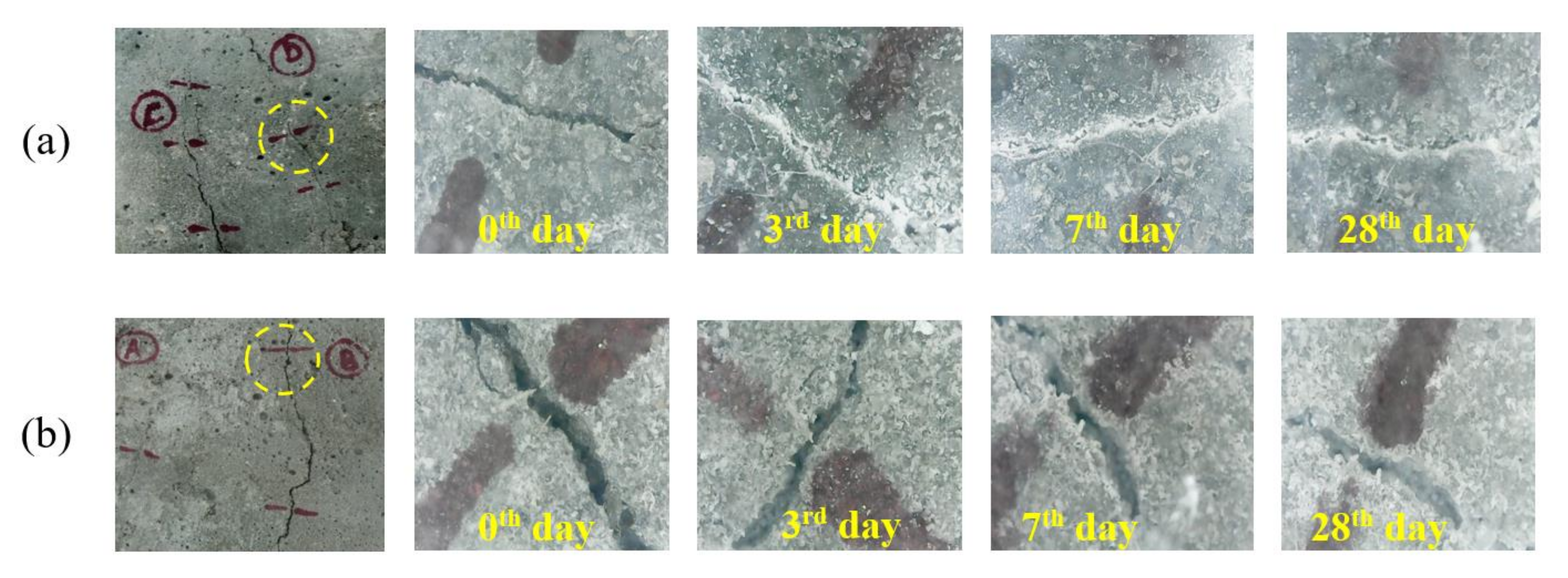
3.7. Crack Healing Capacity

Visual observations were made regularly from the day of inducement of cracks to witness the white powdery precipitate on the cracked areas. The findings were reported on the basis of the rate of precipitation of calcium carbonate in the closing of cracks. Optical microscopic images were taken at regular intervals to analyze crack healing effectiveness for different crack widths.

3.8. Scanning Electron Microscopy (SEM) and X-ray Diffraction (XRD) Analysis

A VEGA 3 TESCAN scanning electron microscope and Bruker D8 Concentrate unit were used to study structural morphology to determine atomic and molecular components such as calcite, aragonite, and vaterite in the white powdery precipitate on the cracked areas of specimens cured for 28 days.

4. Discussion

4.1. Compressive Strength

The compressive strength is calculated by dividing the cracking load with the area at different days of curing. In this study, the mixes were designed to achieve a compressive strength of 40 MPa at 28 days of curing. The most common age of testing is 28 days, but tests can also be carried out at 3, 7, and 14 days.
The compressive strength of specimens is shown in Figure 5 for 3, 7, 14, and 28 days. The addition of bacterial solution into concrete increased compressive strength as compared to the reference specimen (RS). For the specimens cured under 3 days of W-D and F-W conditions, the increase in compressive strength was about 24.1% and 31.2% compared to RS at the age of 3 days. The observed compressive strength for 7-day W-D and F-W cured specimens increased by about 27.4% and 29.0%, respectively, compared to the 7-day RS. There was a further increase in compressive strength at 14 days by about 17.9% and 25.1% for the W-D and F-W cured specimens compared to the 14-day RS. The compressive strength at 28 days for the W-D and F-W cured specimens increased by about 12.8% and 15.81%, respectively, compared to the RS at 28 days. It is clear from the above discussion that the increase in compressive strength in bacterial concrete results from calcium silicate hydrate formation within the matrix’s pores. Additionally, the specimens cured under the F-W condition exhibited a higher compressive strength than the W-D cycles.

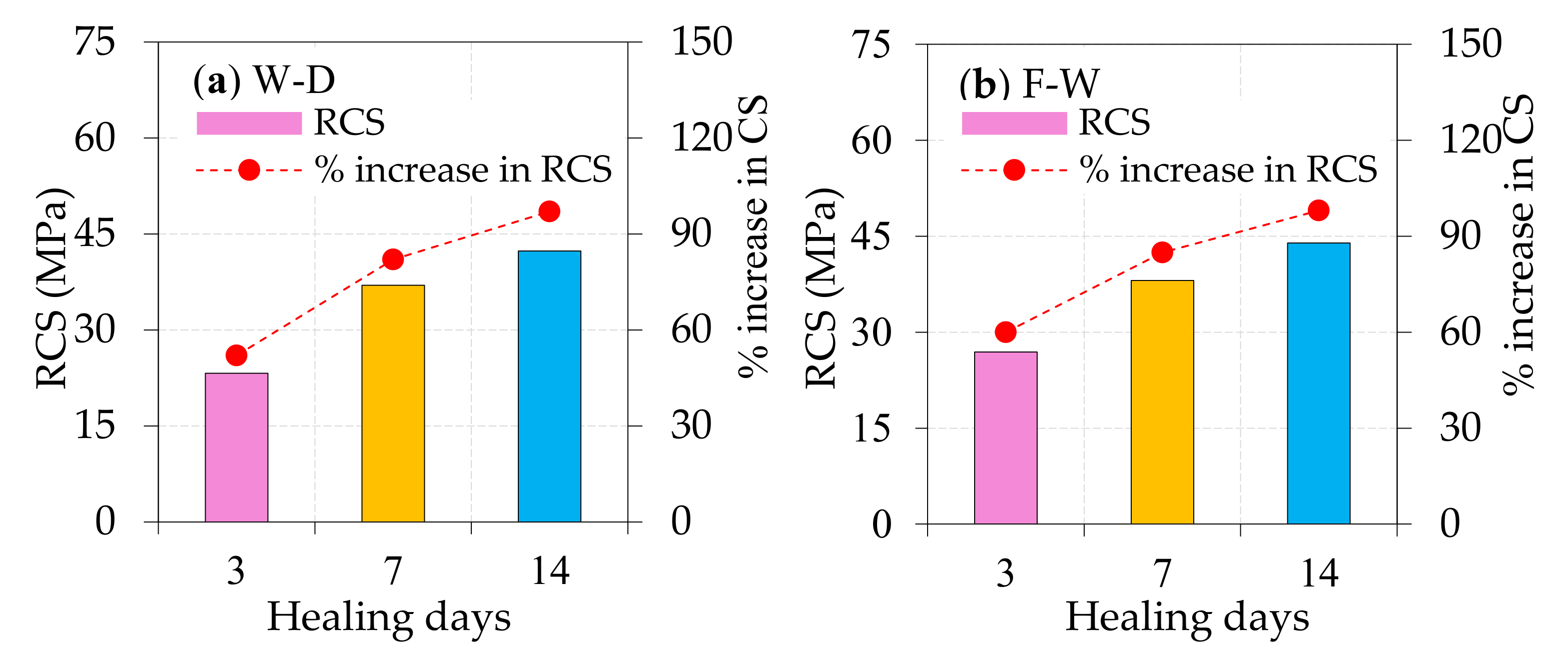
4.2. Regained Compressive Strength

Figure 6a,b illustrates the regained compressive strength of specimens that were loaded until a crack was induced at the age of 3, 7, and 14 days. The regained compressive strength of the W-D cured specimen at 3, 7, and 14 days was increased by 52%, 82%, and 97%, respectively, compared to the corresponding pre-cracked compressive strength of the W-D cured specimen at 28 days. Likewise, for the F-W cured specimens, the observed regained compressive strength was increased by about 60%, 85%, and 98% for 3, 7, and 14 days, respectively, compared to the pre-cracked F-W cured specimens at 28 days. It is inferred from the above discussion that the bacteria’s presence helped the concrete to regain its compressive strength even after the specimen cracked. This phenomenon is due to calcium carbonate production packing the concrete mix and helping it to regain its compressive strength. Furthermore, the regained compressive strength implies that calcium carbonate was produced in the cracks and improved the strength and degree of repair in bacterial concrete.

4.3. Visual Monitoring of Self-Healing

After 28 days of the healing period, the previously cracked specimens at 28 days were collected to visualize self-healing efficiency, and the observed cracks are illustrated in Figure 7. The crack healing was observed at specific intervals of time for 0, 3, 7, and 28 days under W-D and F-W curing. Meanwhile, the cracking width was impossible to control during the loading, leading to variable crack sizes. Three cracks were noted for each specimen to visualize the crack healing, and these cracks were recorded continuously. The images of cracks taken at various time intervals of healing are depicted in Figure 8. With the help of a portable microscope, the pictures were taken, and the cracking width was measured. No notable crack healing was detected in any specimens from the earliest age, as shown in Figure 8. Nevertheless, the observed cracks completely closed at 28 days of healing, further healing the formation of calcium carbonate at the cracked surface. The pores in the concrete act as carriers of bacteria by holding them inactive but alive. They are likely to trigger on experience to water availability and extra oxygen upon the formation of cracks and evolution in concrete. The exposure of bacterial pores with moisture and air resulted in the cracks being repaired due to biosynthesized calcium carbonate deposition in the cracked region.
Table 3 demonstrates the width of cracks that appeared in concrete specimens before and after healing. It is clear from Table 3 that the maximum percentage of crack healing was observed in F-W cured specimens compared to W-D cured specimens. The crack healing percentage for the F-W specimens was about 23% at 3 days, 46% at 7 days, and 90% at 28 days; this indicates that the cracks closed completely near 28 days, and these cracks were not visible to the human eye. Likewise, the percentage of crack healing for the W-D specimens was about 20%, 41%, and 88% for days 3, 7, and 28, respectively.

4.4. Sorptivity

Figure 9 shows the sorptivity determined at 28 days for control and presence of bacteria in cracked and uncracked concrete specimens under two different curing methods. It is clear that the specimen without bacteria, i.e., reference specimen (RS), showed higher sorptivity than specimens with bacteria. Sorptivity decreases with the presence of bacteria in the mix; the structure becomes more compact with W-D and F-W curing methods. Due to the pores’ and voids’ enhanced filling capacity, the sorptivity value decreased due to more calcium silicate hydrate gel and increased healing efficiency in the matrix. The authors of [49,50] have found a similar trend in their work. Therefore, it can be inferred that the bacteria, due to their refined pore structure, best reduce sorptivity for cracked self-healed specimens with the W-D method compared to other curing methods. By comparing with the RS, the percentage difference in sorptivity value measured at 28 days was −6.22%, −17.95%, −6.44%, −7.42% for the W-D, F-W, cracked bacterial concrete (CWD), and CFW specimens, respectively. Due to the formation of calcite precipitate formed by the bacteria, the cracked bacteria specimen’s sorptivity value under W-D is 17.95% less than that of the RS, making the bacterial concrete specimen denser.

4.5. Water Absorption

Figure 10 shows the percentage of water absorption of various mixtures made with and without bacteria for cracked and uncracked specimens for two curing methods at 28 days. The percentage of water absorption decreases for bacterial concrete compared to the RS. This phenomenon is due to the growth of bacteria that allows the accumulation of extra precipitation of calcium carbonate within the pores, leading to the minute pores and voids being filled in the concrete. In contrast to other cracked and uncracked concrete bacteria, because of the spherical-shaped particles in the wet–dry sample, water absorption increased the non-uniform structure’s content. This further leads to the creation of additional pores that lead to the absorption of water. The inclusion of bacteria hampers water absorption due to a change in the system’s structural coherence due to wet–dry curing of extensive bacterial activities. The percentage variation in water absorption was −11.35%, −11.98%, −23.65%, and 10.09% for the W-D, F-W, CWD, and CFW specimens compared to the RS. At 28 days, cracked bacterial concrete (CWD) under wet–dry cycle curing attained higher water absorption (23.65%) than the RS.

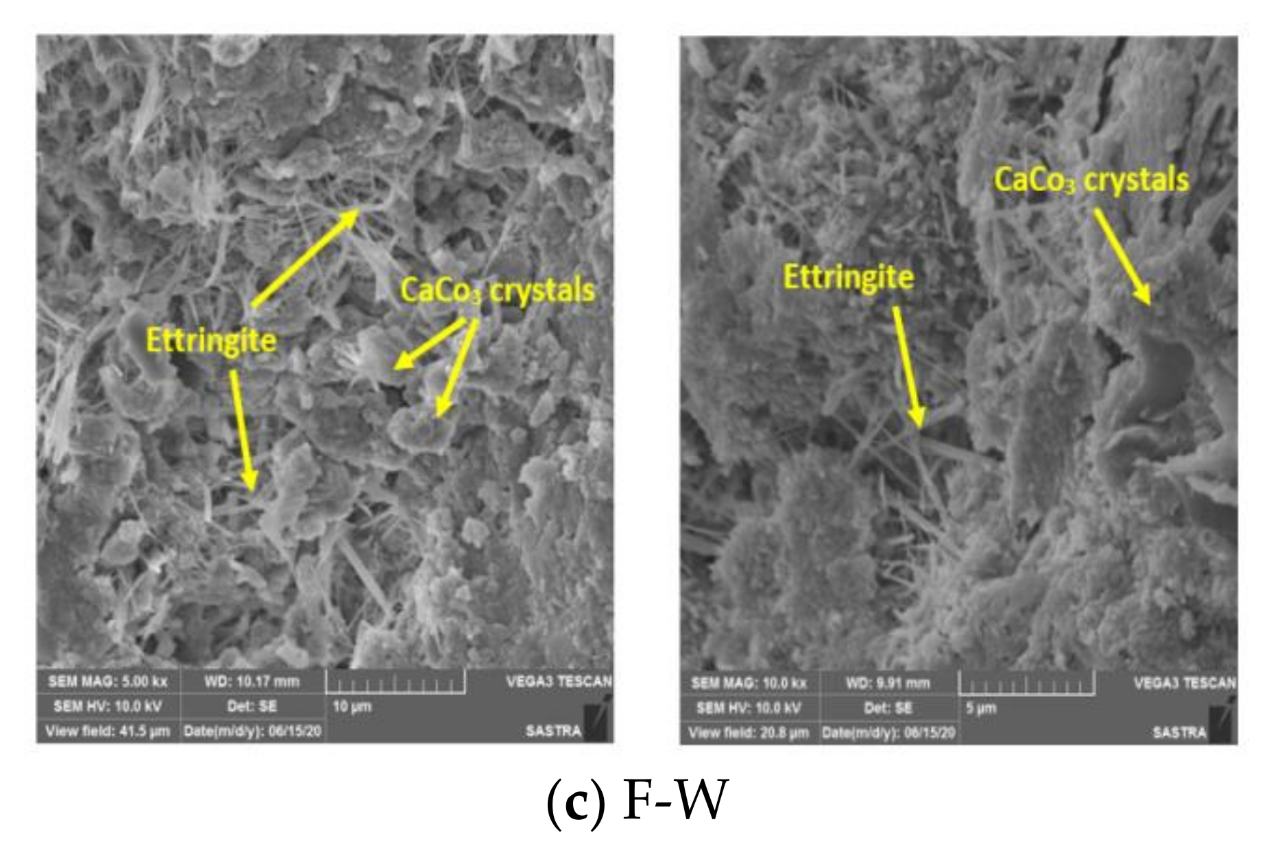
4.6. Scanning Electron Microscopy (SEM)

First, the crack was visually inspected under the portable microscope. The further strengthened structural morphology of the material was investigated using SEM analysis to check the microstructure of the 28-day crushed concrete sample and investigate the type of material precipitated (i.e., calcium carbonate). Samples were taken from the reference specimen and the specimens with bacteria for W-D and F-W samples to compare their microstructures. The SEM images are shown in Figure 11a–c for control, W-D, and F-W specimens, respectively. Figure 11a,b shows the dense formation of calcium carbonate crystals in a rhombohedral shape as lumps, and ettringite fibers can be seen. By comparing the SEM images, we could observe that the internal structure and the precipitated calcium carbonate crystals were similar across all curing processes. Thus, it was concluded that the method of curing does not lead to a significant difference of self-curing and precipitation of calcium carbonate crystals.
Different crystal sizes and hexagonal forms, rhombohedral forms as lumps, and other phases were observed with calcium carbonate spherical shapes in both specimen samples, i.e., W-D and F-W, as shown in Figure 11b,c. This was proven to be calcite ions [51,52] following the previous analysis. It is clear from Figure 11b,c that, with the addition of bacteria, pores are partially filled up by material development. On the surfaces of some particles, the imprints left by bacterial cells were also identified. Pore reduction is due to material growth that undoubtedly increases the strength of the material and makes concrete more durable due to the calcite crystalline structures found within the concrete pores, with the addition of bacteria increasing strength and durability.

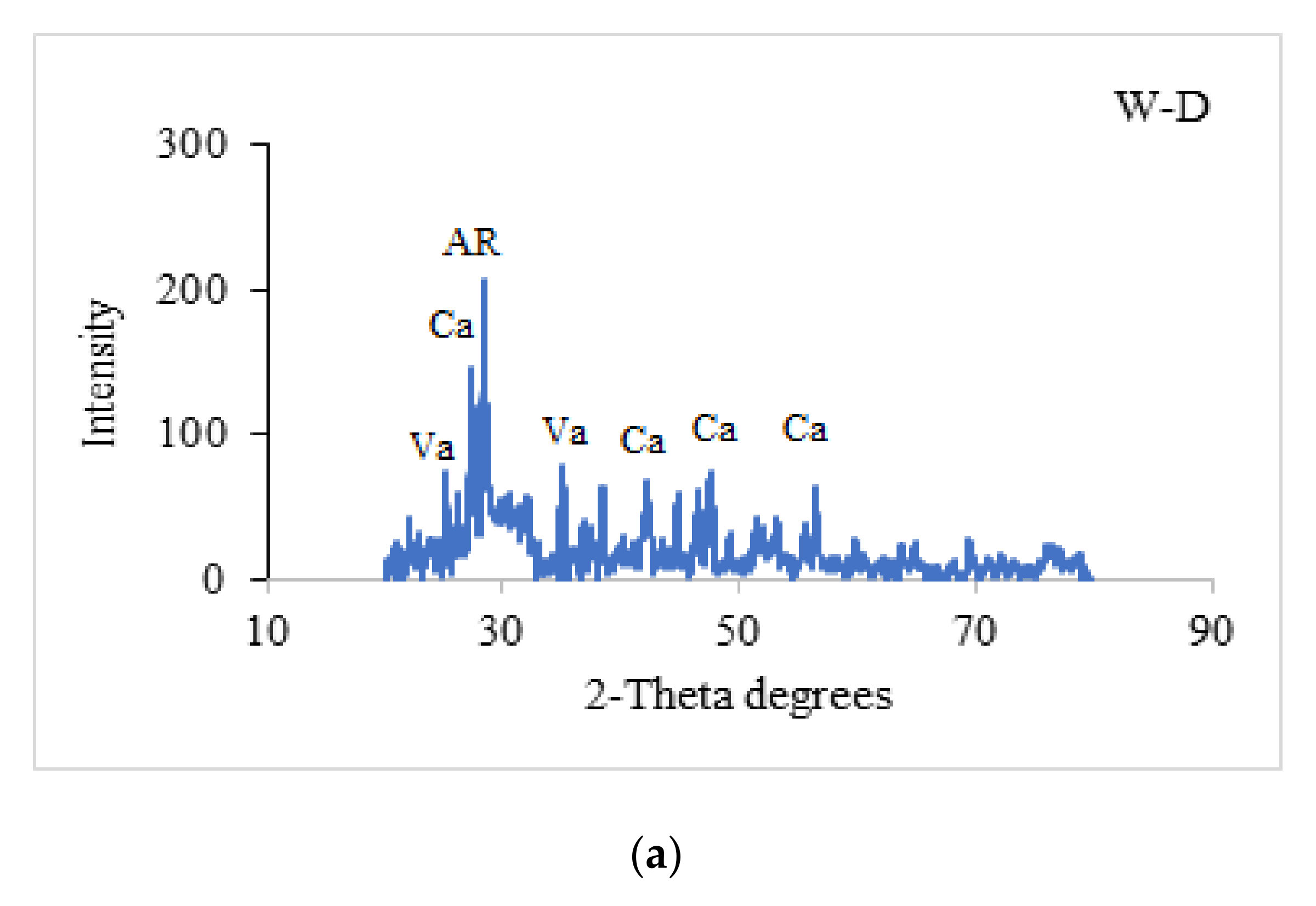
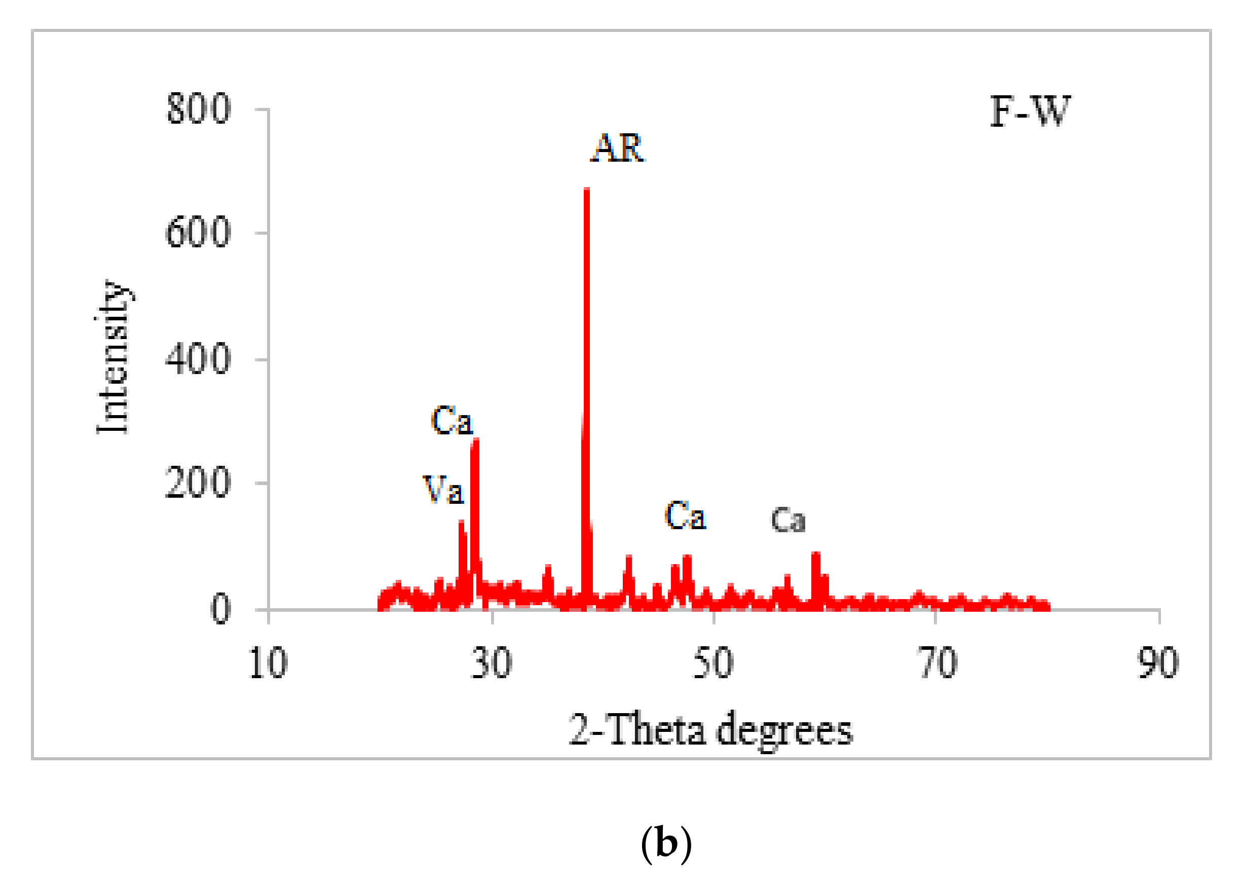
4.7. X-ray Diffraction (XRD)

XRD analysis produced a mixture of mineral precipitation including calcite (Ca), aragonite (AR), and vaterite (Va) caused by bacteria for W-D and F-W, as shown in Figure 12a,b. XRD spectra for W-D and F-W were compared, and the amplitude of peaks in the XRD spectra of the bacteria under W-D is higher than that of F-W. There are distinct peaks for aragonite and maximum peaks for W-D and F-W specimens at 28.511 and 38.447, respectively, and for calcite at 20.298, 25.163, 26.208, 27.358, 47.612, 49.256, 55.605, 56.481, etc. It is noted that a larger calcite formation was found in bacteria specimens, and this is the reason for the reduction of absorption of water from the concrete mix. The results obtained in the present work show a similar trend to those observed and reported by [53], which confirms the presence of calcium carbonate.

5. Scope of Future Work

Developing bacterial concrete by introducing the combination of different types of bacteria by direct application, encapsulation, and immobilization to enhance the regained strength, crack healing efficiency, and durability properties of concrete with various fibers (steel, basalt, polypropylene, glass) could be the scope of future work.

6. Conclusions

The analysis and interpretation of experimental outcomes obtained from the current study led to the following conclusions.
The initial screening revealed that the aerobic, Gram-positive, short rod was a motile Bacilli isolate. This exhibited positive reactions to catalase, triple sugar iron, Simmons’ citrate, and methyl red tests. Adverse reactions to Voges–Proskeur, indole, and MacConkey agar tests were also observed.
The compressive strength for the W-D and F-W cured specimens was increased by about 12.8% and 15.81%, respectively, compared to the RS at 28 days. This phenomenon is due to the formation of calcium silicate hydrate within the matrix pores, contributing to greater compressive strength. Compressive strength was regained more effectively in F-W cured specimens, increasing by about 60%, 85%, and 98% for 3, 7, and 14 days, respectively, compared to 28-day pre-cracked F-W cured specimens. The F-W cured specimens exhibited better regained strength than the W-D cycle cured specimen. Approximately 90% and 88% of surface healing was noticed in the F-W and W-D pre-cracked specimens at 28 days. Agglomeration of calcium carbonate in the concrete microstructure resulted from bacterial activity that plays a crack healing role.
The addition of bacteria in concrete contributes to a decrease in water absorption and sorptivity compared to control concrete. Due to fewer voids, bacterial precipitation tightens the pores, and the mix becomes thick, increasing the durability characteristics of self-healing concrete. SEM and XRD analysis confirmed that the presence of bacteria in concrete could enhance healing efficiency with different types of crystal morphologies of mineral precipitation under W-D and F-W methods compared to control concrete. Bacterially induced mineral precipitation was observed in the form of mixed crystals such as aragonite, calcite, and vaterite.

Author Contributions

Conceptualization, A.S.; methodology, D.G.; software, R.D.L.; validation, G.M.; formal analysis, G.M.; investigation, R.D.L.; resources, R.D.L.; data curation, T.S.G.; writing—G.M.; writing—review and editing, M.A.; visualization, A.S.; supervision, A.S.; project administration, A.S.; funding acquisition, R.F., N.I.V. All authors have read and agreed to the published version of the manuscript.

Funding

This research was financially supported by Peter the Great St. Petersburg Polytechnic University, Russian Academic Excellence Project ”5-100” to cover the article processing charges.

Acknowledgments

We thank the School of Civil Engineering, School of Chemical and Biotechnology, SASTRA Deemed University, for support. The authors gratefully acknowledge the financial support (article processing charge) from Peter the Great Polytechnic University, Saint Petersburg, Russia.

Conflicts of Interest

The authors declare no conflict of interest.

References

  1. Kaur, N.P.; Majhi, S.; Dhami, N.K.; Mistri, A. Healing fine cracks in concrete with bacterial cement for an advanced non-destructive monitoring. Constr. Build. Mater. 2020, 242, 118–151. [Google Scholar] [CrossRef]
  2. Tan, L.; Reeksting, B.; Ferrandiz-Mas, V.; Heath, A.; Gebhard, S.; Paine, K. Effect of carbonation on bacteria-based self-healing of cementitious composites. Constr. Build. Mater. 2020, 257, 119501. [Google Scholar] [CrossRef]
  3. Chen, H.; Qian, C.; Huang, H. Self-healing cementitious materials based on bacteria and nutrients immobilized respectively. Constr. Build. Mater. 2016, 126, 297–303. [Google Scholar] [CrossRef]
  4. Souradeep, G.; Dai, P.S.; Wei, K.H. Autonomous healing in concrete by bio-based healing agents—A review. Constr. Build. Mater. 2017, 146, 419–428. [Google Scholar]
  5. Vijay, K.; Murmu, M.; Deo, S.V. Bacteria based self healing concrete—A review. Constr. Build. Mater. 2017, 152, 1008–1014. [Google Scholar] [CrossRef]
  6. Zhang, J.L.; Wu, R.S.; Li, Y.M.; Zhong, J.Y.; Deng, X.; Liu, B.; Han, N.X.; Xing, F. Screening of bacteria for self-healing of concrete cracks and optimization of the microbial calcium precipitation process. Appl. Microbiol. Biotechnol. 2016, 100, 6661–6670. [Google Scholar] [CrossRef]
  7. Chahal, N.; Siddique, R.; Rajor, A. Influence of bacteria on the compressive strength, water absorption and rapid chloride permeability of fly ash concrete. Constr. Build. Mater. 2012, 28, 351–356. [Google Scholar] [CrossRef]
  8. Siddique, R.; Singh, K.; Kunal; Singh, M.; Corinaldesi, V.; Rajor, A. Properties of bacterial rice husk ash concrete. Constr. Build. Mater. 2016, 121, 112–119. [Google Scholar] [CrossRef]
  9. Kalhori, H.; Bagherpour, R. Application of carbonate precipitating bacteria for improving properties and repairing cracks of shotcrete. Constr. Build. Mater. 2017, 148, 249–260. [Google Scholar] [CrossRef]
  10. Siddique, R.; Jameel, A.; Singh, M.; Barnat-Hunek, D.; Kunal; Aït-Mokhtar, A.; Belarbi, R.; Rajor, A. Effect of bacteria on strength, permeation characteristics and micro-structure of silica fume concrete. Constr. Build. Mater. 2017, 142, 92–100. [Google Scholar] [CrossRef]
  11. Wang, J.; Vandevyvere, B.; Vanhessche, S.; Schoon, J.; Boon, N.; De Belie, N. Microbial carbonate precipitation for the improvement of quality of recycled aggregates. J. Clean. Prod. 2017, 156, 355–366. [Google Scholar] [CrossRef]
  12. Pan, X.; Shi, Z.; Khayat, K.H.; Ling, T.-C.; Li, N. A review on concrete surface treatment Part I: Types and mechanisms. Constr. Build. Mater. 2017, 132, 578–590. [Google Scholar] [CrossRef]
  13. Qiu, J.; Tng, D.Q.S.; Yang, E.-H. Surface treatment of recycled concrete aggregates through microbial carbonate precipitation. Constr. Build. Mater. 2014, 57, 144–150. [Google Scholar] [CrossRef]
  14. Jonkers, H.M.; Thijssen, A.; Muyzer, G.; Copuroglu, O.; Schlangen, E. Application of bacteria as self-healing agent for the development of sustainable concrete. Ecol. Eng. 2010, 36, 230–235. [Google Scholar] [CrossRef]
  15. De Muynck, W.; Debrouwer, D.; De Belie, N.; Verstraete, W. Bacterial carbonate precipitation improves the durability of cementitious materials. Cem. Concr. Res. 2008, 38, 1005–1014. [Google Scholar] [CrossRef]
  16. Wang, J.; Van Tittelboom, K.; De Belie, N.; Verstraete, W. Use of silica gel or polyurethane immobilized bacteria for self-healing concrete. Constr. Build. Mater. 2012, 26, 532–540. [Google Scholar] [CrossRef]
  17. Tziviloglou, E.; Wiktor, V.; Jonkers, H.; Schlangen, E. Bacteria-based self-healing concrete to increase liquid tightness of cracks. Constr. Build. Mater. 2016, 122, 118–125. [Google Scholar] [CrossRef]
  18. Nosouhian, F.; Mostofinejad, F.; Hasheminejad, H. Influence of biodeposition treatment on concrete durability in a sulfate environment. Biosyst. Eng. 2015, 133, 141–152. [Google Scholar] [CrossRef]
  19. Nosouhian, F.; Mostofinejad, D. Reducing Permeability of Concrete by Bacterial Mediation on Surface Using Treatment Gel. ACI Mater. J. 2016, 113, 287–293. [Google Scholar] [CrossRef]
  20. Nosouhian, F.; Mostofinejad, D.; Hasheminejad, H. Concrete Durability Improvement in a Sulfate Environment Using Bacteria. J. Mater. Civ. Eng. 2016, 28, 04015064. [Google Scholar] [CrossRef]
  21. Balam, N.H.; Mostofinejad, D.; Eftekhar, M. Use of carbonate precipitating bacteria to reduce water absorption of aggregates. Constr. Build. Mater. 2017, 141, 565–577. [Google Scholar] [CrossRef]
  22. Balam, N.H.; Mostofinejad, D.; Eftekhar, M. Effects of bacterial remediation on compressive strength, water absorption, and chloride permeability of lightweight aggregate concrete. Constr. Build. Mater. 2017, 145, 107–116. [Google Scholar] [CrossRef]
  23. Tayebani, B.; Mostofinejad, D. Penetrability, Corrosion Potential, and Electrical Resistivity of Bacterial Concrete. J. Mater. Civ. Eng. 2019, 31, 04019002. [Google Scholar] [CrossRef]
  24. Tayebani, B.; Mostofinejad, D. Self-healing bacterial mortar with improved chloride permeability and electrical resistance. Constr. Build. Mater. 2019, 208, 75–86. [Google Scholar] [CrossRef]
  25. Parastegari, N.; Mostofinejad, D.; Poursina, D. Use of bacteria to improve electrical resistivity and chloride penetration of air-entrained concrete. Constr. Build. Mater. 2019, 210, 588–595. [Google Scholar] [CrossRef]
  26. De Muynck, W.; De Belie, D.; Verstraete, W. Improvement of concrete durability with the aid of bacteria. In Proceedings of the 1st International Conference on Self-Healing Materials, Noordwijk aan Zee, The Netherlands, 1 January 2007. [Google Scholar]
  27. Jafarnia, M.S.; Saryazdi, M.K.; Moshtaghioun, S.M. Use of bacteria for repairing cracks and improving properties of concrete containing limestone powder and natural zeolite. Constr. Build. Mater. 2020, 242, 118059. [Google Scholar] [CrossRef]
  28. Ghosh, P.; Mandal, S.; Chattopadhyay, B.; Pal, S. Use of microorganism to improve the strength of cement mortar. Cem. Concr. Res. 2005, 35, 1980–1983. [Google Scholar] [CrossRef]
  29. Bang, S.S.; Galinat, J.K.; Ramakrishnan, V. Calcite precipitation induced by polyurethane-immobilized Bacillus pasteurii. Enzym. Microb. Technol. 2001, 28, 404–409. [Google Scholar] [CrossRef]
  30. Van Tittelboom, K.; De Belie, N.; De Muynck, W.; Verstraete, W. Use of bacteria to repair cracks in concrete. Cem. Concr. Res. 2010, 40, 157–166. [Google Scholar] [CrossRef]
  31. Wiktor, V.; Jonkers, H.M. Quantification of crack-healing in novel bacteria-based self-healing concrete. Cem. Concr. Compos. 2011, 33, 763–770. [Google Scholar] [CrossRef]
  32. Gollapudi, U.; Knutson, C.; Bang, S.; Islam, M. A new method for controlling leaching through permeable channels. Chemosphere 1995, 30, 695–705. [Google Scholar] [CrossRef]
  33. Nemati, M. Modification of porous media permeability, using calcium carbonate produced enzymatically in situ. Enzym. Microb. Technol. 2003, 33, 635–642. [Google Scholar] [CrossRef]
  34. Stocks-Fischer, S.; Galinat, J.K.; Bang, S.S. Microbiological precipitation of CaCO3. Soil Biol. Biochem. 1999, 31, 1563–1571. [Google Scholar] [CrossRef]
  35. DeJong, J.T.; Mortensen, B.M.; Martinez, B.C.; Nelson, D.C. Bio-mediated soil improvement. Ecol. Eng. 2010, 36, 197–210. [Google Scholar] [CrossRef]
  36. Mondal, S.; Das, P.; Chakraborty, A.K. Application of Bacteria in Concrete. Mater. Today Proc. 2017, 4, 9833–9836. [Google Scholar] [CrossRef]
  37. Abudoleh, S.M.; Al Mahayreh, A.; Al Frejat, A.; Al Hulaisy, F.; Hamdan, S.O. Bioconcrete Development Using Calcite-Precipitating Bacteria Isolated from Different Sources. In Proceedings of the 2nd International Conference on Building Materials and Materials Engineering, MATEC Web of Conferences, Lisbon, Portugal, 26–28 September 2018; Available online: https://www.matec-conferences.org/articles/matecconf/abs/2019/27/matecconf_icbmm2018_01011/matecconf_icbmm2018_01011.html (accessed on 30 October 2020).
  38. Chahal, N.; Rajor, R.; Siddique, A. Calcium carbonate precipitation by different bacterial strains. Afr. J. Biotechnol. 2011, 10, 8359–8372. [Google Scholar]
  39. Jagadeesha Kumar, B.G.; Prabhakar, R.; Pushpa, H. Bio mineralization of calcium carbonate by different bacterial strains and their application in concrete crack remediation. Int. J. Adv. Eng. Technol. 2013, 6, 202–213. [Google Scholar]
  40. Zhang, J.; Liu, Y.; Feng, T.; Zhou, M.; Zhao, L.; Zhou, A.; Li, Z. Immobilizing bacteria in expanded perlite for the crack self-healing in concrete. Constr. Build. Mater. 2017, 148, 610–617. [Google Scholar] [CrossRef]
  41. Zhang, J.; Zhou, A.; Liu, Y.; Zhou, B.; Luan, Y.; Wang, S.; Yue, X.; Li, Z. Microbial network of the carbonate precipitation process induced by microbial consortia and the potrntial application to crack healing in concrete. Sci. Rep. 2017, 7, 14600. [Google Scholar] [CrossRef]
  42. Tamura, K.; Dudley, J.; Nei, M.; Kumar, S. MEGA4: Molecular Evolutionary Genetics Analysis (MEGA) Software Version 4.0. Mol. Biol. Evol. 2007, 24, 1596–1599. [Google Scholar] [CrossRef]
  43. Kim, O.-S.; Cho, Y.-J.; Lee, K.; Yoon, S.-H.; Kim, M.; Na, H.; Park, S.-C.; Jeon, Y.S.; Lee, J.-H.; Yi, H.; et al. Introducing EzTaxon-e: A prokaryotic 16S rRNA gene sequence database with phylotypes that represent uncultured species. Int. J. Syst. Evol. Microbiol. 2012, 62, 716–721. [Google Scholar] [CrossRef]
  44. IS. Specification for 53 Grade Ordinary Portland Cement; Bureau of Infdian Standard: New Delhi, India, 2013. [Google Scholar]
  45. IS. Coarse and Fine Aggregate for Concrete—Specification; Bureau of Infdian Standard: New Delhi, India, 2016. [Google Scholar]
  46. IS. Method of Tests for Strength of Concrete; Bureau of Indian Standards: New Delhi, India, 2004. [Google Scholar]
  47. ASTM. Standard Test. Method for Density, Absorption, and Voids in Hardened Concrete; ASTM International: West Conshohocken, PA, USA, 2013. [Google Scholar]
  48. ASTM. Standard Test. Method for Measurement of Rate of Absorption of Water by Hydraulic-Cement Concretes; ASTM International: West Conshohocken, PA, USA, 2020. [Google Scholar]
  49. Achal, V.; Pan, X.; Özyurt, N. Improved strength and durability of fly ash-amended concrete by microbial calcite precipitation. Ecol. Eng. 2011, 37, 554–559. [Google Scholar] [CrossRef]
  50. Khaliq, W.; Ehsan, M.B. Crack healing in concrete using various bio influenced self-healing techniques. Constr. Build. Mater. 2016, 102, 349–357. [Google Scholar] [CrossRef]
  51. Priya, T.S.; Ramesh, N.; Agarwal, A.; Bhusnur, S.; Chaudhary, K. Strength and durability characteristics of concrete made by micronized biomass silica and Bacteria-Bacillus sphaericus. Constr. Build. Mater. 2019, 226, 827–838. [Google Scholar] [CrossRef]
  52. Krishnapriya, S.; Babu, D.L.V.; G., P.A. Isolation and identification of bacteria to improve the strength of concrete. Microbiol. Res. 2015, 174, 48–55. [Google Scholar] [CrossRef]
  53. Nain, N.; Surabhi, R.; Yathish, N.; Krishnamurthy, V.; Deepa, T.; Tharannum, S. Enhancement in strength parameters of concrete by application of Bacillus bacteria. Constr. Build. Mater. 2019, 202, 904–908. [Google Scholar] [CrossRef]
Figure 1. Phylogenetic tree based on the sequence of 16S r RNA indicating the relatedness of Bacillus cohnii.
Sustainability 12 10346 g001
Figure 2. (a) Isolated bacteria show discrete colonies; (b) Gram staining of the isolated bacteria.
Sustainability 12 10346 g002
Figure 3. (a) Streaked agar plates of Bacillus cohnii; (b) Bacteria inoculated in LB medium.
Sustainability 12 10346 g003
Figure 4. Preparation of bacterial cell solution.
Sustainability 12 10346 g004
Figure 5. Compressive strength of specimen at various days of curing.
Sustainability 12 10346 g005
Figure 6. Regained compressive strength of pre-cracked specimens at different days.
Sustainability 12 10346 g006
Figure 7. Visual observations of closure of cracks. (a) Cracks under W-D curing and (b) Cracks under F-W curing.
Sustainability 12 10346 g007
Figure 8. Display of microscopic images of crack healing with respect to crack width (a) Cracks under W-D curing and (b) Cracks under F-W curing.
Sustainability 12 10346 g008
Figure 9. Sorptivity results at 28 days.
Sustainability 12 10346 g009
Figure 10. Water absorption at 28 days.
Sustainability 12 10346 g010
Figure 11. SEM images.
Sustainability 12 10346 g011aSustainability 12 10346 g011b
Figure 12. (a) XRD of bacterial concrete under W-D curing; (b) XRD of bacterial concrete under F-W curing.
Sustainability 12 10346 g012aSustainability 12 10346 g012b
Table 1. Morphological and biochemical characteristics of Bacillus cohnii.
S. NoTestObservation
1.ConfigurationCircular
2.SurfaceSmooth
3.PigmentWhite
4.OpacityOpaque
5.Gram’s reactionGram positive
6.Cell shapeRods
7.ArrangementChains
8.Spore(s)+
9.Motility+
10.Indole+
11.Methyl Red-
12.Voges–Proskauer+
13.Citrate+
14.Triple sugar iron+
15.Catalase+
16.Starch hydrolysis+
17.Casein hydrolysis+
18.Urease+
19.Gelatinase+
Table 2. Mix compositions used in this study.
Mix IdCement (kg/m3)FA (kg/m3)CA (kg/m3)Water (kg/m3)Bacterial Cell Solution (%)Nutrient Solution (%)Curing Method
RS438 710 1110 219 00Full-wet
W-D438 710 1110 219 595Wet–dry cycles
F-W438 710 1110 219 595Full-wet
Table 3. Details of maximum crack width and healing percentage.
Mix IdInitial Crack Width (mm)Crack Healed Size (mm)Crack Healing (%)
3 Days7 Days28 Days3 Days7 Days28 Days
RS0.420.420.420.42000
W-D0.380.3050.2240.045204188
F-W0.370.2850.2010.037234690
Publisher’s Note: MDPI stays neutral with regard to jurisdictional claims in published maps and institutional affiliations.

