Communication

Synthesis, Characterization and Mechanism Study of Green Aragonite Crystals from Waste Biomaterials as Calcium Supplement

1
Resources Recycling Department, University of Science and Technology (UST), 217, Gajeong-ro, Yuseong-gu, Daejeon-34113, Korea
2
Center for Carbon Mineralization, Mineral Resources Research Division, Korea Institute of Geosciences and Mineral Resources (KIGAM), 124 Gwahak-ro, Yuseong-gu, Daejeon-34132, Korea
3
Korea Research Institute on Climate Change, 11, Subyeongongwon-gil, Chuncheon-si 24239, Korea
4
Mechanical Engineering Department, ABES Institute of Technology, Campus 2, NH 24, Ghaziabad-201009, India
5
Department of Earth and Environmental Sciences, Korea University, Seoul-02841, Korea
*
Author to whom correspondence should be addressed.
These authors contributed equally to this study and are co-first authors.
Sustainability 2020, 12(12), 5062; https://doi.org/10.3390/su12125062
Submission received: 22 May 2020 / Revised: 15 June 2020 / Accepted: 19 June 2020 / Published: 22 June 2020
(This article belongs to the Special Issue EARTH 2019-Green Technologies for Resources and Materials Recycling)

Abstract

:
In present work, environmentally benign green aragonite crystals were synthesized from waste chicken eggshells and bivalve seashells through a simple and low-cost wet carbonation method. This method involves a constant stirring of calcium oxide slurry and magnesium chloride suspension in aqueous solution with constraint carbon dioxide injection at 80 °C. The physicochemical properties of the synthesized aragonite were further compared with the aragonite synthesized from commercial calcium oxide. The morphological analysis, such as acicular shape and optimum aspect ratio (~21), were confirmed by scanning electron microscopy. The average crystal size (10–30 µm) and specific surface area (2–18 m2 g−1) were determined by particle size and Brunauer–Emmett–Teller analysis, respectively. Moreover, a schematic crystal growth mechanism was proposed to demonstrate the genesis and progression of aragonite crystal. Green aragonite can bridge the void for numerous applications and holds the potential for the commercial-scale synthesis with eggshells and bivalve seashells as low-cost precursors.

1. Introduction

Precipitated calcium carbonate (PCC) has emerged as a potential inorganic material with numerous industrial applications, particularly as filler in polymers, paints, and paper [1,2]. Among three available polymorphic forms of PCC (i.e., calcite, aragonite, and vaterite), under ambient conditions, calcite is thermodynamically most stable while vaterite can be considered unstable [2]. Aragonite being metastable has been extensively reviewed considering its biocompatible and superior mechanical properties. Aragonite (2.93 g cm−3) is more compact and denser than calcite (2.71 g cm−3), thereby can provide better tensile strength, yield strength, and yield strain when used as filler [3]. Other applications include water treatment [4], replacement and additive to bone [5], and biomedical activities [6]. Synthesis of acicular shaped aragonite is evident in many studies [1,2,7,8]. A study reported that a controlled injection of CO2 in calcium hydroxide slurry suspended magnesium chloride aqueous solution can synthesize needle-shaped aragonite [7]. The remaining magnesium chloride solution can be reused for further aragonite synthesis as Mg2+ and Cl ions are not assimilated into aragonite crystals. During aragonite synthesis, Mg2+ ions act as an impurity ion and promote the formation of aragonite, while simultaneously impeding the nucleation and growth of calcite crystal [7]. Another study suggested that a higher concentration of Mg2+ ions favors crystal growth of aragonite [8]. Numerous theories and empirical relationships have been proposed to rationalize this cognition, such as alteration of surface charge, inhibition of calcite nucleation, calcite poisoning model, and lack understanding related to kinetics of cation dewatering [8,9].
Solid waste management is a hefty encumbrance for a sustainable world. Recycling of solid waste, such as paper, plastic, e-waste, and biomaterials, particularly bivalve seashells and eggshells are prominent [10,11,12,13,14,15]. The exponential rise in human population and their food style has resulted in food waste as an emerging threat to the environment. Waste shells (i.e., bivalve seashells and eggshells) are such waste available in abundance in open dumps and landfills. According to Food and Agricultural Organization of United Nations (2016), China is the leading producer of bivalve seashells (>10 million tonnes) followed by Europe (>632,000 tonnes), Japan (>377,000 tonnes), South Korea (>347,000 tonnes), and Thailand (>210,000 tonnes). In excess of 250,000 tonnes of waste eggshell is estimated to be generated annually worldwide [16]. Dumping these wastes in open fields and public waters can produce severe environmental consequences. For instance, diseases, such as malaria, dengue, and diarrhea, can originate as a result of microbial decomposition of the attached flesh remnants. Moreover, stinky odor due to the release of hazardous gases, particularly hydrogen sulfide, is also evident in previous studies [12,17,18]. Currently, the majority of biomaterial wastes are deposited in landfills and open fields with a slight portion are being utilized in applications including fertilizers and calcium supplements [19,20]. Another factor responsible for minimum reutilization of these biomaterials is the economics involved in the pretreatment, particularly calcination. Therefore, valorization of waste biomaterial, such as bivalve seashells and eggshells, into a product with higher economic value is highly enviable.
The objective of the present study was to synthesize pure, high aspect-ratio and acicular shaped aragonite crystals from biomaterials such as bivalve seashells and eggshells. Further, a comparative parallel study of physicochemical properties was carried out with aragonite synthesized by commercial-grade calcium oxide (COCOMM). An easy-to-perform, low cost, and environment-friendly approach was adopted where simultaneous carbonation and mechanical stirring of calcinated biomaterials suspended in magnesium chloride aqueous solution was carried out. The present study simultaneously focuses on two problems, i.e., inorganic solid food waste management and potential alternative of limestone for the synthesis of aragonite. The economic benefits mainly rely on three factors: (i) MgCl2 solution can be reused again and again for the synthesis of aragonite, (ii) waste biomaterial as a precursor for synthesis of aragonite, (iii) low-temperature calcination of seashells (900 °C). This method also does not generate any by-product, and therefore, no further generation of waste. This environmentally benign avenue of valorization of waste biomaterials through the fabrication of green aragonite with a high aspect ratio can extend and unplug new PCC applications.

2. Experimental

2.1. Materials

Waste bivalve seashells (i.e., abalone, mussel, manila clam, scallop, and oyster shells) and chicken eggshells were obtained from the local supplier in Daejeon, South Korea. Calcium Oxide (96% purity) and magnesium chloride hexahydrate (98% purity) were purchased from Junsei Chemical Ltd., South Korea, while carbon dioxide (>99% purity) was provided by Jeil Gas Company, South Korea. All chemicals were of analytical grade and used without any further modification.

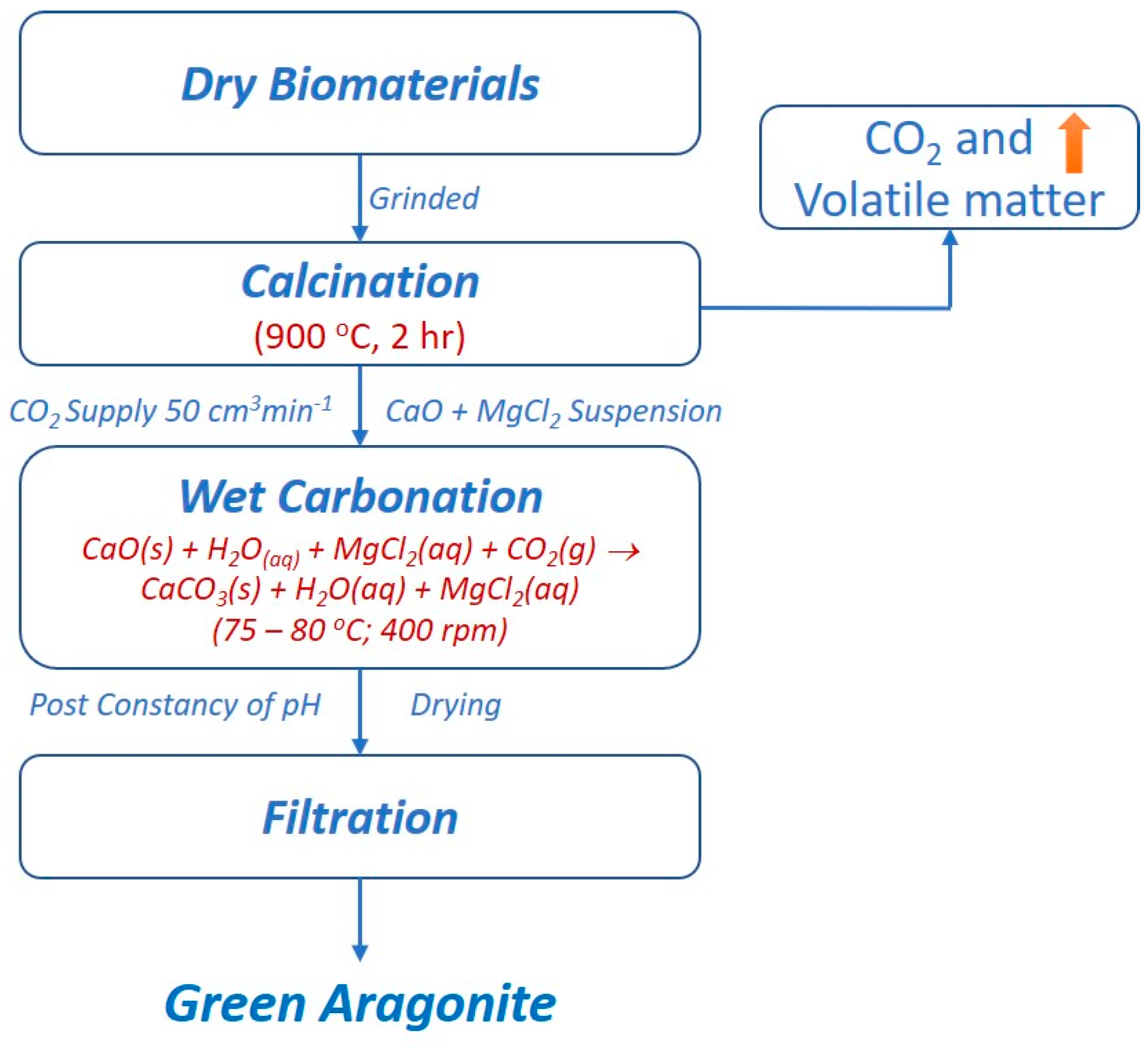
2.2. Synthesis of Green Aragonite

Bivalve seashells and chicken eggshells were first calcinated at 900 °C for 2 h. Calcinated bivalve seashells with similar physicochemical properties were then mixed in a similar proportion. Then, three precursors for the synthesis of green aragonite were obtained, i.e., COCOMM, mixed calcinated seashell, and calcinated chicken eggshell. Green aragonite was effectively synthesized by wet carbonation method. Aqueous suspensions of varying molar ratios (i.e., 1–3) of magnesium chloride hexahydrate and calcium oxide (Mg:Ca) were prepared and stirred continuously at 400 rpm (throughout the reaction). The suspension was then transferred to a 1 L cylindrical Pyrex glass reactor and maintained at 80 °C. Carbon dioxide was injected at a flow rate of 50 cm3 min−1 through a porous ceramic bubbler for a homogeneous reaction. With carbon dioxide injection, the pH gradually decreased, and completion of the reaction was found by observing the constancy of pH. The synthesized green aragonite was washed with ethanol and then with demineralized water followed by filtration and drying at 105 °C for 4 h. The schematic diagram of aragonite synthesis is illustrated in Figure 1.

2.3. Characterization

Investigation of mineralogical and crystallographic phases of bivalve seashells, chicken eggshells, and synthesized green aragonite was conducted by powder X-ray diffraction (XRD) with 2θ ranging from 10° to 90° (BD2745N, Rigaku, Tokyo, Japan). For crystal structure, shape, and morphological analysis, high-resolution microimages were observed by scanning electron microscopy (SEM), (JSM-6330F, JEOL. Co. Ltd., Tokyo, Japan). The specific surface area of the synthesized green aragonite was investigated by Brunauer–Emmett–Teller (BET) (Quadrasorb SI, Quantachrome Instruments, Florida, United States). Particle size distribution was examined by a particle size analyzer (Malvern Zetasizer Nano ZS90, United Kingdom). Surface charge study was conducted through zeta potential analysis (Otsuka ELS-Z, Japan).

3. Result and Discussion
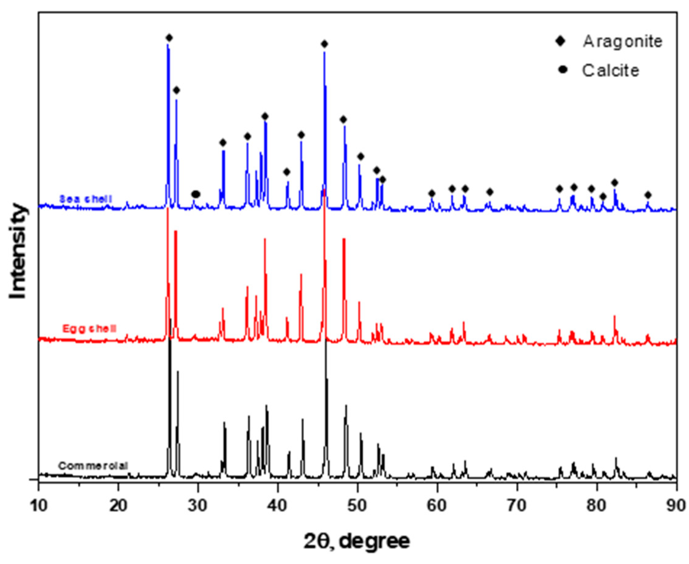
3.1. XRD Analysis

Crystal structures and mineralogical phases of raw bivalve seashells, chicken eggshells, and respectively synthesized green aragonite were determined by powder XRD. As illustrated in Figure S1, calcite and aragonite were found in two phases unevenly distributed in bivalve seashells. For instance, in an abalone shell, the dominant mineralogical phase was observed to be orthorhombic aragonite (~76%) resembling a space group P-mcn (Space 175 Group No. 62; JCPDS PDF Card No. 05-0453) with highest peak at plane <111> and at a 2θ value of 26.24. The minor phase included rhombohedral calcite (~22%) with a space group R-3c (Space Group No. 167; JCPDS PDF Card No. 86-0174). Similar dominancy of the orthorhombic aragonite phase was found in manila clam (~92.5%) with the corresponding minor phase of rhombohedral calcite (~6%). Contrastingly, the major mineralogical phase of oyster, scallop, and mussel shells was identified as rhombohedral calcite with ~92%, ~72%, and ~94%, respectively, while the minor phases constituted orthorhombic aragonite with ~2.5%, ~24.5%, and ~3%, respectively. The XRD patterns of mineralogical phases for manila clam, oyster, scallop, and mussel shells overlapped the same PDF cards as followed by abalone shell. The XRD patterns for chicken eggshell were corresponding to the same rhombohedral calcite phase mentioned earlier. Eventually, as presented in Figure S2, the XRD patterns for calcinated shells and COCOMM aligned and overlapped over the same 2θ resembling a space group Fm-3m (Space Group No. 225; JCPDS PDF Card No.70-4068).
Crystal structures and mineralogical phases of synthesized green aragonite derived from COCOMM displayed the resemblance with acicular shaped aragonite as a major phase with a space group P-mcn (Space 175 Group No. 62; JCPDS PDF Card No. 05-0453), while calcite as a minor impurity. The XRD patterns of synthesized green aragonite with different Mg to Ca molar ratio in reactants are depicted in Figure S3. At Mg:Ca = 1, a meager presence of calcite is evident with visible peaks at 2θ values of 23.07, 29.42, and 39.45. These peaks gradually reduced and became insignificant at Mg:Ca = 1.8 onwards, manifesting the presence and immaculacy of single-phase aragonite. Figure 2 represents the XRD patterns of green aragonite at Mg:Ca = 1.8 synthesized from mixed calcinated bivalve seashells and chicken eggshells, and compared with aragonite synthesized from COCOMM. All the peaks resembling green aragonite (aragonite synthesized from biomaterials) were in correspondence with the aragonite synthesized with COCOMM. This implies that high grade (with negligible calcite) aragonite crystals can be obtained from biomaterials, highlighting the substituting potential for natural resources, such as limestone.

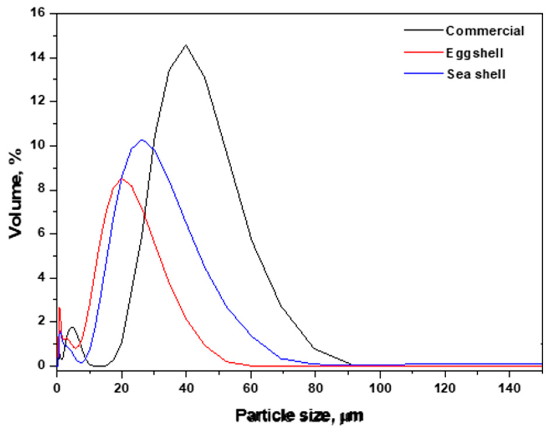
3.2. Particle Size Distribution
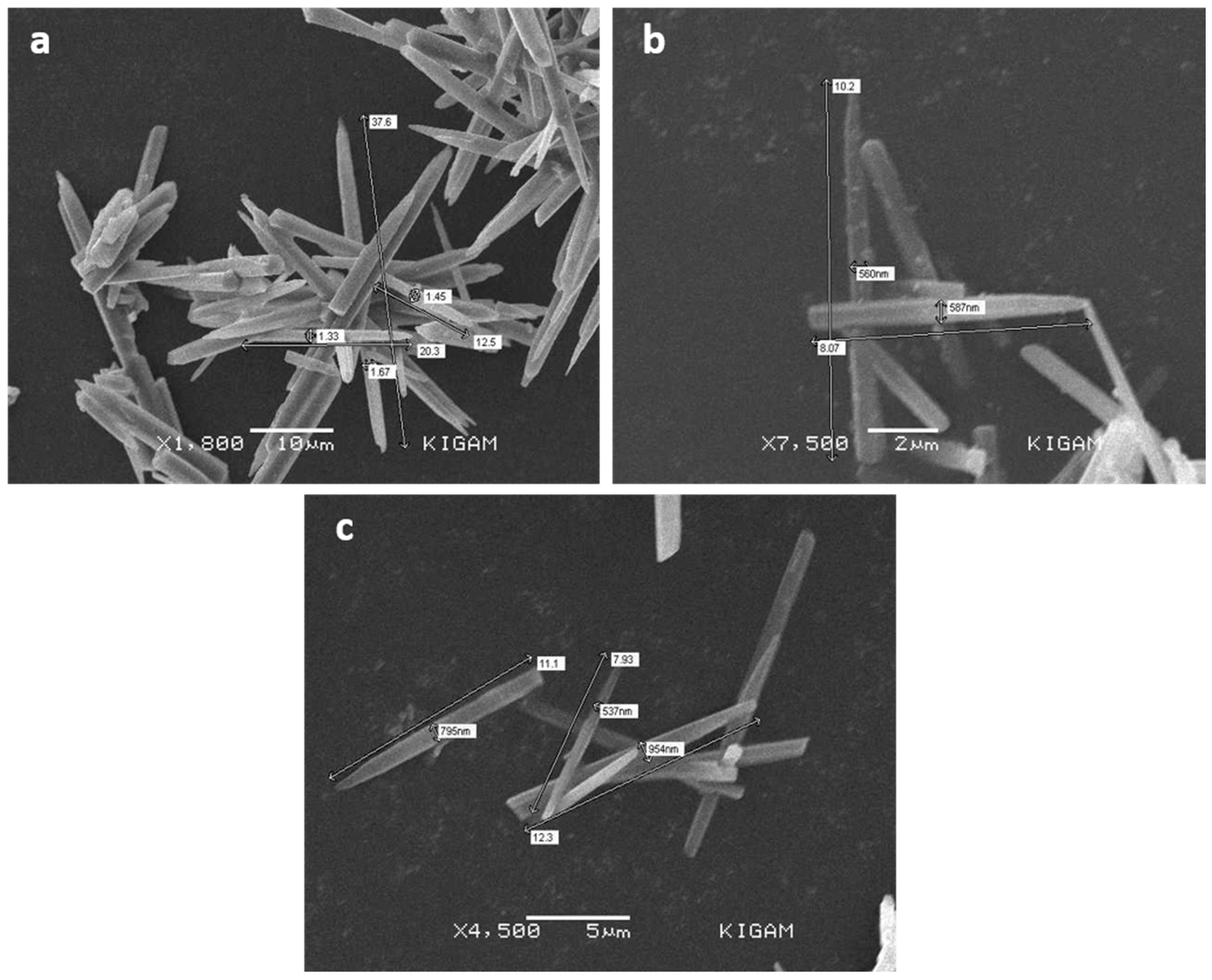
The particle size distribution (Figure 3) for COCOMM was found in the range of 20–80 µm with peaking the volumetric (%) value at ~43 µm. Contrastingly, this range significantly reduced in the case of green aragonite synthesized from bivalve seashells, i.e., between 10 and 65 µm with the highest peak at ~28 µm. The reduction in particle size distribution can be explained with the presence of metallic cations in bivalve seashells. The presence of divalent cations in the form of impurity, such as Sr2+, Fe2+, Al3+, and Ba2+, during the synthesis of aragonite can have a considerable effect on the particle size, however, it does not possess any effect on the resultant shape [21]. Correspondingly, the resultant green aragonite synthesized from chicken eggshell showed an even smaller range of 10–45 µm with the highest peak at ~20 µm. These calcinated eggshells not only include divalent cations but also consist of thermally stable nitrogen compounds (from amino acids/proteins) that affect (through adsorption) simultaneously and resulted in even smaller particle size distribution [22,23].

3.3. SEM Analysis

The surface morphologies of green aragonite synthesized by varying Mg:Ca (i.e., 1–3) are illustrated in Figure S4. The acicular shape was evident, however, it has been identified that purity, particle length, and thickness of green aragonite change with concentration of Mg2+ ions. For instance, at ratio Mg:Ca = 1 (Figure S4a), a mixture of nano-calcite and green aragonite was formed, but as this ratio increases (Figure S4b–g), a gradual decrease in nano-calcite concentration was observed (with pure aragonite at Mg:Ca = 1.8). The concentration of Mg2+ ions was found critical regarding aspect ratio (AR) of green aragonite crystals. As shown in Figure 4, at Mg:Ca = 1.4, the average AR of 12.76 was observed, which gradually increases to 17.99 and 21.06 at Mg:Ca = 1.6 and 1.8, respectively. A further rise in Mg2+ ions negatively influenced the AR as it reduced to 17.86, 13.21, and 12.57 at Mg:Ca = 2.0, 2.4 and 3.0 respectively. Therefore, it can be endorsed that pure green aragonite with optimum AR can be obtained with Mg:Ca = 1.8. These observations indicate that through controlling Mg2+ ions, the morphological behavior of green aragonite can be controlled.
Figure 5 represents the surface morphologies of synthesized green aragonite at Mg:Ca = 1.8 from COCOMM, calcinated chicken eggshell, and calcinated mixed bivalve seashells. It has been observed that the waste shells are well equipped to be used as precursors for green aragonite synthesis as there were no considerable morphological changes when compared with aragonite synthesized from COCOMM. A similar conclusion was reported in the previous study where nano-aragonite particles were successfully synthesized from oyster shells [24]. The green aragonite crystals synthesized from commercial calcium oxide and each calcinated waste shells were acicular in shape with smooth surfaces (negligible pores) and fall under similar average AR (i.e., ~21). Moreover, the presence of possible minute impurities in terms of metallic ions in the shells did not possess any negative effect on green aragonite morphology. Other studies also confirmed the synthesis of needle-shaped aragonite particles through slow sustained precipitation with suspended urea and ammonium carbonate in aqueous solution as carbonate source [21,22]. These results highlighted the potential of waste shells, which can be a possible replacement of limestone in a number of applications (although more detailed investigation of physical and mechanical properties is required, which is not discussed in presented work), especially as a filler in the form of green aragonite.

3.4. BET Analysis

The specific surface area of green aragonite synthesized from bivalve seashells and chicken eggshells (as shown in Figure S5) was determined to be ~2.2 m2 g−1 and ~18.7 m2 g−1, respectively. The extra specific surface area of chicken eggshells derived green aragonite can also be justified by reduced particle size (discussed earlier). These low surface areas indicated the presence of a smooth surface with negligible pores in agreement with the previous study [25].

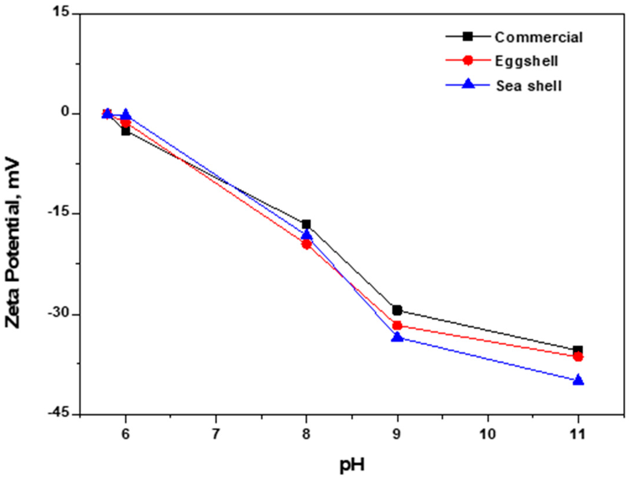
3.5. Zeta Potential Analysis

Zeta potential (ZP) is an indispensable parameter for environmental and biomedical applications [26]. It measures the surface charge characteristics of the charged particles suspended in the aqueous medium. The ZP values with varying pH for synthesized green aragonite are illustrated in Figure 6. The ZP values of synthesized green aragonite when suspended in water were found to be negative with a resulting pH range of 10.5–10.8. Furthermore, the ZP values started shifting towards zero when green aragonite suspension was adjusted to lower pH but particles consequently dissolved at pH < 6.0. Therefore, no positive charge can be obtained on the green aragonite crystal surface at pH < 6.0. This pattern was followed by the aragonite crystals synthesized from all the precursors.

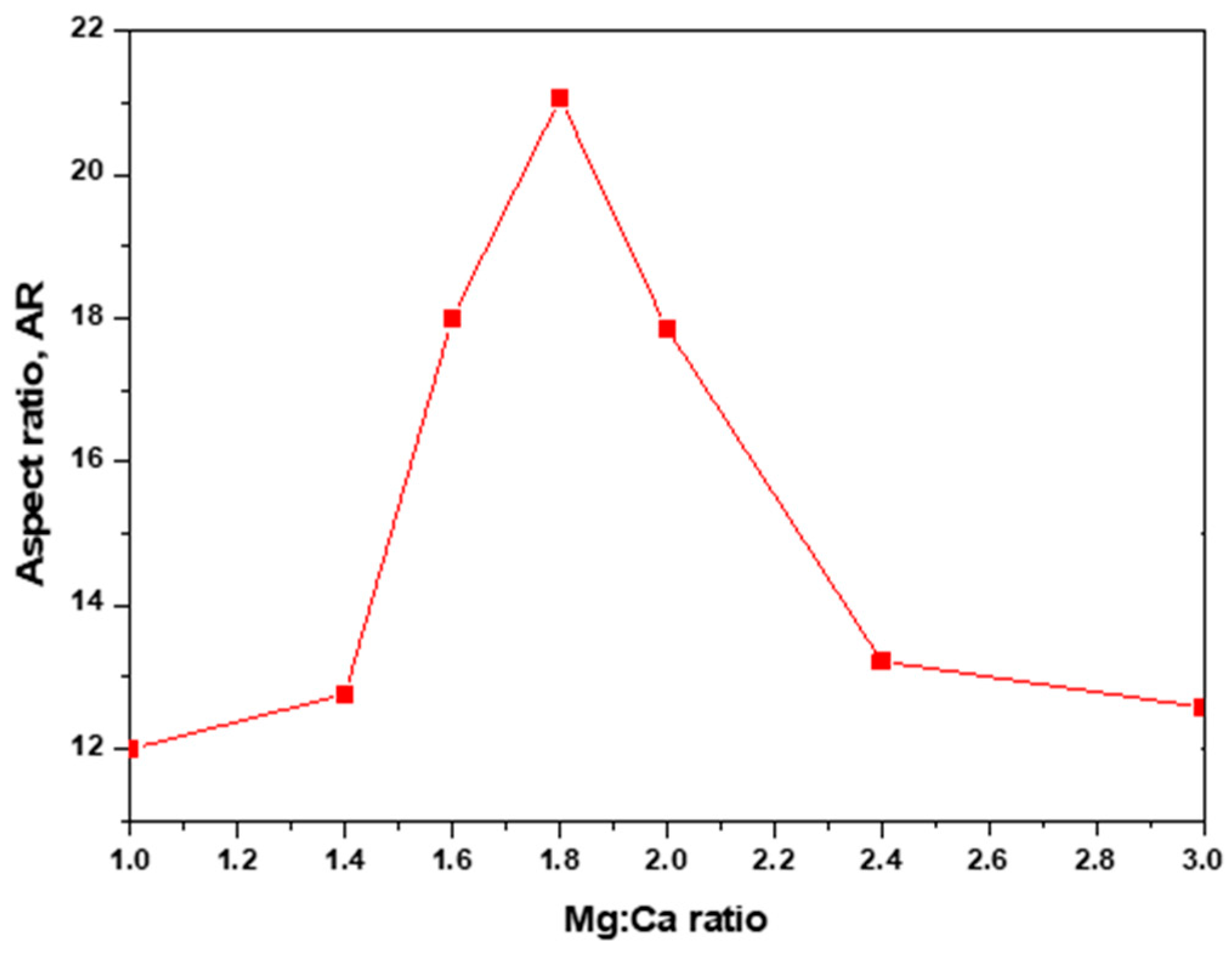
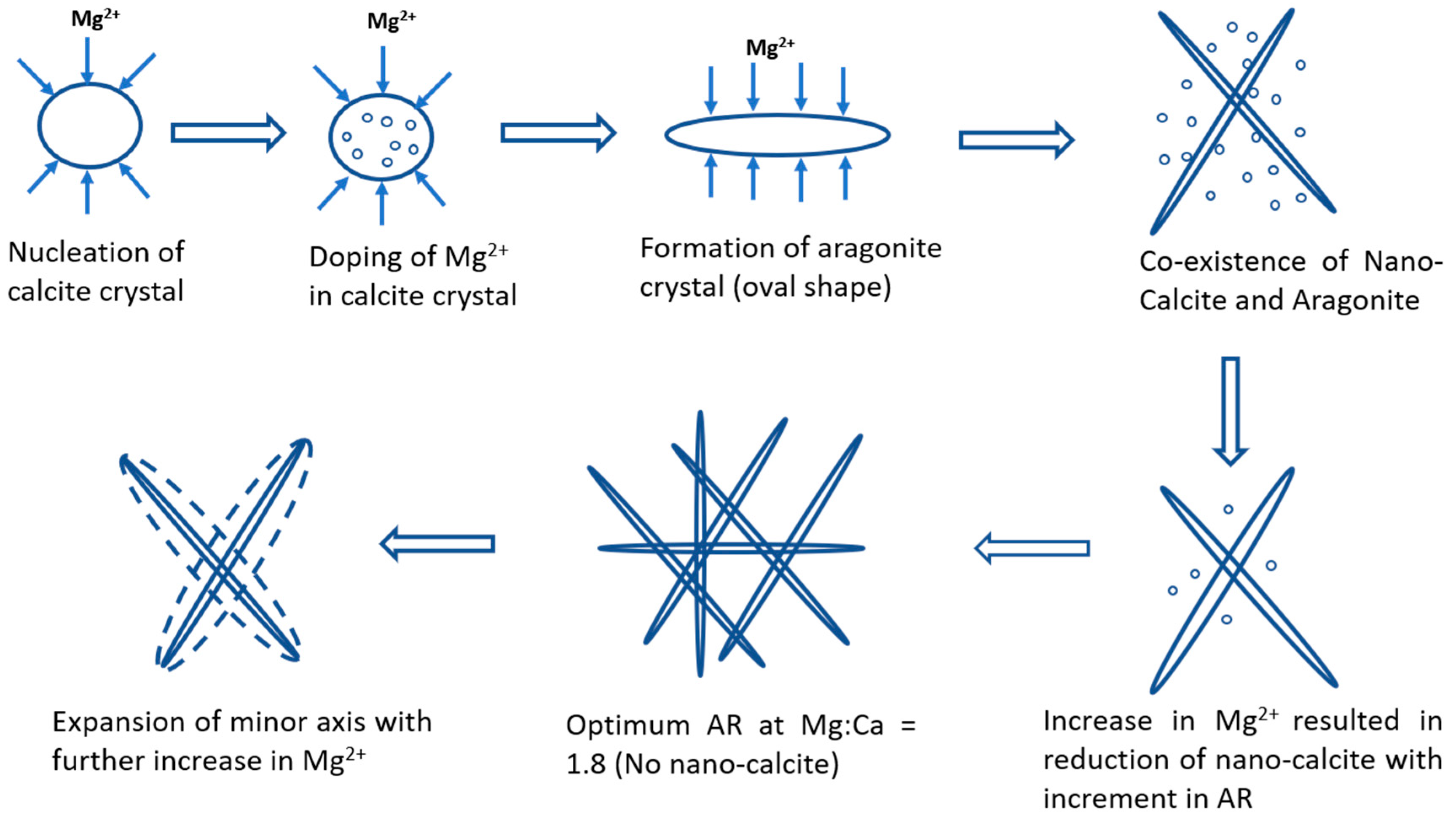
4. Green Aragonite Crystal Growth Mechanism

The wet carbonation method for the synthesis of green aragonite follows the following reaction.
C a O   ( s ) + H 2 O ( l ) C a ( O H ) 2 ( a q ) C a 2 + + 2 O H
C O 2 ( g ) + H 2 O ( l ) H 2 C O 3 ( a q )
H 2 C O 3 ( a q ) H + + H C O 3
H + + H C O 3 2 H + + C O 3 2
C a 2 + + C O 3 2 C a C O 3 ( s )
Overall Reaction:
C a ( O H ) 2 ( a q ) + C O 2 ( g ) C a C O 3 ( s ) + H 2 O ( a q )
It has been discussed in previous studies that Mg2+ ions do not take part in reaction for the synthesis of green aragonite but play a crucial role in inhibiting the nucleation of calcite [8,24,27]. A detailed crystal growth mechanism of green aragonite is depicted in Figure 7. In the beginning, the aqueous solution before carbonation mostly comprises a coexistence of Ca(OH)2 and Mg(OH)2. Homogeneous acicular shaped aragonite formation can only be achieved through controlled carbonation [21]. Therefore, after a series of batch experiments, 50 cm3 min−1 of CO2 injection flowrate was found optimum. Irregular shaped and presence of other polymorphs, such as vaterite and calcite, were observed with the introduction of CO2 at higher flowrates [28,29]. With the initiation of carbonation, the nucleation of calcite particles started as soon as Ca2+ ions encountered CO32− ions. However, some Mg2+ ions can replace Ca2+ ions of calcite to form Mg-doped calcite due to the natural interchangeability of these ions [8]. The rise in Mg2+ concentration tends to decrease the yield of Mg-doped calcite, whereas aragonite crystal growth increases. The Ca2+ and Mg2+ possess an effective ionic radii of 1.00 and 0.72 Å respectively, in the six-fold coordination (i.e., Mg2+ is 28% smaller). In an aqueous system, tenacious hydration coverings are formed on the surface of the developing crystal due to high charge density and adsorption of Mg2+, thereby curtailing the nucleation rate of calcite crystal [30]. Consequently, obstructing the growth of calcite crystal during the whole reaction, Mg2+ may eventually reside in the lattice. Similar conclusions were also reported in a previous study where Mg2+ ions were found culpable for inhibition of calcite crystal growth through poisoning the nucleation sites [31]. This poisoning and inhibition (with low Mg2+ ions) resulted in aragonite crystal possessing oval shape. This implies that the aragonite growth was inhibited in the plane perpendicular to the c-axis. An increase in Mg2+ concentration (Mg:Ca = 1) provided acicular shaped aragonite crystals with a better aspect ratio (AR) along with the coexistence of nano-calcite. Further increase in Mg2+ concentration continued to improve the AR until the concentration reached Mg:Ca = 1.8 (no trace of nano-calcite), beyond which AR decreased again. This indicates that when Mg2+ concentration exceeds a certain limit (i.e., Mg:Ca = 1.8), they start inhibiting the aragonite crystal growth both in the direction perpendicular to c-axis and in the plane of c-axis. Conclusively, it can be presumed that with Mg:Ca < 1.8, Mg2+ promotes the crystal growth in the plane of c-axis through selective side-poisoning mechanism providing it an acicular shape. However, with Mg:Ca > 1.8, the influence of selective side-poisoning mechanism starts reducing and relative expansion of the minor axis come into existence resulted in a gradual decrease in AR. Therefore, the formation of acicular shaped green aragonite with high AR is probably the resultant effect of Mg2+ inhibiting the nucleating sites for calcite formation along with selective side-poisoning mechanism. Further research is still required to provide concrete evidence supporting the effective role of Mg2+ ions aragonite synthesis.

5. Conclusions

Acicular shaped green aragonite crystals were synthesized from biomaterials, such as bivalve seashells and chicken eggshells, through the wet carbonation method. The physicochemical properties of these green aragonite were then compared with aragonite synthesized from COCOMM. It was elucidated from a series of batch experiments that the concentration of Mg2+ ions remarkably affects the aragonite crystal’s shape, size, and AR. The rise of Mg2+ ions, i.e., from Mg:Ca = 1 to Mg:Ca = 3 in the suspension resulted in acicular shaped aragonite with the occurrence of nano-calcite to pure aragonite with high AR at Mg:Ca = 1.8, beyond which AR decreases again. All physicochemical characteristics of green aragonite were found in correspondence with aragonite synthesized from COCOMM, indicating a potential future of these waste biomaterials as aragonite precursor. The genesis and crystal growth mechanism were also discussed, revealing that the Mg2+ ions may control the aragonite crystal growth through the combined effect of Mg2+ inhibiting the nucleating sites for calcite formation along with selective side-poisoning mechanism. The presented work is not only compelling with biomineralization but can also provide significant accession in the synthesis of novel functional materials.

Supplementary Materials

The following are available online at https://www.mdpi.com/2071-1050/12/12/5062/s1, Figure S1: Powder X-ray diffraction patterns of raw waste seashells.

Author Contributions

M.D.K. and L.H.: Conceptualization, methodology, writing-original draft. N.S., A.F., S.-h.J., and M.-h.L.: data curation, visualization, software. J.W.A.: supervision writing-review & editing. All authors have read and agreed to the published version of the manuscript.

Funding

This work was funded by the Ministry of Science and ICT (MSIT), the Ministry of Environment (ME) and the Ministry of trade, Industry and Energy (MOTIE) (2019M3D8A2112963).

Acknowledgments

This work was supported by the National Strategic Project-Carbon Mineralization Flagship Center of the National Research Foundation of Korea (NRF) funded by the Ministry of Science and ICT (MSIT), the Ministry of Environment (ME) and the Ministry of trade, Industry and Energy (MOTIE) (2019M3D8A2112963).

Conflicts of Interest

The authors declare no competing personal relationships and financial interests that could influence the presented work.

References

  1. George, J.; Ishida, H. A review on the very high nanofiller-content nanocomposites: Their preparation methods and properties with high aspect ratio fillers. Prog. Polym. Sci. 2018, 86, 1–39. [Google Scholar] [CrossRef]
  2. Donnelly, F.C.; Purcell-Milton, F.; Dunne, P.; Rulikowska, A.; Alguacil, V.; Gun’Ko, Y.K. Luminescent calcium carbonate micro ‘bow ties’. Mater. Today Commun. 2019, 20, 100590. [Google Scholar] [CrossRef]
  3. Li, H.-Y.; Tan, Y.-Q.; Zhang, L.; Zhang, Y.-X.; Song, Y.; Ye, Y.; Xia, M. Bio-filler from waste shellfish shell: Preparation, characterization, and its effect on the mechanical properties on polypropylene composites. J. Hazard. Mater. 2012, 217, 256–262. [Google Scholar] [CrossRef] [PubMed]
  4. Millero, F.; Huang, F.; Zhu, X.; Liu, X.; Zhang, J.-Z. Adsorption and Desorption of Phosphate on Calcite and Aragonite in Seawater. Aquat. Geochem. 2001, 7, 33–56. [Google Scholar] [CrossRef]
  5. Islam, K.N.; Bin Abu Bakar, Z.; Ali, E.; Hussein, M.Z.; Noordin, M.M.; Loqman, M.; Miah, G.; Wahid, M.H.; Hashim, U. A novel method for the synthesis of calcium carbonate (aragonite) nanoparticles from cockle shells. Powder Technol. 2013, 235, 70–75. [Google Scholar] [CrossRef]
  6. Samanta, A.; Podder, S.; Kumarasamy, M.; Ghosh, C.K.; Lahiri, D.; Roy, P.; Bhattacharjee, S.; Ghosh, J.; Mukhopadhyay, A.K. Au nanoparticle-decorated aragonite microdumbbells for enhanced antibacterial and anticancer activities. Mater. Sci. Eng. C 2019, 103, 109734. [Google Scholar] [CrossRef]
  7. Ota, Y.; Inui, S.; Iwashita, T.; Kasuga, T.; Abe, Y. Preparation of Aragonite Whiskers. J. Am. Ceram. Soc. 1995, 78, 1983–1984. [Google Scholar] [CrossRef]
  8. Park, W.K.; Ko, S.-J.; Lee, S.W.; Cho, K.-H.; Ahn, J.-W.; Han, C. Effects of magnesium chloride and organic additives on the synthesis of aragonite precipitated calcium carbonate. J. Cryst. Growth 2008, 310, 2593–2601. [Google Scholar] [CrossRef]
  9. Nancollas, G.; Sawada, K. Formation of Scales of Calcium Carbonate Polymorphs: The Influence of Magnesium Ion and Inhibitors. J. Pet. Technol. 1982, 34, 645–652. [Google Scholar] [CrossRef]
  10. Hamideh, F.; Akbar, A. Application of eggshell wastes as valuable and utilizable products: A review. Res. Agric. Eng. 2018, 64, 104–114. [Google Scholar] [CrossRef] [Green Version]
  11. Godlewska, J. Recovery and Recycling of Waste Tires in Poland. Procedia Eng. 2017, 182, 229–234. [Google Scholar] [CrossRef]
  12. Khan, M.D.; Ahn, J.W.; Nam, G. Environmental benign synthesis, characterization and mechanism studies of green calcium hydroxide nano-plates derived from waste oyster shells. J. Environ. Manag. 2018, 223, 947–951. [Google Scholar] [CrossRef] [PubMed]
  13. Kumar, A.; Holuszko, M.; Espinosa, D. E-waste: An overview on generation, collection, legislation and recycling practices. Resour. Conserv. Recycl. 2017, 122, 32–42. [Google Scholar] [CrossRef]
  14. Lertwattanaruk, P.; Makul, N.; Siripattarapravat, C. Utilization of ground waste seashells in cement mortars for masonry and plastering. J. Environ. Manag. 2012, 111, 133–141. [Google Scholar] [CrossRef] [PubMed]
  15. Habte, L.; Shiferaw, N.; Mulatu, D.; Thriveni, T.; Chilakala, R.; Ahn, J.W. Synthesis of Nano-Calcium Oxide from Waste Eggshell by Sol-Gel Method. Sustainability 2019, 11, 3196. [Google Scholar] [CrossRef] [Green Version]
  16. Verma, N.; Kumar, V.; Bansal, M.C. Utilization of egg shell waste in cellulase production by Neurospora crassa under Wheat bran-based solid state fermentation. Pol. J. Environ. Stud. 2012, 21, 491–497. [Google Scholar]
  17. Felipe-Sesé, M.A.; Eliche-Quesada, D.; Corpas-Iglesias, F.A. The use of solid residues derived from different industrial activities to obtain calcium silicates for use as insulating construction materials. Ceram. Int. 2011, 37, 3019–3028. [Google Scholar] [CrossRef]
  18. Tsai, W.-T.; Hsien, K.-J.; Hsu, H.-C.; Lin, C.-M.; Lin, K.-Y.; Chiu, C.-H. Utilization of ground eggshell waste as an adsorbent for the removal of dyes from aqueous solution. Bioresour. Technol. 2008, 99, 1623–1629. [Google Scholar] [CrossRef]
  19. Dai, L.; Tan, F.-R.; Li, H.; Zhu, N.; He, M.; Zhu, Q.-L.; Hu, G.Q.; Wang, L.; Zhao, J. Calcium-rich biochar from the pyrolysis of crab shell for phosphorus removal. J. Environ. Manag. 2017, 198, 70–74. [Google Scholar] [CrossRef]
  20. Lu, J.; Cong, X.; Li, Y.; Hao, Y.; Wang, C. Scalable recycling of oyster shells into high purity calcite powders by the mechanochemical and hydrothermal treatments. J. Clean. Prod. 2018, 172, 1978–1985. [Google Scholar] [CrossRef]
  21. Wang, L.; Sondi, I.; Matijević, E. Preparation of Uniform Needle-Like Aragonite Particles by Homogeneous Precipitation. J. Colloid Interface Sci. 1999, 218, 545–553. [Google Scholar] [CrossRef] [PubMed] [Green Version]
  22. Chen, L.; Huang, F.; Li, S.; Shena, Y.; Shena, Y.; Pan, J.; Zhang, Y.; Cai, Y. Biomimetic synthesis of aragonite superstructures using hexamethylenetetramine. J. Solid State Chem. 2011, 184, 2825–2833. [Google Scholar] [CrossRef]
  23. Correia, L.M.; Cecilia, J.A.; Rodríguez-Castellón, E.; Cavalcante, C.L.; Vieira, R.S.; Rodrí Guez- Castelló, N.E.; Loureiro, L. Relevance of the Physicochemical Properties of Calcined Quail Eggshell (CaO) as a Catalyst for Biodiesel Production. J. Chem. 2017, 2017, 1–12. [Google Scholar] [CrossRef]
  24. Ramakrishna, C.; Thenepalli, T.; Han, C.; Ahn, J.W. Synthesis of aragonite-precipitated calcium carbonate from oyster shell waste via a carbonation process and its applications. Korean J. Chem. Eng. 2016, 34, 225–230. [Google Scholar] [CrossRef]
  25. Kezuka, Y.; Kawai, K.; Eguchi, K.; Tajika, M. Fabrication of Single-Crystalline Calcite Needle-Like Particles Using the Aragonite–Calcite Phase Transition. Miner 2017, 7, 133. [Google Scholar] [CrossRef] [Green Version]
  26. Handley-Sidhu, S.; Mullan, T.K.; Grail, Q.; Albadarneh, M.; Ohnuki, T.; Macaskie, L.E. Influence of pH, competing ions and salinity on the sorption of strontium and cobalt onto biogenic hydroxyapatite. Sci. Rep. 2016, 6, 23361. [Google Scholar] [CrossRef] [PubMed] [Green Version]
  27. Ahn, J.-W.; Choi, K.-S.; Yoon, S.-H.; Kim, H. Synthesis of Aragonite by the Carbonation Process. J. Am. Ceram. Soc. 2004, 87, 286–288. [Google Scholar] [CrossRef]
  28. Hosoi, K.; Hashida, T.; Takahashi, H.; Yamasaki, N.; Korenaga, T. Solidification behaviour of calcium carbonate via aragonite-calcite wet transformation with hydrothermal hot pressing. J. Mater. Sci. Lett. 1997, 16, 382–385. [Google Scholar] [CrossRef]
  29. Synthesis of Aragonite-Type Calcium Carbonate by Overgrowth Technique at Atmospheric Pressure. J. Mater. Sci. Lett. 1998, 17, 905–908. [CrossRef]
  30. Kezuka, Y.; Tochigi, E.; Murata, H.; Yoshida, M.; Eguchi, K.; Nakahira, A.; Ikuhara, Y.; Tajika, M. Synthesis of Tunable-Aspect-Ratio Calcite Nanoparticles via Mg2+ Doping. Cryst. Growth Des. 2019, 19, 6784–6791. [Google Scholar] [CrossRef]
  31. Lippmann, F. Versuche zur Aufklärung der Bildungsbedingungen von Calcit und Aragonit, Fortschr. Miner 1960, 38, 156–161. [Google Scholar]
Figure 1. Schematic diagram of aragonite synthesis process from biomaterials.
Sustainability 12 05062 g001
Figure 2. Powder X-ray diffraction patterns of green aragonite synthesized from calcinated mixed bivalve seashell, chicken eggshell and commercial-grade calcium oxide (COCOMM).
Sustainability 12 05062 g002
Figure 3. Particle size distribution of green aragonite synthesized from calcinated mixed bivalve seashell, chicken eggshell and commercial-grade calcium oxide (COCOMM).
Sustainability 12 05062 g003
Figure 4. The aspect ratios (AR) of synthesized green aragonite crystals at different Mg:Ca ratio.
Sustainability 12 05062 g004
Figure 5. SEM images of synthesized green aragonite (a) with Commercial Calcium Oxide (COCOMM), (b) with calcinated chicken eggshell, and (c) with calcinated mixed bivalve seashell at Mg:Ca = 1.8.
Sustainability 12 05062 g005
Figure 6. Zeta potential values of synthesized green aragonite with Commercial Calcium Oxide (COCOMM), calcinated chicken eggshell and calcinated mixed bivalve seashell at Mg:Ca = 1.8.
Sustainability 12 05062 g006
Figure 7. Detailed crystal growth mechanism of green aragonite crystals during the wet carbonation process.
Sustainability 12 05062 g007

