Article

Protective Effect of Ethanolic Extract of Djulis Hull on Indomethacin-Induced Gastric Injury

1
Department of Agricultural Product Technology, Faculty of Agricultural Technology, Brawijaya University, Malang 65145, Indonesia
2
Department of Biological Science and Technology, National Pingtung University of Science and Technology, Pingtung 91201, Taiwan
3
Department of Food Technology, Faculty of Engineering, Bumigora University, Mataram 83127, Indonesia
4
Department of Food Science, National Pingtung University of Science and Technology, Pingtung 91201, Taiwan
5
Department of Nutrition and Health Science, Fooyin University, Kaohsiung 83102, Taiwan
6
Aging and Disease Prevention Research Center, Fooyin University, Kaohsiung 83102, Taiwan
*
Author to whom correspondence should be addressed.
Appl. Sci. 2023, 13(1), 594; https://doi.org/10.3390/app13010594
Submission received: 20 December 2022 / Revised: 28 December 2022 / Accepted: 28 December 2022 / Published: 1 January 2023
(This article belongs to the Special Issue Knowledge-Based Biotechnology for Food, Agriculture and Fisheries)

Abstract

:
Djulis (Chenopodium formosanum), a pseudocereal crop native to Taiwan, is often utilized as a source of grain in the diet because of its high nutritional value. The hull of djulis is discarded as waste during cooking or processing because of its bitter taste. However, recent studies have shown that djulis hull possesses certain benefits, such as antioxidant, blood sugar-lowering, and gut microbiota-regulating properties. Herein, the gastroprotective activity of ethanolic extract of djulis hull (EEDH) against stomach injury caused by indomethacin (IND) in C57BL/6J mice and its mechanism of action was assessed. Preadministration of EEDH significantly attenuated the gastric ulcer caused by IND in a dose-dependent manner (p < 0.05). Additionally, gastric mucosal injury and gastric wall edema within the submucosal layer observed in histopathological examination were improved by administration of EEDH. EEDH preadministration also reinstated the reduction of glutathione (GSH) content and catalase (CAT), and superoxide dismutase (SOD) activities induced by IND, indicating that EEDH can modulate the antioxidant status of gastric mucosa in mice. Moreover, IND-induced decline of gastric COX-1 expression was upregulated in mice of EEDH treatment groups. Administration of IND increased the expression of proinflammatory proteins in the gastric mucosa of mice, including tumor necrosis factor-α (TNF-α) and inducible nitric oxide synthase (iNOS), whereas EEDH treatment significantly decreased their expression (p < 0.05). Consequently, EEDH can improve gastric injury by regulating antioxidant status and inhibiting proinflammatory signaling pathways, and has the potential to be developed as a functional food for gastric protection.

1. Introduction

Gastric ulcer disease is a usual digestive disorder and a global public health problem, and it is thought to occur in approximately 10% of people during their lifetime [1]. Gastric ulceration is caused by an imbalance between mucosal defensive and offensive factors, including genetic factors, gastric acid, Helicobacter pylori, ethanol, and nonsteroidal, antiinflammatory drugs (NSAIDs) [2]. It is associated with mucosal injury, including perforation, loss of mucosal integrity, bleeding of gastric mucosal, apoptosis of mucosal cells, and edema in the submucosal layer [3,4,5]. Patients with severe and frequent gastric ulcer symptoms have a high possibility of developing gastric cancer in the future [6]. The way to prevent or alleviate the symptoms of gastric ulcers from various offensive factors is an important public health issue worthy of attention now.
NSAIDs are a heterogeneous class of commercial drugs extensively used to treat pain and inflammatory diseases (such as chronic pain, osteoarthritis, and rheumatoid arthritis), and have antiinflammatory and antipyretic effects, and are the most commonly used drugs in the world [7,8]. NSAIDs include more than 20 heterogeneous groups of drugs, such as naproxen, aspirin, mefenamic acid, sulindac, ibuprofen, and indomethacin (IND) [9]. Contradictorily, continued and excessive use of NSAIDs produces serious side effects for the stomach, including bleeding, ulceration, and perforation of the gastrointestinal system [10]. The annual incidence of peptic ulcer complications caused by NSAIDs accounts for approximately 4–8%, and it may increase year by year [11]. Among them, IND, an NSAID drug, induces stomach ulcers by inhibiting the expression of cyclooxygenase (COX), reducing the synthesis of prostaglandins (PG) [12]. Furthermore, administration of IND stimulates the generation of reactive oxygen species (ROS), which induces oxidative stress and leads to gastric injury [13]. Oxidative stress initiates numerous transcription factors and results in various chronic diseases. It has been reported that excessive ROS produced during oxidative metabolism initiates the inflammatory process and promotes the activation of tumor necrosis factor-α (TNF-α) as a proinflammatory cytokine [14]. An animal model of IND-induced jejunoileitis suggested that that TNF-α activated the transcription of diverse inflammatory genes, including inducible nitric oxide synthase (iNOS), and thereby caused tissue damage [15]. In addition, disruption of the gastric mucosal antioxidant defense system, including the reduction of glutathione (GSH) levels and superoxide dismutase (SOD) and catalase (CAT) activities was found in rats with gastric ulcers induced by IND [16].
For the purpose of developing functional foods for the prevention or protection of gastric ulcers, the present study used djulis (Chenopodium formosanum Koidz.), a traditional edible grain used in Taiwan for hundreds of years, as the test sample. Djulis has high nutritional value and contains a high content of antioxidant components, exhibiting several health benefits such as anti-adipogenic [17], hypoglycemic [18], anticancer [19], antihypertensive [20], and antioxidant [21] properties. Although certain reports indicate that the hull of djulis contains high amounts of bioactive compounds, including phytosterols, triterpenes, and phenolic compounds [22,23], it is often discarded during cooking or processing because of its bitter taste. Recent studies have shown that the djulis hull had several biological effects, such as antiinflammatory, insulin resistance-improving, and gut microbiota-regulating effects [24,25]. According to the information we have, studies on the relief effects of ethanolic extract of djulis hull (EEDH) against stomach injury caused by IND are limited. Consequently, the purpose of this study was to evaluate the protective effect of EEDH on IND-induced gastric injury and to elucidate its possible mechanism of action.

2. Materials and Methods

2.1. Chemical Reagents

Glycine, glycerol, N,N,N′,N′-tetramethylenediamine (TEMED), tris-base, tris-HCl, and Tween-20 were obtained from BioShop (Burlington, Canada). Ammonium persulfate (NH4)2S2O8, dimethyl sulfoxide (DMSO), secondary antibodies, and RIPA lysis buffer were provided by Merck Millipore (Burlington, MA, USA). Bovine serum albumin (BSA), Coomassie assay protein reagent, Cytiva Amersham™ Hybond™ PVDF membrane, and GE Healthcare Amersham™ ECL prime Western blotting detection reagent were purchased from Thermo-Fisher Scientific (Waltham, MA, USA). Protease inhibitor cocktail was purchased from Fivephoton Biochemicals (San Diego, CA, USA). iNOS and TNF-α antibodies were obtained from Proteintech Group (Rosemont, IL, USA). COX-1 antibody was provided by Cell Signaling Technology (Danvers, MA, USA). Reduced glutathione (GSH) colorimetric assay kit was provided by Elabscience Biotechnology (Houston, TX, USA). Acrylamide/bis-acrylamide and β-actin antibody were purchased from ZEJU Taiwan Bio-Technology (Kaohsiung, Taiwan). Superoxide dismutase (SOD) and catalase (CAT) assay kits were purchased from Cayman Chemical (Ann Arbor, MI, USA). Skim milk was obtained from Fonterra (Auckland, New Zealand). Indomethacin (IND), ethanol (95%), sodium dodecyl sulfate (SDS), and all other chemicals were provided by Sigma-Aldrich (St. Louis, MO, USA).

2.2. Material Preparation

Djulis hull powder was a gift from Dr. Pi-Jen Tsai. Djulis hull powder was extracted with ethanol (95%) at a ratio of 1:20 (w/v) at room temperature for 24 h followed by filtration, and the filtrate was collected. The filtrate was then evaporated with a rotary evaporator to eliminate ethanol, followed by drying with a lyophilizer after freezing to obtain EEDH. The EEDH was then kept at −20 °C and resuspended in saline before being fed to mice.

2.3. Animals and Treatment

Six-week-old C57BL/6JNarl mice were provided by National Laboratory Animal Center (NLAC, Taipei, Taiwan) and kept in a facility with normal conditions (12 h light/12 h dark cycle, temperature 23 ± 2 °C, humidity 50–70%). After a week of adaptation, the mice were randomly divided into the following groups (8 mice per group): (1) Control group (saline); (2) IND group (saline + 120 mg/kg IND); (3) EEDH200 (200 mg/kg EEDH + 120 mg/kg IND); and (4) EEDH500 (500 mg/kg EEDH + 120 mg/kg IND). Saline or EEDH (200 mg/kg and 500 mg/kg) were administered orally once daily for 21 consecutive days. On day 20, the mice were starved for 24 h, followed by administration of normal saline or EEDH, and IND (120 mg/kg) was given to each group except the control group 2 h later. After 6 h, all mice were consequently sacrificed by asphyxiation with CO2. All animal studies were conducted under protocol number NPUST-111-001 approved by the Institutional Animal Care and Use Committee (IUCUC) of NPUST.

2.4. Evaluation of Ulcer Area and Protective Index

In order to evaluate the ulcer area and protective index of EEDH, gastric tissues were harvested and washed with ice-cold saline solution and carefully flattened for imaging. Subsequently, one part of gastric tissues was stored in a −80 °C freezer until further analysis, and the other part was fixed in 10% formalin. The captured images were analyzed and total ulcer area (mm2) for each mouse was measured and counted by using ImageJ® 1.51k software (Bethesda, MD, USA). According to the method of Zhou et al. [26] with a mere modification, the following is the calculation formula of the protective index of EEDH:
Protective   index   ( % ) = Ulcer   area IND   group     Ulcer   area EEDH   group   Ulcer   area IND   group   ×     100 % .

2.5. Histopathological Assessment

The histopathological assessment of stomach tissues in mice was performed according to the procedure in a previous study [27] with certain modifications. In brief, stomach tissues were fixed, processed, and embedded. Paraffin blocks were sectioned at a thickness of 5 μm, and the sections were mounted on slides and stained with hematoxylin and eosin (H&E) after deparaffinization. Finally, all stained sections were photographed and examined by a light microscope equipped with a camera.

2.6. Gastric GSH Determination

A total of 0.03 g of gastric tissue was put into the homogenizing tube filled with beads, and then 270 μL of buffer (containing 12 mM K2HPO4, 8 mM KH2PO4, and 1.5% KCl) was added and homogenized. The supernatant fraction was then obtained by centrifugation of the homogenate for 30 min at 16,400× g (4 °C). Subsequently, the gastric level of GSH was analyzed by using a commercial GSH assay kit following the manufacturer’s guidance as previously mentioned [28].

2.7. Determination of Gastric SOD and CAT Activities

We prepared the homogenate of gastric tissue for SOD activity determination by applying the same method as was used for the determination of GSH. The SOD activity of gastric tissue was analyzed by using the SOD assay kit according to the producer’s recommendations. For the analysis of gastric CAT activity, 0.01 g of gastric tissue was added to 100 μL of buffer containing 50 mM K2HPO4 and 1 mM EDTA (pH 7.0) and homogenized. The supernatant was obtained by centrifugation at 10,000× g for 15 min at 4 °C. Then, the CAT activity of gastric tissue was determined by using a commercial assay kit following the manufacturer’s procedure.

2.8. Analysis of Gastric Protein Expression

The protein of gastric tissue was isolated and analyzed as in our previous study [29]. SDS polyacrylamide gel electrophoresis (SDS-PAGE) was performed to separate the protein samples collected from gastric tissue, and the proteins were further transferred onto a PVDF membrane activated by methanol. Then, 5% skim milk in TBST (Tris-buffered saline with 0.1% Tween-20) was used to soak the PVDF membrane for blocking at 25 °C for 1 h. The blocked membrane was consequently probed with primary antibodies (COX-1, TNF-α, iNOS, and β-actin) overnight at 4 °C. Thereafter, the membrane was washed with TBST followed by incubation in secondary antibody solution for 1–2 h. The signals of the target protein were then generated by ECL and captured by a Luminescence Image System (Hansor, Taichung, Taiwan). The signal intensities were quantified via ImageJ® software.

2.9. Statistical Analysis

Data are represented as means ± standard deviation (SD). The analysis of data was conducted by SPSS 12.0 (Statistical Product and Service Solutions) software (SPSS Inc., Chicago, IL, USA). Analysis of variance (ANOVA) was used for single-factor analysis, and Duncan’s test was used for significance analysis, and a p < 0.05 was determined statistically significant.

3. Results

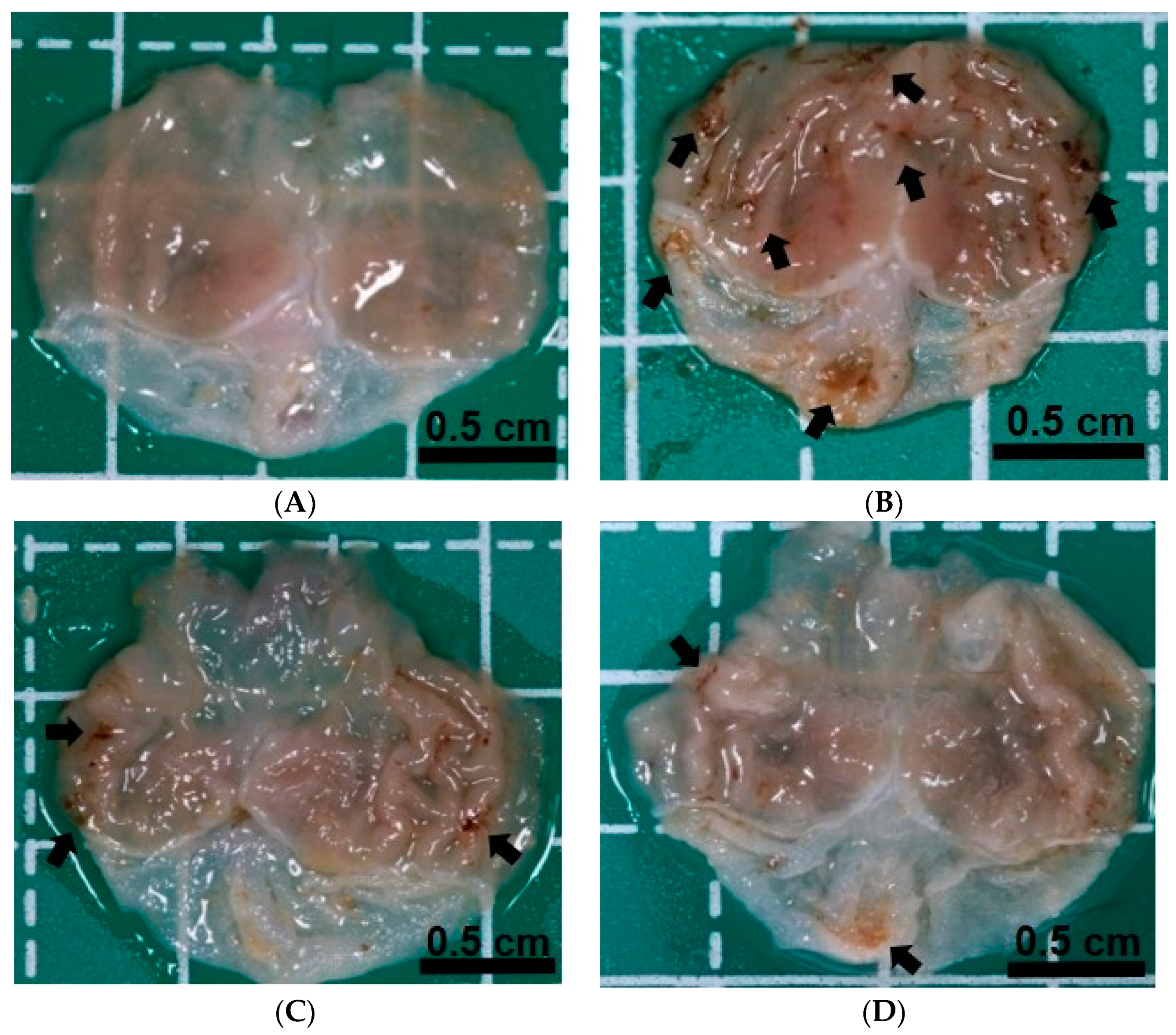
3.1. Effects of EEDH Pretreatment on IND-Induced Gastric Mucosal Damage in Mice

Figure 1 shows representative appearances of the gastric mucosa of mice in each group. It can be found that there is no damage to the gastric mucosa in the control group (Figure 1A), whereas the administration of a single dose of IND (120 mg/kg) caused obvious bleeding ulcers in the gastric mucosa (Figure 1B). It has been revealed in the Figure 1C,D that the gastric mucosal damage of mice in the EEDH200 and EEDH500 groups tended to decrease as the dose of EEDH increased, indicating that administration of EEDH effectively improved gastric mucosal damage and ulcers caused by IND. After the measurement and calculation by the software, the ulcer area of gastric mucosa in mice and the protective index of EEDH are shown in Table 1. It can be seen from the results that after the administration of IND to induce gastric mucosal damage, the gastric ulcer area of the mice in the IND group was as high as 2.7 ± 0.9 mm2, which was significantly different from that in the control group (p < 0.05). In the EEDH treatment groups, after the mice were given 200 mg/kg and 500 mg/kg of EEDH, the ulcer areas were 0.9 ± 0.3 mm2 and 0.5 ± 0.3 mm2, respectively (p < 0.05 and p < 0.01 vs. IND group). It suggests that the treatment of 500 mg/kg EEDH has a more significant efficacy on reducing gastric ulcer than the treatment of 200 mg/kg EEDH, which means that the treatment of EEDH showing gastroprotective effect in a dose-dependent manner. Therefore, the protective index of the EEDH500 group was greater than that of the EEDH200 group (Table 1).

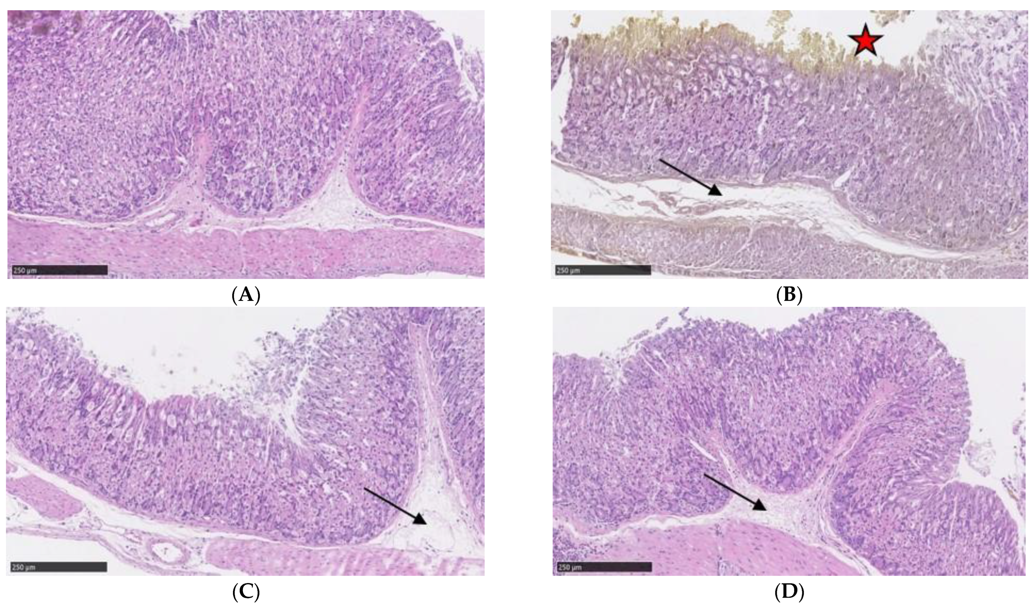
3.2. Effects of EEDH Pretreatment on IND-Induced Changes in Gastric Mucosal Histopathology

From the observation of the appearance of the stomach, it was shown that oral administration of IND caused obvious damage to the gastric mucosa. We further examined the general overview of mouse gastric tissue structure by H&E staining for histopathological evaluation. It has been illustrated in the Figure 2A that mice in the control group had intact gastric mucosal tissue and normal distribution of submucosa, whereas those in the IND group showed severe damage in the gastric epithelial cells, decreased and arranged disorderly in the mucosal glands, and significant inflammatory edema in the submucosal layer (Figure 2B). Different preadministration doses of EEDH (200 mg/kg and 500 mg/kg) revealed protective effects via ameliorating the damage of stomach epithelium and the submucosal edema in mice (Figure 2C,D). Gastric mucosal damage and submucosal edema were the least in mice in the EEDH500 group, suggesting that EEDH pretreatment could reduce gastric mucosal damage in mice in a dose-dependent manner.

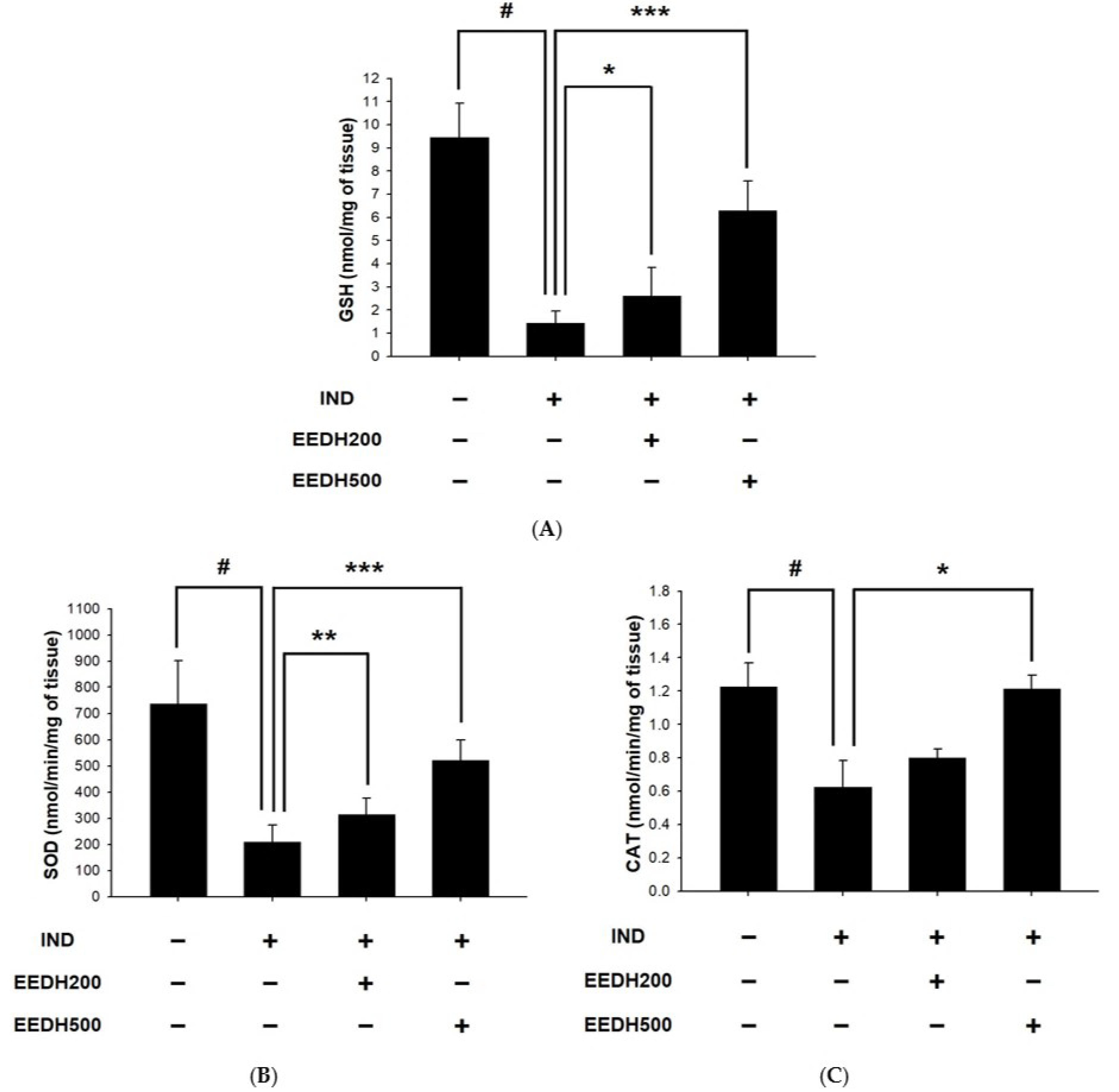
3.3. Effects of EEDH on Gastric Levels of GSH and Activities of SOD and CAT in Mice

To confirm whether orally administered EEDH affects antioxidant system of stomach in mice to alleviate IND-caused gastric mucosal damage, gastric tissues were collected to measure GSH levels and SOD and CAT activities. The results showed that a single dose of IND treatment remarkably declined gastric GSH levels (Figure 3A), while preadministration of EEDH dose-dependently and significantly restored the GSH levels of gastric mucosa in mice (p < 0.05). In terms of the results of enzymatic antioxidants, the SOD and CAT activities of the mice in the IND group were significantly lesser than those in the control group (p < 0.05, Figure 3B,C). The activities of SOD and CAT in the gastric mucosa of mice pretreated with EEDH were significantly improved, especially in the EEDH500 group (p < 0.05). From the above results, it is known that the administration of EEDH can significantly alleviate the decline in the antioxidant status of the gastric mucosa of mice caused by IND.

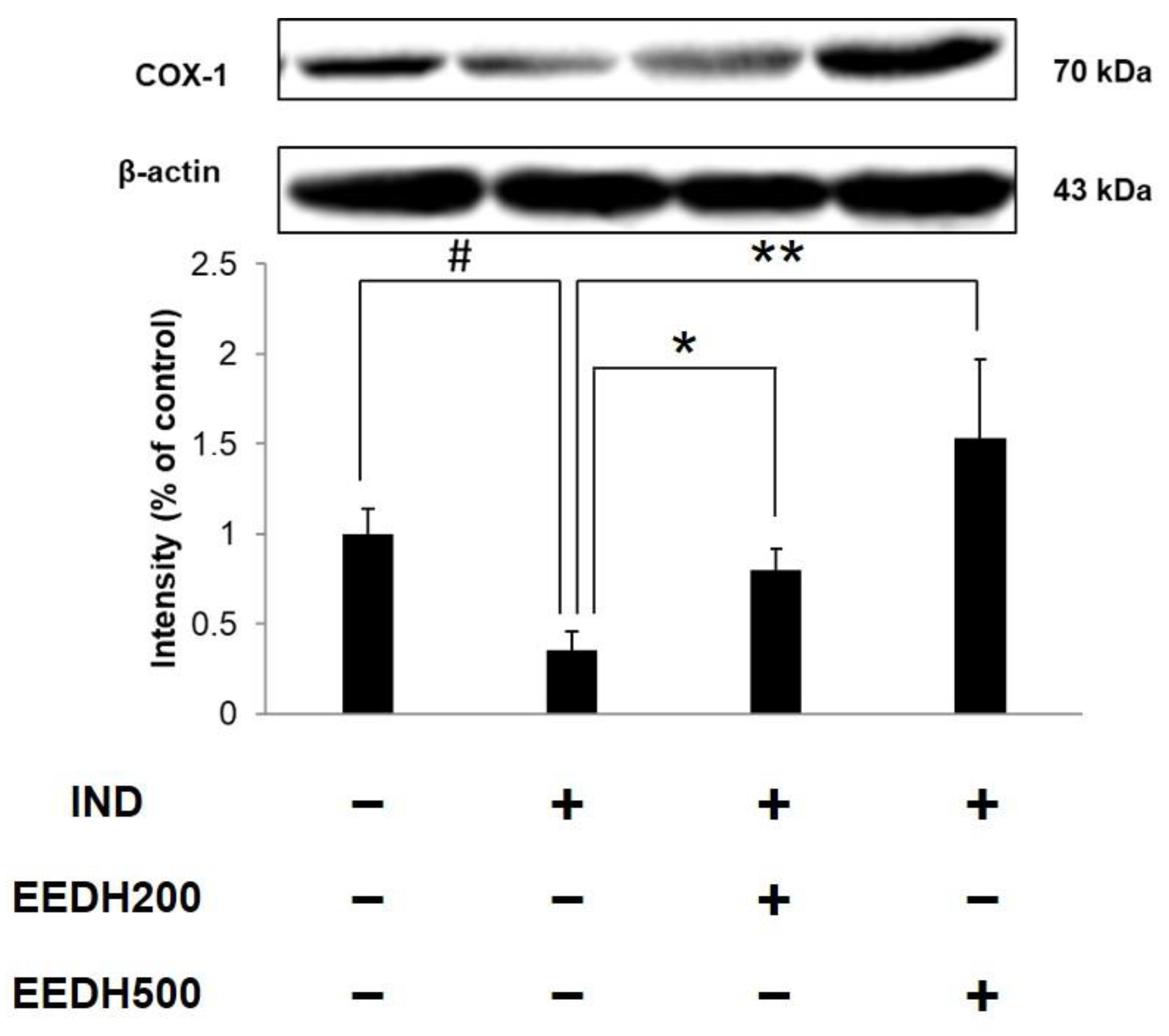
3.4. Effects of EEDH on Gastric COX-1 Protein Expression in Mice

Previous studies have reported that the inhibition of the expression of COX-1 protein in gastric mucosal cells is also one of the major factors for IND to cause ulcers [30,31]. In the present study, we investigated the effects of EEDH on COX-1 expression in IND-treated mice. As is shown in Figure 4, IND led to a significant decline in COX-1 protein expression compared to the control group. The expression of COX-1 protein was significantly greater in the EEDH200 and EEDH500 groups compared to the IND group (p < 0.05), suggesting that EEDH improved gastric mucosal damage via upregulation of COX-1 expression in mice.

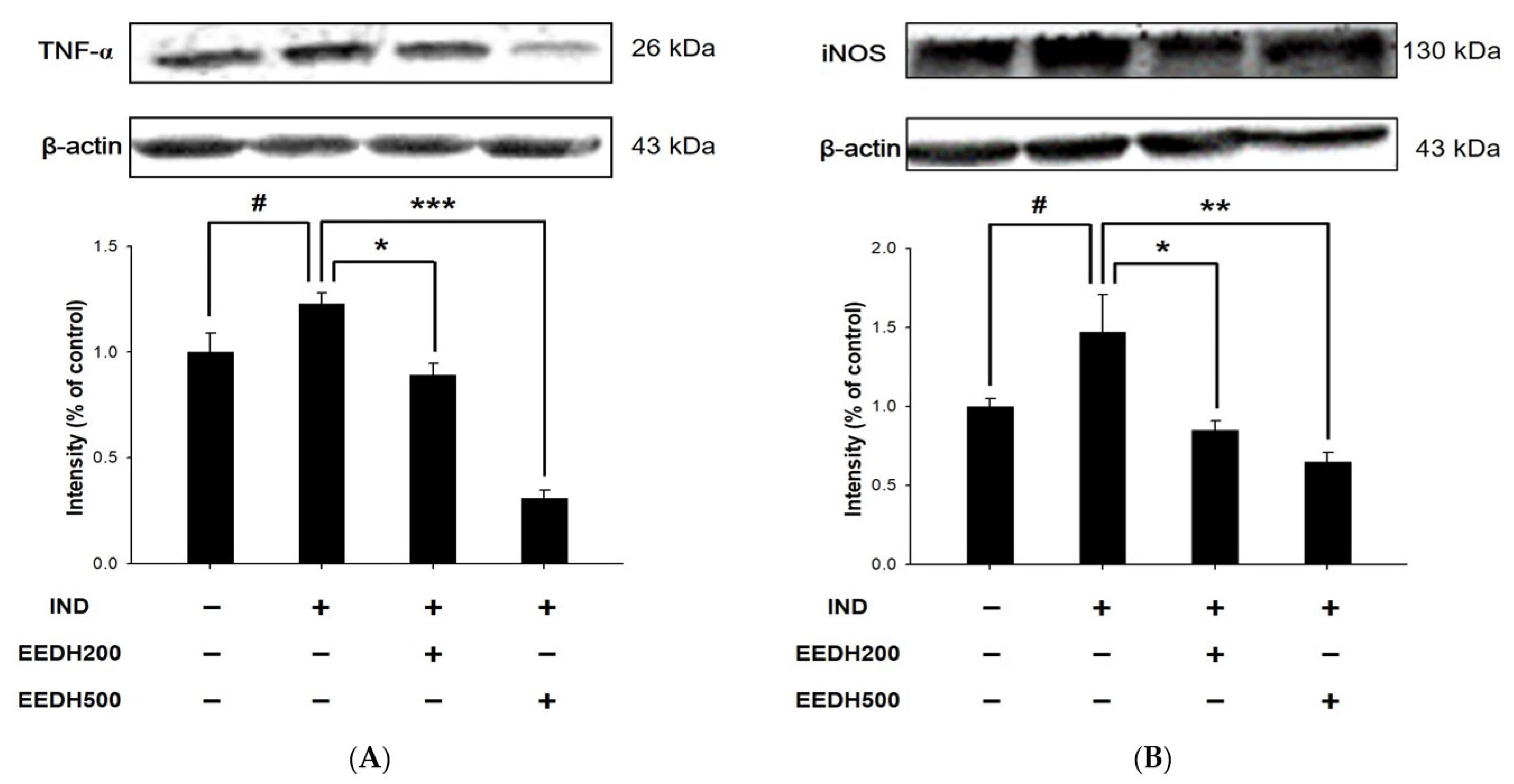
3.5. Effects of EEDH on Gastric Expression of TNF-α and iNOS Proteins in Mice

Figure 5 shows the expression of TNF-α and iNOS proteins of gastric mucosa in IND-treated mice. IND treatment enhanced gastric protein levels of TNF-α and iNOS compared to the control group, indicating that IND promoted the expression of major pro-inflammatory cytokines (TNF-α and iNOS), which resulted in increased the oxidative stress of the gastric mucosa and induced the formation of gastric ulcer. Preadministration of EEDH dose-dependently downregulated the expression of TNF-α and iNOS of gastric mucosal tissues of IND-treated mice. EEDH improved the oxidative stress caused by IND via declining the activation of TNF-α and iNOS proteins, which associates with the elevated antioxidant status in the gastric mucosa.

4. Discussion

NSAIDs are effective in the treatment of inflammation, pain, fever, and clinical and rheumatoid arthritis, and are often used to treat these types of diseases [9]. One of the side effects caused by NSAIDs is gastric ulcer, such as destroying the normal gastric mucosal defense system, allowing gastric acid to cause damage to gastric mucosal cells and causing bleeding and ulcers [10,32,33]. As an NSAID, IND causes gastric ulcer via enhancing gastric acid secretion and triggers oxidative stress through the generation of free radicals [34,35]. Administration of IND inhibits COX expression and reduces PG production, which are among the main causes of gastric mucosal damage [12,13].
The claim of functional foods is to use foods as preventive supplements for diseases, and this study suggests that the benefits of djulis hull may lead to the development of a new antiulcer supplement. Certain previous reports have shown that djulis possesses several kinds of bioactive components, such as betanin, kaempferol, quercetin, and rutin, which can contribute to its biological activities [17,21]. However, djulis hull, which is often discarded as a byproduct, is reported to have three times the betanin and twice the phenolic and flavonoid compounds of djulis seeds [22]. Betaine and flavonoids can inhibit the action of enzymes or transcription factors involved in the inflammatory response and are expected to reduce tissue damage [36,37]. Betanin in djulis is also responsible for the source of red pigment and its associated antioxidant activity [38]. The phenolic components in djulis hull reveal reducing characteristics and act as antioxidants and antiinflammatory agents [39]. All these reasons suggest that djulis hull possesses the capacity to be exploited as a functional food for the prevention of gastric damage due to its active ingredients that can reduce gastric inflammation.
In our preliminary study (n = 3 each group), we found that 120 mg/kg IND treatment caused appropriate gastric ulcer area in C57/BL mice, and preadministration of 200 mg/kg EEDH and 200 mg/kg water extract of djulis hull (WEDH) improved the gastric damage caused by IND. Among them, EEDH revealed better protective activity. That is why we chose 120 mg/kg IND, 200 and 500 mg/kg EEDH for subsequent experiment to elucidate whether a 2.5-fold higher dose of EEDH shows greater gastric gastroprotective effect. In this study, a single dose of IND treatment (120 mg/kg) was conducted to cause gastric damage in C57BL/6J mice, and to investigate whether preadministration of different doses of EEDH (200 mg/kg and 500 mg/kg) for 21 days revealed gastroprotective ability. EEDH significantly reduced the ulcer area of gastric lesions induced by IND with a high ulcer protective index in a dose-response manner (Figure 1 and Table 1). Previous reports have shown that IND inhibits COX-1 expression, which leads to the decease of PG production and the occurrence of gastric mucosal injury [13,40]. Moreover, apart from causing the gastric ulcer, the acute inflammatory response induced by IND also increases proinflammatory mediators such as TNF-α and IL-6 [41]. IND-induced gastric injury was also associated with increased iNOS activity and malondialdehyde (MDA) production [42]. In addition, IND treatment reduced the levels of GSH and the activities of antioxidant enzymes (SOD and CAT) [43]. As antioxidants in the body system, CAT, SOD, and GSH scavenge free radicals and prevent gastric injury caused by oxidative stress. The preadministration of EEDH significantly decreased the protein levels of inflammatory mediators (TNF-α and iNOS) and normalized the activities of antioxidant enzymes (SOD and CAT). Furthermore, the gastric mucosal protective effect of EEDH was also associated with the restoration of COX-1 expression and GSH levels. Histopathological examination (H&E staining) also indicated that EEDH possesses the ability to relieve the wound of mucosal damage. A dose of 200–500 mg/kg for mice is equivalent to the dose of about 16–41 mg/kg for humans, and the daily intake of adults is about 1.0–2.5 g based on an adult body weight of 60 kg. It demonstrated that EEDH has the potential to be exploited as functional foods for gastric prevention or protection.

5. Conclusions

In summary, the findings of this research indicate that oral preadministration of EEDH is an effective approach for preventing gastric mucosal damage induced by IND. EEDH pretreatment significantly showed protective effects on gastric mucosa through the regulation of antioxidant and proinflammatory pathways. According to the information we have, this study is the first to show that EEDH can attenuate the gastric mucosal damage of mice induced by IND. The results found in the present study suggest that EEDH has the potential to be developed as a functional food or nutritional supplement for gastric protection, which would also reduce the problem of djulis hull waste and increase the recycling of waste.

Author Contributions

Conceptualization, F.S.I. and Y.-K.C.; methodology, F.S.I. and N.-C.L.; software, F.S.I. and H.-Y.T.; validation, N.-C.L. and H.-Y.T.; formal analysis, N.-C.L. and H.-Y.T.; investigation, F.S.I. and N.-C.L.; resources, P.-J.T.; data curation, N.-C.L.; writing—original draft preparation, F.S.I.; writing—review and editing, H.-Y.T. and Y.-K.C.; visualization, H.-Y.T. and J.-L.H.; supervision, J.-L.H., A.K.W. and Y.-K.C.; project administration, Y.-K.C.; funding acquisition, Y.-K.C. All authors have read and agreed to the published version of the manuscript.

Funding

This research work was supported by National Science and Technology Council of Taiwan under grant number MOST 111-2637-B-020-009.

Institutional Review Board Statement

The experimental procedures performed on animal were conducted under the guideline and protocol approved by the IACUC of NPUST (approved protocol number NPUST-111-001).

Informed Consent Statement

Not applicable.

Data Availability Statement

Data are contained within the article.

Acknowledgments

We gratefully acknowledge Tzou-Chi Huang and Mei-Li Wu of NPUST for the support of instruments used in the present study.

Conflicts of Interest

The authors declare no conflict of interest.

References

  1. Hafez, A.A.; Tavassoli, E.; Hasanzadeh, A.; Reisi, M.; Javadzade, S.H.; Imanzad, M. Quality of life in peptic ulcer patients referring to Al-Zahra hospital of Isfahan, Iran. Gastroenterol. Hepatol. Bed Bench 2013, 6, S87–S92. [Google Scholar] [PubMed]
  2. Wasman, S.; Mahmood, A.A.; Salehhuddin, H.; Zahra, A.A.; Salmah, I. Cytoprotective activities of Polygonum minus aqueous leaf extract on ethanol-induced gastric ulcer in rats. J. Med. Plants Res. 2010, 4, 2658–2665. [Google Scholar]
  3. Lu, S.Y.; Guo, S.; Chai, S.B.; Yang, J.Q.; Yue, Y.; Li, H.; Sun, P.M.; Zhang, T.; Sun, H.W.; Zhou, J.L.; et al. Autophagy in gastric mucosa: The dual role and potential therapeutic target. Biomed. Res. Int. 2021, 2021, 2648065. [Google Scholar] [CrossRef] [PubMed]
  4. Jang, T.J.; Kim, J.R. Proliferation and apoptosis in gastric antral epithelial cells of patients infected with Helicobacter pylori. J. Gastroenterol. 2000, 35, 265–271. [Google Scholar] [CrossRef] [PubMed]
  5. Shareef, S.H.; Al-Medhtiy, M.H.; Ibrahim, I.A.A.; Alzahrani, A.R.; Jabbar, A.A.; Galali, Y.; Agha, N.F.S.; Aziz, P.Y.; Thabit, M.A.; Agha, D.N.F.; et al. Gastroprophylactic effects of p-cymene in ethanol-induced gastric ulcer in rats. Processes 2022, 10, 1314. [Google Scholar] [CrossRef]
  6. Rawla, P.; Barsouk, A. Epidemiology of gastric cancer: Global trends, risk factors and prevention. Prz. Gastroenterol. 2019, 14, 26–38. [Google Scholar] [CrossRef]
  7. Maddirevula, S.; Abanemai, M.; Alkuraya, F.S. Human knockouts of PLA2G4A phenocopy NSAID-induced gastrointestinal and renal toxicity. Gut 2016, 65, 1575–1577. [Google Scholar] [CrossRef] [Green Version]
  8. Wolfe, M.M.; Lichtenstein, D.R.; Singh, G. Gastrointestinal toxicity of nonsteroidal antiinflammatory drugs. N. Engl. J. Med. 1999, 340, 1888–1899. [Google Scholar] [CrossRef]
  9. Wong, R.S.Y. Role of Nonsteroidal anti-inflammatory drugs (NSAIDs) in cancer prevention and cancer promotion. Adv. Pharmacol. Sci. 2019, 2019, 3418975. [Google Scholar] [CrossRef] [Green Version]
  10. Atchison, J.W.; Herndon, C.M.; Rusie, E. NSAIDs for musculoskeletal pain management:current perspectives and novel strategies to improve safety. J. Manag. Care Pharm. 2013, 19, S3–S19. [Google Scholar]
  11. Griffin, M.R.; Scheiman, J.M. Prospects for changing the burden of nonsteroidal anti-inflammatory drug toxicity. Am. J. Med. 2001, 110, 33S–37S. [Google Scholar] [CrossRef] [PubMed]
  12. Andrews, F.J.; Malcontenti-Wilson, C.; O’Brien, P.E. Effect of nonsteroidal anti-inflammatory drugs on LFA-1 and ICAM-1 expression in gastric mucosa. Am. J. Physiol. 1994, 266, G657–G664. [Google Scholar] [CrossRef] [PubMed]
  13. Yoshikawa, T.; Naito, Y. The role of neutrophils and inflammation in gastric mucosal injury. Free. Radic. Res. 2000, 33, 785–794. [Google Scholar] [CrossRef] [PubMed]
  14. Hussain, T.; Tan, B.; Yin, Y.; Blachier, F.; Tossou, M.C.; Rahu, N. Oxidative stress and inflammation: What polyphenols can do for us? Oxid. Med. Cell. Longev. 2016, 2016, 7432797. [Google Scholar] [CrossRef] [PubMed] [Green Version]
  15. Nandi, J.; Saud, B.; Zinkievich, J.M.; Yang, Z.J.; Levine, R.A. TNF-α modulates iNOS expression in an experimental rat model of indomethacin-induced jejunoileitis. Mol. Cell. Biochem. 2010, 336, 17–24. [Google Scholar] [CrossRef]
  16. Küçükler, S.; Kandemir, F.M.; Yıldırım, S. Protective effect of chrysin on indomethacin induced gastric ulcer in rats: Role of multi-pathway regulation. Biotech. Histochem. 2022, 97, 490–503. [Google Scholar] [CrossRef]
  17. Chyau, C.C.; Chu, C.C.; Chen, S.Y.; Duh, P.D. The inhibitory effects of djulis (Chenopodium formosanum) and its bioactive compounds on adipogenesis in 3T3-L1 adipocytes. Molecules 2018, 23, 1780. [Google Scholar] [CrossRef] [Green Version]
  18. Hsu, B.Y.; Pan, S.Y.; Wu, L.Y.; Ho, C.T.; Hwang, L.S. Hypoglycemic activity of Chenopodium formosanum Koidz. components using a glucose uptake assay with 3T3-L1 adipocytes. Food Biosci. 2018, 24, 9–16. [Google Scholar] [CrossRef]
  19. Lee, C.W.; Chen, H.J.; Xie, G.R.; Shih, C.K. Djulis (Chenopodium formosanum) prevents colon carcinogenesis via regulating antioxidative and apoptotic pathways in rats. Nutrients 2019, 11, 2168. [Google Scholar] [CrossRef] [Green Version]
  20. Chen, S.Y.; Chu, C.C.; Chyau, C.C.; Yang, J.W.; Duh, P.D. Djulis (Chenopodium formosanum) and its bioactive compounds affect vasodilation, angiotensin converting enzyme activity, and hypertension. Food Biosci. 2019, 32, 100469. [Google Scholar] [CrossRef]
  21. Chu, C.C.; Chen, S.Y.; Chyau, C.C.; Wang, S.C.; Chu, H.L.; Duh, P.D. Djulis (Chenopodium formosanum) and its bioactive compounds protect human lung epithelial A549 cells from oxidative injury induced by particulate matter via Nrf2 signaling pathway. Molecules 2021, 27, 253. [Google Scholar] [CrossRef] [PubMed]
  22. Huang, C.Y.; Chu, Y.L.; Sridhar, K.; Tsai, P.J. Analysis and determination of phytosterols and triterpenes in different inbred lines of djulis (Chenopodium formosanum Koidz.) hull: A potential source of novel bioactive ingredients. Food Chem. 2019, 297, 124948. [Google Scholar] [CrossRef] [PubMed]
  23. Chen, J.Y.; Sridhar, K.; Tsai, P.J. Anti-glycation and inhibition of starch hydrolyzing enzymes by enzymatically hydrolysed djulis (Chenopodium formosanum Koidz.) hull, leaf and seedling. Int. J. Food Sci. Technol. 2021, 56, 6601–6610. [Google Scholar] [CrossRef]
  24. Tung, Y.T.; Zeng, J.L.; Ho, S.T.; Xu, J.W.; Lin, I.H.; Wu, J.H. Djulis hull improves insulin resistance and modulates the gut microbiota in high-fat diet (HFD)-induced hyperglycaemia. Antioxidants 2022, 11, 45. [Google Scholar] [CrossRef]
  25. Tung, Y.T.; Zeng, J.L.; Ho, S.T.; Xu, J.W.; Li, S.; Wu, J.H. Anti-NAFLD effect of djulis hull and its major compound, rutin, in mice with high-fat diet (HFD)-induced obesity. Antioxidants 2021, 10, 1694. [Google Scholar] [CrossRef]
  26. Zhou, D.; Yang, Q.; Tian, T.; Chang, Y.; Li, Y.; Duan, L.-R.; Li, H.; Wang, S.-W. Gastroprotective effect of gallic acid against ethanol-induced gastric ulcer in rats: Involvement of the Nrf2/HO-1 signaling and anti-apoptosis role. Biomed. Pharmacother. 2020, 126, 110075. [Google Scholar] [CrossRef] [PubMed]
  27. Beck, P.L.; Xavier, R.; Lu, N.; Nanda, N.N.; Dinauer, M.; Podolsky, D.K.; Seed, B. Mechanisms of NSAID-induced gastrointestinal injury defined using mutant mice. Gastroenterology 2000, 119, 699–705. [Google Scholar] [CrossRef]
  28. Tsai, H.Y.; Yang, J.F.; Chen, H.H.; You, F.N.; Zhao, Y.J.; Lin, Y.H.; Hsu, J.L.; Chang, C.I.; Chen, Y.K. The effect of hot water extract of tilapia on exercise capacity in mice. Appl. Sci. 2022, 12, 2601. [Google Scholar] [CrossRef]
  29. Tsai, H.Y.; Shih, Y.Y.; Yeh, Y.T.; Huang, C.H.; Liao, C.A.; Hu, C.Y.; Nagabhushanam, K.; Ho, C.T.; Chen, Y.K. Pterostilbene and its derivative 3′-hydroxypterostilbene ameliorated nonalcoholic fatty liver disease through synergistic modulation of the gut microbiota and SIRT1/AMPK signaling pathway. J. Agric. Food Chem. 2022, 70, 4966–4980. [Google Scholar] [CrossRef]
  30. Sinha, M.; Gautam, L.; Shukla, P.K.; Kaur, P.; Sharma, S.; Singh, T.P. Current perspectives in NSAID-induced gastropathy. Mediat. Inflamm. 2013, 2013, 258209. [Google Scholar] [CrossRef] [Green Version]
  31. Wallace, J.L. Prostaglandins, NSAIDs, and gastric mucosal protection: Why doesn’t the stomach digest itself? Physiol. Rev. 2008, 88, 1547–1565. [Google Scholar] [CrossRef] [PubMed]
  32. Nemmani, K.V.; Mali, S.V.; Borhade, N.; Pathan, A.R.; Karwa, M.; Pamidiboina, V.; Senthilkumar, S.P.; Gund, M.; Jain, A.K.; Mangu, N.K.; et al. NO-NSAIDs: Gastric-sparing nitric oxide-releasable prodrugs of non-steroidal anti-inflammatory drugs. Bioorg. Med. Chem. Lett. 2009, 19, 5297–5301. [Google Scholar] [CrossRef] [PubMed]
  33. Oren, R.; Ligumsky, M. Indomethacin-induced colonic ulceration and bleeding. Ann. Pharmacother. 1994, 28, 883–885. [Google Scholar] [CrossRef] [PubMed]
  34. Suleyman, H.; Albayrak, A.; Bilici, M.; Cadirci, E.; Halici, Z. Different mechanisms in formation and prevention of indomethacin-induced gastric ulcers. Inflammation 2010, 33, 224–234. [Google Scholar] [CrossRef] [PubMed]
  35. Hamauzu, Y.; Irie, M.; Kondo, M.; Fujita, T. Antiulcerative properties of crude polyphenols and juice of apple, and Chinese quince extracts. Food Chem. 2008, 108, 488–495. [Google Scholar] [CrossRef] [PubMed] [Green Version]
  36. Zhao, G.; He, F.; Wu, C.; Li, P.; Li, N.; Deng, J.; Zhu, G.; Ren, W.; Peng, Y. Betaine in inflammation: Mechanistic aspects and applications. Front. Immunol. 2018, 9, 1070. [Google Scholar] [CrossRef] [Green Version]
  37. Svajger, U.; Jeras, M. Anti-inflammatory effects of resveratrol and its potential use in therapy of immune-mediated diseases. Int. Rev. Immunol. 2012, 31, 202–222. [Google Scholar] [CrossRef]
  38. Tsai, P.J.; Chen, Y.S.; Sheu, C.H.; Chen, C.Y. Effect of nanogrinding on the pigment and bioactivity of djulis (Chenopodium formosanum Koidz.). J. Agric. Food Chem. 2011, 59, 1814–1820. [Google Scholar] [CrossRef]
  39. Huang, H.W.; Cheng, M.C.; Chen, B.Y.; Wang, C.Y. Effects of high pressure extraction on the extraction yield, phenolic compounds, antioxidant and anti-tyrosinase activity of djulis hull. J. Food Sci. Technol. 2019, 56, 4016–4024. [Google Scholar] [CrossRef]
  40. Tanaka, A.; Hase, S.; Miyazawa, T.; Ohno, R.; Takeuchi, K. Role of cyclooxygenase (COX)-1 and COX-2 inhibition in nonsteroidal anti-inflammatory drug-induced intestinal damage in rats: Relation to various pathogenic events. J. Pharmacol. Exp. Ther. 2002, 303, 1248–1254. [Google Scholar] [CrossRef] [Green Version]
  41. Santucci, L.; Fiorucci, S.; Giansanti, M.; Brunori, P.M.; Di Matteo, F.M.; Morelli, A. Pentoxifylline prevents indomethacin induced acute gastric mucosal damage in rats: Role of tumour necrosis factor alpha. Gut 1994, 35, 909–915. [Google Scholar] [CrossRef] [PubMed] [Green Version]
  42. Vivatvakin, S.; Werawatganon, D.; Somanawat, K.; Klaikeaw, N.; Siriviriyakul, P. Genistein-attenuated gastric injury on indomethacin-induced gastropathy in rats. Pharmacogn. Mag. 2017, 13, S306–S310. [Google Scholar] [CrossRef] [PubMed]
  43. Odabasoglu, F.; Cakir, A.; Suleyman, H.; Aslan, A.; Bayir, Y.; Halici, M.; Kazaz, C. Gastroprotective and antioxidant effects of usnic acid on indomethacin-induced gastric ulcer in rats. J. Ethnopharmacol. 2006, 103, 59–65. [Google Scholar] [CrossRef] [PubMed]
Figure 1. Representative gross gastric images of mice. (A) Control group; (B) IND group, treated with IND (120 mg/kg); (C) EEDH200 group, treated with EEDH (200 mg/kg) + IND (120 mg/kg); (D) EEDH500 group, treated with EEDH (500 mg/kg) + IND (120 mg/kg). Mice were administered indicated dose of EEDH orally for 21 consecutive days. 120 mg/kg of IND was then given orally to mice on the last day of experiment and sacrificed 6 h later. The gastric tissue of mice was harvested and photographed. The arrows showed in the figure depict bleeding ulcers.
Applsci 13 00594 g001
Figure 2. The histopathological assessment of gastric mucosa in mice. (H&E staining, scale bars = 250 μm). (A) Control group; (B) IND group, treated with IND (120 mg/kg); (C) EEDH200 group, treated with EEDH (200 mg/kg) + IND (120 mg/kg); (D) EEDH500 group, treated with EEDH (500 mg/kg) + IND (120 mg/kg). The asterisk showed in the figure indicates the damage of the mucosal epithelium accompanied by the destruction of the gland structure. The arrow showed in the figure depicts edema and inflammatory cell infiltration in the submucosa.
Applsci 13 00594 g002
Figure 3. Effects of EEDH on (A) the level of GSH and (B,C) the activities of SOD and CAT of gastric mucosa in mice. Data are represented as means ± SD (n = 8). # p < 0.05 compared with control group. * p < 0.05, ** p < 0.01, and *** p < 0.001 compared with IND-treated group.
Applsci 13 00594 g003
Figure 4. Effects of EEDH pretreatment on the protein levels of COX-1 of gastric mucosa in mice administered with IND. Western blot was applied to determine the expression of COX-1. Data are represented as means ± SD (n = 3). # p < 0.05 compared with control group. * p < 0.05 and ** p < 0.01 compared with IND-treated group.
Applsci 13 00594 g004
Figure 5. Effects of EEDH pretreatment on the protein levels of TNF-α (A) and iNOS (B) of gastric mucosa in mice administered with IND. Western blot was applied to determine the expression of TNF-α and iNOS. Data are represented as means ± SD (n = 3). # p < 0.05 compared with control group. * p < 0.05, ** p < 0.01, and *** p < 0.001 compared with IND-treated group.
Applsci 13 00594 g005
Table 1. Effects of EEDH on gastric ulcer area and protective index in mice treated with IND.
GroupUlcer Area (mm2)Protective Index (%)
Control0
IND2.7 ± 0.9 #
EEDH200 + IND0.9 ± 0.3 *67
EEDH500 + IND0.5 ± 0.3 **80
The ulcer area was quantified by using Image J. Data are expressed as means ± SD (n = 8). # p < 0.05 compared with control group. * p < 0.05 and ** p < 0.01 compared with IND-treated group. Protective index (%) = [(Ulcer areaIND group − Ulcer areaEEDH group)/Ulcer areaIND group] × 100%.
Disclaimer/Publisher’s Note: The statements, opinions and data contained in all publications are solely those of the individual author(s) and contributor(s) and not of MDPI and/or the editor(s). MDPI and/or the editor(s) disclaim responsibility for any injury to people or property resulting from any ideas, methods, instructions or products referred to in the content.

