Technical Note

Direct Suture Repair of Deltoid Ligament Using Barbed Suture: A Cost-Effective Surgical Technique for Ligamentous Repair in Ankle Fractures


Department of Orthopaedic Surgery, University of Virginia, 2280 Ivy Road, Charlottesville, VA 22903, USA
*
Author to whom correspondence should be addressed.
Surg. Tech. Dev. 2026, 15(1), 7; https://doi.org/10.3390/std15010007
Submission received: 16 October 2025 / Revised: 28 January 2026 / Accepted: 7 February 2026 / Published: 10 February 2026

Highlights

What is the main finding?
  • Deltoid ligament repair with barbed PDS suture is a simple, reliable, and cost-effective technique.
What are the implications of the main findings?
  • Surgeons can use this technique without adding significant time, cost, or specialized equipment in cases benefitting from deltoid ligament repair.
  • This technique can be considered for routine use for deltoid ligament repair.

Abstract

Background/Objectives: Deltoid ligament injury has been reported in up to 40% of ankle fractures, as confirmed by arthroscopy. Despite the frequency of this injury, there are multiple methods of fixation, including use of sutures, anchors and grafts, with no consensus in the field on a gold standard of operative technique for deltoid ligament injuries identified in bimalleolar equivalent ankle fractures. This manuscript aims to describe a novel surgical technique for deltoid ligament repair in the setting of bimalleolar equivalent ankle fractures. Surgical Technique: In this technique, following fixation of the fibula in bimalleolar equivalent ankle fractures, barbed polydioxanone (PDS) suture is utilized for direct repair of the deltoid ligament. This technique can be completed with the patient either prone or supine, uses standard fluoroscopy for evaluation of the competency of the deltoid ligament, and utilizes a familiar anteromedial approach to the medial malleolus. When compared to using anchors for repair of the deltoid ligament, barbed PDS suture seems to provide an equivalent and cost-effective strategy for repair. Conclusions: Repair of the deltoid ligament using suture anchors is a widely accepted method that has been demonstrated to provide successful repair. However, these anchors can be costly and rely on appropriate fixation in the bone to provide long-term fixation. This suture technique seems to offer a reliable, cost-effective technique for deltoid ligament repair.

1. Introduction

Deltoid ligament injury is a common component of rotational ankle fractures, with a reported incidence of 40% when confirmed by arthroscopy [1]. The deltoid ligament is composed of superficial and deep fibers [2]. The deep fibers are the primary stabilizers, with fibers originating from the anterior colliculus, posterior colliculus and intercollicular groove of the medial malleolus and inserts on the medial talus [2]. There are a variety of ways to preoperatively and intraoperatively assess deltoid ligament injury to determine the need for ligament repair. Both physical exam and pre-operative radiographs can aid in identification of deltoid injury; however, multiple studies have concluded that physical exam alone to identify swelling, ecchymosis, medial tenderness or other soft tissue predictors is an unreliable indicator of the integrity of the deltoid ligament [2,3,4].
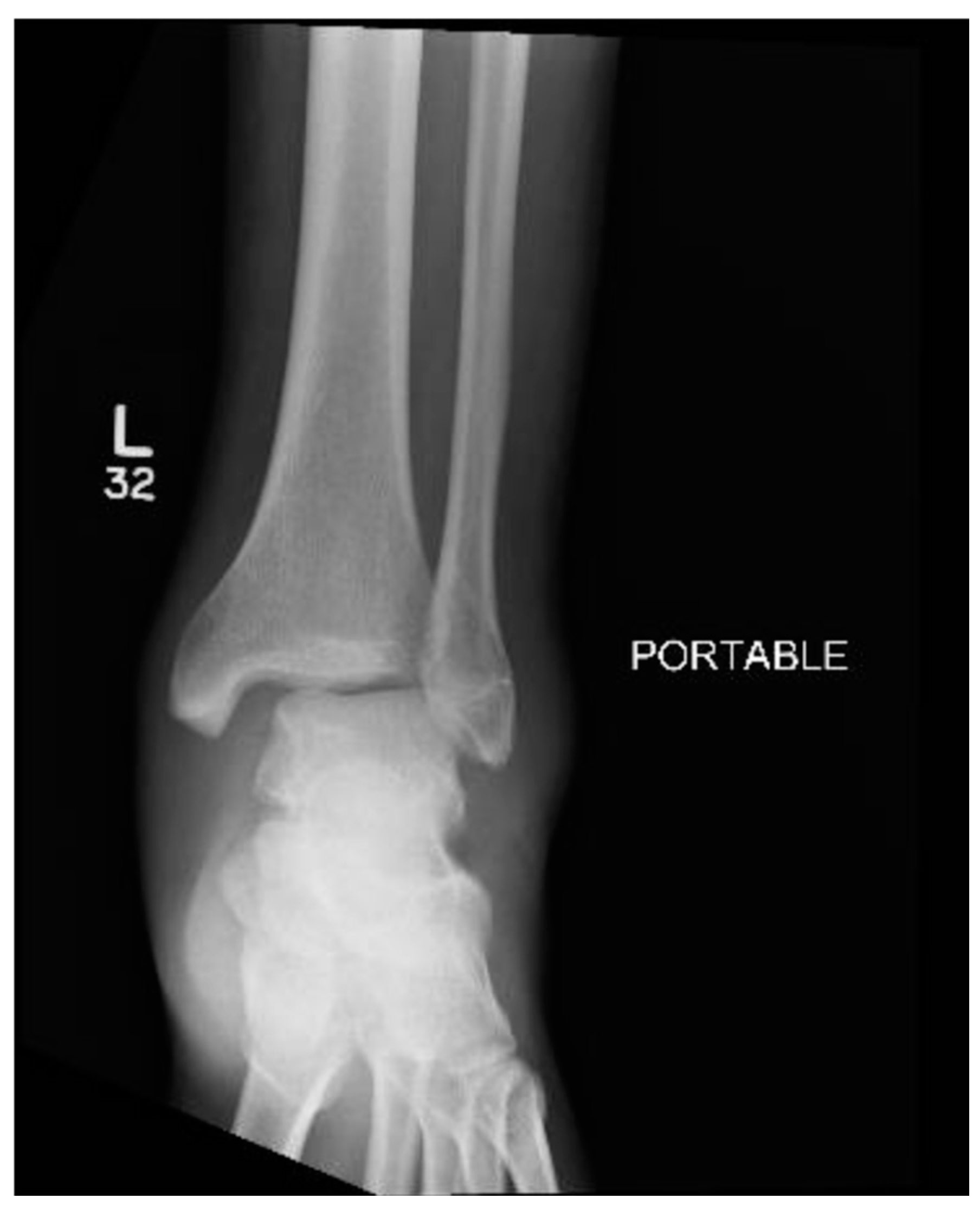
Medial instability is often objectively evaluated by examining the medial clear space (MCS) on plain radiographs to assess deep deltoid ligament integrity (Figure 1). Stress radiography is the preferred approach for preoperative assessment of deltoid ligament integrity [5]. It has been demonstrated that a gravity stress radiograph is non-inferior to a manual stress radiograph in assessing deltoid ligament injury [6]. Some have suggested that adequate stability of the mortise on weight-bearing imaging is sufficient and renders stress images unnecessary. However, congruent weight bearing results in most of the weight passing from the talar dome to the tibial plafond, regardless of the fibular lateral buttress, which does not maximally stress the syndesmosis and is thus not a reliable indicator [7]. The most widely accepted indication of a tear of the deep deltoid ligament is a medial clear space of >5 mm on stress radiographs, and is thus an indication for operative treatment [5,6,8,9].
Usually, open reduction with internal fixation (ORIF) of the fibula through a lateral or posterolateral approach is the first step in the surgical management of bimalleolar equivalent fractures [7]. Necessity of deltoid repair is controversial, and no uniform guidelines exist to determine when deltoid repair is definitively indicated. Additionally, there are multiple methods of repair for the deltoid ligament, with no consensus gold standard [10,11,12,13,14,15,16]. The earliest reported repairs of the deltoid ligament occurred in the 1960s and involved a technique described by Duvries’ Surgery of the Foot [17]. The repair technique involved creating an incision with both vertical and horizontal limbs in the deltoid ligament, forming a cross [17]. The margins were sutured together and left to scar in, which was found to be a favorable repair over the Schoolfield technique present in the 1950s [17]. This repair involved shortening the deltoid ligament through overlapping limbs which was favorable for recurrent sprains, however, less effective with acute injury to the deltoid ligament [17].
Suture anchor techniques have grown in popularity in sports and trauma populations due to ease of use and increased availability of implants [7,16,18,19]. However, studies vary with regard to incision placement, deltoid layer repaired, and the number and position of suture anchors. The general premise of the deltoid ligament repair technique using suture anchors for deltoid avulsion from the medial malleolus is described in detail by Lee et al. [7]. An incision is made over the medial malleolus, and the skin flaps are reflected so that horizontal rents in the superficial and deep deltoid ligaments can be visualized [7]. The posterior tibial tendon is retracted posteriorly and inferiorly, which allows visualization of the talus and medial tibia [7]. Then, proximal deltoid tissue is elevated at the distal tip of the medial malleolus [7]. An appropriately sized hole is drilled into the medial malleolus for placement of the suture anchor [7]. One or more suture anchors are used for fixation of two to four sets of braided nonabsorbable suture. With the anchor secured into the medial malleolus, a “vest-over-pants” technique is used to repair the superficial deltoid, deep deltoid, and capsule [7]. Woo et al. used a similar suture anchor approach with suture anchors while also including aspects of direct suturing in cases of partial superficial deltoid tears or for reinforcement after using anchors for deltoid repair [16].
There are no current studies describing a deltoid repair with barbed suture. The surgical technique detailed below utilizes a direct suturing technique with barbed PDS suture without anchor fixation to repair the deltoid ligament in rotational ankle fractures.

2. Surgical Technique

2.1. Set Up, Positioning, and Initial Fixation

Depending on the pattern of injury and planned fixation, the patient can be placed prone or supine on a regular table. Supine has the advantage of easier access to the medial side. A nonsterile tourniquet is applied to the thigh and used during the procedure. The tourniquet is routinely deflated prior to closure to ensure hemostasis. The operative extremity is elevated to allow for ease of access to both the lateral and medial ankle and remove overlap with the contralateral ankle for ease of fluoroscopy. A large c-arm fluoroscopy unit is placed on the contralateral side of the operating room table. Weight-based cefazolin is given within 30 min prior to skin incision for antibiotic prophylaxis. The lateral (and/or posterior) fractures are generally addressed first with an open reduction and internal fixation (ORIF) of the fractures.

2.2. Indication for Deltoid Repair

The surgeon may determine the need for deltoid repair based on fracture pattern and displacement on the injury films (typically widening of the MCS). If the need for deltoid repair is undecided preoperatively, following fixation of the lateral ankle, the ankle can then be evaluated for deltoid ligament injury intraoperatively. This can be done through fluoroscopic evaluation. An external rotation stress test allows the surgeon to assess potential incompetence of the ankle syndesmosis or deltoid injury depending on the fracture pattern. This is assessed by stabilizing the distal tibia and fibula while applying external rotation stress through the ankle. This applies force through the talus and against the lateral malleolus. Widening at the medial clear space during this maneuver would indicate deltoid injury. Additional assessment of deltoid incompetence can be done by evaluation of talar tilt and medial gapping, with valgus force applied through the tibiotalar joint. This is generally completed following surgical fixation of the fibula. This is an indication for deltoid repair and can be reassessed following repair to evaluate stability of the repair. The cotton test is an additional test that can be used to assess stability; this test provides information regarding both syndesmotic and deltoid stability. To perform a cotton test, a manual distraction force is applied to the fibula with a clamp or bone hook, while a simultaneous equal and opposite force is applied to the tibia to prevent tibial motion. No significant distraction will be radiographically visualized between the fibula and distal tibia if the syndesmotic ligaments are intact [20]. Additionally, no increase in medial clear space should be observed when the cotton test is performed.

2.3. Deltoid Repair

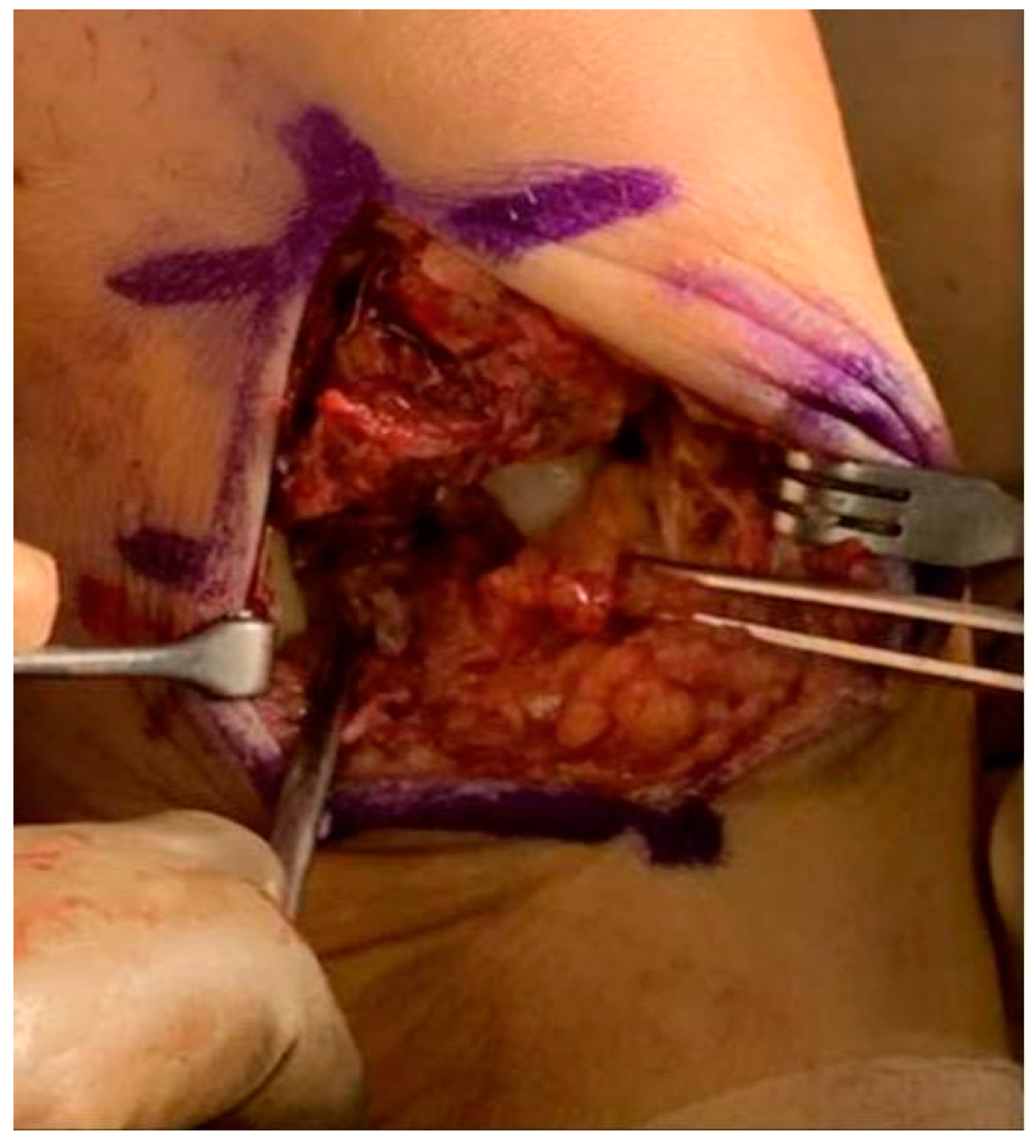
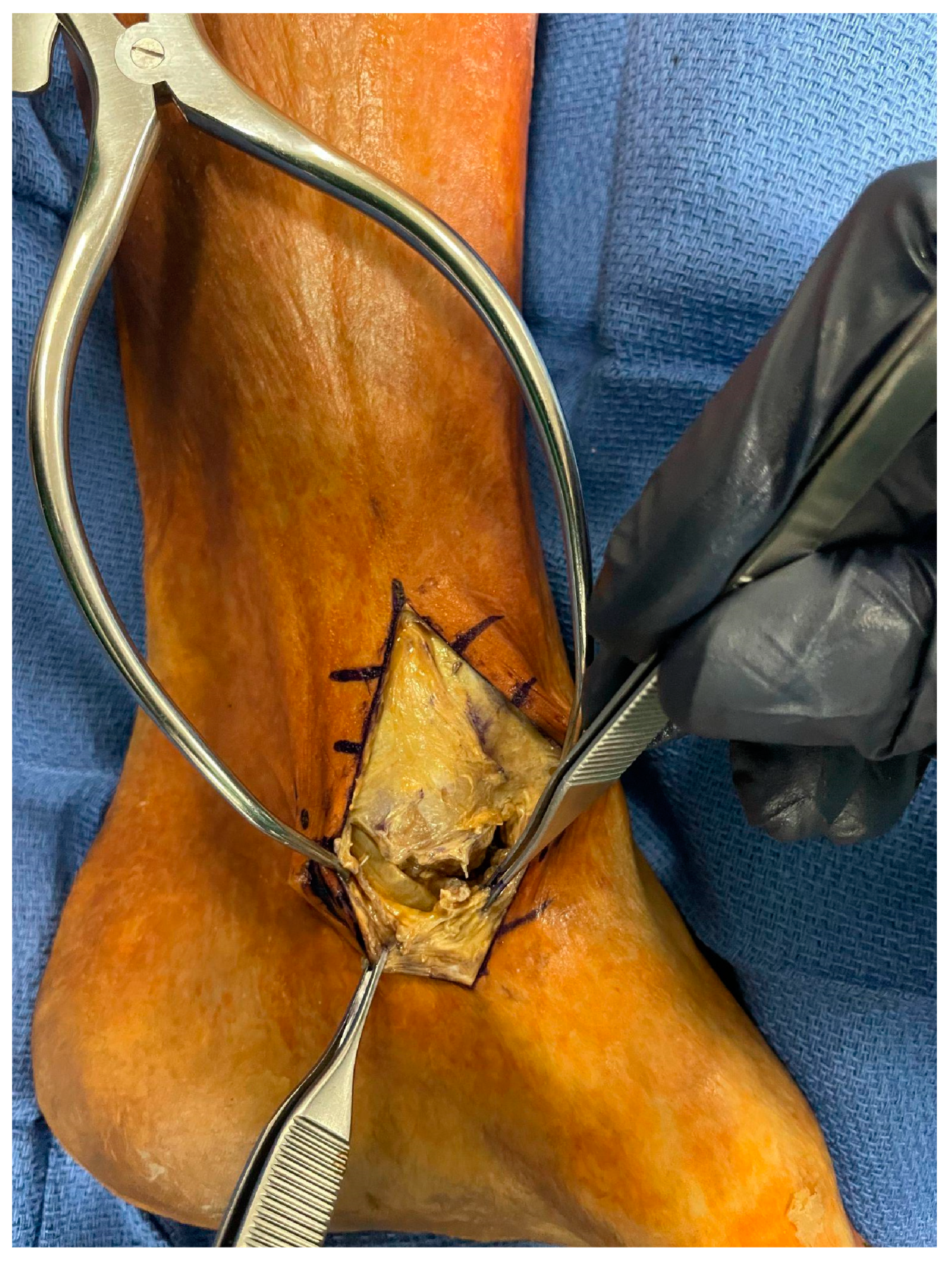
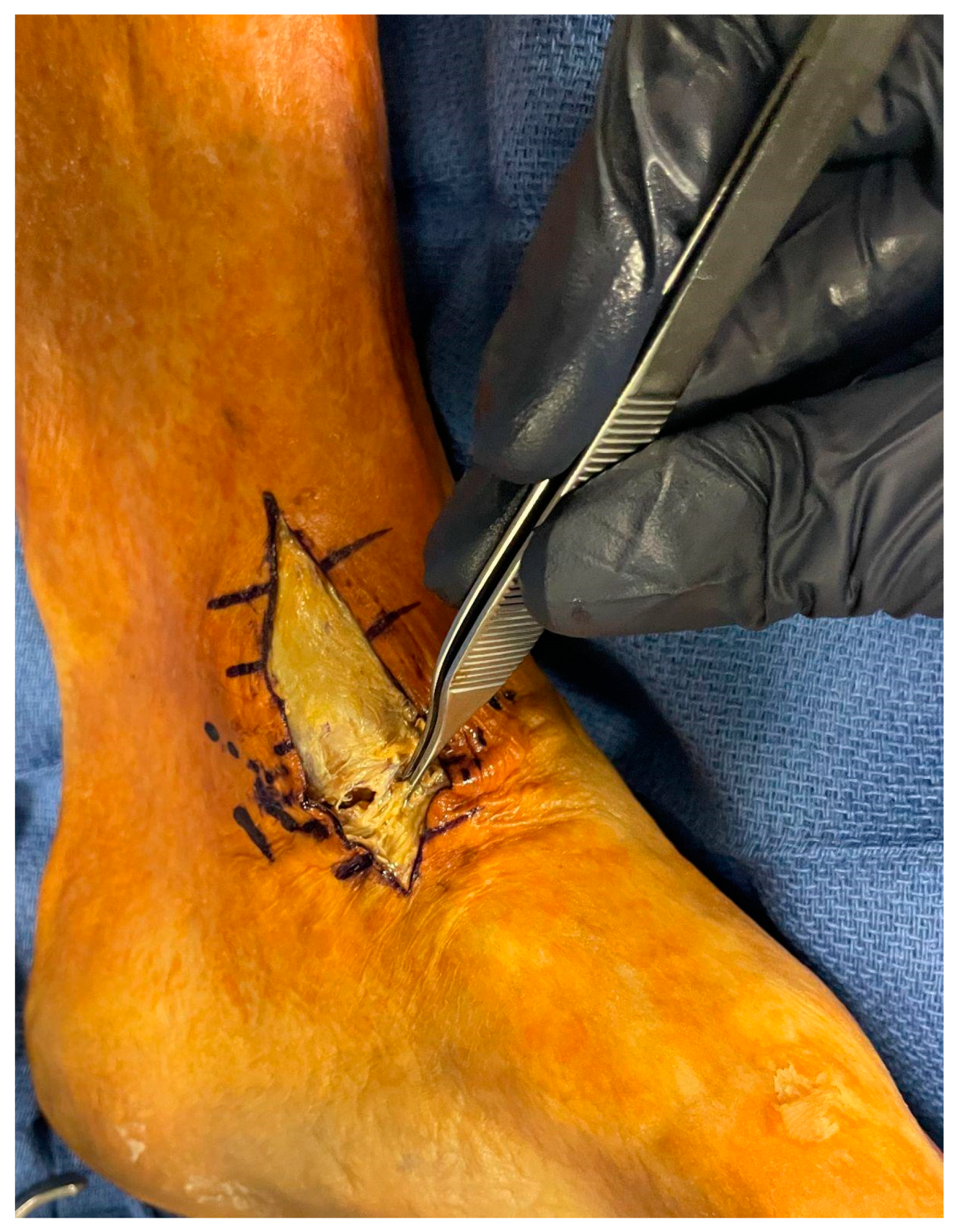
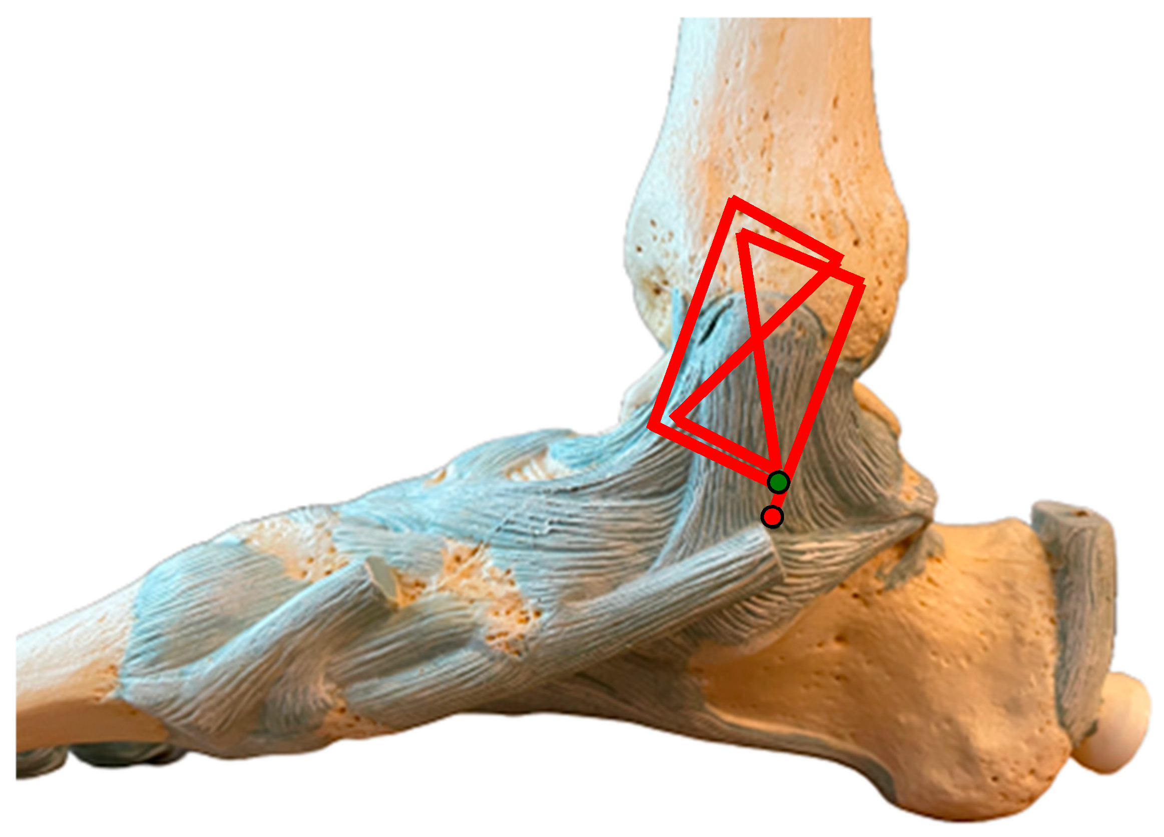
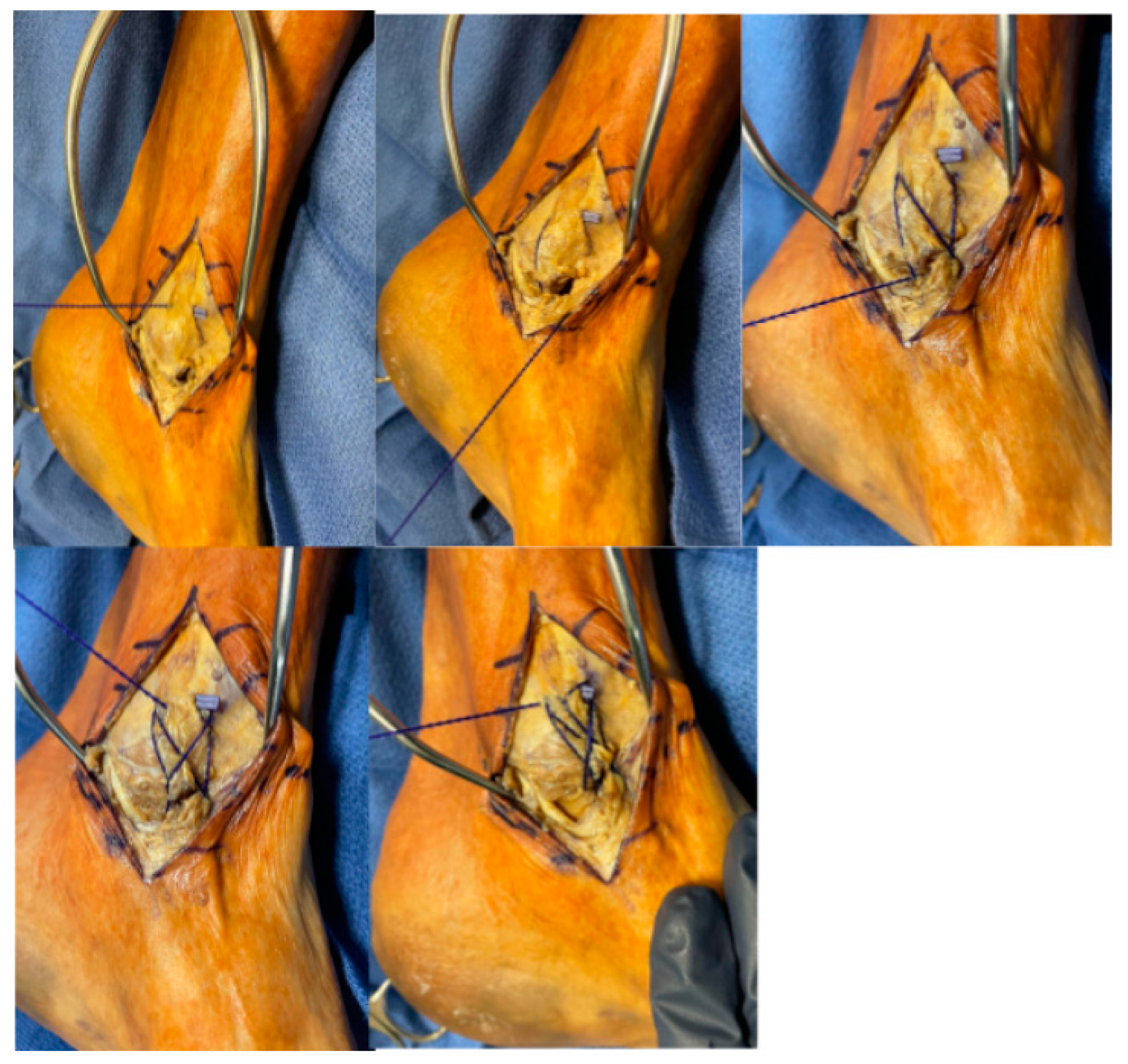
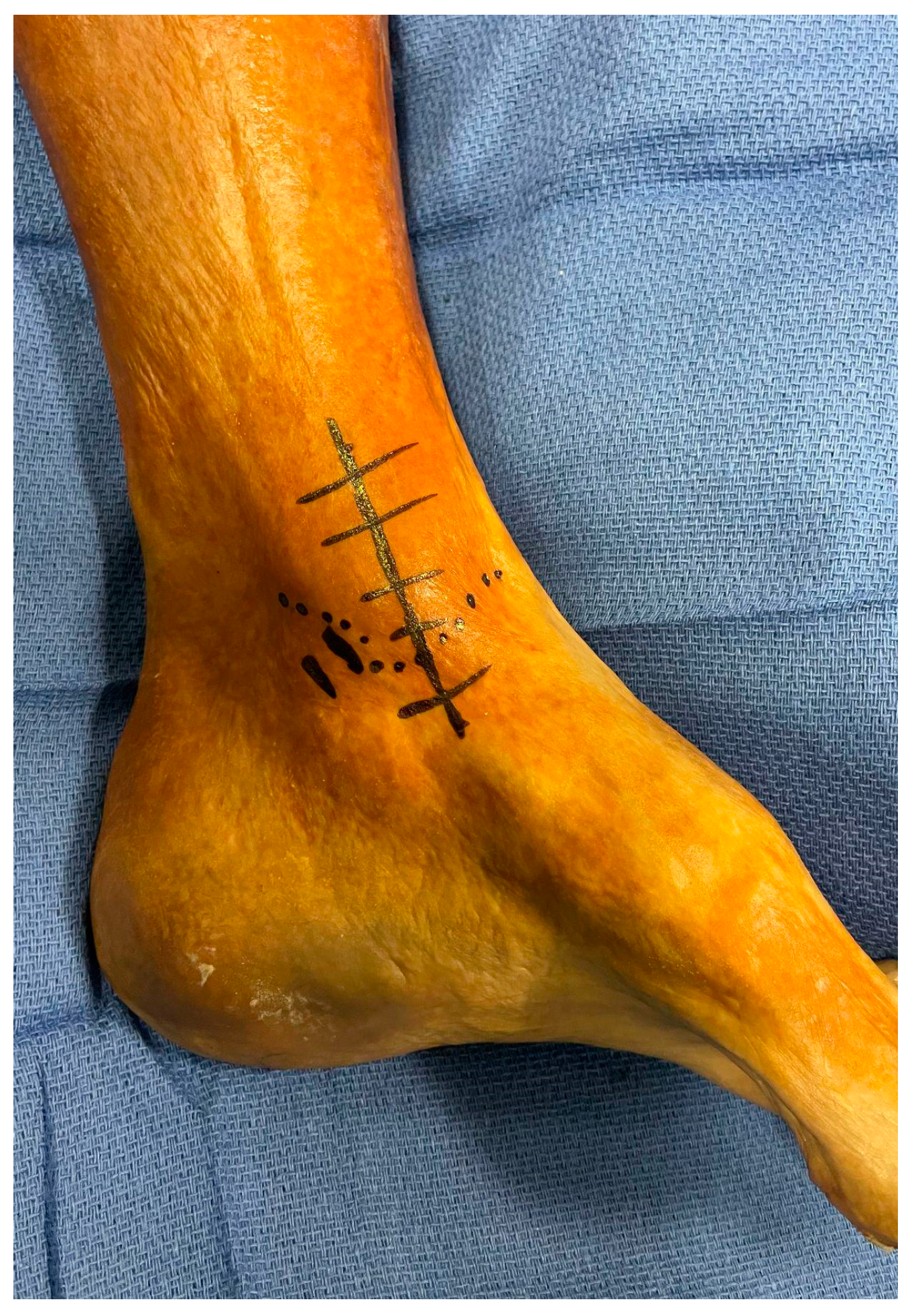
A small anteromedial approach to the ankle centered over the tip of the medial malleolus is used, and incision is carried through skin and subcutaneous tissue (Figure 2). The superficial fibers of the deltoid ligament are identified (Figure 3 and Figure 4). Generally, a tear is identified through the midsubstance of the ligament or as a sleeve of tissue avulsed proximally off the distal aspect of the medial malleolus (Figure 3). Care is taken to ensure no residual deltoid ligament or soft tissue is interposed in the medial gutter. At this time, the medial talus should be exposed through the tear in the ligament, and the joint can be irrigated to minimize the likelihood of an incarcerated loose body. The posterior tibial tendon is identified deep and posterior to the deltoid ligament and is protected during repair (Figure 4). A barbed polydioxanone (PDS) suture (#1 Stratafix [Johnson and Johnson Medtech, Warsaw, IN, USA]) is used to directly repair the deltoid ligament. PDS suture retains 74% of its tensile strength after 2 weeks, 50% after 4 weeks, and 25% after 6 weeks, which allows an appropriate timeframe for ligamentous healing [21]. Gentle internal rotation of the ankle can aid in approximation of the distal limb to the remaining proximal stump, as shown in Figure 5. The suture is first passed through the distal portion of the ligament, followed by crossing limbs through the ligament from the distal tissue to the proximal tissue/periosteum and back. A sample of a possible path for the barbed suture is demonstrated in Figure 6. The goal of the repair is to achieve large transverse passes through the distal soft tissue, followed by similar large transverse passes through the proximal tissue for a minimum total of four crossing suture limbs (Figure 6 and Figure 7). Due to the barbed nature of the suture, this provides a knotless repair. This is done by taking large enough portions of ligament to ensure adequate purchase and applying appropriate tension to the ligamentous repair. Additional passes of suture can be used to reinforce the number of crossing limbs or to provide additional support for more tenuous tissue, as indicated by the surgeon. Fluoroscopy can be used to assess the tension by evaluating the medial clear space and stability with valgus talar tilt following the suture repair. If further stress testing is desired intra-operatively following repair, it should demonstrate stable appearance of the medial clear space following repair.

2.4. Postoperative Protocol

Patients are initially placed in a short leg plaster splint for the first two weeks of the post-operative period. Patients return two weeks post-operatively, and sutures are removed if appropriate. At this time, patients are transitioned to a removable fracture boot but remain non-weight bearing for an additional four weeks. At six weeks, a repeat radiograph is obtained, and patients are then allowed to begin progressive weight bearing as tolerated. Once the patient is comfortable weight bearing in the fracture boot, they are instructed to gradually wean out of the boot as they feel comfortable. They are given a home exercise program (AAOS home exercise program) at the two-week postoperative mark, which includes Achilles stretching, ankle range of motion, foot dexterity drills, and ankle strengthening with resistance bands [22]. At six weeks, patients then begin outpatient physical therapy.

2.5. Pearls and Pitfalls

Similar to the traditional surgical techniques with suture anchors or allografts, this technique is accomplished through an anteromedial approach to the medial malleolus, which is familiar to the majority of orthopedic surgeons. This approach allows adequate exposure of the remaining deltoid tissue. Care should be taken during dissection to ensure the posterior tibialis tendon is identified deep and posterior to the deltoid fibers to ensure the tendon is not captured in the repair. Additionally, on the tibial attachment of the deltoid, inclusion of the periosteum into the repair by taking passes close to bone can add to the robustness of the repair.

3. Discussion

Over the past few decades, multiple improvements and variations to surgical anchors and specialized suture devices have become available to surgeons. Anchor types include metal implants, biocomposite implants such as polyether ether ketone (PEEK), and all-suture options. It has been demonstrated in the literature for many repair types, such as rotator cuff repair, patellar tendon repair, and quadriceps tendon repair, that suture anchors possess equivalent strength compared to previously used methods [23,24,25,26]. These anchors have demonstrated the capability for higher cyclic loads without gap formation compared to transosseous tunnels in both quadriceps tendon and patellar tendon repair [25,26]. Additionally, the development of all-suture anchors has led to smaller drill holes while maintaining the same cyclic load to failure as other metal and biocomposite anchors [27].
However, there are limitations to consider when using anchors, especially in elderly patient populations with lower bone density. Tingart et al. demonstrated that with both metal and biocomposite anchor types, there was increased failure associated with decreased bone density [28]. The use of barbed suture negates the need for fixation within the bone, which is particularly useful for the elderly, osteoporotic population, patients with significant metabolic comorbidities, or neuropathic patients. Additionally, this suture repair is most ideal in cases of midsubstance tears or when there is a stump of ligament remaining for repair. Additionally, there is a large cost difference between suture anchor devices and barbed suture. On average, a suture anchor costs $578 with a standard deviation of $220 according to a meta-analysis completed by Bacak et al. in 2023 [29]. For deltoid ligament repair, surgeons will often utilize two anchors, resulting in over $1000 in anchors alone. In comparison, a single barbed suture costs approximately $20 per suture opened [30]. At this institution, a single barbed PDS suture costs approximately $25.02 USD per suture pack compared to an average of $342–552 USD per suture anchor depending on the type of anchor used. The cost for suture anchors can double, as noted in the literature, if two anchors are required for adequate repair. At this institution, generally two anchors or one barbed PDS suture are used. This results in a cost of $25.02 compared to $684–1104 for suture anchors (range present due to variability in type of anchor used). Direct suture repair with barbed sutures provides an avenue to alleviate a significant cost burden from the patient.
Barbed sutures have been studied extensively in both general surgery, plastic surgery and orthopedic literature in regard to deep tissue closure. These suture types provide an efficient method of closure compared to traditional sutures by eliminating the need to tie after each throw. Studies have demonstrated no increased rate of surgical site infection or wound dehiscence when utilizing barbed sutures for deep tissue and fascial closure [30,31,32]. Although these are not directly comparable to ligamentous repair, they demonstrate that the strength of these sutures is equivalent to other suture types.
There are no long-term studies available demonstrating a comparison between suture anchor and direct barbed suture repair of the deltoid ligament. Therefore, there is no long-term data regarding the potential attenuation of the suture repair over time or acute failure rates when compared to suture anchor repair. Additionally, there is no comparison of patient demographic factors or injury patterns that could prove more amenable to this repair technique. Further research should be conducted comparing these methods of repair to further support this technique as a cost-effective means of addressing deltoid ligament injury.

4. Conclusions

While repair of the deltoid ligament was once deemed unnecessary during ankle fracture fixation, recent evidence supports operative fixation. Biomechanical analysis supports the importance of an intact deltoid ligament in providing rotational stability and restoring physiologic joint motion. Repair of the deltoid ligament using suture anchors is a widely accepted method; however, these anchors can be costly and rely on appropriate fixation in the bone to provide long-term fixation. This technique may represent a feasible, low-cost alternative for deltoid ligament repair, though further biomechanical and prospective clinical studies are needed to confirm its equivalence to suture anchor fixation. Further evaluation of deltoid repair techniques, including a high powered, randomized, controlled trial evaluating long-term clinical outcomes, may identify an optimal treatment method.

Author Contributions

C.V., data curation, investigation, methodology, project administration, visualization, writing—original draft, writing—review and editing; A.A., data curation, investigation, methodology, visualization, writing—original draft; T.H., conceptualization, investigation, visualization, validation, writing—original draft, writing—review and editing; S.Y., conceptualization, methodology, visualization, writing—review and editing; M.H., supervision, writing—review and editing. All authors have read and agreed to the published version of the manuscript.

Funding

This research received no external funding.

Institutional Review Board Statement

Institutional Review Board permission was obtained and this study was found to be exempt.

Informed Consent Statement

Due to the retrospective nature of the study, this study is exempt from individual consent by the Institutional Review Board.

Data Availability Statement

The data is not located in a public repository; however, it is available for review upon request.

Conflicts of Interest

No conflict of interests are present for any contributing authors to this study.

References

  1. Hintermann, B.; Regazzoni, P.; Lampert, C.; Stutz, G.; Gächter, A. Arthroscopic findings in acute fractures of the ankle. J. Bone Jt. Surg. Br. 2000, 82, 345–351. [Google Scholar] [CrossRef]
  2. van den Bekerom, M.P.J.; Mutsaerts, E.L.A.R.; vanDijk, C.N. Evaluation of the integrity of the deltoid ligament in supination external rotation ankle fractures: A systematic review of the literature. Arch. Orthop. Trauma. Surg. 2009, 129, 227–235. [Google Scholar] [CrossRef] [PubMed]
  3. McConnell, T.; Creevy, W.; Tornetta, P., 3rd. Stress examination of supination external rotation-type fibular fractures. J. Bone Jt. Surg. 2004, 86, 2171–2178. [Google Scholar] [CrossRef] [PubMed]
  4. DeAngelis, N.A.; Eskander, M.S.; French, B.G. Does medial tenderness predict deep deltoid ligament incompetence in supination-external rotation type ankle fractures? J. Orthop. Trauma 2007, 21, 244–247. [Google Scholar] [CrossRef]
  5. Egol, K.A.; Amirtharajah, M.; Tejwani, N.C.; Capla, E.L.; Koval, K.J. Ankle stress test for predicting the need for surgical fixation of isolated fibular fractures. J. Bone Jt. Surg. 2004, 86, 2393–2398. [Google Scholar] [CrossRef]
  6. Gill, J.B.; Risko, T.; Raducan, V.; Grimes, J.S.; Schutt, R.C.J. Comparison of manual and gravity stress radiographs for the evaluation of supination-external rotation fibular fractures. J. Bone Jt. Surg. 2007, 89, 994–999. [Google Scholar] [CrossRef]
  7. Lee, S.; Lin, J.; Hamid, K.S.M.; Bohl, D.D.M. Deltoid Ligament Rupture in Ankle Fracture: Diagnosis and Management. J. Am. Acad. Orthop. Surg. 2019, 27, e648–e658. [Google Scholar] [CrossRef]
  8. Petrisor, B.A.; Poolman, R.; Koval, K.; Tornetta, P.; Bhandari, M.; Evidence-Based Orthopaedic Trauma Working Group. Management of displaced ankle fractures. J. Orthop. Trauma 2006, 20, 515–518. [Google Scholar] [CrossRef]
  9. Michelson, J.D.; Varner, K.E.; Checcone, M. Diagnosing deltoid injury in ankle fractures: The gravity stress view. Clin. Orthop. 2001, 387, 178–182. [Google Scholar] [CrossRef]
  10. Bluman, E.M. Peritalar Symposia Articles Deltoid Ligament Injuries in Ankle Fractures: Should I Leave It or Fix It? Foot Ankle Int. 2012, 33, 236–238. [Google Scholar] [CrossRef]
  11. Hintermann, B.; Knupp, M.; Pagenstert, G.I. Deltoid ligament injuries: Diagnosis and management. Foot Ankle Clin. 2006, 11, 625–637. [Google Scholar] [CrossRef] [PubMed]
  12. Jones, C.R.; Nunley, J.A. Deltoid ligament repair versus syndesmotic fixation in bimalleolar equivalent ankle fractures. J. Orthop. Trauma 2015, 29, 245–249. [Google Scholar] [CrossRef] [PubMed]
  13. McCollum, G.A.; van den Bekerom, M.P.J.; Kerkhoffs, G.M.M.J.; Calder, J.D.F.; van Dijk, C.N. Syndesmosis and deltoid ligament injuries in the athlete. Knee Surg. Sports Traumatol. Arthrosc. Off. J. ESSKA. 2013, 21, 1328–1337. [Google Scholar] [CrossRef] [PubMed]
  14. Strömsöe, K.; Höqevold, H.E.; Skjeldal, S.; Alho, A. The repair of a ruptured deltoid ligament is not necessary in ankle fractures. J. Bone Jt. Surg. Br. 1995, 77, 920–921. [Google Scholar] [CrossRef]
  15. Tornetta, P. Competence of the Deltoid Ligament in Bimalleolar Ankle Fractures After Medial Malleolar Fixation. J. Bone Jt. Surg. Am. 2000, 82, 843–848. [Google Scholar] [CrossRef]
  16. Woo, S.H.; Bae, S.-Y.; Chung, H.-J. Short-Term Results of a Ruptured Deltoid Ligament Repair During an Acute Ankle Fracture Fixation. Foot Ankle Int. 2017, 39, 35–45. [Google Scholar] [CrossRef]
  17. DuVries, H. Burns and Freezing; Foreign Bodies and Gunshot Wounds; Sprains and Athletic Injuries. In Surgery of the Foot, 2nd ed.; The C.V. Mosby Company: St. Louis, MO, USA, 1959; pp. 117–119. [Google Scholar]
  18. Pellegrini, M.J.; Torres, N.; Cuchacovich, N.R.; Huertas, P.; Muñoz, G.; Carcuro, G.M. Chronic deltoid ligament insufficiency repair with Internal BraceTM augmentation. Foot Ankle Surg. Off. J. Eur. Soc. Foot Ankle Surg. 2019, 25, 812–818. [Google Scholar] [CrossRef]
  19. Schairer, W.W.; Nwachukwu, B.U.; Dare, D.M.; Drakos, M.C. Arthroscopically Assisted Open Reduction-Internal Fixation of Ankle Fractures: Significance of the Arthroscopic Ankle Drive-through Sign. Arthrosc. Tech. 2016, 5, e407–e412. [Google Scholar] [CrossRef]
  20. Mizel, M.S. Technique Tip: A Revised Method of the Cotton Test for Intra-Operative Evaluation of Syndesmotic Injuries. Foot Ankle Int. 2003, 24, 86–87. [Google Scholar] [CrossRef]
  21. Dart, A.J.; Dart, C.M. Suture material: Conventional and stimuli responsive. Compr. Biomater. 2011, 6, 573–587. [Google Scholar] [CrossRef]
  22. Foot and Ankle Conditioning Program—Orthoinfo—Aaos. Foot and Ankle Conditioning Program. Available online: https://orthoinfo.aaos.org/globalassets/pdfs/2017-rehab_foot-and-ankle.pdf (accessed on 1 November 2025).
  23. Pietschmann, M.F.; Fröhlich, V.; Ficklscherer, A.; Hausdorf, J.; Utzschneider, S.; Jansson, V.; Müller, P.E. Pullout strength of suture anchors in comparison with transosseous sutures for rotator cuff repair. Knee Surg. Sports Traumatol. Arthrosc. 2008, 16, 504–510. [Google Scholar] [CrossRef] [PubMed]
  24. Dankert, J.F.; Mehta, D.D.; Remark, L.H.; Leucht, P. Transosseous tunnels versus suture anchors for the repair of acute quadriceps and patellar tendon ruptures: A systematic review and meta-analysis of biomechanical studies. J. Orthop. Sci. 2023, 28, 821–828. [Google Scholar] [CrossRef] [PubMed]
  25. O’Donnell, R.; Lemme, N.J.; Marcaccio, S.; Walsh, D.F.; Shah, K.N.; Owens, B.D.; DeFroda, S.F. Suture Anchor Versus Transosseous Tunnel Repair for Inferior Pole Patellar Fractures Treated With Partial Patellectomy and Tendon Advancement: A Biomechanical Study. Orthop. J. Sports Med. 2021, 9, 23259671211022245. [Google Scholar] [CrossRef] [PubMed] [PubMed Central]
  26. Sherman, S.L.; Black, B.; Mooberry, M.A.; Freeman, K.L.; Gulbrandsen, T.R.; Milles, J.L.; Evans, L.; Flood, D.; Pfeiffer, F. Biomechanical Evaluation of Suture Anchor versus Transosseous Tunnel Patellar Tendon Repair Techniques. J. Knee Surg. 2019, 32, 825–832. [Google Scholar] [CrossRef] [PubMed]
  27. Yang, Y.S.; Shih, C.A.; Fang, C.J.; Huang, T.T.; Hsu, K.L.; Kuan, F.C.; Su, W.R.; Hong, C.K. Biomechanical comparison of different suture anchors used in rotator cuff repair surgery-all-suture anchors are equivalent to other suture anchors: A systematic review and network meta-analysis. J. Exp. Orthop. 2023, 10, 45. [Google Scholar] [CrossRef] [PubMed] [PubMed Central]
  28. Tingart, M.J.; Apreleva, M.; Lehtinen, J.; Zurakowski, D.; Warner, J.J.P. Anchor Design and Bone Mineral Density Affect the Pull-Out Strength of Suture Anchors in Rotator Cuff Repair: Which Anchors are Best to use in Patients with Low Bone Quality? Am. J. Sports Med. 2004, 32, 1466–1473. [Google Scholar] [CrossRef]
  29. Bacak, C.; Zisling, H.; Sandler, A.; Tompkins, R.; Childs, B.; Parnes, N. Anchor Down: The Cost of Surgical Anchors. J. Orthop. Bus. 2023, 3, 13–15. [Google Scholar] [CrossRef]
  30. Glener, S.R.; Patel, P.D.; Serva, S.N.; Self, D.M.; Heller, J.E. Prospective analysis of STRATAFIX™ symmetric PDS plus suture for fascial closure in spinal surgery: A pilot study. Neurosurg. Rev. 2024, 47, 438. [Google Scholar] [CrossRef]
  31. Tang, A.R.; Steinle, A.M.; Chanbour, H.; Emeka-Ibe, G.; Stephens, B.F.; Zuckerman, S.L.; Abtahi, A.M. Barbed Suture versus Interrupted Suture in Posterior Cervical Spine Surgery: Are They Equivalent? Spine Surg. Relat. Res. 2022, 6, 645–653. [Google Scholar] [CrossRef] [PubMed] [PubMed Central]
  32. Song, M.; Cho, Y. A Prospective Randomized Controlled Study of Stratafix versus Standard-of-Care for Deep Tissue Closure in Orthopedic Surgery. Clin. Orthop. Surg. 2024, 16, 820–826. [Google Scholar] [CrossRef] [PubMed] [PubMed Central]
Figure 1. AP radiograph of a left ankle with medial clear space (MCS) widening.
Std 15 00007 g001
Figure 2. Incision for medial approach to the ankle. Medial malleolus and posterior tibialis tendon marked as landmarks. The incision is located centered on the medial malleolus.
Std 15 00007 g002
Figure 3. Demonstration of deltoid ligament tear through an anteromedial approach to the ankle. Distal portion of deltoid ligament grasped with forceps at the inferior portion of incision. Top (superior), bottom (inferior).
Std 15 00007 g003
Figure 4. The deltoid ligament tear is then identified deep to the skin and subcutaneous tissues. Through the tear, the joint is generally visible. Forceps identify the distal portion of the deep deltoid, grasping both superficial and deep fibers. The posterior tibial tendon is seen posterior to the medial malleolus.
Std 15 00007 g004
Figure 5. This figure demonstrates the approximation of the proximal and distal fibers with internal rotation of the ankle.
Std 15 00007 g005
Figure 6. Deltoid ligament repair is undertaken with barbed PDS suture, following the path (red line) starting at the green circle and ending at the red circle.
Std 15 00007 g006
Figure 7. This sequence of images represents the stepwise passes of the barbed PDS suture, with the first image (top left) demonstrating the first pass within the proximal tibia and the last image (bottom right) demonstrating completion of the pattern described in Figure 6.
Std 15 00007 g007
Disclaimer/Publisher’s Note: The statements, opinions and data contained in all publications are solely those of the individual author(s) and contributor(s) and not of MDPI and/or the editor(s). MDPI and/or the editor(s) disclaim responsibility for any injury to people or property resulting from any ideas, methods, instructions or products referred to in the content.