Article

Exploring the Role of Salicylic Acid in Regulating the Colonization Ability of Bacillus subtilis 26D in Potato Plants and Defense against Phytophthora infestans

1
The Institute of Biochemistry and Genetics, Ufa Federal Research Center of the Russian Academy of Sciences, 450054 Ufa, Russia
2
The Faculty of Natural Geographic, Bashkir State Pedagogical University Named after M. Akmullah, 450000 Ufa, Russia
*
Author to whom correspondence should be addressed.
Int. J. Plant Biol. 2023, 14(1), 242-253; https://doi.org/10.3390/ijpb14010020
Submission received: 11 January 2023 / Revised: 16 February 2023 / Accepted: 16 February 2023 / Published: 20 February 2023
(This article belongs to the Section Plant Physiology)

Abstract

:
Plant colonization by endophytic bacteria is mediated by different biomolecules that cause dynamic changes in gene expression of both bacteria and plant. Phytohormones, in particular, salicylic acid, play a key role in the regulation of endophytic colonization and diversity of bacteria in methaphytobiome. For the first time it was found that salicylic acid influenced motility in biofilms and transcription of the surfactin synthetase gene of the endophytic strain Bacillus subtilis 26D in vitro. Treatment of Solanum tuberosum plants with salicylic acid, along with B. subtilis 26D, increased the number of endophytic cells of bacteria in potato internal tissues and level of transcripts of bacterial surfactin synthetase gene and decreased transcription of plant PR genes on the stage of colonisation with endophytes. Thus, the production of surfactin plays an important role in endophytic colonization of plants, and salicylic acid has an ability to influence this mechanism. Here we firstly show that plants treated with salicylic acid and B. subtilis 26D showed enhanced resistance to the late blight pathogen Phytophthora infestans, which was accompanied by increase in transcriptional activity of plant PR-genes and bacterial surfactin synthetase gene after pathogen inoculation. Therefore, it is suggested that salicylic acid can modulate physiological status of the whole plant–endophyte system and improve biocontrol potential of endophytic strains.

1. Introduction

The close relationship between microorganisms and other inhabitants of the biosphere is currently beyond doubt. The plant should be considered as a system of the host plant and a set of microorganisms inhabiting its surface (phylloplane, rhizosphere) and internal tissues [1,2,3,4,5,6]. The community of endophytes is composed of non-pathogenic strains of different microorganisms living inside a plant and capable of coexisting with it without causing harm. Some of them bring certain benefits, acting as plant-growth promoting microorganisms (PGPM) [1,2,3]. Bacterial endophytes are of great interest both from the point of view of the fundamental basis of the symbiosis of microorganisms with higher plants, and the practical application of such relationships. Many endophytes, such as facultative symbionts from the genus Bacillus, do not form specific anatomical structures inside plants [2,4], but apparently play an important role in the formation of the plant phenotype, regulating growth processes and resistance to different stress factors [1,2,3,4]. Communities of endophytic microorganisms are less diverse than those of the soil and the rhizosphere [1,5]. Endophytic pool has low representation of Acidobacteria, Bacteroidetes, and Verrucomicrobia and an enrichment of Actinobacteria and Firmicutes [5], which indicates selectivity in the establishment of symbioses by plant host [5,6,7] or endophytic bacteria [8,9].
Plant colonization by endophytes is a multifaceted process consisting of some steps that are mediated by plant and/or bacterial molecular patterns and driving significant changes in gene expression of both bacterial and plant genes. Bacterial surfactants such as lipopeptides (LPs) take part in the processes of biofilm formation and persisting [10]. Of particular interest is the family of surfactins, characteristic of bacteria of the genus Bacillus. Surfactin is involved in lateral expansion of colonies, bacterial colonization of plant surfaces [11,12,13] and induced systemic resistance (ISR) [10]. B. subtilis UMAF6614 formed biofilms in the melon phylloplane due to production of surfactin [12]. This strain produced lipopeptides bacillomycin and fengycin, which effectively suppress the development of phytopathogens [12]. In vitro tests confirmed the inhibitory effect of B. subtilis plipastatin alone or in combination with surfactin against Fusarium species [13].
The antagonistic potential of two strains of B. velezensis BBC023 and BBC047 in vitro was almost identical, but the production of surfactin in BBC047 was 1/3 higher than in BBC023 [14]. Accordingly, the high surfactin producing strain forms stable biofilms and contributes priming of immune responses both in the underground and aboveground parts of plant; low producing strain can do it only in the root system [14]. B. velezensis GA1 quickly colonizes tomato plantlets and forms visible biofilm-like structures. This process is correlated with consistent srfAA gene expression and surfactin production rate in the cell population [15]. The analysis of the nucleotide sequences of lipopeptide synthetases of the endophytic strain B. subtilis 26D showed the presence of the BsSfp surfactin synthetase gene responsible for the synthesis of surfactin; liquid chromatography of the culture filtrate of the B. subtilis 26D revealed an LP with Rf 0.65, which was similar in mobility to commercial surfactin [16]. This strain showed the ability to induce systemic resistance in potato and wheat plants to insect pests and phytopathogens [17,18].
Therefore, production of surfactin can play a role in both plant–endophyte interaction and plant protection against pathogens. ISR induced by PGPM is modulated by signal transduction networks in which SA can take part [19,20]. However, these signaling molecules control the interaction of plants with all environmental microorganisms, including endophytic bacteria [21,22,23]. The decrease in the diversity in the endophytic community was shown in lines of A. thaliana with increased expression of PR-genes [5]. The relative abundance of Firmicutes (including Bacillus species) was lower in immunocompromised insusceptible to SA mutants jar1 ein2 npr1, ein2 npr1, and npr1 jar1 [5]. A. thaliana line with impaired SA-mediated defense responses was hypercolonized by Klebsiella pneumoniae 342 and Salmonella enterica serovar Typhimurium 14,028 [7]. Higher internal colonization of Gluconacetobacter diazotrophicus PAL5 and S. enterica 14 028 was also found in an A. thaliana mutant that was defective in SA-mediated defense reactions [21]. P. putida BP25 strain stimulated the expression of genes which are involved in the implementation of SA-dependent reactions, then colonized A. thaliana [6]. It was found that SA enhanced cell adhesion in biofilms formed by bacteria B. cereus [24], which, apparently, was one of the important factors of plant colonization. Thus, the initial attachment of P. putida lapA strain, deficient on LapA protein, to the roots of Medicágo satíva was similar to that of the wild type, but the formation of microcolonies and the subsequent development of the mature biofilm was impaired, resulting in weaker root colonization [25,26]. Consequently, SA can influence both plants and bacteria physiological parameters during their interaction.
At the moment, the most limiting productive capacity factor for S. tuberosum is late blight caused by Phytophthora infestans (Mont.) de Bary infection. If left uncontrolled, late blight causes massive yield losses annually, especially under favorable conditions for P. infestans dispersion [27]. Earlier, it was found that endophytic strain B. subtilis 26D effectively protected potato plants against the late blight causal agent due to its direct antagonistic effect against the oomycete and the establishment of ISR [17].
PR1 gene, which encodes protein PR1, was reported as the marker of SA-dependent reactions in some plant species [28,29,30,31,32,33]. PR-1 proteins are involved in the cell wall thickening and thereby prevent the spread of the pathogens in the apoplast [28,30]. The role of PR1 in plant resistance to oomycetes [31] and bacterial infections [32,33] was demonstrated previously. Hydrolases produced by Phytophthora sp. are important factors of its aggressiveness, which ensure the penetration of the pathogen into tissues, for example, due to the destruction of plant cell walls and the degradation of protective proteins [34]. The defense reaction of plants against hydrolase-producing oomycetes is accompanied by the synthesis of inhibitors of these enzymes, in particular, PR-6 proteins [29,35]. Phytopathogenic bacteria also inject effector proteins, including proteases, into plant cells, presumably to colonize their hosts [36], and endophytic bacteria B. subtilis 26D produce proteases in vitro [18], but the function of these proteins inside plant tissues has remained a mystery [36]. Thus, proteinase inhibitors (PR6) and PR1 proteins from plants potentially can prevent invasion of both pathogens and endophytes.
Our hypothesis is that a high rate of endophytic colonization of plants with bacteria is essential to support the resistance of the whole plant/endophyte system to phytopathogens. Thus, the aim of this work is to investigate the role of bacterial surfactin in the colonization of potato plants by B. subtilis 26D and the role of SA in the regulation of this process.

2. Materials and Methods

2.1. Plant and Microbe Material

Plants: sterile Solanum tuberosum L. plants cultivar Early Rose obtained by microcloning technology and grown in tubes with Murashige and Skoog medium in the KBW E6 climatic chamber (Binder GmbH, Tuttlingen, Germany) with a 16 h light period at 20–22 °C for 14 days prior to use.
Bacteria: Gram-positive aerobic B. subtilis 26D strain from the collection of the Laboratory of Biochemistry of Plant Immunity of the Institute of Biochemistry and Genetics UFRC RAS [37] were used. Bacteria were grown on liquid lysogeny broth (LB) medium (1% tryptone, 0.5% yeast extract, 0.5% NaCl) at 20–22 °C using laboratory shakers (120 rpm).
Pathogen: P. infestans ISM isolated from late blight-affected potato tubers of the Udacha variety (Russia, Bashkortostan, Birsky district, 55°26′06.0 N 55°36′22.9 E) in 2018 was used to infect potato plants. To activate the aggressiveness of the isolate, mycelium sections were transferred under aseptic conditions to disks from the middle part of potato tubers (Rannyaya Rosa variety), and were placed in Petri dishes with moist filter paper and incubated at 18 °C for 7–10 days. Part of the tissue from the affected areas of the tuber was transferred on potato-dextrose agar and incubated at 18 °C for 7 days. At 7 days after placement on PDA, the surface of the mycelium in Petri dishes was filled with sterile water and incubated at 4 °C for 30 min to release the spores; the spore titer was counted in a Fuchs–Rosenthal chamber. Spore suspension was diluted to a concentration of 1 × 10⁵ spores/mL with sterile distilled water.

2.2. Models of Plant–Microbe Interaction

Model “Plant+endophyte”: 14-days-old plants growing on Murashige and Skoog medium were inoculated with 5 μL of B. subtilis 26D suspension (108 cells/mL) on distilled water or 1 μM SA. The drop of B. subtilis 26D suspension was applied on the stem neighbor the zone of formation of adventitious roots, according to the previously described method [24]. A part of the plants was treated with 5 μL of distilled water (distiller A1110, LLC Liston, Zhukov, Russia) and was used as control ones in all experiments.
Model “Plant+endophyte+pathogen”: Plants were grown in a gnotobiotic system for 7 days. Parts of control plants and plants, containing endophytes, were treated with 5 μL of distilled water. All others were inoculated with a 5 µL of 1 × 10⁵ spores/mL zoospore suspension of P. infestans per plant leaf. Observations for the symptoms of the disease continued for 12 days after inoculation, in each of the variants. Photographs were taken using the C-4000 Zoom camera (Olympus, Tokyo, Japan); images were analyzed using the GNU Image Manipulation Program (NIH, Bethesda, MD, USA) to calculate the percentage of affected leaf area.

2.3. Bacillus Motility

Petri dishes with LB containing 0.2%, 0.4% and 0.8% agar were prepared fresh, and the following day were dried for 30 min in a laminar flow hood [16]. Half of the agar plates contained 1 μM SA. Each plate was toothpick inoculated from an overnight colony and scored for swimming and swarming motility after 18 h (0.2% agar) and 32 h (0.4% and 0.8% agar) incubation at 37 °C. In these timepoints cells from the edge and the center of growing colonies were collected for RNA extraction.

2.4. RNA Isolation and the Reverse Transcription Quantitative Polymerase Chain Reaction (qPCR)

Total RNA was isolated from bacterial cultures with 1 mL of RNA extraction kit Lira® (Biolabmix, Novosibirsk, Russia) supplemented with 30 μL 50 mg/mL lysozyme in TE buffer (0.01 M Tris-HCl, 0.001 M EDTA, pH 8.0). Total RNA was isolated from plants 24 h post inoculation with B. subtilis 26D, from control plants and plants containing endophytes after 24 h post infection with P. infestans with the same reagents. RNA concentration was measured using the Smart Spec Plus spectrophotometer (Bio-Rad, Hercules, CA, USA). Concentrations of RNA in samples were equalized. The first cDNA strand was synthesized using oligonucleotide primers and M-MLV reverse transcriptase (Thermo Scientific, Madrid, Spain).
The obtained cDNA was diluted five-fold and used for qPCR. The qPCR was performed by polymerase chain reaction in real time using a set of predefined reagents SYBR Green I (Synthol, Moscow, Russia) and the device CFX Connect Real-Time PCR Detection System (Bio-Rad, Hercules, CA, USA). The qPCR was run according to the following program: 50 °C for 2 min; 95 °C for 10 min; 40 cycles of 95 °C for 15 s and 60 °C for 1 min. After the final qPCR cycle, a melting curve analysis was conducted to determine the specificity of the reaction (at 95 °C for 15 s, 60 °C for 1 min and 95 °C for 15 s). The efficiency of primer pairs was evaluated using 10-fold cDNA dilution series. The expression of plant genes is presented as fold change normalized to the reference gene StAct (Potato Actin), B. subtilis 26D gene—to the reference Bs16S gene (16S rRNA) and relative to the untreated control sample. The iCycler iQ5 Real-Time Detection System software (Bio-Rad, Hercules, CA, USA) was used for data analysis. The primers used are given in Table 1.

2.5. Analysis of the Number of B. subtilis 26D Cells in Internal Plant Tissues

Number of colony-forming units (CFU) microorganisms in shoot and root tissues of plants were determined 7 days post inoculation with bacteria [17]. For this, 40 mg of plant material was surface sterilized with 95% ethanol for 3 min and washed with distilled water twice. Plant samples were homogenized in sterile porcelain mortars and diluted in 40 mL of sterile water. Aliquots (30 μL) of the homogenate after dilution were distributed over the surface of the agarized LB medium with a Drygalsky spatula until completely dry. Petri dishes were incubated at 28 °C in the thermostat TS-1/20 SPU (SKTB SPU, Smolensk, Russia) for 24 h. Number of CFU were counted up in no fewer than 10 plates, and their number was recalculated per 1 g of wet weight of shoots or roots [17].

2.6. Statistical Analysis

In total, 30 leaves of potato plants were used in each variant for late blight lesions estimation. Morphology of B. subtilis 26D colonies in each variant was investigated in 10 independent plates. A total of 10 plants were used in each variant for the investigation of the number of endophytic bacteria (shoots and roots of plants were sampled for each repetition). At least three biological replications in three technical repetitions each were examined in all experiments for qPCR. Data presented were mean values with standard errors (±SE). Means were compared using analysis of variance (ANOVA) followed by Student’s test with p ≤ 0.05. The program Statistica 12.0 (Stat Soft, Tulsa, OK, USA) was used.

3. Results

3.1. Influence of Salicylic Acid on B. subtilis 26D Motility and Surfactin Syntetase Transcription In Vitro

On a semi-liquid medium (0.2% agar), B. subtilis 26D formed breaks in the medium with a liquefied center and edges growing on the surface of the medium 24 h after loading (Figure 1A). When SA was added to the culture medium the size of the colonies was two times higher than the control values. Colonies of B. subtilis 26D on a medium containing 0.4% agar also had an irregular shape and formed breaks on the surface of the medium, and also spread radially. On the medium with the addition of SA, the motility of bacteria increased, and the halo, indicating the invasion of bacteria, spread over most of the surface of the 0.4% agar. Whereas on the medium containing 0.8% agar B. subtilis 26D formed colonies with dendritic edges and a characteristic wrinkled surface, asymmetric spread of colonies by swarming was also observed. Under the influence of the SA, there was a significant increase in migrating parts of colonies (Figure 1A).
SA did not affect the transcriptional activity of the surfactin synthetase gene BsSfp in the zone of active promotion of bacterial colonies on a medium containing 0.2% agar (Figure 1B). On 0.4% agar, the addition of SA to the culture medium increased the transcriptional activity of this gene by 3 times in relation to colonies not affected by SA. The highest transcriptional activity of the BsSfp gene was observed on 0.8% agar, which was two orders of magnitude higher than in colonies developing on 0.2–0.4% agar. At the same time, in the presence of SA in culture medium, the transcriptional activity of surfactin synthetase gene increased by another 75%. These data can be the reason for the effect of SA on swarming motility of B. subtilis 26D cells.

3.2. Impact of Salicylic Acid on Population of B. subtilis 26D

On the 7th day after post-inoculation of sterile test-tube potato plants with a suspension of bacterial cells, 44.9 ± 10.0 × 10⁴ CFU of B. subtilis 26D/g of fresh weight was present in the tissues of shoots, and half as much in the tissues of the roots (Table 2). Under the influence of SA, 75.0 ± 10.42 × 10⁴ CFU of endophytes/g of fresh mass of shoots was found. No differences in endophyte content were observed in roots of water- and SA-treated plants.
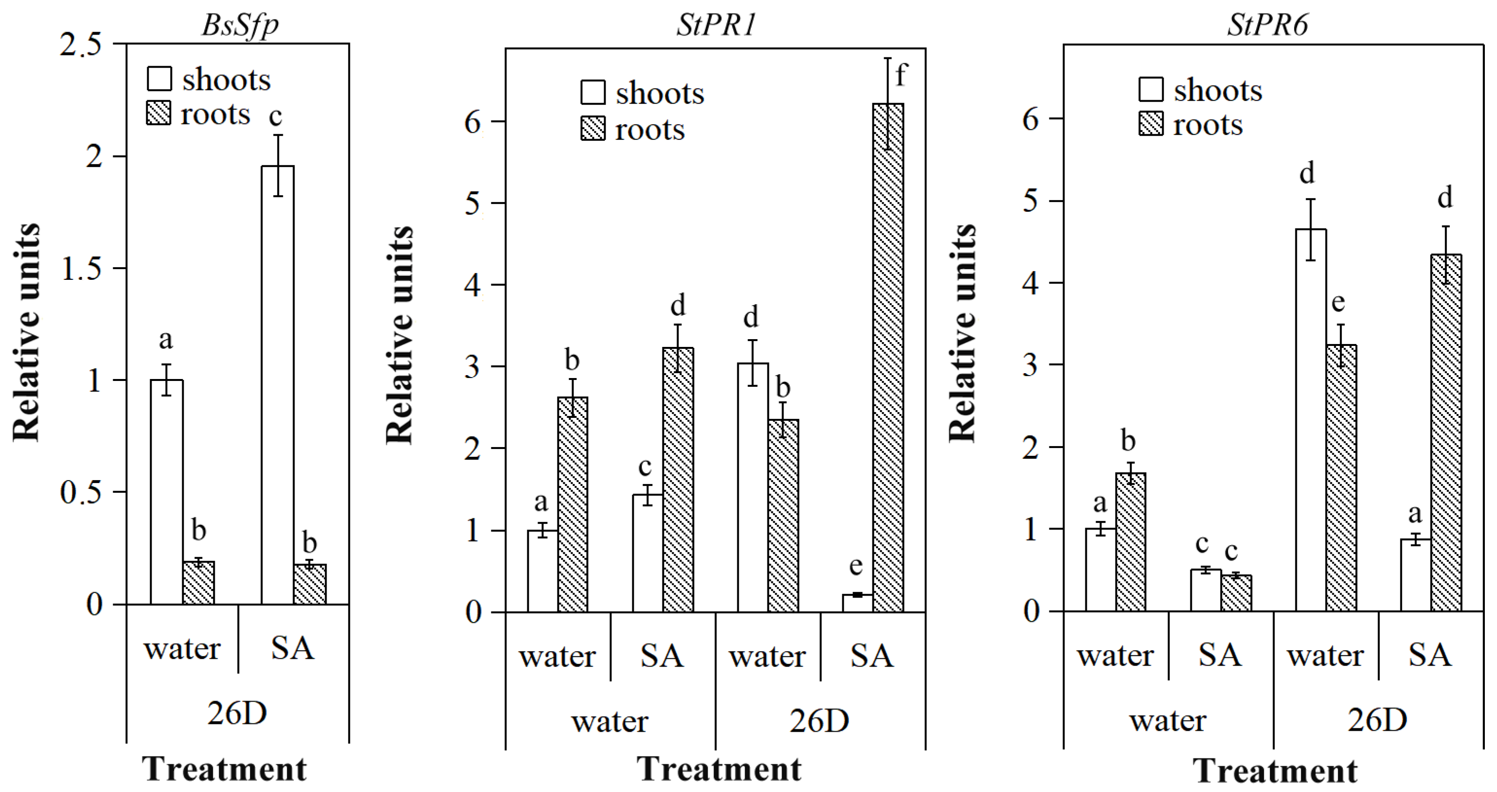
Transcriptional activity of the B. subtilis 26D BsSfp gene was observed in potato plants 24 h after post-inoculation with bacteria. In shoots it was 5 times higher than in roots (Figure 2). Under the influence of SA, the transcriptional activity of the bacterial gene in shoots increased twofold and did not change in roots.
In water-treated plants more StPR1 and StPR6 gene transcripts were observed in roots than in shoots (Figure 2). Under the influence of SA alone, the transcriptional activity of StPR1 gene increased by about 20%, while of StPR6 decreased twofold in both cases. Inoculation of plants with endophytic bacteria increased the transcriptional activity of StPR1 3-fold in shoots but did not affect this parameter in roots; however, the relative amount of StPR6 transcripts increased 5-fold in shoots and twice in roots relative to water-treated, endophyte-free control ones. At the same time, the impact of SA led to a significant decrease in the content of StPR1 gene transcripts, without a significant change in the content of StRR6 transcripts in shoots of plants inoculated with B. subtilis 26D. In roots of plants exposed to SA, after inoculation with endophytes, were also observed a 6-fold increase in the number of StPR1 transcripts and a 4-fold increase in the number of StPR6 transcripts.

3.3. Salicylic Acid and B. subtilis 26D Enhance Plant Resistance to P. infestans

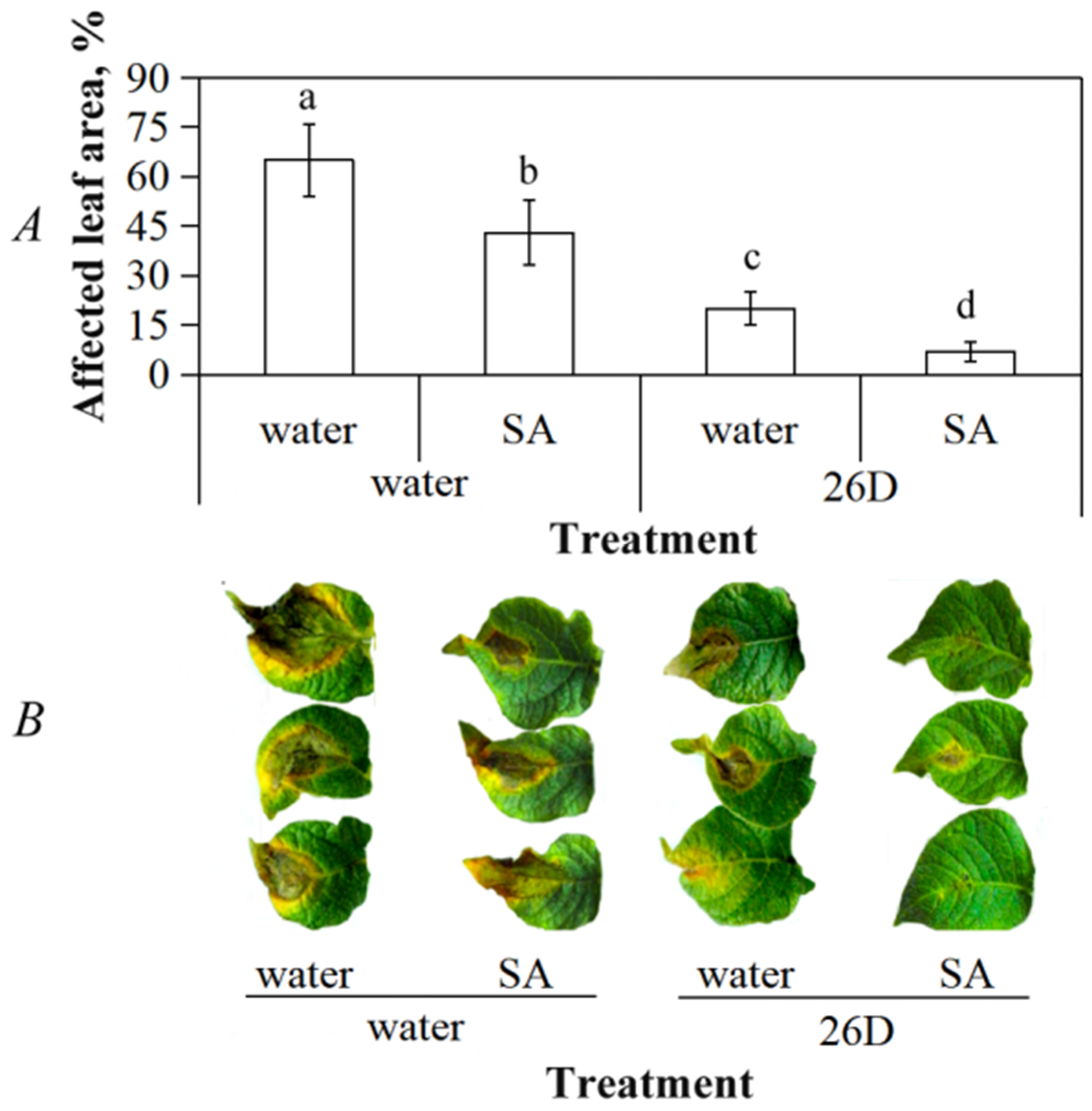
Inoculation of potato plants with B. subtilis 26D cell suspension decreased the percentage of late blight damaged sites on potato leaves near twofold, whereas the effect of SA was less significant (Figure 3). Simultaneous treatment with B. subtilis 26D and SA led to the most substantial decrease in lesions area compared with control ones (about 8 times less than on leaves of water-treated plants).
B. subtilis 26D and SA alone did not cause an increase in the content of transcripts of plant gene StPR6, but jointly they increased this parameter by 20% relative to control ones (Figure 4). Solitary treatment with B. subtilis 26D did not lead to an increase in the content of StPR1 gene transcripts, but SA led to a 30% increase in this parameter. In plants treated with B. subtilis 26D+SA transcriptional activity of StPR1 was three times higher than in water-treated ones. Thus, long-term coexistence of plants with endophytes led to transcriptional changes over time.
The relative level of transcripts of the StPR6 gene was 2-fold lower than thereof in control plants 24 h after infection with the late blight pathogen (Figure 4); in plants treated with SA it was 20% higher than in non-infected plants treated with water. The presence of B. subtilis 26D in plants increased by 80% the transcriptional activity of the proteinase inhibitor gene StPR6, and 3 times relative to control ones in combination with SA. Transcriptional activity of the gene StPR1 was approximately the same as in control plants 24 h after infection with late blight pathogen. Treatment with SA and inoculation with B. subtilis 26D alone increased the transcriptional activity of the StPR1 gene in infected plants by 40%, while co-treatment increased this indicator 4-fold relative to uninfected control plants.

4. Discussion

Coevolution of plants with microorganisms started, according to [38], with the colonization of the land by ancestral plants 450 million years ago. Endophytes are the most closely related with plant microorganisms, but we have the least information on basic principles of organization of this symbiose [1,2,3].
Some properties such as motility, chemotaxis and adherence were considered crucial for initiating endophytic colonization. After the adhesion of the bacteria to the roots microcolonies are formed involving the essential participation of exopolysaccharides and lipopolysaccharides, as it has been reported on the example of the colonization of rice roots by Herbaspirillum seropedicae [39] and Gluconacetobacter diazotrophicus [40]. Azoarcus sp. strains harbor a disruptional insertion of the fliM gene and show a significantly diminished motility phenotype and plant colonization ability [41]. The pathogenesis of P. aeruginosa and its ability to colonize environments depend on its swarming and twitching abilities [42].
In order to move with the help of swarming migration, the cells must be in a layer of water. Movement on a denser agar becomes more difficult and bacteria need to attract water with the help of osmotic agents [16]. It is possible to overcome these difficulties with the help of surfactants such as lipopeptides. In our work 1 μM SA stimulated bacterial motility, in particular on semi-solid (0.8% agar) medium, in our opinion due to the promotion of transcriptional activity of the surfactin synthetase gene of endophytic B. subtilis 26D. Importantly, treatment of plants with suspension of B. subtilis 26D in 1 μM SA (then both plant and bacteria were under the influence of SA) led to increase in the number of endophytic cells in plant shoots. Earlier, it was shown that 1000 μg mL−1 SA decreased swimming motility and did not affect swarming motility of B. cereus and P. fluorescens [24]. Increase in the growth rate due to increase in motility of bacterial colonies on a semi-solid nutrient medium with 1–100 μg/mL of another phenolic compound, ferulic acid, allowed us to make an assumption about the movement of endophyte cells to developing plant organs, where high ferulic acid concentrations and gradual lignification occurred [43].
Despite the fact that B. subtilis 26D successfully colonized the aerial part of plants (shoots) and, to a lesser extent, roots, treatment with these microorganisms causes defence reactions in plants after contact. It was expressed in an increase in the transcriptional activity of the salicylate-dependent StPR1 gene in both shoots and roots, and jasmonate-dependent StPR6 in roots. Earlier, [44] demonstrated that B. phytofirmans PsJN perception by grapevine cells triggers a local immune response. However, the defence responses are significantly weaker than those occurring in a non-host interaction [44]. Co-treatment with SA reduced the transcriptional activity of StPR1 and StPR6 genes in colonized shoots and increases it in the roots, thereby spatially separating the manifestation of defense reactions on the endophyte invading. Observed supression of plant immune reactions probably led to the increase in the number of endophytic cells of B. subtilis 26D in plant shoots. Apparently, such phytohormone-regulated titers of bacterial cells in tissues are important factors in the subsequent formation of their resistance [1,2,13].
In our study, the joint treatment of plants with SA and inoculation with B. subtilis 26D cell reduced the intensity of late blight symptoms on plant leaves. A distinctive feature of systemic resistance mediated by PGPM is the priming mechanism [1,10,20], which may be based on the observed initial protective reaction against the introduction of the endophyte that represents a weak pathogen [44]. The decrease in the intensity of manifestation of late blight symptoms on the leaves of potato plants treated with SA and the bacterial strain, in which we found the accumulation of StPR6 gene transcripts, indicates the importance of proteinase inhibitors in protecting potatoes from the late blight pathogen.
In this work we found that the strain of endophytic bacteria under study promoted the accumulation of transcripts of genes encoding StPR1 proteins to the same extent as SA, and composition of B. subtilis 26D+SA had a cumulative effect on this parameter in plants infected with P. infestans. Lastochkina O. et al. [45] also showed that the joint treatment of potato tubers with B. subtilis 10-4 and SA before storage increased their resistance to P. infestans and F. oxysporum, reducing the pathogen-induced accumulation of proline and the intensity of lipid peroxidation in tubers. In addition, the combination B. subtilis 10-4+SA reduced root rot pathogens infection of wheat plants, which indicates the possibility of a protective effect of such compositions on many crops [46]. In this context, it was important that the treatment of Nicotiana benthamiana plants with SA-producing rhizospheric bacteria P. tremae EB-44 and Curtobacterium herbarum EB-47 induced resistance of plants to pathogenic bacteria P. syringae pv. tabaci [47]. We demonstrated that the effect of SA-producers can be achieved by combined treatment of plants with endophytic bacteria and exogenic SA.
It was shown for the first time that in the host plant/endophyte system there was a microsymbiont response to the introduction of the pathogen. An increase in the transcriptional activity of the key gene for the biosynthesis of surfactin of B. subtilis 26D, whose function can be both a direct antagonistic effect against the causative agent of the late blight and maintaining the stability and motility of bacteria in biofilms under biotic stress, was observed.
Our data indicate the possibility of a synergistic activation of the components of the defense system in plants regulated on the one hand by SA and, on the other hand, by endophytic bacteria B. subtilis 26D. We demonstrate the way to manipulate the process of endophytic colonization of plants using SA. Taking into account the insecticidal activity of strain under consideration against aphids Schizaphis graminum and wheat pathogen Stagonospora nodorum [18] and Leptinotarsa decemlineata [17], as well as the effectiveness of SA against plant viral diseases [22,27], co-treatment with bacteria B. subtilis 26D and SA can open one of the approaches in artificial development of targeting plant microbiome systems and an integrated protection of plants from biotic influences.

5. Conclusions

(1)
Plants develop defense reactions on endophytic bacteria;
(2)
SA promotes endophytic colonization of shoots by down-regulation of plant defense reactions and up-regulation of the surfactin synthase gene of B. subtilis 26D in vitro and in planta;
(3)
High level of endophytic cells in SA+B. subtilis 26D treated plants leads to improvement in potato plants resistance to P. infestans;
(4)
Pathogen attack initiates activity of both plant PR genes and surfactin synthase gene of B. subtilis 26D and SA increase these parameters.

Author Contributions

Conceptualization, A.S. and I.M.; methodology, A.S.; validation, G.B. and A.G.; investigation, G.B. and A.G.; writing—original draft preparation, A.S.; writing—review and editing, I.M. All authors have read and agreed to the published version of the manuscript.

Funding

This research was funded by RCF, grant number 20-76-00003.

Institutional Review Board Statement

Not appreciable.

Informed Consent Statement

Not appreciable.

Data Availability Statement

Not appreciable.

Conflicts of Interest

The authors declare no conflict of interest.

References

  1. Kumawat, K.C.; Razdan, N.; Saharan, K. Rhizospheric Microbiome: Bio-based Emerging Strategies for Sustainable Agriculture Development and Future Perspectives. Microbiol. Res. 2022, 254, 126901. [Google Scholar] [CrossRef]
  2. Pinski, A.; Betekhtin, A.; Hupert-Kocurek, K.; Mur, L.A.J.; Hasterok, R. Defining the Genetic Basis of Plant–Endophytic Bacteria Interactions. Int. J. Mol. Sci. 2019, 20, 1947. [Google Scholar] [CrossRef] [PubMed] [Green Version]
  3. Sharma, M.; Mallubhotla, S. Diversity, Antimicrobial Activity, and Antibiotic Susceptibility Pattern of Endophytic Bacteria Sourced from Cordia dichotoma L. Front. Microbiol. 2022, 13, 879386. [Google Scholar] [CrossRef] [PubMed]
  4. Praca, L.B.; Gomes, A.C.M.M.; Cabral, G.; Martins, E.S.; Sujii, E.H.; Monnerat, R.G. Entophytic Colonization by Brazilian Strains of Bacillus thuringiensis on Cabbage Seedlings Grown in vitro. Bt Res. 2012, 3, 11–19. [Google Scholar] [CrossRef]
  5. Lebeis, S.L.; Paredes, S.H.; Lundberg, D.S.; Breakfield, N.; Gehring, J.; McDonald, M.; Malfatti, S.; Glavina del Rio, T.; Jones, C.D.; Tringe, S.G.; et al. Salicylic Acid Modulates Colonization of the Root Microbiome by Specific Bacterial Taxa. Science 2015, 349, 860–864. [Google Scholar] [CrossRef] [PubMed] [Green Version]
  6. Sheoran, N.; Kumar, A.; Munjal, V.; Nadakkakath, A.V.; Eapen, S.J. Pseudomonas putida BP25 Alters Root Phenotype and Triggers Salicylic Acid Signaling as a Feedback Loop in Regulating Endophytic Colonization in Arabidopsis thaliana. Physiol. Mol. Plant. Pathol. 2016, 93, 99–111. [Google Scholar] [CrossRef]
  7. Iniguez, A.L.; Dong, Y.; Carter, H.D.; Ahmer, B.M.; Stone, J.M.; Triplett, E.W. Regulation of Enteric Endophytic Bacterial Colonization by Plant Defenses. Mol. Plant Microbe Interact. 2005, 18, 169–178. [Google Scholar] [CrossRef] [Green Version]
  8. Trda, L.; Fernandez, O.; Boutrot, F.; Heloir, M.C.; Kelloniemi, J.; Daire, X. The Grapevine Flagellin Receptor VvFLS2 Differentially Recognizes Flagellin-derived Epitopes from the Endophytic Growth-promoting Bacterium Burkholderia phytofirmans and Plant Pathogenic Bacteria. New Phytol. 2014, 201, 1371–1384. [Google Scholar] [CrossRef]
  9. Martínez-Gil, M.; Yousef-Coronado, F.; Espinosa-Urgel, M. LapF, the Second Largest Pseudomonas putida Protein, Contributes to Plant Root Colonization and Determines Biofilm Architecture. Mol. Microbiol. 2010, 77, 549–561. [Google Scholar] [CrossRef]
  10. Maksimov, I.V.; Singh, B.P.; Cherepanova, E.A.; Burkhanova, G.F.; Khairullin, R.M. Prospects and Applications of Lipopeptide-producing Bacteria for Plant Protection (Review). App. Biochem. Microbiol. 2020, 56, 15–28. [Google Scholar] [CrossRef]
  11. Otto, S.B.; Martin, M.; Schäfer, D.; Hartmann, R.; Drescher, K.; Brix, S.; Kovács, Á.T. Privatization of Biofilm Matrix in Structurally Heterogeneous Biofilms. mSystems 2020, 5, e00425-20. [Google Scholar] [CrossRef] [PubMed]
  12. Zeriouh, H.; de Vicente, A.; Pérez-García, A.; Romero, D. Surfactin Triggers Biofilm Formation of Bacillus subtilis in Melon Phylloplane and Contributes to the Biocontrol Activity. Environ. Microbiol. 2014, 16, 2196–2211. [Google Scholar] [CrossRef]
  13. Munakata, Y.; Heuson, E.; Daboudet, T.; Deracinois, B.; Duban, M.; Hehn, A.; Coutte, F.; Slezack-Deschaumes, S. Screening of Antimicrobial Activities and Lipopeptide Production of Endophytic Bacteria Isolated from Vetiver Roots. Microorganisms 2022, 10, 209. [Google Scholar] [CrossRef]
  14. Stoll, A.; Salvatierra-Martínez, R.; González, M.; Araya, M. The Role of Surfactin Production by Bacillus velezensis on Colonization, Biofilm Formation on Tomato Root and Leaf Surfaces and Subsequent Protection (ISR) against Botrytis cinerea. Microorganisms 2021, 9, 2251. [Google Scholar] [CrossRef] [PubMed]
  15. Hoff, G.; Arguelles Arias, A.; Boubsi, F.; Pršić, J.; Meyer, T.; Ibrahim, H.M.M.; Steels, S.; Luzuriaga, P.; Legras, A.; Franzil, L.; et al. Surfactin Stimulated by Pectin Molecular Patterns and Root Exudates Acts as a Key Driver of the Bacillus-Plant Mutualistic Interaction. mBio 2021, 12, e0177421. [Google Scholar] [CrossRef]
  16. Cherepanova, E.A.; Galyautdinov, I.V.; Burkhanova, G.F.; Maksimov, I.V. Isolation and Identification of Lipopeptides of Bacillus subtilis 26D. App. Biochem. Microbiol. 2021, 57, 636–642. [Google Scholar] [CrossRef]
  17. Sorokan, A.; Benkovskaya, G.; Burkhanova, G.; Blagova, D.; Maksimov, I. Endophytic Strain Bacillus subtilis 26DCryChS Producing Cry1Ia Toxin from Bacillus thuringiensis Promotes Multifaceted Potato Defense against Phytophthora infestans (Mont.) de Bary and Pest Leptinotarsa decemlineata Say. Plants 2020, 9, 1115. [Google Scholar] [CrossRef] [PubMed]
  18. Maksimov, I.V.; Blagova, D.K.; Veselova, S.V.; Sorokan, A.V.; Burkhanova, G.F.; Cherepanova, E.A.; Sarvarova, E.R.; Rumyantsev, S.D.; Alekseev, V.Y.; Khayrullin, R.M. Recombinant Bacillus subtilis 26DCryChS Line with Gene Btcry1Ia Encoding Cry1Ia Toxin from Bacillus thuringiensis Prom otes Integrated Wheat Defense Against Pathogen Stagonospora nodorum Berk. and Greenbug Schizaphis graminum Rond. BioControl 2020, 144, 104242. [Google Scholar] [CrossRef]
  19. Veselova, S.V.; Burkhanova, G.F.; Rumyantsev, S.D.; Blagova, D.K.; Maksimov, I.V. Strains of Bacillus ssp. Regulate Wheat Resistance to Greenbug Schizaphis graminum Rond. App. Biochem. Microbiol. 2019, 55, 46–52. [Google Scholar] [CrossRef]
  20. Riera, N.; Wang, H.; Li, Y.; Li, J.; Pelz-Stelinski, K.; Wang, N. Induced Systemic Resistance Against Citrus Canker Disease by Rhizobacteria. Phytopathology 2018, 108, 1038–1045. [Google Scholar] [CrossRef] [Green Version]
  21. De Souza, A.R.; De Souza, S.; De Oliveira, M.; Ferraz, T.; Figueiredo, F.; Da Silva, N.; Rangel, P.; Panisset, C.; Olivares, F.; Campostrini, E. Endophytic Colonization of Arabidopsis thaliana by Gluconacetobacter diazotrophicus and its Effect on Plant Growth Promotion, Plant Physiology, and Activation of Plant Defense. Plant Soil 2016, 399, 257–270. [Google Scholar] [CrossRef]
  22. Li, N.; Han, X.; Feng, D.; Yuan, D.; Huang, L.-J. Signaling Crosstalk between Salicylic Acid and Ethylene/Jasmonate in Plant Defense: Do We Understand What They Are Whispering? Int. J. Mol. Sci. 2019, 20, 671. [Google Scholar] [CrossRef] [PubMed] [Green Version]
  23. Mosher, S.; Moeder, W.; Nishimura, N.; Jikumaru, Y.; Joo, S.H.; Urquhart, W.; Klessig, D.F.; Kim, S.K.; Nambara, E.; Yoshioka, K. The Lesion-Mimic Mutant cpr22 Shows Alterations in Abscisic Acid Signaling and Abscisic Acid Insensitivity in a Salicylic Acid-Dependent Manner. Plant Physiol. 2010, 152, 1901–1913. [Google Scholar] [CrossRef] [PubMed] [Green Version]
  24. Lemos, M.; Borges, A.; Teodósio, J.; Araújo, P.; Mergulhão, F.; Melo, L.; Simões, M. The Effects of Ferulic and Salicylic Acids on Bacillus cereus and Pseudomonas fluorescens Single- and Dual-species Biofilms. Int. Biodeterior. Biodegrad. 2014, 86, 42–51. [Google Scholar] [CrossRef] [Green Version]
  25. Puhm, M.; Hendrikson, J.; Kivisaar, M.; Teras, R. Pseudomonas putida Biofilm Depends on the vWFa-Domain of LapA in Peptides-Containing Growth Medium. Int. J. Mol. Sci. 2022, 23, 5898. [Google Scholar] [CrossRef] [PubMed]
  26. Chen, X.; Marszałkowska, M.; Reinhold-Hurek, B. Jasmonic Acid, Not Salicylic Acid Restricts Endophytic Root Colonization of Rice. Front. Plant. Sci. 2020, 10, 3–15. [Google Scholar] [CrossRef] [Green Version]
  27. Mesterházy, Á.; Oláh, J.; Popp, J. Losses in the Grain Supply Chain: Causes and Solutions. Sustainability 2020, 12, 2342. [Google Scholar] [CrossRef] [Green Version]
  28. Ramos, O.F.; Smith, C.M.; Fritz, A.K.; Madl, R.L. Salicylic Acid-Mediated Synthetic Elicitors of Systemic Acquired Resistance Administered to Wheat Plants at Jointing Stage Induced Phenolics in Mature Grains. Crop Sci. 2017, 57, 3122–3129. [Google Scholar] [CrossRef]
  29. Van Loon, L.C.; Rep, M.; Pieterse, C.M. Significance of Inducible Defense-related Proteins in Infected Plants. Annu. Rev. Phytopathol. 2006, 44, 135–151. [Google Scholar] [CrossRef] [Green Version]
  30. Breen, S.; Williams, S.; Outram, M.; Kobe, B.; Solomon, P.S. Emerging Insights Into the Functions of Pathogenesis-Related Protein1. Trends Plant Sci. 2017, 22, 871–879. [Google Scholar] [CrossRef]
  31. Kattupalli, D.; Srinivasan, A.; Soniya, E.V. A Genome-wide Analysis of Pathogenesis-Related Protein-1 (PR-1) Genes from Piper nigrum Reveals its Critical Role During Phytophthora capsici Infection. Genes 2021, 12, 1007. [Google Scholar] [CrossRef]
  32. Shin, S.H.; Pak, J.-H.; Kim, M.J.; Kim, H.J.; Oh, J.S.; Choi, H.K.; Jung-Hun, P.; Chung, Y.S. An Acidic PATHOGENESIS-RELATED1 Gene of Oryza grandiglumis Is Involved in Disease Resistance Response Against Bacterial Infection. Plant Pathol. J. 2014, 30, 208–214. [Google Scholar] [CrossRef] [PubMed] [Green Version]
  33. Lincoln, J.E.; Sanchez, J.P.; Zumstein, K.; Gilchrist, D.G. Plant and Animal PR1 Family Members Inhibit Programmed Cell Death and Suppress Bacterial Pathogens in Plant Tissues. Mol. Plant Pathol. 2018, 19, 2111–2123. [Google Scholar] [CrossRef] [PubMed] [Green Version]
  34. Zhang, Q.; Li, W.; Yang, J.; Xu, J.; Meng, Y.; Shan, W. Two Phytophthora parasitica Cysteine Protease Genes, PpCys44 and PpCys45, Trigger Cell Death in Various Nicotiana spp. and Act as Virulence Factors. Mol. Plant. Pathol. 2020, 21, 541–554. [Google Scholar] [CrossRef] [PubMed] [Green Version]
  35. Wang, Y.; Mostafa, S.; Zeng, W.; Jin, B. Function and Mechanism of Jasmonic Acid in Plant Responses to Abiotic and Biotic Stresses. Int. J. Mol. Sci. 2021, 22, 8568. [Google Scholar] [CrossRef] [PubMed]
  36. Hotson, A.; Mudgett, M.B. Cysteine Proteases in Phytopathogenic Bacteria: Identification of Plant Targets and Activation of Innate Immunity. Curr. Opin. Plant. Biol. 2004, 7, 384–390. [Google Scholar] [CrossRef]
  37. IBG UFRC RAS Endophytes Catalogue. Available online: http://ibg.anrb.ru/wp-content/uploads/2019/04/Katalog-endofit.doc (accessed on 2 February 2023).
  38. Hassani, M.A.; Durán, P.; Hacquard, S. Microbial Interactions within the Plant Holobiont. Microbiome 2018, 6, 58. [Google Scholar] [CrossRef] [Green Version]
  39. Balsanelli, E.; Tuleski, T.R.; de Baura, V.A.; Yates, M.G.; Chubatsu, L.S.; de Oliveira Pedrosa, F.; de Souza, E.M.; Monteiro, R.A. Maize Root Lectins Mediate the Interaction with Herbaspirillum seropedicae via N-acetyl Glucosamine Residues of Lipopolysaccharides. PLoS ONE 2013, 8, e77001. [Google Scholar] [CrossRef]
  40. Meneses, C.H.S.G.; Rouws, L.F.M.; Simoes-Araujo, J.L.; Vidal, M.S.; Baldani, J.I. Exopolysaccharide Production Is Required for Biofilm Formation and Plant Colonization by the Nitrogen-fixing Endophyte Gluconacetobacter diazotrophicus. Mol. Plant Microbe Interact. 2011, 24, 1448–1458. [Google Scholar] [CrossRef] [Green Version]
  41. Fernández-Llamosas, H.; Díaz, E.; Carmona, M. Motility, Adhesion and c-di-GMP Influence the Endophytic Colonization of Rice by Azoarcus sp. CIB. Microorganisms 2021, 9, 554. [Google Scholar] [CrossRef]
  42. Yeung, A.T.; Torfs, E.C.; Jamshidi, F.; Bains, M.; Wiegand, I.; Hancock, R.E.; Overhage, J. Swarming of Pseudomonas aeruginosa Is Controlled by a Broad Spectrum of Transcriptional Regulators, Including MetR. J. Bacteriol. 2009, 191, 5592–5602. [Google Scholar] [CrossRef] [Green Version]
  43. Sarvarova, E.R.; Khairullin, R.M.; Maksimov, I.V. Effect of Ferulic Acid on the Colony Growth and Cell Reproduction of the Endophytic Bacterial Strain Bacillus subtilis 26D. Appl. Biochem. Microbiol. 2021, 57, 508–513. [Google Scholar] [CrossRef]
  44. Bordiec, S.; Paquis, S.; Lacroix, H.; Dhondt, S.; Ait Barka, E.; Kauffmannx, S.; Jeandet, P.; Mazeyrat-Gourbeyre, F.; Clément, C.; Baillieul, F.; et al. Comparative Analysis of Defence Responses Induced by the Endophytic Plant Growth-promoting Rhizobacterium Burkholderia phytofirmans Strain PsJN and the Non-host Bacterium Pseudomonas syringae pv. pisi in Grapevine Cell Suspensions. J. Exp. Bot. 2011, 62, 595–603. [Google Scholar] [CrossRef] [Green Version]
  45. Lastochkina, O.; Baymiev, A.; Shayahmetova, A.; Garshina, D.; Koryakov, I.; Shpirnaya, I.; Pusenkova, L.; Mardanshin, I.; Kasnak, C.; Palamutoglu, R. Effects of Endophytic Bacillus subtilis and Salicylic Acid on Postharvest Diseases (Phytophthora infestans, Fusarium oxysporum) Development in Stored Potato Tubers. Plants 2020, 9, 76. [Google Scholar] [CrossRef] [PubMed] [Green Version]
  46. Lastochkina, O.; Garshina, D.; Allagulova, C.; Fedorova, K.; Koryakov, I.; Vladimirova, A. Application of Endophytic Bacillus subtilis and Salicylic Acid to Improve Wheat Growth and Tolerance under Combined Drought and Fusarium Root Rot Stresses. Agronomy 2020, 10, 1343. [Google Scholar] [CrossRef]
  47. Islam, M.N.; Ali, M.S.; Choi, S.J.; Park, Y.I.; Baek, K.H. Salicylic Acid-Producing Endophytic Bacteria Increase Nicotine Accumulation and Resistance against Wildfire Disease in Tobacco Plants. Microorganisms 2019, 8, 31. [Google Scholar] [CrossRef] [PubMed] [Green Version]
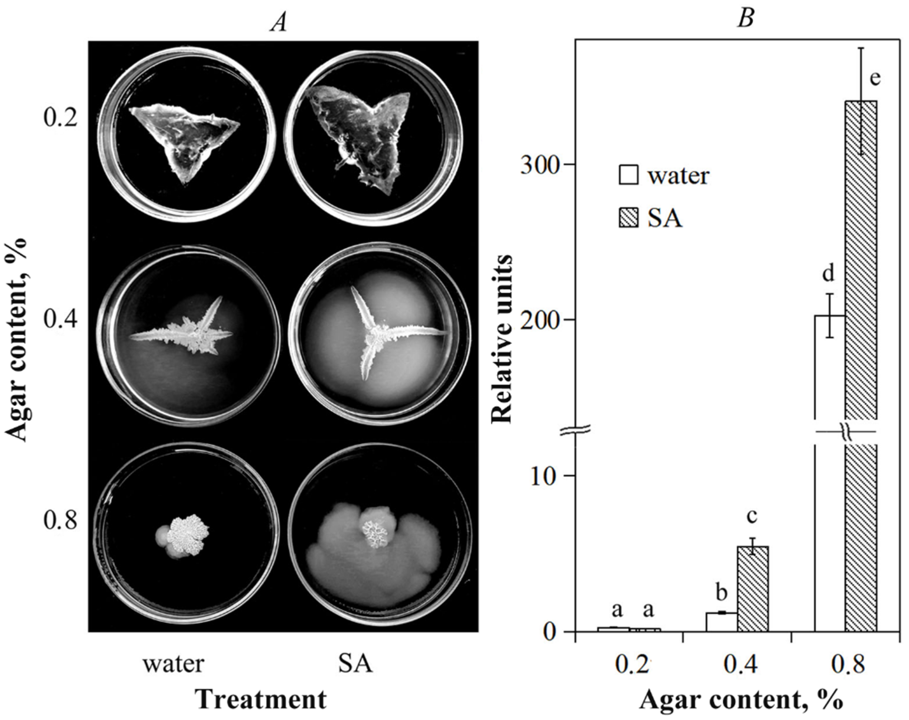
Figure 1. The influence of SA on motility of B. subtilis 26D (A) and transcriptional activity of the surfactin synthetase gene BsSfp (B) on plates with 0.2%, 0.4% and 0.8% agar. The expression of target gene is presented as a fold change normalized to the reference gene encoding 16S rRNA. Values followed by the same alphabet are not significantly different from each other by Student test p < 0.05.
Ijpb 14 00020 g001
Figure 2. Influence of SA on the activity of transcription of the B. subtilis 26D BsSfp gene (surfactin synthetase), potato genes StPR6 (trypsin inhibitor), StPR1 (basic protective protein) in potato plants 24 h after post-inoculation with B. subtilis 26D suspension. The expression of each target gene is presented as a fold change normalized to the relevant reference gene. Values followed by the same alphabet are not significantly different from each other by Student test p < 0.05.
Ijpb 14 00020 g002
Figure 3. Impact of SA and B. subtilis 26D on development of late blight disease symptoms on potato leaves on the 12th day after P. infestans inoculation. (A) Percentage of leaf area with disease symptoms. (B) Late blight lesions on potato leaves. Values followed by the same alphabet are not significantly different from each other by Student test p < 0.05.
Ijpb 14 00020 g003
Figure 4. Influence of SA and B. subtilis 26D on the activity of transcription of the BsSfp gene (surfactin synthetase), potato genes StPR6 (trypsin inhibitor), StPR1 (basic protective protein) in potato plants 24 h after infection with P. infestans spores. The expression of each target gene is presented as a fold change normalized to the relevant reference gene. Values followed by the same alphabet are not significantly different from each other by Student test p < 0.05.
Ijpb 14 00020 g004
Table 1. Primers used for qPCR.
GeneNCBI Access NumberGene ProductPrimers
StPR1AY050221SA-responsive pathogenesis related protein 1 (PR1 protein) of potatoF: 5′_tgggtggtggttcatttcttgt_3′
R: 5′_catttaattccttacacatcataag_
StPR6NW_006239045.1Proteinase inhibitor of potato (PR6)F: 5′_gggaaagaatatgctcaagttat_3′
R: 5′_aattctccatcatcttccactg_3′
BsSrfEU882341.1Surfactin synthetase of B. subtilisF: 5′_atcttcccgacgctcatttc_3′
R: 5′_atctcaaggctgatcggtttc
StActX55749.1Actin, potato housekeeping gene F: 5′_gatggtgtcagccacac_3′
R: 5′_attccagcagcttccattcc_3′
Bs16SNR_112116.216S rRNA, B. subtilis housekeeping gene F: 5′_accagaaagccacggctaactac_3′
R: 5′_ggcggaaaccccctaacact_3′
Table 2. Population of B. subtilis 26D in roots and shoots of potato plants (7 days after post- inoculation).
Content of CFU × 104/g in Internal Plant TissuesTreatment
26D26D+SA
shoots44.9 ± 10.0 a75.0 ± 10.42 b
roots19.8 ± 5.15 a21.4 ± 7.6 a
Note: Data represented as mean values ± standard error, values followed by the same alphabet in lines are not significantly different from each other by Student test p < 0.05.
Disclaimer/Publisher’s Note: The statements, opinions and data contained in all publications are solely those of the individual author(s) and contributor(s) and not of MDPI and/or the editor(s). MDPI and/or the editor(s) disclaim responsibility for any injury to people or property resulting from any ideas, methods, instructions or products referred to in the content.