Abstract
Microorganisms represent a viable option for the control of phytopathogens. From the surface of healthy mangoes, different bacteria were isolated. For all isolated bacterial strains, we determined their antimicrobial activity against a fungal strain that caused anthracnose in mangoes and against Alternaria alternata, both in the culture medium and directly on mangoes. The bacterial strains with the highest antifungal activity were identified by sequencing the 16s rRNA gene. Two species of Serratia were identified: marcescens and nematodiphila. Finally, the chitinolytic, glucanolytic, and cellulolytic activity and prodigiosin production of bacteria with antifungal activity was determined. Five fungal strains were isolated from mangoes with anthracnose. Only one strain was responsible for anthracnose in mangoes. This fungal strain was identified as Colletotrichum siamense. Against C. siamense and A. alternata in vitro and in mango selected strains of Serratia showed antifungal activity. Finally, the Serratia strains produced chitinases, glucanases, cellulases and prodigiosin, and the two S. marcescens strains did not produce hemolysins. The three Serratia strains isolated in this study can potentially be used in the biological control of anthracnose caused by C. siamense and A. alternata on mango.
1. Introduction
The genus Colletotrichum comprises several important species that are among the most common fungal pathogens that damage various tropical and subtropical fruit and vegetables. Most crops around the world are susceptible to one or more Colletotrichum species. Alternaria alternata is another fungus that causes damage to fruit and vegetables around the world. Colletotrichum spp. and A. alternata are associated with economically important diseases in cereals, legumes, vegetables, and perennial crops, including fruit trees [1]. The importance of Colletotrichum, A. alternata, and the anthracnose disease they cause in horticultural products is a growing threat to global agriculture, and more than 50% of fruit and vegetable losses are caused by Colletotrichum spp. and A. alternata [2]. The late onset of fungus disease symptoms in a crop is often devastating and leads to significant losses after harvest. Tropical fruit crops in which Colletotrichum and A. alternata cause severe losses include papaya, avocado, and mango. The high susceptibility of fruit, such as mango, to Colletotrichum and A. alternata exposes the need for alternative Colletotrichum and A. alternata prevention and control methods [3].
Anthracnose is considered the most important disease affecting mango (Magnifera indica L.) crops. It is characterised by the presence of dark spots on leaves, flowers, and peduncles; on fruit, dark sunken irregular lesions appear. The disease manifests a high degree of severity under a humid climate, reaching the pulp of the fruit, which generates a bitter taste [4]. Mango anthracnose is generally attributed to species of the genus Colletotrichum and, to a lesser degree, to the genus Alternaria. In the case of Colletotrichum, most cases of anthracnose are attributed to only two species: gloeosporioides and asianum. However, there are reports that other Colletotrichum species also cause anthracnose in mangoes, including C. alienum, C. fructicola, C. tropicale, and C. siamense [5]. Recently, Tovar-Pedraza et al. [5] reported that C. siamense is a highly virulent species on mango in Mexico.
The most common method of combating these phytopathogenic fungi is the use of chemical pesticides. However, it is widely documented that pesticides are toxic to humans, animals, and plants [6]. In Mexico, the most commonly used pesticides to combat anthracnose in mangoes are azoxystrobin, trifloxystrobin (extremely toxic to aquatic plants and animals, as well as humans if inhaled), pyraclostrobin (toxic to aquatic plants and animals), cyprodinil (toxic to fish, bees, and aquatic invertebrates), fludioxonil (highly toxic to aquatic organisms, with long-lasting effects), thiabenzadol (toxic to humans), mancozeb (acute toxicity), chlorothalonil (harmful for human health, toxic to fish), and copper derivatives [7].
Therefore, it is necessary to search for non-toxic control alternatives for phytopathogenic fungi. One approach is the use of microorganisms that inhibit the growth of phytopathogenic fungi. In general, biological control is based on the action of living organisms (animals, plants, or microorganisms) that can reduce the populations of a pest. There are reports of different microorganisms that exert an inhibitory or antagonistic effect on different phytopathogenic fungi, representing a viable option for the control of phytopathogens. The use of different bacteria, such as Bacillus subtilis [8], and Brevundimonas diminuta [9] can control anthracnose in mango, caused mainly by C. gloeosporioides. However, the antagonistic effect of other microorganisms, such as strains of the genus Serratia, on Colletotrichum spp. or A. alternata has not been reported. There are some reports of the inhibitory effect of Serratia on other fungal genera [10,11]. It is possible that strains of the genus Serratia could be used for biological control of Colletotrichum spp. and A. alternata.
In addition, it should be noted that most studies on the antagonistic effects of bacteria to control anthracnose have been carried out with C. gloeosporioides. Currently, there are only two studies on the in vitro antagonistic effects of bacteria on C. siamense [12,13]. However, there are limited reports on the use of Serratia species as a biological control agent or on its antifungal activity, and no studies have investigated the antagonistic effects of species of the genus Serratia against C. siamense or A. alternata. In this context, the objective of this study was to isolate bacteria with in vitro antifungal activity against C. siamense and A. alternata, molecularly identify them, determine their chitinolytic, glucanolytic, and cellulolytic activity and prodigiosin, and determine their antagonistic effects against C. siamense and A. alternata in mangoes.
2. Materials and Methods
2.1. Isolation and Identification of Mango Phytopathogenic Fungi
2.1.1. Isolation of Fungi from Mangoes with Anthracnose
Ataulfo mangoes with evident anthracnose damage were obtained from a public market in the city of Pachuca, Hidalgo. The mangoes were transported to the laboratory under aseptic conditions and in sterile plastic bags. In the laboratory, the mangoes were disinfected by immersion in a sodium hypochlorite solution at 200 mg/mL for 10 min. Subsequently, the mangoes were washed with sterile tap water and dried in a laminar flow hood for 10 min. Using sterile dissecting forceps, the cuticle was removed from the area damaged by anthracnose; the damaged mango pulp was then minced with a sterile platinum loop, and the sample was streaked onto sterile potato dextrose agar (PDA) in Petri dishes. The plates were incubated at 28 °C for 7 days, and conidia production was determined microscopically with cotton blue staining with lactophenol [14]. The fungal strains that showed conidia formation were planted on potato dextrose agar plates and incubated at 28 °C for 7 days. The fungal cultures were stored and refrigerated until use.
2.1.2. Verification of the Pathogenicity of the Isolated Fungal Strains
Preparation of the Mangoes
Physiologically ripe mangoes were bought at a market in the city of Pachuca, Hidalgo. The Ataulfo mangoes showed no bumps or signs of anthracnose and were transported to the laboratory under aseptic conditions in sterile plastic bags. In the laboratory, the mangoes were wiped with a clean cloth to remove dirt and subsequently disinfected and dried, as described above.
Preparation of the Fungal Inoculum
Each fungal strain that formed conidia was used to inoculate a 250 mL rectangular dilution bottle with 40 mL of PDA agar in an inclined position. The bottles were incubated at 28 °C for 14 days, and subsequently, 50 mL of a sterile solution of Tween 20 (0.1%) in distilled water was added to each bottle; the mixture was stirred for 1 min to release the conidia. The obtained suspension was filtered through sterile cotton gauze, and the number of conidia in the filtered suspension was estimated using a Neubauer chamber. For each fungus, a conidia suspension in sterile distilled water of approximately 1 × 10⁵ conidia per mL was prepared.
Inoculation of Mangoes and Detection of Anthracnose Signs
Disinfected mangoes were wounded with a bodkin to a depth of 1 mm, and each wound was inoculated with 20 µL of the conidia suspension (1 × 10⁵ conidia/mL) of each fungal strain. As a negative control, wounds were inoculated only with sterile distilled water. Inoculated mangoes were stored at 28 °C for 14 days, and five mangoes were used for each suspension of conidia of each fungus and five for the control. Inoculated mangoes were checked daily for visible signs of anthracnose. Only one fungal strain, called MID, caused anthracnose in mangoes.
2.1.3. Molecular Identification of the Mid Fungal Strain
DNA was extracted from approximately 500 mg biomass of strain MID using a ZR Fungal/Bacterial DNA MiniPrep kit. The amount of DNA obtained was measured using an Epoch spectrophotometer system (Agilent Technologies Mexico, Mexico City, Mexico). From the total DNA obtained, the 18S-ITS1-5.8S-ITS2-26S gene fragment was amplified with oligonucleotides ITS5 and ITS4, according to the conditions previously described by White [15]. A 1% agarose gel was used to check the size of the expected fragment (500 nt) at 100 V for 1 h. Sequencing of the 18S-ITS1-5.8S-ITS2-26S gene fragment was carried out by the CINVESTAV National Laboratory of Experimental Services, and two sequences were obtained for each oligonucleotide. The sequences were manually edited using Chromas 2.4, (2012), forming a consensus sequence for each isolate. The consensus sequences of each of the samples were compared with the BLAST program of the National Center for Biotechnology Information (NCBI, Bethesda, MD, USA) to determine similarities among biological sequences. The consensus sequence was aligned with the BLAST search results using Clustal W built-in MEGA 6.0 [16]. A dendrogram was generated using the neighbor joining method to observe phylogenetic relationships with different holotype sequences of the defined species.
2.2. Isolation and Identification of Bacteria with Antifungal Activity
2.2.1. Bacterial Isolation
Physiologically ripe Ataulfo mangoes were bought at a market in Pachuca, Hidalgo. The mangoes showed no bumps or anthracnose lesions and were transported to the laboratory under septic conditions. In the laboratory, each mango was placed in a plastic bag, and 100 mL of sterile peptone diluent (0.1%) was added and rubbed onto the bag for 1 min. Subsequently, via surface extension, Petri dishes containing agar for standard methods (SMA) were streaked and incubated for 24–48 h at 35 °C. Plates with 20 to 100 bacterial colonies were selected for isolation. Colonies with different morphologies were selected and Gram-stained. The selected strains were streaked in tubes with slanted SMA and incubated at 35 °C for 24 h. The tubes with the cultures were stored in a refrigerator until use.
2.2.2. In Vitro Antifungal Effect of Isolated Bacteria
Alternaria alternata ATCC 42012 and C. siamense, which were isolated from mangoes with anthracnose, were used. The studies were carried out using the dual growth method in culture plates, applying PDA according to the methodology described by Saha et al. [17]. For this, each of the bacterial strains was cultured in a tube with 3 mL of trypticase soy broth (CST) at 35 °C for 48 h. Subsequently, the cultures were centrifuged at 1507× g for 30 min, and the bacterial supernatant was collected in sterile tubes. In the central parts of PDA plates, C. siamense or A. alternata were streaked; then, in these plates, 50 µL aliquots of the supernatant of the same bacterium were placed on the PDA agar at four equidistant points to form the corners of an imaginary square surrounding the C. siamense or A. alternata inoculum. Each spot where the supernatant was placed was 2 cm from the C. siamense or A. alternata streaking point. Once the supernatant was absorbed by the PDA agar, the plates were incubated at 28 °C for 14 days. Daily, the radial growth of C. siamense or A. alternata against the supernatant of each bacterial type was measured. PDA plates streaked with C. siamense or A. alternata were included as a control, but sterile distilled water was used instead of the bacterial supernatant. The studies were conducted in triplicate.
2.2.3. Identification of Bacterial Strains with Antifungal Activity
Seven bacterial strains (B1, B2, B3, B4, B5, B6, B7) were identified, four of which (B4, B5, B6, B7) were biochemically identified using the automated VITEK 2 kit (bioMérieux, Marcy-l’Étoile, France) following the manufacturer’s instructions. The other three strains (B1, B2, B3) were molecularly identified. For this, DNA was extracted from approximately 500 mg biomass of each strain using a ZR Fungal/Bacterial DNA MiniPrep kit. The amount of DNA obtained was measured using an Epoch spectrophotometer system (Agilent Technologies Mexico, Mexico City, Mexico). From the total DNA obtained, the 16s rRNA gene was amplified with the universal oligonucleotides 27F (5′AGAGTTTGATCMTGGCTCAG3′) and 1492R (5′TACGGYTACCTTGTTACGACTT3′), according to the conditions previously described by White [15]. A 1% agarose gel was used to check the size of the expected fragment (1500 nt) at 100 V for 1 h. Two sequences were obtained for each oligonucleotide and manually edited using Chromas v2.4, (Technelysium Pty. Ltd., South Brisbane, Australia, 2012), forming a consensus sequence for each isolation. The consensus sequences of each of the samples were compared with the BLAST programme of the National Center for Biotechnology Information (NCBI, Bethesda, MD, USA) to determine similarities among biological sequences. The consensus sequence was aligned with the BLAST search results with Clustal W built-in MEGA 6.0 [16]. A dendrogram was generated using the neighbor joining method to observe the phylogenetic relationships with different holotype sequences of the defined species. The biochemical identification in VITEK 2 and the sequencing of the 16s rRNA gene were both carried out by the CINVESTAV National Laboratory of Experimental Services, Mexico. Molecular study identified two strains of Serratia marcescens and one of S. nematodiphila.
2.3. Antifungal Effect of S. marcescens and S. nematodiphila Strains against the Conidia of Colletotrichum and Alternaria
The determination of the antifungal effect of the Serratia strains against C. siamense and A. alternata ATCC 42012 conidia was determined following Gutiérrez-Román et al. [18]. Briefly, equal volumes of a conidial suspension of C. siamense or A. alternata in sterile tap water (1 × 10⁶ conidia/mL) and cells of Serratia strains (two S. marcescens or S. nematodiphila) grown in Luria Bertani medium (LB) to a density of 1 × 10⁹ cfu/mL were mixed. The control contained the conidial suspension of C. siamense or A. alternata mixed with sterile LB medium. Twenty microliters of each mixed was applied to a glass slide and incubated in a humid chamber at 30 °C in the dark. Slides were observed by microscopy and photographed every 6 h for 24 h. The percentage of conidia germination was estimated by counting a total of 100 conidia per slide under a microscope at 100× magnification. Percent germination of conidia were evaluated, and the images were captured with a Zeiss AxioCam ERc 5s. The percent conidial germination (±standard error) was calculated from mean values as: CG % = (C − T)/C × 100; Where, CG = conidial germination, C = percentage of germinated conidia in control, and T = percentage of germinated conidia in treated samples. The studies were conducted in triplicate.
2.4. Chitinolytic Activity Assay
2.4.1. Preparation of Colloidal Chitin
Colloidal chitin was prepared as reported by Zarei et al. [11]. Briefly, 24 g chitin powder obtained from shrimp shell (C7170, Sigma-Aldrich Co., St. Louis, MO, USA) was added to 790 mL concentrated HCl and left at 4 °C overnight with vigorous stirring. The mixture was added to 8 L of ice-cold ethanol 95% (v/v) with rapid stirring and maintained at 4 °C overnight. The precipitate was collected by centrifugation at 5000× g for 20 min at 4 °C and was then washed with sterile distilled water until the colloidal chitin had a pH of 7.0.
2.4.2. Chitinolytic Activity
The assay was carried out using the agar diffusion technique. Tubes containing 3 mL of trypticase soy broth (TSB; Bioxon, Becton Dickinson, Ciudad de México, Mexico) were inoculated with individual Serratia strains (two S. marcescens and S. nematodiphila) and incubated at 35 °C for 24 h. The cultures were washed twice in sterile isotonic saline solution (ISS; 0.85% of NaCl) by centrifuging at 3500× g for 20 min and resuspending the pellets in ISS at about 109 CFU/mL. However, according to Zarei et al. [11], a minimum culture (MC) medium containing colloidal chitin was prepared with the following ingredients and proportions: 0.5% colloidal chitin, 0.03% peptone, 0.03% yeast extract, 0.07% K2HPO4, 0.03% KH2PO4, 0.05% MgSO4.7H2O, 1.5% agar, 0.2% NH4NO3, 0.1% NaCl (w/v), and 0.1% (v/v) trace elements (pH 7). The culture medium was sterilised by autoclaving and poured into Petri dishes. Separately, 20-µL aliquots of washed cultures of Serratia strains were placed on Petri dishes containing the culture media with colloidal chitin. Each washed culture was inoculated in triplicate. Once the washed cultures were absorbed by the agar, inoculated plates were incubated at 35 °C for 2 days. After incubation, the appearance of a translucent zone around the inoculum in the culture medium was considered chitinolytic activity [11]. The diameter (mm) of the resulting translucent zone for each Serratia strain were measured, and the average was obtained for each strain.
2.5. Glucanolytic Activity
Glucanase enzyme production of Serratia strains was determined following the procedure described by Renwick et al. [19]. The MC medium described above was now prepared containing a substrate of β-1,3-glucanase: laminarin (0.01%, Sigma-Aldrich, St. Louis, MO, USA). The Serratia strains (two S. marcescens and one S. nematodiphila) were cultured on the MC medium containing laminarin and incubated at 35 °C for 2 days. After incubation, 0.1% w/v Congo red solution (Sigma-Aldrich, St. Louis, MO, USA) was applied to cultivated plates, and the formation of clear zones around colonies was considered positive to β-1,3-glucanase activity by depolymerisation of laminarin [19].
2.6. Cellulolytic Activity
The cellulolytic enzyme production assay of Serratia strains was performed according to Ashwini and Srividya [20]. The MC medium described above was now prepared containing a substrate of β-1,4 cellulase: carboxy methyl cellulose (1%, Sigma-Aldrich, St. Louis, MO, USA). The Serratia strains (two S. marcescens and S. nematodiphila) were cultured on the MC medium containing carboxy methyl cellulose and incubated at 35 °C for 2 days. After incubation, 0.1% (w/v) Congo red solution (Sigma-Aldrich, St. Louis, MO, USA) was applied to cultivated plates, and the formation of clear zones around colonies was considered positive for β-1,4 cellulase activity [20].
2.7. Prodigiosin Production
Prodigiosin production was determined following Slater et al. [21]. The Serratia strains (two S. marcescens and one S. nematodiphila) were inoculated in tubes containing Luria Bertani medium (LB) and incubated at 20 °C for 18 h to reach a density of approximately 1 × 10⁹ cfu/mL. Separately, 1 mL of each culture was placed in a clean tube and centrifuged at 10,000× g for 10 min. The supernatant was discarded, and the pellet was resuspended in acidified ethanol (4% 1 M HCl in ethanol) to extract prodigiosin from the cells. Cell debris was removed by centrifugation at 10,000× g for 10 min, and the supernatant was transferred to a cuvette for measurement of absorbance at 534 nm. The relative prodigiosin concentration was expressed per cell (A534 mL−1 OD600 unit).
2.8. Antagonistic Effects of S. marcescens and S. nematodiphila against C. siamense and A. alternata on Mango
Physiologically ripe “Ataulfo” mangoes were used; they were cleaned and sanitised, as described above. A 1 × 10⁴ conidia/mL-suspension of C. siamense or A. alternata was prepared as described above. Simultaneously, supernatants collected from 48 h cultures at 37 °C in CST of the two strains of S. marcescens (B1 and B3) and of the strain of S. nematodiphila (B2) were obtained as described above. Two inoculation methods were tested: (a) inoculation in the mango wound and (b) inoculation of the mango on the integral or undamaged surface of the mangoes. For the first case, (a) the mangoes were wounded with a bodkin to a depth of 1 mm, and the wounds were inoculated with 20 µL of the supernatant of S. marcescens B1, S. marcescens B3, or S nematodiphila B2. The mangoes were kept in a laminar flow hood for 30 min so that the supernatant could be absorbed by the pulp of the mangoes or dried. Subsequently, 20 µL of the conidia suspension (final concentration of 102 conidia per mango) was placed onto the same spot and left for another 30 min in a laminar flow hood at room temperature. For A. alternata, only this procedure was tested. For the second case, (b) the entire process mentioned above in section a was carried out, but instead of causing a wound, the three supernatants of Serratia strains and C. siamense were inoculated on the intact surface. In all cases, as a negative control, mangoes were inoculated only with sterile distilled water, and other mangoes inoculated only with the supernatant of each of the 3 Serratia strains were used.
Finally, for both cases, the inoculated mangoes were stored for 14 days in a bioclimatic chamber at 30 °C and 90% relative humidity. All treatments were performed in triplicate. The mangoes were checked daily for visible signs of anthracnose. Damage to mangoes related to anthracnose was recorded according to the eight-level hedonic scale reported by Corkidi et al. [22]. This hedonic scale considers 5 levels of damage: 0–1% (1), 1–5% (2), 6–9% (3), 10–49% (4), 50–100% (5). The experiment was repeated three times.
2.9. Hemolytic Activity of Serratia Marcescens Strains
Hemolytic activity of the S. marcescens strain was assessed according to Gerhardt et al. [23]. Briefly, S. marcescens strains were inoculated separately in 3 mL of trypticase soy broth (TSB; Bioxon, Becton Dickinson, Ciudad de México, Mexico) and incubated at 35 ± 2 °C for 18 h. Then, S. marcescens strains were streaked on tryptone soya agar (TSA) plates supplemented with 5% goat blood and incubated at 30 °C for 48 h. After incubation, the plates were observed against light for transparent zones formation around the bacterial growth. A clear zone indicates β-hemolysin production, dark colored zone indicates α hemolysin production and green colored zone indicate γ hemolysin production [23].
2.10. Statistical Analyses
All experiments on the antagonistic effect, both in vitro and in mangoes, and antifungal effect on conidia, chitinolytic, glucanolytic, cellulolytic, and prodigiosin assays were repeated three times separately. Differences between the means were determined using ANOVA and Tukey’s test (p < 0.05) with Statistica v6.0 (Statsoft, Inc., Tulsa, OK, USA, 1997). In some cases, the means and standard deviations were plotted.
3. Results
3.1. Isolation and Identification of Mango Phytopathogenic Fungi
From Ataulfo mangos with anthracnose, four fungal strains that presented conidia formation were isolated. These strains were inoculated on healthy Ataulfo mangoes to determine their pathogenic potential. Only one fungal strain, called MID, caused typical signs of anthracnose in inoculated mangoes. Molecular identification of the MID fungal strain by amplifying the consensus sequence of the 18S-ITS1-5.8S-ITS2-26S fragment resulted in a total of 508 nucleotides (Sequence A1 in Appendix A).
According to the information contained in the BLAST of the National Center for Biotechnology Information (NCBI), the sequence obtained from the MID strain corresponded to C. siamense. The most similar sequence corresponded to C. siamense strain HLX5, with an accession number of MN860116.1, with 100% sequence coverage and 99% sequence identity.
3.2. Antifungal Activity of Isolated Bacteria
We isolated 30 bacterial strains from mangoes; all isolates were Gram-negative rods. Using the dual growth technique in culture plates, it was determined that only seven bacterial strains (B1 to B7) had an antifungal effect against C. siamense (Figure 1). As seen in Figure 1, of these bacterial strains, three (B1, B2, and B3) showed a marked antifungal effect against C. siamense. These three bacterial strains also showed antifungal effects against A. alternata in the culture medium (Figure 2).
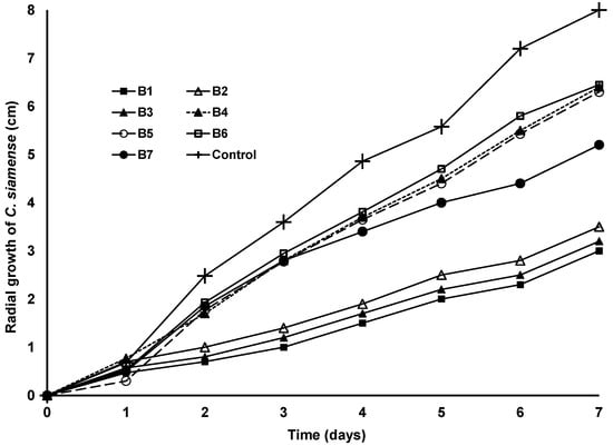
Figure 1.
Antifungal effect of seven bacterial strains (B1 to B7) against C. siamense.
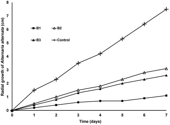
Figure 2.
Antifungal effect of 3 bacterial strains against A. alternata in culture medium.
Identification of Bacterial Strains with Antifungal Activity
Of the seven bacterial strains (B1 to B7) that showed antifungal activity in in vitro tests, four (B4, B5, B6, and B7) were identified by biochemical tests, and four were of the species Serratia marcescens. These were identified only with biochemical tests because they had a moderate antifungal effect in in vitro studies.
The other three strains (B1, B2, and B3) with the greatest antifungal effect in in vitro studies were molecularly identified by the 16s ribosomal RNA gene. The sequence most similar to the B1 strain, according to the BLAST search of the National Center for Biotechnology Information (NCBI), was Serratia marcescens, with 100% sequence coverage and 99% identity, sequence of the 16S fragment of strain B1 with a total of 1453 nucleotides (Sequence A2).
In the case of strain B2, the most similar sequence according to the BLAST search corresponded to Serratia nematodiphila, with 100% sequence coverage and 99% identity (Sequence A3).
Finally, in the case of strain B3, the most similar sequence according to the BLAST search corresponded to S. marcescens (Sequence A4).
3.3. Antifungal Effect of S. marcescens and S. Nematodiphila Strains against the Conidia of Colletotrichum and Alternaria
In general, marked inhibition was observed in the germination of C. siamense and A. alternata conidia at around 90 and 82% with the three Serratia strains, for 6 and 24 h, respectively (Table 1).

Table 1.
Antifungal effect of Serratia marcescens B1 and B3 and S. nematodiphila B2 strains against conidia germination of C. siamense and A. alternata.
3.4. Chitinolytic, Glucanolytic and Cellulolytic Activity
The three strains of Serratia showed chitinolytic activity on the culture medium with translucent zones for S. marcescens B1 and B3 and S. nematodiphila B2 (Table 2). Laminarin is a low molecular weight β-glucan storage polysaccharide present in brown algae. Laminarin is frequently used to determine the production of glucanases by microorganisms. In our study, all three Serratia strains caused depolymerisation of laminarin, indicating glucanase production (Table 2). All three Serratia strains showed evidence of glucanase production (Table 2). The three strains hydrolysed carboxy methyl cellulose in the culture medium (Table 2). The three Serratia strains produced prodigiosin at an average concentration of 1.8 to 2.5 A534 mL−1 OD600 unit (Table 2).

Table 2.
Production of compounds with antifungal activity by S. marcescens and S. nematodiphila strains.
3.5. Antagonistic Effects of S. marcescens and S. nematodiphila on Mango
C. siamense was able to grow and cause anthracnose in mangoes infected with 102 conidia, both in fruit with lesions and in lesions inoculated with conidia of C siamense, as well as in those fruit with undamaged surfaces and inoculated with C siamense (Figure 3 and Figure 4).
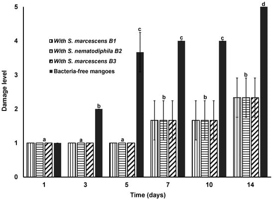
Figure 3.
Mangoes with lesions inoculated with C. siamense and treated with strains of Serratia. The damage level means, 1: between 0 to 1% of damage; 2: between 1 to 5% of damage; 3: between 6 to 9% of damage; 4: between 10 to 49% of damage and 5: between 50 to 100% of damage. Different letters in the columns on the different days of treatment mean a statistically significant difference (p < 0.05).
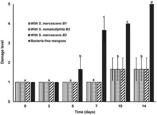
Figure 4.
Mangoes with undamaged surfaces inoculated with C. siamense and treated with strains of Serratia. The damage level means, 1: between 0 to 1% of damage; 2: between 1 to 5% of damage; 3: between 6 to 9% of damage; 4: between 10 to 49% of damage and 5: between 50 to 100% of damage. Different letters in the columns on the different days of treatment mean a statistically significant difference (p < 0.05).
In addition, 102 conidia of A. alternata also caused anthracnose in mangoes (Figure 5). However, when the mangoes were treated simultaneously with C. siamense or A. alternata and with each of the three strains of Serratia, the appearance of anthracnose signs in the mangoes was delayed, and the progression of C. siamense or A. alternata infection was slower (Figure 3, Figure 4 and Figure 5). It should be noted that the mangoes used as negative controls did not show spots or anthracnose during the 14 days of the study.
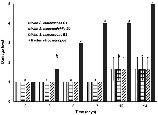
Figure 5.
Mangoes inoculated with A. alternata and treated with strains of Serratia. The damage level means, 1: between 0 to 1% of damage; 2: between 1 to 5% of damage; 3: between 6 to 9% of damage; 4: between 10 to 49% of damage and 5: between 50 to 100% of damage. Different letters in the columns on the different days of treatment mean a statistically significant difference (p < 0.05).
3.6. Hemolytic Activity
The test for hemolytic activity on blood-supplemented TSA plates revealed that the isolate S. marcescens (B1 and B2 strains) lacked hemolytic activity since no transparent zone formation occurred around the bacterial colonies.
4. Discussion
Although C. siamense is one of the species that causes anthracnose in mangoes in different parts of the world [24,25], in Mexico, this species has been little studied as a causal agent of anthracnose. There is only one recently published article in which C. siamense was isolated from mangoes with anthracnose grown in Mexico [5]. Our results confirm that C. siamense is a species that causes anthracnose in mangoes grown in Mexico. Furthermore, the results suggest that C. siamense is more widespread in Mexico than previously thought and causes disease in mango fruit. In this case, further studies are necessary in Mexico to determine the real distribution of C. siamense and its impact on mango crops and other fruit. In addition, there is limited information available in the scientific literature on the antagonistic effects of bacteria on C. siamense and A. alternata. Recently, Xu et al. [13] described the in vitro antagonistic effect of a Bacillus subtilis strain on C. siamense. In another study, Fantinel et al. [12] reported that Bacillus thuringiensis var. Kurstak had a moderate antimicrobial effect on C. siamense in in vitro studies. The antagonistic effects of endophytic fungi on C. siamense have also been reported, for example, for fungi of the genera Chaetomium, Fusarium, Phomopsis, Cladosporium, Periconia, Curvularia, Leptosphaeria, and Penicillium [26].
In our study, two strains of S. marcescens and one of S. nematodiphila were identified, with remarkable antifungal effects in vitro against C. siamense. It has been reported that the genus Serratia can produce different chemical compounds with bactericidal effects [27]. However, there are no reports available in the scientific literature on the antifungal effect of Serratia on the genus Colletotrichum. Therefore, our study describes the antifungal effect of Serratia species on C. siamense for the first time (Table 1). Our results agree with those reported by Gutiérrez-Román et al. [18], who reported a high inhibition in the germination of C. gloeosporioides conidia by different strains of S. marcescens. In our study, the three Serratia strains had a prominent antifungal effect against C. siamense and A. alternata. The observed antifungal effect may be due to different agents, such as enzymes (chitinase, for example) or chemical compounds, e.g., prodigiosin. Due to this, it was determined if the Serratia strains produced any enzymes or chemical compounds that could be responsible for the observed inhibitory effect.
The three Serratia strains that we isolated in this study showed chitinolytic, glucanolytic, and cellulolytic activity (Table 2). Previous studies have reported the chitinolytic activity of strains of S. marcescens and strains of S. nematodiphila [28,29]. In different studies, it has been reported that one of the mechanisms by which some bacteria inhibit the growth or activity of fungi is by the production of chitinases [30]. Currently, to control phytopathogenic fungi, chitinolytic bacteria are used [30]. Chitinolytic bacteria of the genus Serratia are well known for their chitinase production [11]. Their ability to produce these hydrolytic enzymes could be considered an important factor for contributing significantly as an effective biocontrol agent [11]. Consequently, having bacterial strains that produce chitinases represents an alternative for the prevention and control of phytopathogenic fungi. However, as mentioned above, there are limited reports on the use of Serratia species as a biological control agent or on its antifungal activity, and no studies have investigated the antagonistic effects of species of the genus Serratia against C. siamense.
One of the main components of the wall of phytopathogenic fungi is the glucan; between 50 and 60% of the fungal wall is glucan [31]. Having bacteria that produce enzymes that lyse the glucan in the wall of phytopathogenic fungi becomes a viable biocontrol resource. Different authors have reported the isolation of glucanase-producing Serratia strains [32]. However, the isolation of non-glucanase-producing strains has also been reported [32]. This shows that Serratia strains may have different inhibition strategies or mechanisms against phytopathogenic fungi. There are many reports on the cellulolytic capacity of S. marcescens [33]; however, there is limited information on the cellulolytic activity of S. nematodiphila. In this case, our study contributes to the increase in information on the cellulolytic activity of S. nematodiphila strains.
Prodigiosin is a chemical compound with antibacterial and antifungal effects [34]. It is a secondary metabolite produced by some bacteria of the genera Serratia, Pseudomonas, Vibrio, Alteromonas, Rugamonas, Streptoverticillium, and Streptomyces [34]. However, Serratia marcescens is the species that is most frequently related to the production of prodigiosin and is the most studied bacterium on this topic [18]. Although it has been reported that other species of the genus Serratia can produce prodigiosin, such as S. plymuthica [33] and S. nematodiphila [35], reports on strains of Serratia other than S. marcescens that can produce prodigiosin are limited. However, there are also strains of S. marcescens that do not produce prodigiosin [36]. As mentioned, in our studies, both S. marcescens and S. nematodiphila produced prodigiosin (Table 2). As observed, S. marcescens B1, S. marcescens B3, and S nematodiphila B2, isolated from healthy mangoes in this study, were capable of producing chitinases, glucanases, cellulases, and prodigiosin. This strongly suggests that these three Serratia strains have at least four different strategies or mechanisms to inhibit phytopathogenic fungi, such as C. siamense and A. alternata.
As it could be observed, the three Serratia strains that were tested presented an antifungal effect against C. siamense and A. alternata (Figure 3, Figure 4 and Figure 5). This clearly shows that the three Serratia strains examined showed a remarkable effect against C. siamense and A. alternata in mangoes, regardless of the type of inoculation or contamination of the mangoes (Figure 3 and Figure 4).
Currently, there are no reports on the minimum inoculum level required of C. siamense and A. alternata conidia to cause infection in mangoes. However, in the present study, an inoculum concentration of approximately 102 conidia was sufficient to cause infection in mango. In addition, the first signs of anthracnose became evident on the third day after inoculation (Figure 3 and Figure 5), suggesting that the strains of C. siamense used in the study (and that were isolated from mangoes with anthracnose) and A. alternata have high virulence. In different studies, other species, such as C. gloeosporioides, have been reported to cause anthracnose signs in contaminated mangoes six days after inoculation of mangoes with conidia [9]. In our study, signs of anthracnose became evident on the third day after inoculation, which suggests greater virulence of C siamense than C. gloeosporioides. In fact, it has recently been reported that strains of C. siamense isolated from mango in Mexico were more virulent than other strains of different species of the genus Colletotrichum that were also isolated from mangoes in Mexico, including C. gloeosporioides [5].
As previously mentioned, there is currently no information available on the antifungal effect of the genus Serratia on the genus Colletotrichum or A. alternata. However, there are some reports of the inhibitory effect of Serratia on other fungal genera. For example, Guo et al. [10] reported the antifungal activity of S. marcescens against Fusarium proliferatum; the authors observed a strong inhibition of conidia germination and mycelial growth. Furthermore, they observed that S. marcescens suppressed fumonisin synthesis. Guo et al. [10] also reported that S. marcescens strain SerEW01 could synthesize chitinases that degrade the cell wall of the fungal mycelium. In another study, Zarei et al. [37] reported that S. marcescens inhibited the growth of Rhizoctonia solani, Bipolaris sp., Alternaria raphanin, and Alternaria brassicicola through the expression of a chitinase.
In our study, the inhibitory effect of the three strains of Serratia can be attributed to chitinolytic, glucanolytic, and cellulolytic activity and prodigiosin production. However, it would be appropriate to explore other possible mechanisms of inhibition of these three Serratia strains on C. siamense and A. alternata, as well as to evaluate the incorporation of these strains of Serratia from pre-harvest mangoes.
It is important to note that S. marcescens has been related to cases of disease in some people, which is why it is considered an opportunistic pathogen. Therefore, it is essential to appraise the risk of every potential biocontrol bacterium to avoid using potential human pathogens as agricultural inputs. Pathogenic strains of S. marcescens are reported to produce hemolysins [38]. In our study, the S. marcescens strains did not present hemolytic activity, which strongly suggests that they are not pathogenic bacteria. However, additional testing is needed to determine that S. marcescens strains do not have virulence genes and they are safe for use.
5. Conclusions
To the best of our knowledge, this is the first report showing that an inoculum concentration of 102 conidia of C. siamense and A. alternata can initiate infection of mango fruit, causing anthracnose. It is also the first report showing that Serratia strains have an antifungal effect on C. siamense and A. alternata, both in the growing medium and in mango. In addition, it is the first report of the isolation of a strain of S. nematodiphila that simultaneously produces chitinases, glucanases, cellulases, and prodigiosin. In addition, the S. marcescens strains did not show hemolytic activity which is a general feature of pathogenic S. marcescens strains. However, additional testing is needed to determine that S. marcescens strains are safe for use. Therefore, after considering the safety and biocontrol potential of Serratia strains, it may be suggested that the three Serratia strains isolated in this study (S. marcescens B1, S. nematodiphila B2, and S. marcescens B3) can potentially be used in the biological control of anthracnose caused by C. siamense and A. alternata on mango.
Author Contributions
Conceptualization, J.A.T.-L. and J.C.-R.; methodology, J.A.T.-L. and E.R.-V.; investigation, J.A.T.-L., C.A.G.-A., E.R.-V., J.R.V.-I., O.A.A.-S. and R.N.F.-C.; resources, writing—original draft preparation, funding acquisition and supervision, J.C.-R.; writing review and editing, J.A.T.-L., E.R.-V., C.A.G.-A.; formal analysis, J.A.T.-L., J.R.V.-I., O.A.A.-S., R.N.F.-C. All authors have read and agreed to the published version of the manuscript.
Funding
This research received no external funding.
Institutional Review Board Statement
Not applicable.
Informed Consent Statement
Not applicable.
Data Availability Statement
Not applicable.
Acknowledgments
The authors thank the Autonomous University of the Hidalgo State and the National Council of Science and Technology (CONACYT) of Mexico for the scholarship for doctorate studies awarded to J. Alexander Trejo-López.
Conflicts of Interest
The authors declare no conflict of interest.
Appendix A
- Sequence A1. Nucleotide sequence of the identified Colletotrichum siamense. According to the information contained in the BLAST of the National Center for Biotechnology Information (NCBI), the sequence obtained from the MID strain corresponded to Colletotrichum siamense. The most similar sequence corresponded to C. siamense strain HLX5, with an accession number of MN860116.1, with 100% sequence coverage and 99% sequence identity.
- ATTACTGAGTTTACGCTCTACAACCCTTTGTGAACATACCTATAACTGTTGCTTCGGCGGGTAGGGTCTCCGCGACCCTCCCGGCCTCCCGCCTCCGGGCGGGTCGGCGCCCGCCGGAGGATAACCAAACTCTGATTTAACGACGTTTCTTCTGAGTGGTACAAGCAAATAATCAAAACTTTTAACAACGGATCTCTTGGTTCTGGCATCGATGAAGAACGCAGCGAAATGCGATAAGTAATGTGAATTGCAGAATTCAGTGAATCATCGAATCTTTGAACGCACATTGCGCCCGCCAGCATTCTGGCGGGCATGCCTGTTCGAGCGTCATTTCAACCCTCAAGCTCTGCTTGGTGTTGGGGCCCTACAGCTGATGTAGGCCCTCAAAGGTAGTGGCGGACCCTCTCGGAGCCTCCTTTGCGTAGTAACTTTACGTCTCGCACTGGGATCCGGAGGGACTCTTGCCGTAAAACCCCCCAATTTTCCAAAGGTTGACCTCGGATCAGGT.
- Sequence A2. Nucleotide sequence of the identified Serratia marcescens. The sequence most similar to the B1 strain, according to the BLAST search of the National Center for Biotechnology Information (NCBI), was with an accession number of CP053572.1 with 100% sequence coverage and 99% identity (sequence of the 16S fragment of strain B1 with a total of 1453 nucleotides).
- TGGCGGCAGGCCTAACACATGCAAGTCGAGCGGTAGCACAGGGGAGCTTGCTCCCTGGGTGACGAGCGGCGGACGGGTGAGTAATGTCTGGGAAACTGCCTGATGGAGGGGGATAACTACTGGAAACGGTAGCTAATACCGCATAACGTCGCAAGACCAGAGGGGGACCTTCGGGCCTCTTGCCATCAGATGTGCCCAGATGGGATTAGTGTGGCTCACCTAGGCGACGATCCCTAGCTGGTCTGAGAGGATGACCAGCCACACTGGAACTGAGACACGGTCCAGACTCCTACGGGAGGCAGCAGTGGGGAATATTGCACAATGGGCGCAAGCCTGATGCAGCCATGCCGCGTGTGTGAAGAAGGCCTTCGGGTTGTAAAGCACTTTCAGCGAGGAGGAAGGTGGTGAACTTAATACGTTCATCAATTGACGTTACTCGCAGAAGAAGCACCGGCTAACTCCGTGCCAGCAGCCGCGGTAATACGGAGGGTGCAAGCGTTAATCGGAATTACTGGGCGTAAAGCGCACGCAGGCGGTTTGTTAAGTCAGATGTGAAATCCCCGGGCTCAACCTGGGAACTGCATTTGAAACTGGCAAGCTAGAGTCTCGTAGAGGGGGGTAGAATTCCAGGTGTAGCGGTGAAATGCGTAGAGATCTGGAGGAATACCGGTGGCGAAGGCGGCCCCCTGGACGAAGACTGACGCTCAGGTGCGAAAGCGTGGGGAGCAAACAGGATTAGATACCCTGGTAGTCCACGCTGTAAACGATGTCGATTTGGAGGTTGTGCCCTTGAGGCGTGGCTTCCGGAGCTAACGCGTTAAATCGACCGCCTGG.GGAGTACGGCCGCAAGGTTAAAACTCAAATGAATTGACGGGGGCCCGCACAAGCGGTGGAGCATGTGGTTTAATTCGATGCAACGCGAAGAACCTTACCTACTCTTGACATCCAGAGAACTTAGCAGAGATGCTTTGGTGCCTTCGGGAACTCTGAGACAGGTGCTGCATGGCTGTCGTCAGCTCGTGTTGTGAAATGTTGGGTTAAGTCCCGCAACGAGCGCAACCCTTATCCTTTGTTGCCAGCGGTTCGGCCGGGAACTCAAAGGAGACTGCCAGTGATAAACTGGAGGAAGGTGGGGATGACGTCAAGTCATCATGGCCCTTACGAGTAGGGCTACACACGTGCTACAATGGCATATACAAAGAGAAGCGACCTCGCGAGAGCAAGCGGACCTCATAAAGTATGTCGTAGTCCGGATTGGAGTCTGCAACTCGACTCCATGAAGTCGGAATCGCTAGTAATCGTAGATCAGAATGCTACGGTGAATACGTTCCCGGGCCTTGTACACACCGCCCGTCACACCATGGGAGTGGGTTGCAAAAGAAGTAGGTAGCTTAACCTTCGGGAGGGCGCTTACCACTTTGTGATTCATGACTGGGG.
- Sequence A3. Nucleotide sequence of the identified Serratia nematodiphila. In the case of strain B2, the most similar sequence according to the BLAST search corresponded to Serratia nematodiphila, with an accession number of KP318499.1 with 100% sequence coverage and 99% identity.
- AGMTTGYTSCCCGGGTGACGAGCGSCGGACGGGTGAGYAATGTCTAGGRAAACTGCCTGAAGCTTGCTCCCCGGGTGACGAGCGGCGGACGGGTGAGTAATGTCTGGGAAACTGCCTGATGGAGRGGGATAACTACTGGAAACGGTAGCTAATACCGCATAACGTCGCAAGACCAAAGATGGAGGGGGATAACTACTGGAAACGGTAGCTAATACCGCATAACGTCGCAAGACCAAAGAGGGGGACCTTCGGGCCTCTTGCCATCAGATGTGCCCAGATGGGATTAGCTAGTAGGTGGGGGGGGACCTTCGGGCCTCTTGCCATCAGATGTGCCCAGATGGGATTAGCTAGTAGGTGGGGTAATGGCTCACCTAGGCGACGATCCCTAGCTGGTCTGAGAGGATGACCAGCCACACTGGGTAATGGCTCACCTAGGCGACGATCCCTAGCTGGTCTGAGAGGATGACCAGCCACACTGGAACTGAGACACGGTCCAGACTCCTACGGGAGGCAGCAGTGGGGAATATTGCACAATGGGCAACTGAGACACGGTCCAGACTCCTACGGGAGGCAGCAGTGGGGAATATTGCACAATGGGCGCAAGCCTGATGCAGCCATGCCGCGTGTGTGAAGAAGGCCTTCGGGTTGTAAAGCACTTTGCAAGCCTGATGCAGCCATGCCGCGTGTGTGAAGAAGGCCTTCGGGTTGTAAAGCACTTTCAGCGAGGAGGAAGGTGGTGAGCTTAATACGYTCATCAATTGACGTTACTCGCAGAAGAACAGCGAGGAGGAAGGTGGTGAGCTTAATACGTTCATCAATTGACGTTACTCGCAGAAGAAGCACCGGCTAACTCCGTGCCAGCAGCCGCGGTAATACGGAGGGTGCAAGCGTTAATCGGAGCACCGGCTAACTCCGTGCCAGCAGCCGCGGTAATACGGAGGGTGCAAGCGTTAATCGGAATTACTGGGCGTAAAGCGCACGCAGGCGGTTTGTTAAGTCAGATGTGAAATCCCCGGGCTATTACTGGGCGTAAAGCGCACGCAGGCGGTTTGTTAAGTCAGATGTGAAATCCCCGGGCTCAACCTGGGAACTGCATTTGAAACTGGCAAGCTAGAGTCTCGTAGAGGGGGGTAGAATTCCAACCTGGGAACTGCATTTGAAACTGGCAAGCTAGAGTCTCGTAGAGGGGGGTAGAATTCCAGGTGTAGCGGTGAAATGCGTAGAGATCTGGAGGAATACCGGTGGCGAAGGCGGCCCCCCAGGTGTAGCGGTGAAATGCGTAGAGATCTGGAGGAATACCGGTGGCGAAGGCGGCCCCCTGGACGAAGACTGACGCTCAGGTGCGAAAGCGTGGGGAGCAAACAGGATTAGATACCCTGTGGACGAAGACTGACGCTCAGGTGCGAAAGCGTGGGGAGCAAACAGGATTAGATACCCTGGTAGTCCACGCTGTAAACGATGTCGATTTGGAGGTTGTGCCCTTGAGGCGTGGCTTCCGGGTAGTCCACGCTGTAAACGATGTCGATTTGGAGGTTGTGCCCTTGAGGCGTGGCTTCCGGAGCTAACGCGTTAAATCGACCGCCTGGGGAGTACGGCCGCAAGGTTAAAACTCAAATGAAAGCTAACGCGTTAAATCGACCGCCTGGGGAGTACGGCCGCAAGGTTAAAACTCAAATGAATTGACGGGGGCCCGCACAAGCGGTGGAGCATGTGGTTTAATTCGATGCAACGCGAAGAACTTGACGGGGGCCCGCACAAGCGGTGGAGCATGTGGTTTAATTCGATGCAACGCGAAGAACCTTACCTACTCTTGACATCCAGAGAACTTWCCAGAGATGCWTTGGTGCCTTCGGGAACTCCTTACCTACTCTTGACATCCAGAGAACTTTCCAGAGATGCATTGGTGCCTTCGGGAACTCTGAGACAGGTGCTSCATGGCTGTCGTCAGCTCGTGTTGTGAAATGTTGGGGTTAAGTCCCTGAGACAGGTGCTGCATGGCTGTCGTCAGCTCGTGTTGTGAAATGTTGGGTTAAGTCCCGCAACGAGCGCAACCATATCCTTTGCTGCCAGCGGTCCGTCGGACTCAAGGAGACGCAACGAGCGCAACCCTTATCCTTTGTTGCCAGCGGTTCGGCCGGGAACTCAAAGGAGACTGCCAGTGATAAACTGGAGGAAG.
- Sequence A4. Nucleotide sequence of the identified Serratia marcescens. In the case of strain B3, the most similar sequence according to the BLAST search corresponded to S. marcescens strain S14, with an accession number of MK346258.1 with 100% sequence coverage and 99% identity.
- TGGGTGACGTKYGGGGACGATGAGCAACGTCAGGAAAACTGCCTGATGGAGGGGGATATGGGTGACGAGCGGCGGACGGGTGAGTAATGTCTGGGAAACTGCCTGATGGAGGGGGATAACTACTGGAAACGGTAGCTAATACCGCATAACGTCGCAAGACCAAAGAGGGGGACCTTCGACTACTGGAAACGGTAGCTAATACCGCATAACGTCGCAAGACCAAAGAGGGGGACCTTCGGGCCTCTTGCCATCAGATGTGCCCAGATGGGATTAGCTAGTAGGTGGGGTAATGGCTCACGGCCTCTTGCCATCAGATGTGCCCAGATGGGATTAGCTAGTAGGTGGGGTAATGGCTCACCTAGGCGACGATCCCTAGCTGGKCTCTAGGCGACGATCCCTAGCTGGTCTGAGAGGATGACCAGCCACACTGGAACTGAGACACGTCCMGAMTYCYTACGGGARGSAGCRSKGGGRAWATTGCACAWGGGSGCAGCCTGATGTCCAGACTCCTACGGGAGGCAGCAGTGGGGAATATTGCACAATGGGCGCAAGCCTGATGCAKCMTGSCGCGKGKGTGAAGAGGCTTCSGGTGCAGCCATGCCGCGTGTGTGAAGAAGGCCTTCGGGT.
References
- Dean, R.; Van Kan, J.A.; Pretorius, Z.A.; Hammond-Kosack, K.E.; Di Pietro, A.; Spanu, P.D.; Rudd, J.J.; Dickman, M.; Kahmann, R.; Ellis, J.; et al. The top 10 fungal pathogens in molecular plant pathology. Mol. Plant Pathol. 2012, 13, 414–430. [Google Scholar] [CrossRef] [PubMed]
- Awang, Y.; Ghani, M.A.A.; Sijam, K.; Mohamad, R.B. Effect of calcium chloride on anthracnose disease and postharvest quality of red-flesh dragon fruit (Hylocereus polyrhizus). Afr. J. Microbiol. Res. 2011, 5, 5250–5259. [Google Scholar] [CrossRef]
- Siddiqui, Y.; Asgar, A. Colletotrichum gloeosporioides (Anthracnose). In Postharvest Decay: Control Strategies; Chapter 11; Bautista-Baños, S., Ed.; Academic Press: Cambridge, MA, USA; Elsevier Inc.: London, UK, 2014; pp. 337–371. [Google Scholar] [CrossRef]
- Díaz-Medina, A.R.; Arboleda-Zapata, T.; Ríos-Osorio, L.A. Biological control strategies used for the management of antracnosis caused by Colletotrichum gloeosporioides in mango fruits: A systematic review. Trop. Subtrop. Agroecosyst. 2019, 22, 595–611. [Google Scholar]
- Tovar-Pedraza, J.M.; Mora-Aguilera, J.A.; Nava-Díaz, C.; Lima, N.B.; Michereff, S.J.; Sandoval-Islas, J.S.; Leyva-Mir, S.G. Distribution and pathogenicity of Colletotrichum species associated with mango anthracnose in Mexico. Plant Dis. 2020, 104, 137–146. [Google Scholar] [CrossRef]
- Köhler, H.R.; Triebskorn, R. Wildlife ecotoxicology of pesticides: Can we track effects to the population level and beyond? Science 2013, 341, 759–765. [Google Scholar] [CrossRef] [PubMed]
- Zhan, Y.; Zhang, M. Spatial and temporal patterns of pesticide use on California almonds and associated risks to the surrounding environment. Sci. Total Environ. 2014, 472, 517–529. [Google Scholar] [CrossRef] [PubMed]
- Touré, Y.; Ongena, M.; Jacques, P.; Guiro, A.; Thonart, P. Role of lipopeptides produced by Bacillus subtilis GA1 in the reduction of grey mould disease caused by Botrytis cinerea on apple. J. Appl. Microbiol. 2004, 96, 1151–1160. [Google Scholar] [CrossRef]
- Kefialew, Y.; Ayalew, A. Postharvest biological control of anthracnose (Colletotrichum gloeosporioides) on mango (Mangifera indica). Postharvest Biol. Technol. 2008, 50, 8–11. [Google Scholar] [CrossRef]
- Guo, Z.; Zhang, X.; Wu, J.; Yu, J.; Xu, M.; Chen, D.; Zhang, Z.; Li, X.; Chi, Y.; Wan, S. In vitro inhibitory effect of the bacterium Serratia marcescens on Fusarium proliferatum growth and fumonisins production. Biol. Control 2020, 19, 202–219. [Google Scholar] [CrossRef]
- Zarei, M.; Aminzadeh, S.; Zolgharnein, H.; Safahieh, A.; Ghoroghi, A.; Motallebi, A.; Daliri, M.; Sahebghadam-Lotfi, A. Serratia marcescens B4A chitinase product optimization using Taguchi approach. Iran. J. Biotechnol. 2010, 8, 252–262. [Google Scholar]
- Fantinel, V.S.; Muniz, M.F.B.; Poletto, T.; Dutra, A.F.; Krahn, J.T.; Favaretto, R.F.; Sarzi, J.S. Biocontrole in vitro de Colletotrichum siamense utilizando Trichoderma spp. e Bacillus thuringiensis var. kurstaki. Rev. Ciência Agríc. 2018, 16, 43–50. [Google Scholar] [CrossRef]
- Xu, J.X.; Li, Z.Y.; Lv, X.; Yan, H.; Zhou, G.Y.; Cao, L.X.; He, Y.H. Isolation and characterization of Bacillus subtilis strain 1-L-29, an endophytic bacteria from Camellia oleifera with antimicrobial activity and efficient plant-root colonization. PLoS ONE 2020, 15, e0232096. [Google Scholar] [CrossRef] [PubMed]
- Živković, S.; Stevanović, M.; Đurović, S.; Ristić, D.; Stošić, S. Antifungal activity of chitosan against Alternaria alternata and Colletotrichum gloeosporioides. Pestic. Phytomed. 2018, 33, 197–204. [Google Scholar] [CrossRef]
- White, T.J.; Bruns, T.; Lee, S.; Taylor, J. Amplification and direct sequencing of fungal ribosomal RNA genes for phylogenetics. In PCR Protocols: A Guide to Methods and Applications; Innis, M.A., Gelfand, D.H., Sninsky, J.J., White, T.J., Eds.; Academic Press Inc.: New York, NY, USA, 1990; pp. 315–322. [Google Scholar] [CrossRef]
- Tamura, K.; Stecher, G.; Peterson, D.; Filipski, A.; Kumar, S. Molecular evolutionary genetics analysis version 6.0. Mol. Biol. Evol. 2013, 30, 2725–2729. [Google Scholar] [CrossRef]
- Saha, D.; Purkayastha, G.D.; Ghosh, A.; Isha, M.; Saha, A. Isolation and characterization of two new Bacillus subtilis strains from rhizosphere of eggplant as potential biocontrol agents. J. Plant Pathol. 2012, 94, 109–127. [Google Scholar] [CrossRef]
- Gutiérrez-Román, M.I.; Holguín-Meléndez, F.; Bello-Mendoza, R.; Guillén-Navarro, K.; Dunn, M.F.; Huerta-Palacios, G. Production of prodigiosin and chitinases by tropical Serratia marcescens strains with potential to control plant pathogens. World J. Microbiol. Biotechnol. 2012, 28, 145–153. [Google Scholar] [CrossRef]
- Renwick, A.; Campbell, R.; Coe, S. Assessment of in vivo screening systems for potential biocontrol agents of Gaeumannomyces graminis. Plant Pathol. 1991, 40, 524–532. [Google Scholar] [CrossRef]
- Ashwini, N.; Srividya, S. Potentiality of Bacillus subtilis as biocontrol agent for management of anthracnose disease of chilli caused by Colletotrichum gloeosporioides OGC1. 3 Biotech 2014, 4, 127–136. [Google Scholar] [CrossRef]
- Slater, H.; Crow, M.; Everson, L.; Salmond, G.P. Phosphate availability regulates biosynthesis of two antibiotics, prodigiosin and carbapenem, in Serratia via both quorum-sensing-dependent and-independent pathways. Mol. Microbiol. 2003, 47, 303–320. [Google Scholar] [CrossRef]
- Corkidi, G.; Balderas-Ruíz, K.A.; Taboada, B.; Serrano-Carreón, L.; Galindo, E. Assessing mango anthracnose using a new three-dimensional image-analysis technique to quantify lesions on fruit. Plant Pathol. 2006, 55, 250–257. [Google Scholar] [CrossRef]
- Gerhardt, P.; Murray, R.; Wood, W.; Krieg, N. Methods for General and Molecular Bacteriology; American Society of Microbiology: Washington, DC, USA, 1994; pp. 21–42. [Google Scholar]
- Lima, N.B.; Batista, M.V.D.A.; De Morais, M.A.; Barbosa, M.A.G.; Michereff, S.J.; Hyde, K.D.; Câmara, M.P.S. Five Colletotrichum species are responsible for mango anthracnose in northeastern Brazil. Fungal Divers. 2013, 61, 75–88. [Google Scholar] [CrossRef]
- Sharma, G.; Kumar, N.; Weir, B.S.; Hyde, K.D.; Shenoy, B.D. The ApMat marker can resolve Colletotrichum species: A case study with Mangifera indica. Fungal Divers. 2013, 61, 117–138. [Google Scholar] [CrossRef]
- Du, W.; Yao, Z.; Li, J.; Sun, C.; Xia, J.; Wang, B.; Ren, L. Diversity and antimicrobial activity of endophytic fungi isolated from Securinega suffruticosa in the Yellow River Delta. PLoS ONE 2020, 15, e0229589. [Google Scholar] [CrossRef]
- Lapenda, J.C.; Silva, P.A.; Vicalvi, M.C.; Sena, K.X.F.R.; Nascimiento, S.C. Antimicrobial activity of prodigiosin isolated from Serratia marcescens UFPEDA. World J. Microbiol. Biotechnol. 2015, 31, 399–406. [Google Scholar] [CrossRef] [PubMed]
- Basharat, Z.; Tanveer, F.; Yasmin, A.; Shinwari, Z.K.; He, T.; Tong, Y. Genome of Serratia nematodiphila MB307 offers unique insights into its diverse traits. Genome 2018, 61, 469–476. [Google Scholar] [CrossRef] [PubMed]
- Han, K.I.; Patnaik, B.B.; Cho, A.R.; Lim, H.K.; Lee, J.M.; Jang, Y.G.; Jeong, Y.S.; Yoo, T.K.; Lee, G.S.; Han, M.D. Characterization of chitinase-producing Serratia and Bacillus strains isolated from insects. Entomol. Res. 2014, 44, 109–120. [Google Scholar] [CrossRef]
- Veliz, E.A.; Martínez-Hidalgo, P.; Hirsch, A.M. Chitinase-producing bacteria and their role in biocontrol. AIMS Microbiol. 2017, 3, 689–705. [Google Scholar] [CrossRef] [PubMed]
- Bowman, S.M.; Free, S.J. The structure and synthesis of the fungal cell wall. Bioessays 2006, 28, 799–808. [Google Scholar] [CrossRef]
- Kalbe, C.; Marten, P.; Berg, G. Strains of the genus Serratia as beneficial rhizobacteria of oilseed rape with antifungal properties. Microbiol. Res. 1996, 151, 433–439. [Google Scholar] [CrossRef]
- Sethi, S.; Datta, A.; Gupta, B.L.; Gupta, S. Optimization of cellulase production from bacteria isolated from soil. Int. Sch. Res. Notices 2013, 2013, 985685. [Google Scholar] [CrossRef]
- Darshan, N.; Manonmani, H.K. Prodigiosin and its potential applications. J. Food Sci. Technol. 2015, 52, 5393–5407. [Google Scholar] [CrossRef] [PubMed]
- Manas, N.H.A.; Yee, C.L.; Tesfamariam, Y.M.; Zulkharnain, A.; Mahmud, H.; Mahmod, D.S.A.; Fuzi, S.F.Z.M.; Azelee, N.I.W. Effects of oil substrate supplementation on production of prodigiosin by Serratia nematodiphila for dye-sensitized solar cell. J. Biotechnol. 2020, 317, 16–26. [Google Scholar] [CrossRef] [PubMed]
- Grimont, P.A.; Grimont, F.; Starr, M.P. Serratia species isolated from plants. Curr. Microbiol. 1981, 5, 317–322. [Google Scholar] [CrossRef]
- Zarei, M.; Aminzadeh, S.; Zolgharnein, H.; Safahieh, A.; Daliri, M.; Noghabi, A.; Ghoroghi, A.; Motallebi, A. Characterization of a chitinase with antifungal activity from a native Serratia marcescens B4A. Braz. J. Microbiol. 2011, 42, 1017–1029. [Google Scholar] [CrossRef]
- Kurz, C.L.; Chauvet, S.; Andres, E.; Aurouze, M.; Vallet, I.; Michel, G.P.; Uh, M.; Celli, J.; Filloux, A.; De Bentzmann, S.; et al. Virulence factors of the human opportunistic pathogen Serratia marcescens identified by in vivo screening. EMBO J. 2003, 22, 1451–1724. [Google Scholar] [CrossRef]
Publisher’s Note: MDPI stays neutral with regard to jurisdictional claims in published maps and institutional affiliations. |
© 2022 by the authors. Licensee MDPI, Basel, Switzerland. This article is an open access article distributed under the terms and conditions of the Creative Commons Attribution (CC BY) license (https://creativecommons.org/licenses/by/4.0/).