
A Century of Post-Traumatic Appendicitis: A Comprehensive Review with an Illustrative Case
 ,
,
Abstract
1. Introduction and Clinical Significance
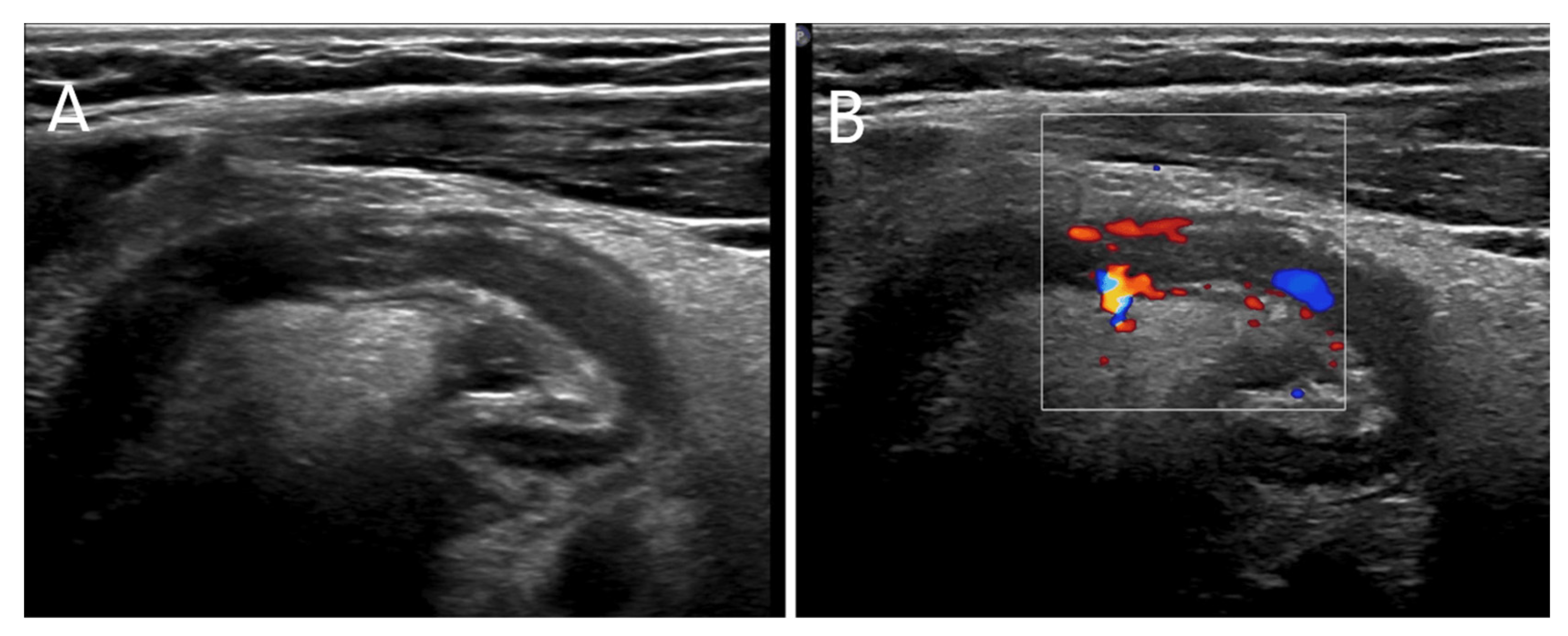
2. Case Presentation
3. Materials and Methods
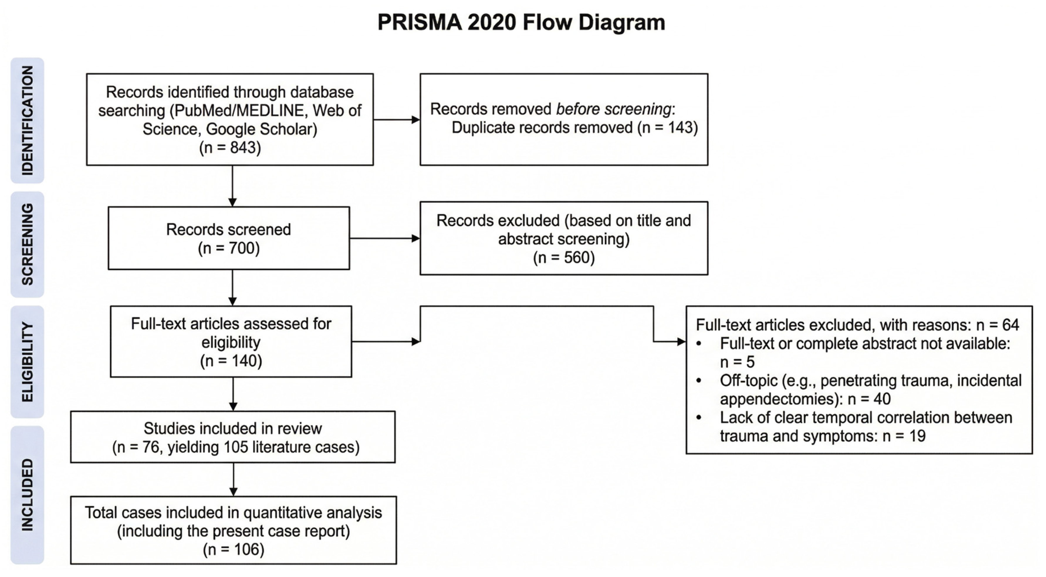
3.1. Search Strategy and Selection Criteria
3.2. Inclusion and Exclusion Criteria
3.3. Data Extraction
3.4. Statistical Analysis
4. Results of the Comprehensive Literature Review
4.1. Baseline and Trauma Characteristics
4.2. Clinical Presentation and Diagnostic Findings
4.3. Complication Patterns Across a Century of Practice
5. Discussion
Limitations to the Study
6. Conclusions
Author Contributions
Funding
Institutional Review Board Statement
Informed Consent Statement
Data Availability Statement
Conflicts of Interest
Abbreviations
| CT | Computed Tomography |
| PTA | Post-traumatic Appendicitis |
| AIR | Appendicitis Inflammatory Response |
| PAS | Pediatric Appendicitis Score |
Appendix A

| Paper | Year | Cases (n) | Age (Years) | Sex | Cause of Trauma | Diagnosis | Appendicolith | Complications |
|---|---|---|---|---|---|---|---|---|
| Pediatric Cases | ||||||||
| Ramsook C [2] | 2001 | 1 | 12 | M | kick in the abdomen | CT scan | yes | no |
| Pisoni et al. [12] | 2006 | 1 | 10 | F | blunt abdominal trauma | no | periappendicitis | |
| Salinas-Castro et al. [13] | 2022 | 1 | 14 | F | soccer ball trauma | CT scan | no | no |
| Habachi et al. [14] | 2023 | 1 | 12 | M | fall | US | no | perforation, abscess |
| Etensel et al. [16] | 2005 | 5 | 10 (5–14) | M (5) | MVC (4), fall/polytrauma (1) | surgery (5) | no (5/5) | perforation/pneumoperitoneum (1) |
| Taylor CJ [18] | 1923 | 1 | 18 | M | hit by an elbow while playing football | surgery | no | no |
| Bissell et al. [19] | 1928 | 3 | 14 (10–18) | M (3) | fall (2), kick (1) | surgery (3) | yes (3/3) | perforation (2), abscess (1), peritonitis (2) |
| Marcus et al. [20] | 1931 | 5 | 11 (9–14) | M (4), F (1) | kick (1), MVC (1), fall (3) | surgery (5) | yes (1/5) | perforation (2), peritonitis (1), gangrenous (1) |
| Shutkin et al. [21] | 1936 | 1 | 14, N/A | M (2) | kick (1), fall (1) | surgery (2) | yes (1/2) | gangrenous (2), perforation (2) |
| Rhodes et al. [22] | 1940 | 3 | 11 (11–13) | M (1), F (2) | hit by a volley ball (1), hit by a baseball (1), fall (1) | surgery (3) | no (3/3) | gangrenous (1), perforation (2), hemorrhageshemorragies (1) |
| Bhajekar et al. [23] | 1942 | 1 | 13 | M | fall | surgery | no | abscess |
| Black WR [24] | 1948 | 2 | 14, 14 | M (1), F (1) | bicycle fall (1), fall (1) | surgery (2) | yes (2/2) | abscess, gangrenous (2) |
| Hennington et al. [25] | 1991 | 1 | 12 | M | bicycle fall | surgery | no | no |
| Musemeche et al. [26] | 1995 | 1 | 4 | M | road car accident | CT scan | no | |
| Ciftci et al. [27] | 1996 | 3 | 11 (7–14) | M (2), F (1) | hit by a ball (1), MVC (1), assault (1) | surgery (1), CT scan (2) | no (3/3) | perforation (3) |
| Serour et al. [28] | 1996 | 3 | 8 (7–11) | M (3) | punch (1), fall (1), fight (1) | CT scan (1), surgery (2) | yes (1/3) | gangrenous/periappendicitis (1), perforation/abscess (1) |
| Osterhoudt KC [29] | 2000 | 1 | 9 | M | hit by a car | US | no | perforation |
| Paul G [30] | 2000 | 1 | 16 | M | seatbelt collision | surgery | no | perforation, auto-amputation |
| Houry et al. [31] | 2001 | 1 | 5 | M | fall | CT scan | no | perforation |
| Ramesh et al. [32] | 2002 | 1 | 11 | M | bicycle fall | US | no | perforation, appendicitis |
| Sumer et al. [33] | 2008 | 1 | 8 | M | fall | perforation | ||
| Amir et al. [34] | 2009 | 1 | 10 | M | trauma from a desk’s corner | CT scan | no | no |
| Toumi et al. [35] | 2010 | 1 | 11 | M | hit by an elbow | CT scan | no | peritonitis, abscess |
| Torres-Grau et al. [36] | 2012 | 1 | 15 | M | bicycle fall | surgery | no | non |
| Paschos et al. [37] | 2012 | 1 | 17 | F | bicycle fall | US | no | |
| Wani I [38] | 2013 | 4 | 13 (9–17) | M (2), F (2) | bicycle fall (1), kick in the abdomen (2), fall (1) | US/CT scan (4) | yes (2/4) | mesoappendicular hematoma (1) |
| Reyes-Hernández et al. [39] | 2013 | 1 | 14 | F | bullying | US | no | |
| Goldman et al. [40] | 2013 | 1 | 11 | M | punch in the abdomen | MRI | no | no |
| Moslemi et al. [41] | 2013 | 1 | 13 | M | bicycle fall | CT scan | no | auto-amputation, pneumoperitoneum |
| Ahmed et al. [42] | 2014 | 1 | 12 | M | trauma from a desk’s corner | US | no | perforation, shock, hemoperitoneum, peritonitis |
| Jensen et al. [43] | 2016 | 1 | 7 | M | bicycle fall | CT scan | yes | auto-amputation |
| Cobb [44] | 2017 | 1 | 17 | M | MVC | CT scan | no | peritonitis |
| Klis et al. [45] | 2017 | 1 | 12 | F | bicycle fall | US | no | perforation |
| Singh et al. [46] | 2018 | 1 | 10 | M | bicycle fall | surgery | no | no |
| Çağlar [47] | 2018 | 1 | 12 | M | fall from a swing | RX/CT scan | no | no |
| Zvizdic et al. [48] | 2020 | 1 | 7 | M | hors hoof kick | CT scan | no | perforation, peritonitis |
| Sharma et al. [49] | 2021 | 1 | 11 | M | bicycle fall | CT scan | no | perforation, pneumoperitoneum, appendiceal auto-amputation |
| Chalh et al. [50] | 2021 | 1 | 8 | M | fall on a boulder | US | yes | nno |
| Kerkeni et al. [51] | 2022 | 1 | 8 | M | bicycle fall | surgery | no | auto-amputation, perforation, pneumoperitoneum |
| Albalawi et al. [52] | 2023 | 1 | 10 | M | Fall fall from a trampoline | CT scan | no | no |
| Jaramillo et al. [53] | 2024 | 1 | 18 | F | MVC | CT scan | no | no |
| DuBois et al. [54] | 2025 | 1 | 13 | F | MVC | CT scan | yes | no |
| Namdeo S [55] | 2025 | 1 | 11 | M | hit by a football | surgery | no | no |
| Gupta et al. [56] | 2016 | 2 | 9, 11 | M (1), F (1) | bicycle fall (1), fall on a boulder (1) | US (1), surgery (1) | yes (2/2) | perforation with abscess (1) |
| Adult Population | ||||||||
| Alabdulaaly et al. [15] | 2024 | 1 | 21 | F | Road road traffic accident | CT scan | no | perforation |
| Behan RJ [57] | 1927 | 1 | 28 | M | fall on a tomato patch | surgery | no | perforation, peritonitis |
| Bissell et al. [19] | 1928 | 1 | 19 | M | hit on the abdomen during a fight | surgery | yes | perforation, peritonitis |
| Marcus et al. [20] | 1931 | 1 | 47 | M | hit by a carbon brick | surgery | no | peritonitis |
| Maes et al. [58] | 1935 | 1 | 27 | M | calf kick | surgery | no | haemorragies |
| Rhodes et al. [22] | 1940 | 4 | 27 (21–37) | M (4) | fall (2), hit by a timber (1), hit by a knee (1) | surgery (4) | yes (1/4) | perforation (4), peritonitis (2), abscess (1) |
| Gatewood et al. [59] | 1956 | 1 | 39 | F | MVC | surgery | auto-amputation | |
| Fiorini et al. [60] | 1974 | 1 | 21 | M | MVC | surgery | no | perforation |
| Geer et al. [61] | 1975 | 1 | 39 | M | MVC | surgery | no | auto-amputation, appendiceal infarct |
| Thomas DF [62] | 1978 | 1 | 36 | M | MVC | surgery | yes | auto-amputation |
| Hennington et al. [25] | 1991 | 1 | 46 | M | trauma due to car fall | surgery | no | gangrenosa |
| Bangs RG [63] | 1991 | 1 | 20 | M | MVC | surgery | no | no |
| Stephenson et al. [64] | 1995 | 1 | 32 | F | seat belt collision | surgery | no | no |
| Edwards et al. [65] | 1999 | 1 | 41 | M | MVC | CT scan | no | auto-amputation |
| Takagi et al. [66] | 2000 | 1 | 45 | F | seat belt collision | surgery | no | periappendicitis |
| Tekin et al. [67] | 2001 | 1 | 36 | M | punch and hit | surgery | no | perforation, hemorragieshemorrhages |
| Hagger et al. [68] | 2002 | 1 | 60 | M | fall from a ladder | CT scan | no | abscess |
| Karavokyros et al. [69] | 2004 | 1 | 21 | M | robbed and hit | surgery | no | no |
| Derr et al. [70] | 2009 | 1 | 41 | M | fall while cliff diving | US | no | no |
| Atalla et al. [71] | 2010 | 1 | 53 | M | fall on a car door | CT scan | no | no |
| Faulkner et al. [72] | 2010 | 1 | 28 | F | MVC | CT scan | no | appendicular-cutaneous fistula |
| O’Kelly et al. [73] | 2012 | 1 | 40 | M | blunt abdominal trauma | surgery | no | perforation |
| Wani I [38] | 2013 | 4 | 49 (34–64) | M (2), F (2) | fall (3), kick (1) | US/CT scan (4) | yes (3/4) | appendicular lump (1), perforation (1), mesoappendicular haematoma (1) |
| Altunkaş et al. [74] | 2015 | 1 | 32 | F | fall | US | yes | caecal hematomas |
| Ross et al. [75] | 2016 | 1 | 59 | F | MVC | CT scan | yes | no |
| Go et al. [76] | 2016 | 1 | 23 | M | seat belt collision | surgery | no | auto-amputation |
| Latorre et al. [77] | 2017 | 1 | 26 | M | MVC | surgery | no | perforation, peritonitis |
| Khilji et al. [78] | 2017 | 1 | 43 | M | MVC | CT scan | no | no |
| AlJaberi et al. [79] | 2018 | 1 | 24 | M | seat belt collision | CT scan | no | auto-amputation |
| Siddiqui J [80] | 2018 | 1 | 22 | M | Fall from a ladder (3 m) | CT scan | no | no |
| Toffaha et al. [81] | 2021 | 1 | 35 | M | camel kick | CT scan | no | no |
| Alharmoodi et al. [82] | 2022 | 1 | 53 | M | MVC | CT scan | no | hemoperitoneum, auto-amputation |
| Demir et al. [83] | 2024 | 1 | 65 | M | seat belt collision | CT scan | no | periappendicitis |
| Shawasha et al. [84] | 2024 | 1 | 28 | M | hit by a knee (fall) | CT scan | no | perforation, abscess |
| Karapolat et al. [85] | 2018 | 1 | 36 | M | bicycle fall | US/CT scan | no | no |
| Our Case | 2026 | 1 | 10 | M | hit by a hockey stick | US | no | no |
References
- He, R.; Lai, J.; Jiang, O.; Li, J. The incidence and temporal trend of appendicitis in children: An analysis from the Global Burden of Disease. J. Gastrointest. Surg. 2025, 29, 101935. [Google Scholar] [CrossRef] [Scilit] [PubMed]
- Ramsook, C. Traumatic appendicitis: Fact or fiction? Pediatr. Emerg. Care 2001, 17, 264–266. [Google Scholar] [CrossRef] [Scilit]
- Fowler, R.H. The rare incidence of acute appendicitis resulting from external trauma. Ann. Surg. 1938, 107, 529–539. [Google Scholar] [CrossRef] [Scilit]
- Naumann, D.N.; Barker, T. Traumatic appendicitis is probably not real: An illustrative analysis of coincidental occurrences in nature. Trauma. Surg. Acute Care Open 2023, 8, 001093. [Google Scholar] [CrossRef] [Scilit]
- Andersson, R.E.; Stark, J. Diagnostic value of the appendicitis inflammatory response (AIR) score Asystematic review meta-analysis. World J. Emerg. Surg. 2025, 20, 12. [Google Scholar] [CrossRef] [Scilit]
- Andersson, M.; Kolodziej, B.; Andersson, R.E. Validation of the Appendicitis Inflammatory Response (AIR) Score. World J. Surg. 2021, 45, 2081–2091. [Google Scholar] [CrossRef] [Scilit]
- Bai, S.; Hu, S.; Zhang, Y.; Guo, S.; Zhu, R.; Zeng, J. The Value of the Alvarado Score for the Diagnosis of Acute Appendicitis in Children: A Systematic Review and Meta-Analysis. J. Pediatr. Surg. 2023, 58, 1886–1892. [Google Scholar] [CrossRef] [Scilit]
- Ebell, M.H.; Shinholser, J. What are the most clinically useful cutoffs for the Alvarado and Pediatric Appendicitis Scores? A systematic review. Ann. Emerg. Med. 2014, 64, 365–372. [Google Scholar] [CrossRef] [Scilit]
- Samuel, M. Pediatric appendicitis score. J. Pediatr. Surg. 2002, 37, 877–881. [Google Scholar] [CrossRef] [Scilit] [PubMed]
- Salahuddin, S.M.; Ayaz, O.; Jaffer, M.; Naeem, R.; Tikmani, S.S.; Mian, A.I. Pediatric Appendicitis Score for Identifying Acute Appendicitis in Children Presenting With Acute Abdominal Pain to the Emergency Department. Indian Pediatr. 2022, 59, 774–777. [Google Scholar] [CrossRef] [Scilit] [PubMed]
- Sala, L. Post-Traumatic Appendicitis. Arch. Sci. Med. 1964, 117, 254–257. [Google Scholar]
- Pisoni, L.; Castoldi, M.; Merlini, D.A.; Via, A.; Morandi, E. Post-traumatic acute appendicitis. A case report. Ann. Ital. Chir. 2006, 77, 441–442. [Google Scholar] [PubMed]
- Salinas-Castro, K.J.; Mejía-Quiñones, V.; Zúñiga-Londoño, N.Y. Acute appendicitis after closed abdominal trauma: A case report. Radiol. Case Rep. 2022, 18, 631–634. [Google Scholar] [CrossRef] [Scilit] [PubMed]
- Habachi, G.; Aziza, B.; Ben-Ammar, S. Acute peritonitis secondary to post-traumatic appendicitis: A case report and literature review. World J. Clin. Cases 2023, 11, 6491–6497. [Google Scholar] [CrossRef] [Scilit]
- Alabdulaaly, N.I.; Alghamdi, S.; Albahli, M.; Alabdullah, H.; Alghuliga, A. A case of post-traumatic acute appendicitis: Uncommon consequence of blunt abdominal trauma. J. Surg. Case Rep. 2024, 12, 761. [Google Scholar] [CrossRef] [Scilit]
- Etensel, B.; Yazici, M.; Gürsoy, H.; Ozkisacik, S.; Erkus, M. The effect of blunt abdominal trauma on appendix vermiformis. Emerg. Med. J. 2005, 22, 874–877. [Google Scholar] [CrossRef] [Scilit]
- Sobczak, J.; Burzyńska, M.; Sikora, A.; Wysocka, A.; Karawani, J.; Sikora, J.P. Post-Traumatic Stress Response and Appendicitis in Children-Clinical Usefulness of Selected Biomarkers. Biomedicines 2023, 11, 1880. [Google Scholar] [CrossRef] [Scilit]
- Taylor, C.J. Trauma and Appendicitis. Br. Med. J. 1923, 1, 17. [Google Scholar] [CrossRef] [Scilit]
- Bissell, A.H. Trauma as a factor in acute appendicitis. Arch. Surg. 1928, 17, 672–675. [Google Scholar] [CrossRef] [Scilit]
- Marcus, M. Tatsachen und Theorie der traumatischen Appendicitis. Dtsch. Z. Chir. 1931, 232, 372–389. [Google Scholar]
- Shutkin, M.W.; Wetzler, S.H. Traumatic appendicitis. Am. J. Surg. 1936, 31, 514–520. [Google Scholar] [CrossRef] [Scilit]
- Rhodes, G.K.; Birnbaum, W.D. Traumatic Appendicitis. Calif. West. Med. 1940, 53, 162–165. [Google Scholar]
- Bhajekar, M.V. A Case of Traumatic Appendicitis. Ind. Med. Gaz. 1942, 77, 32. [Google Scholar]
- Black, W.R. The role of abdominal trauma in acute appendicitis. Br. Med. J. 1948, 2, 424. [Google Scholar] [CrossRef] [Scilit]
- Hennington, M.H.; Tinsley, E.A., Jr.; Proctor, H.J.; Baker, C.C. Acute appendicitis following blunt abdominal trauma. Incidence or coincidence? Ann. Surg. 1991, 214, 61–63. [Google Scholar] [CrossRef] [Scilit]
- Musemeche, C.A.; Baker, J.L. Acute appendicitis: A cause of recurrent abdominal pain in pediatric trauma. Pediatr. Emerg. Care 1995, 11, 30–31. [Google Scholar] [CrossRef] [Scilit]
- Ciftci, A.O.; Tanyel, F.C.; Büyükpamukçu, N.; Hiçsönmez, A. Appendicitis after blunt abdominal trauma: Cause or coincidence? Eur. J. Pediatr. Surg. 1996, 6, 350–353. [Google Scholar] [CrossRef] [Scilit]
- Serour, F.; Efrati, Y.; Klin, B.; Shikar, S.; Weinberg, M.; Vinograd, I. Acute appendicitis following abdominal trauma. Arch. Surg. 1996, 131, 785–786. [Google Scholar] [CrossRef] [Scilit]
- Osterhoudt, K.C. Hocus-pocus: A case of abdominal pain after blunt abdominal trauma. Pediatr. Ann. 2000, 29, 93–96. [Google Scholar] [CrossRef] [Scilit] [PubMed]
- Paul, G. Blunt trauma to the appendix. Aust. N. Z. J. Surg. 2000, 70, 238–239. [Google Scholar] [CrossRef] [Scilit] [PubMed]
- Houry, D.; Colwell, C.; Ott, C. Abdominal pain in a child after blunt abdominal trauma: An unusual injury. J. Emerg. Med. 2001, 21, 239–241. [Google Scholar] [CrossRef] [Scilit]
- Ramesh, G.; Ho, P.W.; Ng, K.L.; Jegan, T. Appendicitis following blunt abdominal trauma. Med. J. Malays. 2002, 57, 123–124. [Google Scholar]
- Sumer, A.; Çağlayan, K.; Altinli, E.; Urlu, M. Post-Traumatic Perforated Appendicitis: A Case Report. IKSST Derg. 2008, 1, 27–29. [Google Scholar]
- Amir, A.; Amir, L.; Waisman, Y. Acute appendicitis after a blunt perineal trauma: An illustrative case. Pediatr. Emerg. Care 2009, 25, 184–185. [Google Scholar] [CrossRef] [Scilit]
- Toumi, Z.; Chan, A.; Hadfield, M.B.; Hulton, N.R. Systematic review of blunt abdominal trauma as a cause of acute appendicitis. Ann. R. Coll. Surg. Engl. 2010, 92, 477–482. [Google Scholar] [CrossRef] [Scilit]
- Torres-Grau, J.; Monkhouse, S.J.W. Trauma induced appendicitis…a real entity. Am. Med. J. 2012, 3, 124–125. [Google Scholar]
- Paschos, K.A.; Boulas, K.; Liapis, A.; Georgiou, E.; Vrakas, X. Traumatic appendicitis in minor blunt abdominal injury. Emerg. Med. Australas. 2012, 24, 343–346. [Google Scholar] [CrossRef] [Scilit]
- Wani, I. Post-traumatic retrocaecal appendicitis. OA Case Rep. 2013, 2, 1–3. [Google Scholar] [CrossRef] [Scilit]
- Reyes-Hernández, U.; Castell-Roldán, E.P.; Hernández-Lira, I.; Reyes-Gómez, U.; Reyes-Hernández, K.L.; Reyes-Hernández, D.P.; Jiménez-Martínez, J.A.; Lira-Rodríguez, C.; Perea-Martínez, A.; Urrutia, J.C. Una escolar con apendicitis traumática por bullying. Rev. Mex. Pediatr. 2013, 80, 154–157. [Google Scholar]
- Goldman, S.; Canastra, N.; Genisca, A. Appendicitis Following Blunt Abdominal Trauma: An Illustrative Case. Rhode Isl. Med. J. 2013, 105, 37–38. [Google Scholar]
- Moslemi, S.; Forootan, H.R.; Tahamtan, M. Co-incidence of acute appendicitis and appendiceal transection after blunt abdominal trauma: A case report. Iran. J. Med. Sci. 2013, 38, 343–346. [Google Scholar]
- Ahmed, S.T.; Ranjan, R.; Saha, S.B.; Singh, B. Traumatic appendicitis misdiagnosed as a case of haemoperitoneum. BMJ Case Rep. 2014, 2014, bcr2013202082. [Google Scholar] [CrossRef] [Scilit]
- Jensen, A.; Baertschiger, R.; Hackworth, J.; Rescorla, F. Blunt abdominal trauma with handlebar injury: A rare cause of traumatic amputation of the appendix associated with acute appendicitis. J. Pediatr. Surg. Case Rep. 2017, 7, 13–15. [Google Scholar] [CrossRef] [Scilit]
- Cobb, T. Appendicitis following blunt abdominal trauma. Am. J. Emerg. Med. 2017, 35, 1386.e5–1386.e6. [Google Scholar] [CrossRef] [Scilit]
- Klis, S.; Trang, T.L.; Lutke Holzik, M.F. Posttraumatische appendicitis. Ned. Tijdschr. Geneeskd. 2017, 161, D548. [Google Scholar] [PubMed]
- Singh, A.P.; Gupta, A.K.; Pardeshi, R.; Barolia, D.K. Blunt Trauma Abdomen-Induced Jejunal Perforation with Appendicitis. Med. J. Dr. D.Y. Patil Vidyapeeth 2018, 11, 371–373. [Google Scholar] [CrossRef] [Scilit] [PubMed]
- Çağlar, A.; Er, A.; Atacan, Ö.; Gülcü, P.; Genç, S.; Çağlar, İ.; Apa, H. A Child with Acute Appendicitis Secondary to Blunt Abdominal Trauma: A Case Report and Review of the Literature. J. Pediatr. Emerg. Intensive Care Med. 2018, 5, 140–143. [Google Scholar] [CrossRef] [Scilit]
- Zvizdic, Z.; Pasic-Sefic, I.; Vranic, S. Acute perforated appendicitis after blunt abdominal trauma: A report from a 7-year-old. Am. J. Emerg. Med. 2020, 38, 408.e1–408.e2. [Google Scholar] [CrossRef] [Scilit] [PubMed]
- Sharma, K.; Tomar, S.; Sharma, S.; Bajpai, M. Floating appendix: Post-traumatic amputation of the appendix as sequela or complication?: A case report. J. Med. Case Rep. 2021, 15, 192. [Google Scholar] [CrossRef] [Scilit]
- Chalh, O.; El Haddad, S.; Choayb, S.; Allali, N.; Chat, L. Traumatic Appendicitis in Children. Glob. Pediatr. Health 2021, 8, 2333794X21992168. [Google Scholar] [CrossRef] [Scilit]
- Kerkeni, Y.; Thamri, F.; Zouaoui, A.; Houidi, S.; Jouini, R. Is pneumoperitoneum specific of post-traumatic appendicitis? Case report and literature review. Ann. Pediatr. Surg. 2022, 18, 29. [Google Scholar] [CrossRef] [Scilit]
- Albalawi, N.; Alhazmi, M.; ALqahtani, A.; Aloboudi, A.; Mesawa, A.; Alotaibi, N.; Babiker, A. Appendicitis Post Fall in the Pediatric Population: A Case Report. Cureus 2023, 15, e49603. [Google Scholar] [CrossRef] [Scilit] [PubMed]
- Jaramillo, J.D.; Mosquera, B.R.; Botello, Y.P. Retrocecal Appendicitis Post-blunt Abdominal Trauma: A Case Report. Cureus 2024, 16, e61839. [Google Scholar] [CrossRef] [Scilit]
- DuBois, S.M.; Ullrich, L.A. Acute appendicitis after motor vehicle collision. Trauma Case Rep. 2025, 58, 101207. [Google Scholar] [CrossRef] [Scilit]
- Namdeo, S. Traumatic Twist: A Case of Appendicitis Post-Trauma. BJS 2025, 112, znaf128.379. [Google Scholar] [CrossRef] [Scilit]
- Gupta, R.; Mathur, V.; Bansal, J.; Bhandari, A.; Mathur, P.; Sharma, S.B.; Gupta, A.K. Acute appendicitis following blunt abdominal trauma in children: By chance or a cause? Formos. J. Surg. 2016, 49, 17–22. [Google Scholar] [CrossRef] [Scilit]
- Behan, R.J. Traumatic appendicitis. Ann. Surg. 1927, 85, 263–268. [Google Scholar] [CrossRef] [Scilit] [PubMed]
- Maes, U.; McFetridge, E. Traumatic appendicitis. Am. J. Surg. 1935, 30, 478–482. [Google Scholar] [CrossRef] [Scilit]
- Gatewood, J.W.; Russum, W.J. Injuries to the appendix secondary to blunt trauma. Am. J. Surg. 1956, 91, 558–560. [Google Scholar] [CrossRef] [Scilit]
- Fiorini, G.T. Acute appendicitis following motor vehicle accident. Can. Fam. Physician 1974, 20, 59–60. [Google Scholar]
- Geer, D.A.; Armanini, G.; Guernsey, J.M. Trauma to the appendix. A report of two cases. Arch. Surg. 1975, 110, 446–447. [Google Scholar] [CrossRef] [Scilit]
- Thomas, D.F. Injury to the appendix after blunt abdominal trauma. Br. Med. J. 1978, 2, 474. [Google Scholar] [CrossRef] [Scilit]
- Bangs, R.G. Acute appendicitis following blunt abdominal trauma. Ann. Surg. 1992, 216, 100. [Google Scholar] [CrossRef] [Scilit]
- Stephenson, B.M.; Shandall, A.A. Seat-belt compression appendicitis. Br. J. Surg. 1995, 82, 325. [Google Scholar] [CrossRef] [Scilit]
- Edwards, R.A.; Paul, D.B.; Anderson, G.L.; McCarthy, M.C. Transection of the appendix: A seat belt injury. Arch. Surg. 1999, 134, 90–91. [Google Scholar] [CrossRef] [Scilit]
- Takagi, Y.; Yasuda, K.; Abe, T. Seat belt compression appendicitis. J. Clin. Gastroenterol. 2000, 31, 184. [Google Scholar] [CrossRef] [Scilit]
- Tekin, K.; Ozek, M.C. Acute appendicitis after blunt abdominal trauma. Ulus. Travma Derg. 2001, 7, 207–209. [Google Scholar] [PubMed]
- Hagger, R.; Constantinou, J.; Shrotria, S. Acute appendicitis after a fall from a ladder: A traumatic aetiology? Emerg. Med. J. 2002, 19, 366–367. [Google Scholar] [CrossRef] [Scilit] [PubMed]
- Karavokyros, I.; Pikoulis, E.; Karamanakos, P. A case of blunt abdominal trauma and posttraumatic acute appendicitis. Ulus. Travma Acil Cerrahi Derg. 2004, 10, 60–62. [Google Scholar] [PubMed]
- Derr, C.; Goldner, D.E. Posttraumatic appendicitis: Further extending the extended Focused Assessment with Sonography in Trauma examination. Am. J. Emerg. Med. 2009, 27, 632.e5–632.e7. [Google Scholar] [CrossRef] [Scilit]
- Atalla, M.A.; Carangan, M.; Rozen, W.M. Acute traumatic appendicitis following blunt abdominal trauma. ANZ J. Surg. 2010, 80, 572–573. [Google Scholar] [CrossRef] [Scilit]
- Faulkner, G.; Pettit, S.H. Post-traumatic appendicular-cutaneous fistula: A case report. Ann. R. Coll. Surg. Engl. 2010, 92, W6–W7. [Google Scholar] [CrossRef] [Scilit]
- O’Kelly, F.; Lim, K.T.; Hayes, B.; Shields, W.; Ravi, N.; Reynolds, J.V. The Houdini effect--an unusual case of blunt abdominal trauma resulting in perforative appendicitis. Ir. Med. J. 2012, 105, 86–87. [Google Scholar]
- Altunkaş, A.; Özçağlayan, T.İ.; Çelik, A.O.; Sümbül, A. Posttravmatik apandisit; gerçek mi söylenti mi? Abant Med. J. 2015, 4, 67–69. [Google Scholar] [CrossRef] [Scilit]
- Ross, S.W.; Bouchez, J.D.; Fischer, P.E.; Brintzenhoff, R.A.; Sing, R.F.; Christmas, A.B.; Thomas, B.W. A Violation of Occam’s Razor: Acute Appendicitis after Motor Vehicle Collision. Am. Surg. 2016, 82, e281–e283. [Google Scholar] [CrossRef] [Scilit] [PubMed]
- Go, S.J.; Sul, Y.H.; Ye, J.B.; Kim, J.S. Appendiceal transection associated with seat belt restraint. Ann. Surg. Treat. Res. 2016, 91, 93–95. [Google Scholar] [CrossRef] [Scilit]
- Latorre, S.I.; Bustos, J.A.; Villate, J.P. Apendicitis aguda posterior a un trauma abdominal cerrado. ¿Casualidad o causalidad? Rev. Fac. Med. 2017, 65, 161–163. [Google Scholar] [CrossRef] [Scilit]
- Khilji, M.F.; Zia Ullah, Q. Seat Belt Compression Appendicitis following Motor Vehicle Collision. Case Rep. Emerg. Med. 2017, 2017, 8245046. [Google Scholar] [CrossRef] [Scilit]
- AlJaberi, L.M.; Salameh, A.E.K.; Almarzooqi, R.M.; Emar, M.F.; Salhab, R. Transection of the appendix and omentum following a seat belt injury; case report and literature review. Ann. Med. Surg. 2018, 34, 11–13. [Google Scholar] [CrossRef] [Scilit]
- Siddiqui, J.; Oliphant, R.; Yeh, Z.Y.; Suen, M. Appendicitis: A Traumatic Etiology. J. Case Rep. 2018, 8, 176–178. [Google Scholar] [CrossRef] [Scilit]
- Toffaha, A.; Al-Yahri, O.; Hijawi, Z.; Al-Mudares, S.; Al-Tarakji, M.; Shahid, F.; Ali, S.M. Appendicitis Secondary to Trauma following a Camel Kick: Case Report and Review of Literature. Case Rep. Surg. 2021, 2021, 6667873. [Google Scholar] [CrossRef] [Scilit]
- Alharmoodi, F.; Alsharhan, F.; Alafari, H.; Bekdache, O. Blunt Abdominal Trauma Causing Transection of the Appendix: A Case Report. Am. J. Med. Case Rep. 2022, 10, 89–92. [Google Scholar]
- Demir, U.; Kafadar, H. Acute appendicitis in a vehicle driver after a traffic accident: A case report. Ann. Clin. Anal. Med. 2024, 15, 582–584. [Google Scholar] [CrossRef] [Scilit]
- Shawasha, B.A.; Awaysa, A.M.; Smerat, M.I.; Fakhouri, S.N.F. Traumatic appendicitis with mass formation, a rare case report. J. Surg. Case Rep. 2024, 2024, rjae265. [Google Scholar] [CrossRef] [Scilit] [PubMed]
- Karapolat, B.; Kucuk, H. Traumatic acute appendicitis: A case report. Medeni. Med. J. 2018, 33, 342–345. [Google Scholar] [CrossRef] [Scilit]


| At Admission | 2nd Day | Normal Values | |
|---|---|---|---|
| White blood cells (/mm3) | 8640 | 5900 | 4500–13,500/mm3 |
| Neutrophils (/mm3) | 5790 | 2830 | 1500–8000/mm3 |
| C-reactive protein (g/L) | 46.7 | 49.8 | <5 g/L |
| Symptom/Sign—AIR Score | 1st Episode | 2nd Episode |
|---|---|---|
| Right iliac fossa pain | 1/1 | 1/1 |
| Vomiting | 0/1 | 0/1 |
| Rebound tenderness | 0/3 | 0/3 |
| Fever of 38.5 °C or more | 0/1 | 0/1 |
| Leukocytosis (>15,000/mm3) | 0/2 | 0/2 |
| Neutrophils (>85%) | 0/2 | 0/2 |
| C-reactive protein (>50 g/L) | 1/2 | 1/2 |
| Total | 2/12 | 2/12 |
| Symptom/Sign—Alvarado Score | 1st Episode | 2nd Episode |
|---|---|---|
| Abdominal pain that migrates to the right iliac fossa | 1/1 | 1/1 |
| Anorexia (loss of appetite) or ketones in the urine | 0/1 | 0/1 |
| Nausea or vomiting | 0/1 | 0/1 |
| Tenderness in the right iliac fossa | 1/2 | 1/2 |
| Rebound tenderness | 0/1 | 0/1 |
| Fever of 37.3 °C or more | 1/1 | 0/1 |
| Leukocytosis (>10,000/mm3) | 0/2 | 0/2 |
| Neutrophils (>75%) | 0/1 | 0/1 |
| Total | 3/10 | 2/10 |
| Symptom/Sign—PAS Score | 1st Episode | 2nd Episode |
|---|---|---|
| Abdominal pain that migrates to the right iliac fossa | 1/1 | 1/1 |
| Anorexia | 0/1 | 0/1 |
| Nausea or vomiting | 0/1 | 0/1 |
| Tenderness in the right iliac fossa | 1/2 | 1/2 |
| Tenderness with coughing/percussion/hopping | 0/2 | 0/2 |
| Fever of 38 °C or more | 0/1 | 0/1 |
| Leukocytosis (>10,000/mm3) | 0/1 | 0/1 |
| Polymorphonuclear leukocytes (>75%) | 0/1 | 0/1 |
| Total | 2/10 | 2/10 |
| At Admission | At Discharge | Normal Values | |
|---|---|---|---|
| White blood cells (/mm3) | 5480 | 5150 | 4500–13,500/mm3 |
| Neutrophils (/mm3) | 2220 | 2660 | 1500–8000/mm3 |
| C-reactive protein (g/L) | 21.4 | 11.2 | <5 g/L |
| Variable | Overall (N = 106) | Pediatrics (N = 65) | Adults (N = 41) | p-Value |
|---|---|---|---|---|
| Sex (Male), n (%) [95% CI] | 82 (77.4) [69.4–85.4] | 50 (76.9) [66.7–87.1] | 32 (78.0) [65.3–90.7] | 1.00 |
| High-Energy Trauma, n (%) [95% CI] | 32 (30.2) [21.5–38.9] | 12 (18.5) [9.1–27.9] | 20 (48.8) [33.5–64.1] | 0.001 |
| Low-Energy Trauma, n (%) [95% CI] | 74 (69.8) [61.1–78.5] | 53 (81.5) [72.1–90.9] | 21 (51.2) [35.9–66.5] | 0.001 |
| Primary Diagnosis Modality, n (%) [95% CI] | ||||
| - Computed Tomography (CT) | 40 (37.7) [28.5–46.9] | 22 (33.8) [22.3–45.3] | 18 (43.9) [28.7–59.1] | 0.312 |
| - Ultrasound (US) | 11 (10.4) [4.6–16.2] | 9 (13.8) [5.4–22.2] | 2 (4.9) [0.0–11.5] | 0.197 |
| - Surgical | 52 (49.1) [39.6–58.6] | 31 (47.7) [35.6–59.8] | 21 (51.2) [35.9–66.5] | 0.842 |
| Presence of Appendicolith, n (%) [95% CI] | 23/102 (22.5) [14.4–30.6] | 15/62 (24.2) [13.5–34.9] | 8/40 (20.0) [7.6–32.4] | 0.809 |
| Overall Complications, n (%) [95% CI] | 70 (66.0) [57.0–75.0] | 40 (61.5) [49.7–73.3] | 30 (73.2) [59.7–86.7] | 0.293 |
| Need of Surgery, n (%) [95% CI] | 105 (99.1) [97.3–100.0] | 64 (98.5) [95.5–100.0] | 41 (100.0) [100.0–100.0] | 1.00 |
| Variable | Overall | Pediatrics | Adults | p-Value |
|---|---|---|---|---|
| Abdominal Pain, n (%) [95% CI] | 105/106 (99.1) [97.3–100.0] | 65/65 (100) [100.0–100.0] | 40/41 (97.6) [92.9–100.0] | 0.386 |
| Vomiting, n (%) [95% CI] | 34/91 (37.4) [27.5–47.3] | 25/56 (44.6) [31.6–57.6] | 9/35 (25.7) [11.2–40.2] | 0.079 |
| Fever, n (%) [95% CI] | 25/93 (26.9) [17.9–35.9] | 19/58 (32.8) [20.7–44.9] | 6/35 (17.1) [4.6–29.6] | 0.147 |
| Variable | Overall | Pediatrics | Adults | N Total |
|---|---|---|---|---|
| WBC (cells/μL) | 14,800 | 16,050 | 13,750 | 40 |
| Neutrophils (cells/μL) | 11,500 | 10,000 | 14,250 | 11 |
| C-Reactive Protein (mg/dL) | 3.52 | 2.28 | 4.75 | 10 |
| Complication Type | Historical Era (N = 29) (1925–1980) | Transition Era (N = 13) (1981–2000) | Modern Era (N = 63) (2001–2025) | p-Value |
|---|---|---|---|---|
| Uncomplicated, n (%) [95% CI] | 2 (6.9) [0.0–16.1] | 3 (23.1) [0.2–46.0] | 29 (46.0) [33.7–58.3] | <0.001 |
| Perforation, n (%) [95% CI] | 15 (51.7) [33.5–69.9] | 6 (46.2) [19.1–73.3] | 17 (27.0) [16.0–38.0] | 0.033 |
| Peritonitis, n (%) [95% CI] | 8 (27.6) [11.3–43.9] | 0 (0.0) [0.0–0.0] | 5 (7.9) [1.2–14.6] | 0.021 |
| Intra-abdominal Abscess, n (%) [95% CI] | 4 (13.8) [1.3–26.3] | 1 (7.7) [0.0–22.2] | 5 (7.9) [1.2–14.6] | 0.456 |
| Auto-amputation, n (%) [95% CI] | 3 (10.3) [0.0–21.4] | 2 (15.4) [0.0–35.0] | 7 (11.1) [3.3–18.9] | 1.00 |
| Gangrene, n (%) [95% CI] | 7 (24.1) [8.5–39.7] | 2 (15.4) [0.0–35.0] | 0 (0.0) [0.0–0.0] | <0.001 |
| Hematoma, n (%) [95% CI] | 1 (3.4) [0.0–10.0] | 0 (0.0) [0.0–0.0] | 4 (6.3) [0.3–12.3] | 1.00 |
Disclaimer/Publisher’s Note: The statements, opinions and data contained in all publications are solely those of the individual author(s) and contributor(s) and not of MDPI and/or the editor(s). MDPI and/or the editor(s) disclaim responsibility for any injury to people or property resulting from any ideas, methods, instructions or products referred to in the content. |
© 2026 by the authors. Licensee MDPI, Basel, Switzerland. This article is an open access article distributed under the terms and conditions of the Creative Commons Attribution (CC BY) license.
Share and Cite
Pasquinucci, M.; Marangoni, I.; Battistella, V.; Pinto, M.E.; Pasinato, A.; Chiarenza, F.S.; Meneghesso, D. A Century of Post-Traumatic Appendicitis: A Comprehensive Review with an Illustrative Case. Pediatr. Rep. 2026, 18, 79. https://doi.org/10.3390/pediatric18030079
Pasquinucci M, Marangoni I, Battistella V, Pinto ME, Pasinato A, Chiarenza FS, Meneghesso D. A Century of Post-Traumatic Appendicitis: A Comprehensive Review with an Illustrative Case. Pediatric Reports. 2026; 18(3):79. https://doi.org/10.3390/pediatric18030079
Chicago/Turabian StylePasquinucci, Mattia, Irene Marangoni, Veronica Battistella, Maria E. Pinto, Alessandra Pasinato, Fabio S. Chiarenza, and Davide Meneghesso. 2026. "A Century of Post-Traumatic Appendicitis: A Comprehensive Review with an Illustrative Case" Pediatric Reports 18, no. 3: 79. https://doi.org/10.3390/pediatric18030079
APA StylePasquinucci, M., Marangoni, I., Battistella, V., Pinto, M. E., Pasinato, A., Chiarenza, F. S., & Meneghesso, D. (2026). A Century of Post-Traumatic Appendicitis: A Comprehensive Review with an Illustrative Case. Pediatric Reports, 18(3), 79. https://doi.org/10.3390/pediatric18030079