Article

Genetic Characterization of Avian Influenza Virus A (H1N1) Isolated from a Fieldfare Turdus pilaris in Ukraine

1
State Institution Kyiv City Center for Diseases Control and Prevention for the Ministry of Health of Ukraine, 03190 Kyiv, Ukraine
2
National Scientific Center Institute of Experimental and Clinical Veterinary Medicine, 61023 Kharkiv, Ukraine
3
Centre for Ecology and Evolution in Microbial Model Systems, Linnaeus University, 39231 Kalmar, Sweden
*
Authors to whom correspondence should be addressed.
Microbiol. Res. 2026, 17(1), 19; https://doi.org/10.3390/microbiolres17010019
Submission received: 19 November 2025 / Revised: 7 January 2026 / Accepted: 8 January 2026 / Published: 14 January 2026

Abstract

Avian influenza viruses are predominantly associated with waterfowl and shorebirds, and are rarely detected in other avian hosts in nature. In 2021, an H1N1 virus was isolated from a Fieldfare Turdus pilaris in Zaporizhzhia Oblast, Ukraine. A phylogenetic analysis revealed that all eight gene segments belonged to the Eurasian low-pathogenic avian influenza lineages. The highest nucleotide identity of the HA gene was observed with viruses detected in Georgia, Sweden, and Ukraine (99.11%), while the NA gene showed the greatest identity to viruses from Western Europe (99.14–99.57%). Genetic analysis of the HA cleavage site showed a sequence (PSIQSR↓GLF) that contained a single basic amino acid. No deletions were detected in the stalk region of NA gene, and no specific mutations in PB2 protein were found. However, several amino acid substitutions were identified in the HA gene (D204E, S207T, and D239G) that may affect the binding affinity to specific antibodies. The occurrence of this virus in a wild, seemingly healthy thrush indicate that additional surveillance in poorly studied ecological groups such as Passeriformes is warranted.

1. Introduction

Influenza virus is a single-stranded, negative-sense RNA virus belonging to the family Orthomyxoviridae. Currently, four types of influenza viruses are recognized, namely A, B, C, and D [1], but among them influenza A viruses (IAVs) pose the greatest threat, causing disease in various animal species, including humans and birds, often with severe consequences. Influenza A possesses a number of characteristics—such as a segmented genome, a high mutation rate, and the ability to undergo reassortments—which enable it to infect a wide range of hosts [1]. Influenza A viruses exhibit high antigenic diversity, with 19 hemagglutinin (HA) and 11 neuraminidase (NA) subtypes identified to date [2]. The subtypes H1–H16 and H19 have been detected in a broad spectrum of hosts (birds, mammals, and humans) [2], whereas H17N10, H18N11, and a newly described H9N2-like subtype have been found exclusively in bats [3,4]. Not all IAV subtypes have the same epidemiological or veterinary importance. In the poultry industry, viruses of the H5 and H7 subtypes—both highly pathogenic (HPAI) and low-pathogenic (LPAI) variants—are of particular concern, as they have been responsible for the majority of epizootics among domestic and wild birds in recent decades [5]. Since 2020–2021, the world has been facing a panzootic caused by HPAI H5Nx viruses, resulting in the death of approximately half a billion domestic birds and hundreds of thousands of wild birds representing 356 species from 21 orders [1]. Outbreaks of this HPAI H5 virus have been reported on all continents, with the exception of Australia [6]. For human health, the most significant IAV subtypes are H1, H2, and H3, which are responsible for seasonal epidemics and several major pandemics—including the Spanish H1N1 pandemic of 1918, the Asian H2N2 pandemic of 1957, the Hong Kong H3N2 pandemic of 1968 [6,7], and the Mexican H1N1 pandemic of 2009 [8]. In addition, IAVs of subtypes H1 and H3 actively circulate among domestic mammals, primarily in swine (H1, H3) [9], as well as in horses and dogs (H3) [10].
In recent years, there has been a trend toward an expansion of the natural host range of IAVs of various subtypes, with viruses overcoming interspecies barriers causing infections in atypical hosts. Consequently, IAVs are regarded as potential candidates for future pandemic emergence. Striking examples of this include infections of domestic and wild mammals (including carnivores and ruminants) with avian influenza viruses of the H5Nx subtype since 2020, as well as the large-scale epizootic of HPAI H5N1 among dairy cattle accompanied by human infections in the United States [11]. Other notable examples involve human infections with H7N9 [12], H9N2 [13], and H3N8 [14] viruses. Overall, at least 16 influenza A subtypes (H1N2, H3N8, H5N1, H5N6, H5N8, H6N1, H7N2, H7N3, H7N4, H7N7, H7N9, H9N2, H10N3, H10N5, H10N7, and H10N8) have been associated with sporadic, direct transmission events from birds (or other hosts) to humans [1].
Although wild waterfowl and shorebirds are still considered the main natural reservoirs of IAVs in nature, monitoring and studying viruses circulating in other ecological groups of birds remains equally important. Particular attention should be given to H1 subtype viruses, which can be transmitted from birds to mammals and are capable of co-circulation among wild birds, domestic pigs, and humans. An important aspect of such studies is the investigation of genetic changes in receptor-binding sites of surface glycoproteins, as these mutations may influence viral binding affinity to host cell receptors and alter antibody sensitivity.
During active surveillance of influenza viruses in wild birds in the Azov–Black Sea region of Ukraine, an avian influenza A (H1N1) virus was isolated in Zaporizhzhia Oblast in 2021 from a Fieldfare (Turdus pilaris) during the wintering period—A/Fieldfare/Bogatyr-Ukraine/M218914/86-90/24-02/2021. Given the epidemiological relevance of this subtype to both human and animal health (particularly to the swine industry), and the fact that the virus was isolated from an asymptomatic passerine, a group of birds that are not typical hosts of influenza viruses, the aim of this study was to conduct genetic and phylogenetic analyses of this virus to determine its origin and to assess its potential risk to humans, domestic animals, and poultry.

2. Materials and Methods

2.1. Sample Collection

For virological investigation, faecal samples were collected in February 2021 from Fieldfares (Turdus pilaris, order Passeriformes) in Zaporizhzhia Oblast, Ukraine. Individual fecal samples were placed into 2.0 mL cryotubes containing 1 mL of viral transport medium (VTM) supplemented with antibiotics—penicillin (2000 U/mL) and streptomycin (2000 U/mL) [15,16,17]. The samples were immediately stored and transported in liquid nitrogen. Upon arrival at the laboratory, the samples were stored at −80 °C until further processing.

2.2. Virus Isolation

Virus isolation was performed according to the classical WOAH (World Organisation for Animal Health) procedure [15,17,18]. In short, samples were inoculated into the allantoic cavity of 9–10-day-old specific-pathogen-free (SPF) embryonated chicken eggs (Valo BioMedia GmbH, Osterholz-Scharmbeck, Germany) and incubated at 37 °C. After embryo death or after 120 h of incubation the eggs were chilled at 4 °C overnight. Allantoic fluids were harvested and tested for hemagglutinating activity (HA) using 1% chicken red blood cells. Each sample underwent three serial passages in embryonated eggs to confirm viral replication. Samples positive for hemagglutination were further identified by hemagglutination inhibition (HI) assay using reference antisera. The following antisera were used: H1N1, H2N3, H3N8, H4N6, H4N8, H5N1, H5N2, H5N3, H5N8, H6N2, H6N8, H7N1, H7N2, H7N3, H8N4, H9N2, H9N7, H10N1, H10N7, H10N9, H11N6, H11N9, H12N5, H13N6, H14N5, H14N6, H15N9, H16N3, and avian paramyxoviruses (APMV) types 1–9. Reference antisera were provided by the Veterinary Laboratories Agency (Weybridge, UK) and the Istituto Zooprofilattico Sperimentale delle Venezie (Padova, Italy).

2.3. Sequencing and Genetic Analysis

The avian influenza virus H1N1 isolated from the Fieldfare was subjected to full-genome sequencing at the Swedish Veterinary Agency (Uppsala, Sweden). The eight genomic segments were amplified using the Uni/Inf primer set [19] and the SuperScript™ III One-Step RT-PCR System with Platinum™ Taq High Fidelity (Invitrogen, Waltham, MA, USA). The quality and concentration of the generated amplicons were assessed using a Qubit 2.0 Fluorometer and the Qubit™ 1X dsDNA HS (High Sensitivity) Assay Kit (Thermo Fisher Scientific, Waltham, MA, USA), and concentrations were normalized to 0.2 ng/µL. Indexed paired-end libraries were prepared using a Nextera XT DNA Sample Preparation Kit (Illumina Inc., San Diego, CA, USA) following the manufacturer’s instructions. The libraries were purified using AMPure XP beads (Beckman Coulter, Indianapolis, IN, USA), and their quality was verified with an Agilent 2100 Bioanalyzer (Agilent Technologies, Santa Clara, CA, USA). Sequencing was performed on an Illumina MiSeq platform using a MiSeq Reagent Kit v3 (600-cycle, paired-end run). Raw sequence data were analyzed using CLC Genomics Workbench v21 (CLC bio, Aarhus, Denmark). Low-quality reads and adaptor sequences were trimmed prior to assembly. Comparative sequences of influenza viruses from other countries were retrieved from the GISAID (Global Initiative on Sharing All Influenza Data) database (https://platform.gisaid.org, accessed on 1 October 2025) using BLAST analysis. The final dataset sequences were aligned for each gene segment using MAFFT, and alignments were manually trimmed to the open reading frame (ORF) using Geneious Prime 2024.1 software. Phylogenetic trees were constructed using the neighbor-joining (NJ) method with 1000 bootstrap replicates [20]. Three-dimensional structures of hemagglutinin (HA) and neuraminidase (NA) proteins were visualized using tools provided by the GISAID platform. Mutation analysis was performed based on the available sequence data from GISAID.

2.4. Egg Infection and Lethal Dose

To determine the 50% egg infectious dose (EID50) and 50% egg lethal dose (ELD50), serial 10-fold dilutions of the viral isolate were prepared in phosphate-buffered saline. A volume of 100 μL from each dilution was inoculated into the allantoic cavity of 10-day-old embryonated chicken eggs, with four eggs per dilution. The eggs were incubated at 37 °C for 120 h and then chilled at 4 °C overnight. The EID50 values were calculated from the hemagglutination results of harvested allantoic fluids using the Reed and Muench method [18].

3. Results

3.1. Virological Study

During virological examination of fecal samples collected from Fieldfares, two hemagglutinating isolates were obtained. These were subsequently identified as avian influenza A viruses of subtypes H1N1 (A/Fieldfare/Bogatyr-Ukraine/M218914/86-90/24-02/2021) and H7N1 (A/Fieldfare/Bogatyr-Ukraine/M2110904/15-18/24-02/2021). The local infection rate among Fieldfares was 11.1%. Subsequent analyses focused on the H1N1 isolate, which is described in detail in this study. To assess certain biological properties and the replication ability of the virus in embryonated chicken eggs, titration experiments were performed.
The results demonstrated that the H1N1 virus isolated from Fieldfares replicated efficiently in embryonated chicken eggs, with hemagglutination titers ranging from 1:256 to 1:1024. The EID50 was determined to be 7.7 log10/0.1 mL, while the virus has a lethal dose (ELD50) of 6.74 log10/0.1 mL (Table 1).

3.2. Molecular and Genetic Characterization

Full-genome sequencing of all eight gene segments of the H1N1 Fieldfare virus enabled comparative analysis of the obtained sequences with publicly available data in the GenBank and GISAID databases. Based on the analysis of the HA cleavage site (PSIQSR↓GLF), the virus was classified as a LPAI strain. The results of sequence homology analysis are summarized in Table 2.
The A/Fieldfare/Bogatyr-Ukraine/M218914/86-90/24-02/2021 (H1N1) isolate has a genome that contains gene segments derived from avian influenza viruses of different subtypes originating from wild waterfowl across Eurasia. The HA gene showed the highest similarity (99.11%) to A/Mallard Duck/Republic of Georgia/18/2018 (H1N5) circulating in the Caucasus region. The NA gene was most closely related (99.57% identity) to A/Mallard/Novosibirsk region/3445k/2020 (H1N1) from Western Siberia. Among the internal genes, PB2, PB1, NP, and M exhibited the highest sequence identity with viruses isolated from Eastern and Northern Europe and the Caucasus, including: A/Goose/Czech Republic/13440-1/2023 (H9N2)—98.77% (PB2), A/Mallard/Sweden/SVA250219SZ0371/FB045884/H-2024 (H3N8)—98.94% (PB1), A/Mallard Duck/Republic of Georgia/2/2018 (H10N7)—99.33% (NP), and A/Common Teal/Republic of Georgia/1/2018 (H4N6)—99.64% (M). In contrast, the PA gene showed the highest similarity (99.53%) to A/Wild Duck/Novosibirsk region/5194k/2021 (H5N1) from Western Siberia, while the NS gene demonstrated 99.88% identity with A/Mallard/Novosibirsk region/3541k/2020 (H12N5) from the same region. It is noteworthy that for the majority of genes (PB2, PB1, HA, NP, NA, M, and NS), the Ukrainian isolate shared the greatest homology with LPAI viruses of various subtypes. However, the PA gene showed the highest similarity to a HPAI A(H5N1) virus of clade 2.3.4.4b, which circulated among wild waterfowl in Western Siberia in 2021.

3.3. Phylogenetic Analysis

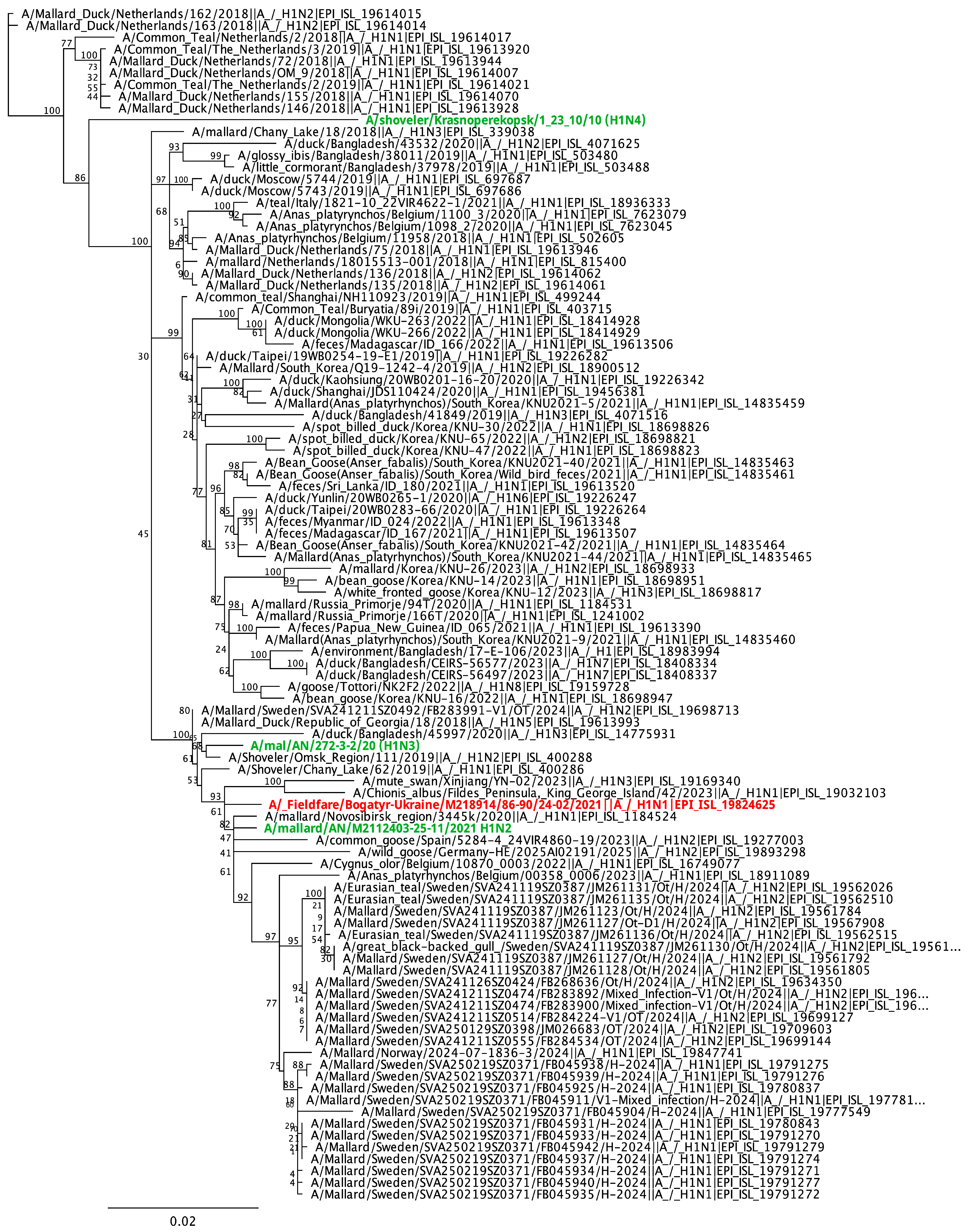
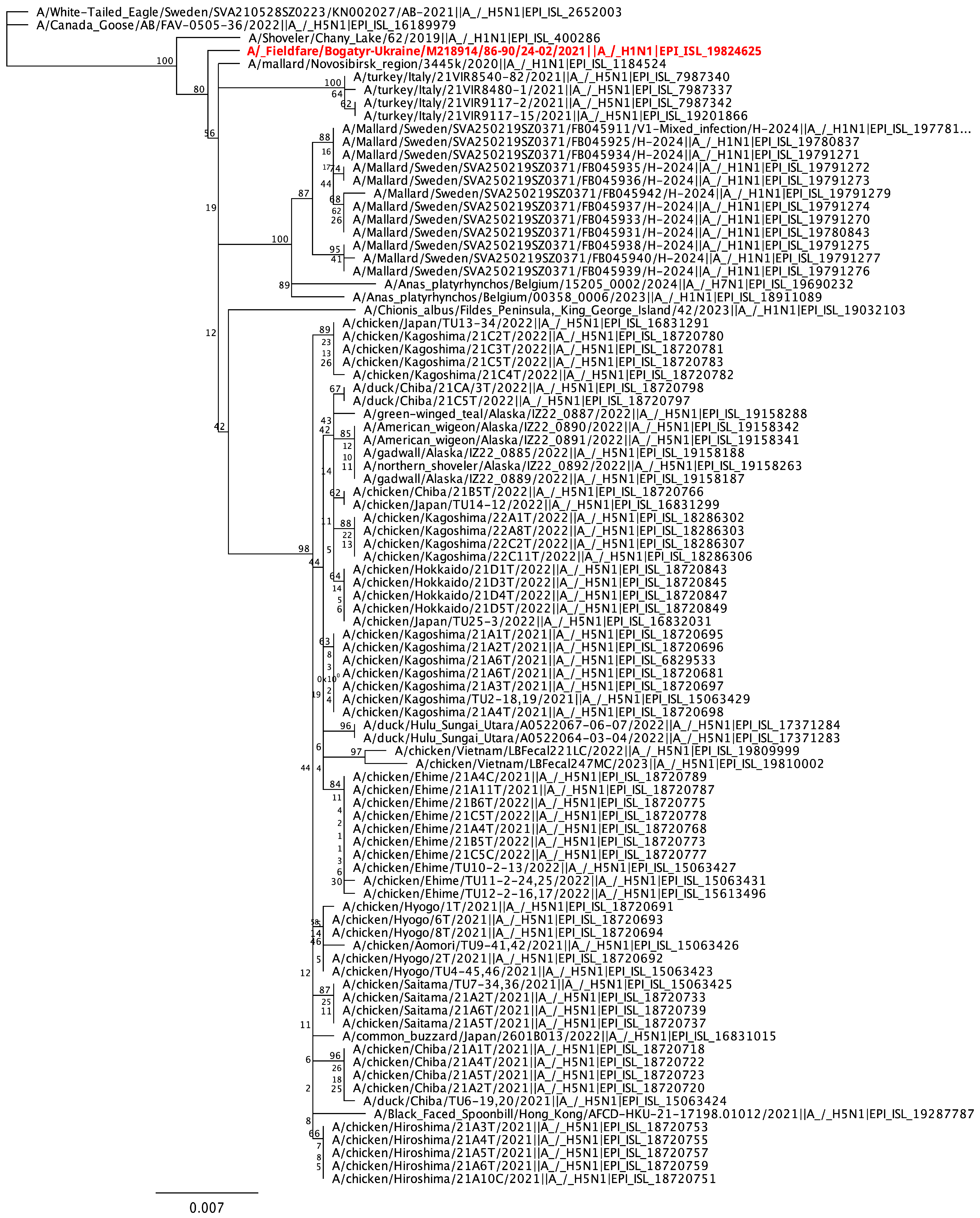
To determine the origin of the A/Fieldfare/Bogatyr-Ukraine/M218914/86-90/24-02/2021 (H1N1) virus and to assess its genetic relationships with other influenza viruses circulating in Ukraine, neighboring countries, and the broader Eurasian region, we conducted phylogenetic analysis for each of the eight gene segments. Phylogenetic trees were constructed using the most closely related influenza virus sequences obtained from GenBank and GISAID databases. The results demonstrated that all gene segments of the Ukrainian virus belong to the Eurasian lineage of LPAI viruses (Figure 1 and Figure 2 and Supplementary Figures S1–S7).
For the PB2 and PB1 genes (Supplementary Figures S2 and S3), the Fieldfare virus clustered with influenza viruses isolated from wild waterfowl in Central and Eastern Europe. Notably, based on the PB1 gene, the virus grouped within a cluster containing wild bird isolates from Sweden and Italy (2024–2025), as well as HPAI A(H5N1) viruses isolated from chickens in Germany in 2025. In contrast, the PA gene (Supplementary Figure S4) of the Ukrainian virus was positioned within a cluster of Asian influenza viruses. Phylogenetic analysis of the genes encoding the HA and NA surface glycoproteins revealed that the H1N1 virus from Fieldfare is closely related to avian influenza viruses that circulated among wild waterfowl in Ukraine and Siberia during 2020–2021. It is noteworthy that Ukrainian H1 subtype viruses isolated in 2020–2021 from wild waterfowl (H1N2, H1N3) and Fieldfares (H1N1) clustered together within a single major clade, whereas an H1N4 virus isolated from wild waterfowl in 2010 in Ukraine was positioned separately, indicating genetic divergence. For the NP gene (Supplementary Figure S5), the Ukrainian isolate grouped within a large cluster comprising influenza viruses of various subtypes from Central and Western Europe, which also included another Ukrainian wild bird isolate (H7N2). The M gene (Supplementary Figure S6) clustered with viruses originating from the Caucasus and Western Europe, while the NS gene of the Fieldfare virus was positioned within a cluster of viruses circulating in Asia (Bangladesh).

3.4. Molecular Analysis

A detailed molecular characterization of all amino acid sequences of the A/Fieldfare/Bogatyr-Ukraine/M218914/86-90/24-02/2021 (H1N1) was conducted to assess the potential risk of this virus to mammals. The analysis revealed that the amino acid sequence at the hemagglutinin (HA) cleavage site is PSIQSR↓GLF, containing a single basic amino acid, which is characteristic of LPAI viruses. Using the FluSurver analytical tools available on the GISAID platform, we examined the presence of critical amino acid substitutions in various gene segments (Table 3).
We combined the mutations data in HA of virus A/Fieldfare/Bogatyr-Ukraine/M218914/86-90/24-02/2021 in antigenic sites into Table 4 for better understanding the influence of mutations for virus functions and properties. Therefore the key mutations were identified in 3 antigenic sites among 5, namely Sa, Ca1 and Cb, as we can see in Table 4.
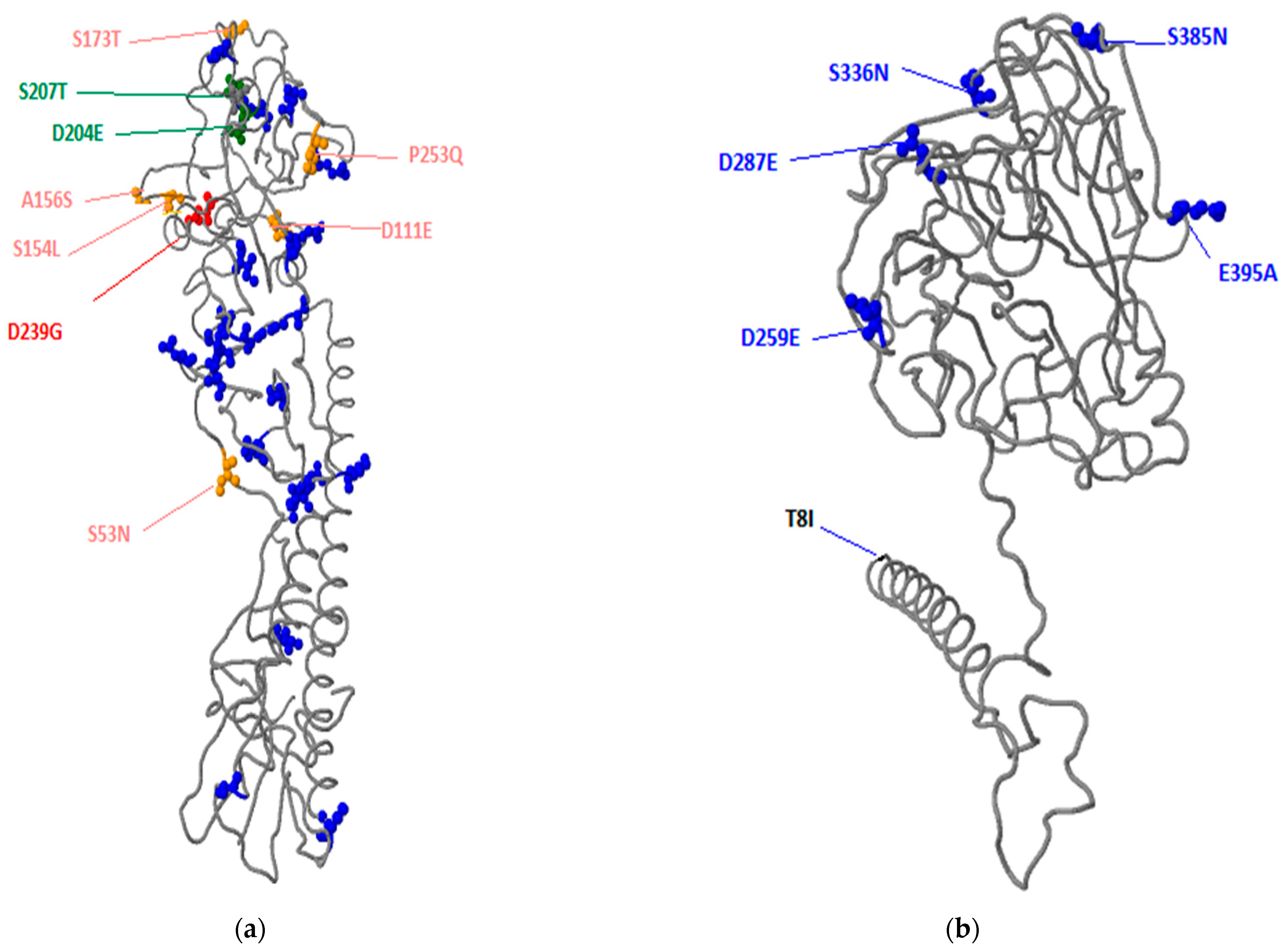
Among the 37 mutations identified in the hemagglutinin (HA) gene of the influenza A virus A/Fieldfare/Bogatyr-Ukraine/M218914/86-90/24-02/2021, particular attention should be given to the substitutions S173T, D204E, S207T, and D239G located within the antigenic site Ca1. The presence of these mutations may influence the virus’s ability to bind to host cells. Consequently, amino acid changes at these positions could alter the strength and nature of antibody binding, potentially affecting antigen–antibody interactions and immune recognition.
In Figure 3a, the amino acid substitutions detected in the HA protein of the influenza A virus A/Fieldfare/Bogatyr-Ukraine/M218914/86-90/24-02/2021 are presented as a three-dimensional (3D) structural model. Different colors indicate amino acid substitutions associated with distinct functional categories (see Table 3 for details).
Regarding the NA of the same virus, mutational analysis identified six amino acid substitutions; however, these mutations are not considered critical and are unlikely to have a significant impact on neuraminidase functionality. The spatial distribution of these mutations is illustrated in Figure 3b as a 3D model, with color-coded residues corresponding to their putative functional roles (see Table 3).
The analyses of the remaining gene segments and their corresponding proteins revealed one significant amino acid substitution—N319K in the nucleoprotein (NP). This mutation has been reported in A(H5N1) viruses and is associated with a shift in host specificity and increased virulence in mammals. In the remaining genes, either no mutations were detected (M, NS, PB1) or only minor substitutions were found (PB2, PA) that are not considered to affect protein functionality.

4. Discussion

Here we report the genetic characterization of an influenza A (H1N1) virus isolated from passerine birds in Ukraine. It is generally accepted that the main natural reservoir of IAVs in nature comprises wild aquatic and shorebirds, primarily species belonging to the orders Anseriformes and Charadriiformes. However, the role of other ecological groups of wild birds, including Passeriformes, in maintaining the natural circulation and further spread remains a subject of debate [21,22,23]. Some researchers argue that passerines do not participate in or play any role in the ecology of avian influenza viruses [24,25], while others suggest that these birds may contribute to the maintenance and transmission of the virus [22,26,27,28,29,30,31]. Experimental infection studies have demonstrated that passerine species can be infected with both LPAI and HPAI strains, confirming that the virus is capable of replicating in their organisms [32,33,34]. It has also been proposed that passerine birds in the farm environment may serve as an ecological “bridge species” in the transmission of influenza viruses among different avian groups [35], including domestic poultry, wild waterfowl, chickens, and turkeys [36].
The literature data indicate that approximately 0.1% of passerine birds possess antibodies against influenza virus [37], although some studies report much higher seroprevalence rates ranging from 6% to 76% [38]. Virus isolation rates have averaged 0.5% [37], while PCR positivity has ranged from 0.1–1% [27,37,39,40,41,42], to 2.3–17.5% [22,23,34,43,44,45,46], and in some cases, up to 50% [27]. It should be noted that ecological and epidemiological settings of wild passerine surveillance studies varies markedly, in whether they were conducted in resident or migratory birds, in farm environments or natural habitats, in times of HPAI outbreak situations or not, and whether designed as active or passive surveillance; such factors, and others such as species choice and sample size, may influence the generality of the findings. Widespread surveillance of wild birds, including Passeriformes, intensified during the global HPAI panzootic beginning in 2020. Reports indicate that 1.1–6.5% of passerine birds tested positive for influenza A virus by PCR, mostly corresponding to detections of HPAI H5 strains [47].
In Ukraine, surveillance of wild passerines has a long history, dating back to 2004, and has included serological, virological, and molecular–genetic investigations. Between 2004 and 2025, a comprehensive survey of 65 passerine species from 20 families was carried out across 15 regions of Ukraine (manuscript submitted). Antibodies against influenza virus were detected in a small proportion of wild passerines from seven species, with an average seroprevalence of 1.24–8.94%. Virological studies resulted in the isolation of two viruses—H1N1 and H7N1—from Fieldfares (Turdus pilaris), including the A/Fieldfare/Bogatyr-Ukraine/M218914/86-90/24-02/2021 strain described in this paper. The overall infection rate among passerines was 0.15%, while the local infection rate among Fieldfares reached 11.1%. The mean PCR positivity among passerines was 3.61% (manuscript submitted).
Our data show no substantial differences between this isolate and the classical Eurasian LPAI H1N1 viruses circulating among wild waterfowl, nor any significant alterations in receptor-binding sites. No behavioral abnormalities or mortality among wild Fieldfares were observed during monitoring. However, the virus demonstrated efficient replication in embryonated chicken eggs and caused embryo death, indicating notable biological activity in an experimental system.
The route of infection for Fieldfares remains unclear. It may represent an isolated spillover event resulting from ecological overlap with wild waterfowl at shared wintering, feeding, and watering sites, or there could be intraspecific transmission among Fieldfares. The biological and ecological characteristics of Passeriformes further highlight their potential epidemiological role: many species are highly abundant and synanthropic [29], living in close proximity to human settlements and domestic animals (including species of the family Turdidae, to which the Fieldfare belongs). This proximity increases the potential for cross-species pathogen exchange between humans, domestic poultry, livestock, and passerines [48,49]. Additionally, many passerines are migratory, capable of long-distance dispersal of pathogens, thereby contributing to the geographic spread of infectious agents.
The phylogenetic analysis revealed that the Ukrainian A(H1N1) virus (A/Fieldfare/Bogatyr-Ukraine/M218914/86-90/24-02/2021) is genetically related to other Eurasian LPAI viruses of various subtypes. The only exception was the PA gene, which showed close similarity to HPAI A(H5N1) viruses circulating in Western Siberia and China. The detection of a multi-gene reassortant, with one segment (PA) related to a contemporary HPAI H5N1 virus, in a healthy passerine underscores the dynamic nature of viral gene pools in wild birds and the potential for unexpected gene combinations to emerge in new host types.
Mutation analysis across all genomic segments demonstrated that the highest number of mutations occurred in the surface glycoproteins—37 in HA and 6 in NA—consistent with their roles in host–cell interaction and exposure to immune selection pressure. Other gene segments contained only single or no substitutions. Most HA mutations were associated with internal viral processes, such as protein oligomerization, while several substitutions had unknown or negligible functional effects. This includes, in particular, the PA polymerase complex mutations D272E, E382D, Y535H, and I543L, which are not currently linked to functional alterations.
Our data support the inclusion of synanthropic and migratory passerines, particularly in regions of waterfowl overlap, in active influenza surveillance programs to better assess the risk of viral spillover and reassortment.

Supplementary Materials

The following supporting information can be downloaded at: https://www.mdpi.com/article/10.3390/microbiolres17010019/s1, Figure S1. List of Supplementary Files. Figure S2. Phylogenetic tree of the PB2 gene of the influenza A virus A/Fieldfare/Bogatyr-Ukraine/M218914/86-90/24-02/2021 (H1N1). Figure S3. Phylogenetic tree of the PB1 gene of the influenza A virus A/Fieldfare/Bogatyr-Ukraine/M218914/86-90/24-02/2021 (H1N1). Figure S4. Phylogenetic tree of the PA gene of the influenza A virus A/Fieldfare/Bogatyr-Ukraine/M218914/86-90/24-02/2021 (H1N1). Figure S5. Phylogenetic tree of the NP gene of the influenza A virus A/Fieldfare/Bogatyr-Ukraine/M218914/86-90/24-02/2021 (H1N1). Figure S6. Phylogenetic tree of the M gene of the influenza A virus A/Fieldfare/Bogatyr-Ukraine/M218914/86-90/24-02/2021 (H1N1). Figure S7. Phylogenetic tree of the NS gene of the influenza A virus A/Fieldfare/Bogatyr-Ukraine/M218914/86-90/24-02/2021 (H1N1).

Author Contributions

Conceptualization, A.M. and D.M.; methodology, A.M. and D.M.; formal analysis, N.M., N.T., L.R. and A.P.; investigation, N.M., N.T., L.R. and A.P.; resources, D.M.; data curation, D.M.; writing—original draft preparation, A.M. and D.M.; writing—review and editing, A.M., D.M. and J.W.; visualization, A.M.; project administration, D.M.; funding acquisition, D.M. All authors have read and agreed to the published version of the manuscript.

Funding

This research was funded by National Research Foundation of Ukraine; grant number #2021.01/0006 “Study of circulation of zoonotic influenza A viruses in a natural reservoir, assessment of their epidemic risks and danger to human health in Ukraine” (virological study, phylogenetic analyses and 3D modeling). Funded by the European Union under grant agreement (101084171)—(Kappa-Flu). Views and opinions expressed are however those of the author(s) only and do not necessarily reflect those of the European Union or REA. Neither the European Union nor the granting authority can be held responsible for them. The manuscript was written and finalized by Denys Muzyka during the period of his project “Virus on wings: toward an early warning system for detection of avian influenza virus”, funded by The Knowledge Foundation (Sweden).

Institutional Review Board Statement

The study was reviewed and approved by the Institutional Animal Care and Use Committee of the National Scientific Center Institute of Experimental and Clinical Veterinary Medicine (protocol #1-23, 19 April 2023).

Informed Consent Statement

Not applicable.

Data Availability Statement

The sequencing of eight genes of Ukrainian viruses is available at GISAID platform (https://platform.gisaid.org, accessed on 31 October 2025).

Acknowledgments

We gratefully acknowledge all data contributors, i.e., the Authors and their Originating laboratories responsible for obtaining the specimens, and their Submitting laboratories for generating the genetic sequence and metadata and sharing via the GISAID Initiative, on which this research is based. The authors gratefully acknowledge Zohari Siamak (Department of Virology, Immunobiology and Parasitology, National Veterinary Institute, Swedish Veterinary Agency, Uppsala, Sweden) for help with sequencing of virus.

Conflicts of Interest

The authors declare no conflicts of interest.

References

  1. Bi, Y.; Yang, J.; Wang, L.; Ran, L.; Gao, G.F. Ecology and evolution of avian influenza viruses. Curr. Biol. 2024, 34, R716–R721. [Google Scholar] [CrossRef] [Scilit]
  2. Bellido-Martín, B.; Rijnink, W.F.; Iervolino, M.; Kuiken, T.; Richard, M.; Fouchier, R.A.M. Evolution, spread and impact of highly pathogenic H5 avian influenza A viruses. Nat. Rev. Microbiol. 2025, 24, 45–60. [Google Scholar] [CrossRef] [Scilit] [PubMed]
  3. Wu, Y.; Wu, Y.; Tefsen, B.; Shi, Y.; Gao, G.F. Bat-derived influenza-like viruses H17N10 and H18N11. Trends Microbiol. 2014, 22, 183–191. [Google Scholar] [CrossRef] [Scilit]
  4. Yang, W.; Schountz, T.; Ma, W. Bat Influenza Viruses: Current Status and Perspective. Viruses 2021, 13, 547. [Google Scholar] [CrossRef] [Scilit] [PubMed]
  5. Verhagen, J.H.; Fouchier, R.A.M.; Lewis, N. Highly Pathogenic Avian Influenza Viruses at the Wild-Domestic Bird Interface in Europe: Future Directions for Research and Surveillance. Viruses 2021, 13, 212. [Google Scholar] [CrossRef] [Scilit]
  6. Ryding, S.; Ross, T.A.; Klaassen, M.; Wille, M. Impacts of a Potential HPAI H5N1 Incursion on Australian Wildlife. Austral Ecology 2025, 50, e70048. [Google Scholar] [CrossRef] [Scilit]
  7. Russell, C.A.; Kasson, P.M.; Donis, R.O.; Riley, S.; Dunbar, J.; Rambaut, A.; Asher, J.; Burke, S.; Davis, C.T.; Garten, R.J.; et al. Improving pandemic influenza risk assessment. eLife 2014, 3, e03883. [Google Scholar] [CrossRef] [Scilit]
  8. Dawood, F.S.; Jain, S.; Finelli, L.; Shaw, M.W.; Lindstrom, S.; Garten, R.J.; Gubareva, L.V.; Xu, X.; Bridges, C.B.; Uyeki, T.M. Novel Swine-Origin Influenza A (H1N1) Virus Investigation Team, Emergence of a novel swine-origin influenza A (H1N1) virus in humans. N. Engl. J. Med. 2009, 360, 2605–2615. [Google Scholar] [CrossRef] [Scilit]
  9. Yoon, S.W.; Webby, R.J.; Webster, R.G. Evolution and ecology of influenza A viruses. Curr. Top. Microbiol. Immunol. 2014, 385, 359–375. [Google Scholar] [CrossRef] [Scilit]
  10. Wasik, B.R.; Voorhees, I.E.H.; Parrish, C.R. Canine and Feline Influenza. Cold Spring Harb. Perspect. Med. 2021, 11, a038562. [Google Scholar] [CrossRef] [Scilit]
  11. Krammer, F.; Hermann, E.; Rasmussen, A.L. Highly pathogenic avian influenza H5N1: History, current situation, and outlook. J. Virol. 2025, 99, e0220924. [Google Scholar] [CrossRef] [Scilit] [PubMed]
  12. Pu, Z.; Xiang, D.; Li, X.; Luo, T.; Shen, X.; Murphy, R.W.; Liao, M.; Shen, Y. Potential Pandemic of H7N9 Avian Influenza A Virus in Human. Front. Cell. Infect. Microbiol. 2018, 8, 414. [Google Scholar] [CrossRef] [Scilit] [PubMed]
  13. Song, W.; Qin, K. Human-infecting influenza A (H9N2) virus: A forgotten potential pandemic strain? Zoonoses Public Health 2020, 67, 203–212. [Google Scholar] [CrossRef] [Scilit] [PubMed]
  14. Yang, R.; Sun, H.; Gao, F.; Luo, K.; Huang, Z.; Tong, Q.; Song, H.; Han, Q.; Liu, J.; Lan, Y.; et al. Human infection of avian influenza A H3N8 virus and the viral origins: A descriptive study. Lancet. Microbe 2022, 3, e824–e834. [Google Scholar] [CrossRef] [Scilit]
  15. Spackman, E. Avian Influenza Virus, 1st ed.; Methods in Molecular Biology, 436; Humana Press: Totowa, NJ, USA, 2008. [Google Scholar]
  16. Spackman, E.; Pedersen, J.C.; McKinley, E.T.; Gelb, J., Jr. Optimal specimen collection and transport methods for the detection of avian influenza virus and Newcastle disease virus. BMC Vet. Res. 2013, 9, 35. [Google Scholar] [CrossRef] [Scilit]
  17. Spackman, E. (Ed.) Animal Influenza Virus, 2nd ed.; Springer: New York, NY, USA, 2014. [Google Scholar]
  18. Athens, G.A. A Laboratory Manual for The Isolation, Identification and Characterization of Avian Pathogens, 5th ed; American Association of Avian Pathologists: Jacksonville, FL, USA, 2008. [Google Scholar]
  19. Zhou, B.; Wentworth, D.E. Influenza A virus molecular virology techniques. Methods Mol. Biol. 2012, 865, 175–192. [Google Scholar] [CrossRef] [Scilit]
  20. Byrne, A.M.P.; James, J.; Mollett, B.C.; Meyer, S.M.; Lewis, T.; Czepiel, M.; Seekings, A.H.; Mahmood, S.; Thomas, S.S.; Ross, C.S.; et al. Investigating the Genetic Diversity of H5 Avian Influenza Viruses in the United Kingdom from 2020–2022. Microbiol. Spectr. 2023, 11, e0477622. [Google Scholar] [CrossRef] [Scilit]
  21. Causey, D.; Edwards, S.V. Ecology of avian influenza virus in birds. J. Infect. Dis. 2008, 197, S29–S33. [Google Scholar] [CrossRef] [Scilit]
  22. Peterson, A.T.; Bush, S.E.; Spackman, E.; Swayne, D.E.; Ip, H.S. Influenza A virus infections in land birds, People’s Republic of China. Emerg. Infect. Dis. 2008, 14, 1644–1646. [Google Scholar] [CrossRef] [Scilit]
  23. Cumming, G.S.; Caron, A.; Abolnik, C.; Cattoli, G.; Bruinzeel, L.W.; Burger, C.E.; Cecchettin, K.; Chiweshe, N.; Mochotlhoane, B.; Mutumi, G.L.; et al. The ecology of influenza A viruses in wild birds in southern Africa. EcoHealth 2011, 8, 4–13. [Google Scholar] [CrossRef] [Scilit]
  24. Morishita, T.Y.; Aye, P.P.; Ley, E.C.; Harr, B.S. Survey of pathogens and blood parasites in free-living passerines. Avian Dis. 1999, 43, 549–552. [Google Scholar] [CrossRef] [Scilit]
  25. Munster, V.J.; Baas, C.; Lexmond, P.; Waldenström, J.; Wallensten, A.; Fransson, T.; Rimmelzwaan, G.F.; Beyer, W.E.; Schutten, M.; Olsen, B.; et al. Spatial, temporal, and species variation in prevalence of influenza A viruses in wild migratory birds. PLoS Pathog. 2007, 3, e61. [Google Scholar] [CrossRef] [Scilit]
  26. Chang, H.; Dai, F.; Liu, Z.; Yuan, F.; Zhao, S.; Xiang, X.; Zou, F.; Zeng, B.; Fan, Y.; Duan, G. Seroprevalence survey of avian influenza A (H5) in wild migratory birds in Yunnan Province, Southwestern China. Virol. J. 2014, 11, 18. [Google Scholar] [CrossRef] [Scilit]
  27. Fuller, T.L.; Saatchi, S.S.; Curd, E.E.; Toffelmier, E.; Thomassen, H.A.; Buermann, W.; DeSante, D.F.; Nott, M.P.; Saracco, J.F.; Ralph, C.; et al. Mapping the risk of avian influenza in wild birds in the US. BMC Infect. Dis. 2010, 10, 187. [Google Scholar] [CrossRef] [Scilit] [PubMed]
  28. Kou, Z.; Lei, F.M.; Yu, J.; Fan, Z.J.; Yin, Z.H.; Jia, C.X.; Xiong, K.J.; Sun, Y.H.; Zhang, X.W.; Wu, X.M.; et al. New genotype of avian influenza H5N1 viruses isolated from tree sparrows in China. J. Virol. 2005, 79, 15460–15466. [Google Scholar] [CrossRef] [Scilit]
  29. Shriner, S.A.; Root, J.J. A Review of Avian Influenza A Virus Associations in Synanthropic Birds. Viruses 2020, 12, 1209. [Google Scholar] [CrossRef] [Scilit]
  30. Lipkind, M.; Shihmanter, E.; Shoham, D. Further characterization of H7N7 avian influenza virus isolated from migrating starlings wintering in Israel. Zentralblatt Veterinarmedizin. Reihe B 1982, 29, 566–572. [Google Scholar] [CrossRef] [Scilit] [PubMed]
  31. Račnik, J.; Slavec, B.; Trilar, T.; Zadravec, M.; Dovč, A.; Krapež, U.; Barlič-Maganja, D.; Zorman Rojs, O. Evidence of avian influenza virus and paramyxovirus subtype 2 in wild-living passerine birds in Slovenia. Eur. J. Wildl. Res. 2008, 54, 529–532. [Google Scholar] [CrossRef] [Scilit]
  32. Root, J.J.; Bosco-Lauth, A.M.; Marlenee, N.L.; Bowen, R.A. Viral shedding of clade 2.3.4.4 H5 highly pathogenic avian influenza A viruses by American robins. Transbound. Emerg. Dis. 2018, 65, 1823–1827. [Google Scholar] [CrossRef] [Scilit]
  33. Breithaupt, A.; Kalthoff, D.; Dale, J.; Bairlein, F.; Beer, M.; Teifke, J.P. Neurotropism in blackcaps (Sylvia atricapilla) and red-billed queleas (Quelea quelea) after highly pathogenic avian influenza virus H5N1 infection. Vet. Pathol. 2011, 48, 924–932. [Google Scholar] [CrossRef] [Scilit]
  34. Qin, Z.; Clements, T.; Wang, L.; Khatri, M.; Pillai, S.P.; Zhang, Y.; Lejeune, J.T.; Lee, C.W. Detection of influenza viral gene in European starlings and experimental infection. Influenza Other Respir. Viruses 2011, 5, 268–275. [Google Scholar] [CrossRef] [Scilit]
  35. Caron, A.; Grosbois, V.; Etter, E.; Gaidet, N.; de Garine-Wichatitsky, M. Bridge hosts for avian influenza viruses at the wildlife/domestic interface: An eco-epidemiological framework implemented in southern Africa. Prev. Vet. Med. 2014, 117, 590–600. [Google Scholar] [CrossRef] [Scilit]
  36. Ayala, A.J.; Yabsley, M.J.; Hernandez, S.M. A Review of Pathogen Transmission at the Backyard Chicken-Wild Bird Interface. Front. Vet. Sci. 2020, 7, 539925. [Google Scholar] [CrossRef] [Scilit]
  37. Slusher, M.J.; Wilcox, B.R.; Lutrell, M.P.; Poulson, R.L.; Brown, J.D.; Yabsley, M.J.; Stallknecht, D.E. Are passerine birds reservoirs for influenza A viruses? J. Wildl. Dis. 2014, 50, 792–809. [Google Scholar] [CrossRef] [Scilit]
  38. Hadipour, M.; Vosoughi, A.; Fakhrabadipour, M.; Azad, F.; Khademi, I. Serological evaluation for supporting the potential role of house sparrows in LPAIV (H9N2) transmission. Int. J. Anim. Vet. Adv. 2011, 3, 189–192. [Google Scholar]
  39. Hesterberg, U.; Harris, K.; Stroud, D.; Guberti, V.; Busani, L.; Pittman, M.; Piazza, V.; Cook, A.; Brown, I. Avian influenza surveillance in wild birds in the European Union in 2006. Influenza Other Respir. Viruses 2009, 3, 1–14. [Google Scholar] [CrossRef] [Scilit] [PubMed]
  40. Cappelle, J.; Servan de Almeida, R.; Fofana, B.; Dakouo, M.; Balança, G.; Gil, P.; Albina, E.; Gaidet, N. Circulation of avian influenza viruses in wild birds in Inner Niger Delta, Mali. Influenza Other Respir. Viruses 2012, 6, 240–244. [Google Scholar] [CrossRef] [Scilit] [PubMed]
  41. Kou, Z.; Li, Y.; Yin, Z.; Guo, S.; Wang, M.; Gao, X.; Li, P.; Tang, L.; Jiang, P.; Luo, Z.; et al. The survey of H5N1 flu virus in wild birds in 14 Provinces of China from 2004 to 2007. PLoS ONE 2009, 4, e6926. [Google Scholar] [CrossRef] [Scilit]
  42. Williams, R.A.; Segovia-Hinostroza, K.; Ghersi, B.M.; Gonzaga, V.; Peterson, A.T.; Montgomery, J.M. Avian Influenza infections in nonmigrant land birds in Andean Peru. J. Wildl. Dis. 2012, 48, 910–917. [Google Scholar] [CrossRef] [Scilit]
  43. Borovská, P.; Kabát, P.; Ficová, M.; Trnka, A.; Svetlíková, D.; Betáková, T. Prevalence of avian influenza viruses, Mycobacterium avium, and Mycobacterium avium, subsp. paratuberculosis in marsh-dwelling passerines in Slovakia, 2008. Biologia 2011, 66, 282–287. [Google Scholar] [CrossRef] [Scilit]
  44. Gronesova, P.; Ficova, M.; Mizakova, A.; Kabat, P.; Trnka, A.; Betakova, T. Prevalence of avian influenza viruses, Borrelia garinii, Mycobacterium avium, and Mycobacterium avium subsp. paratuberculosis in waterfowl and terrestrial birds in Slovakia, 2006. Avian. Pathol. 2008, 37, 537–543. [Google Scholar] [CrossRef] [Scilit] [PubMed]
  45. Gronesova, P.; Kabat, P.; Trnka, A.; Betakova, T. Using nested RT-PCR analyses to determine the prevalence of avian influenza viruses in passerines in western Slovakia, during summer 2007. Scand. J. Infect. Dis. 2008, 40, 954–957. [Google Scholar] [CrossRef] [Scilit]
  46. Thinh, T.V.; Gilbert, M.; Bunpapong, N.; Amonsin, A.; Nguyen, D.T.; Doherty, P.F., Jr.; Huyvaert, K.P. Avian influenza viruses in wild land birds in northern Vietnam. J. Wildl. Dis. 2012, 48, 195–200. [Google Scholar] [CrossRef] [Scilit] [PubMed]
  47. European Food Safety Authority; European Centre for Disease Prevention and Control. The European Union One Health 2021 Zoonoses Report. EFSA J. 2022, 20, e07666. [Google Scholar] [CrossRef] [Scilit]
  48. Günther, A.; Pohlmann, A.; Globig, A.; Ziegler, U.; Calvelage, S.; Keller, M.; Fischer, D.; Staubach, C.; Groschup, M.H.; Harder, T.; et al. Continuous surveillance of potentially zoonotic avian pathogens detects contemporaneous occurrence of highly pathogenic avian influenza viruses (HPAIV H5) and flaviviruses (USUV, WNV) in several wild and captive birds. Emerg. Microbes Infect. 2023, 12, 2231561. [Google Scholar] [CrossRef] [Scilit] [PubMed]
  49. Gibb, R.; Redding, D.W.; Chin, K.Q.; Donnelly, C.A.; Blackburn, T.M.; Newbold, T.; Jones, K.E. Zoonotic host diversity increases in human-dominated ecosystems. Nature 2020, 584, 398–402. [Google Scholar] [CrossRef] [Scilit] [PubMed]
Figure 1. Phylogenetic tree of the HA gene of the influenza A virus A/Fieldfare/Bogatyr-Ukraine/M218914/86-90/24-02/2021 (H1N1), which is designated in red color. (Other Ukrainian avian influenza viruses were designated in green color).
Microbiolres 17 00019 g001
Figure 2. Phylogenetic tree of the NA gene of the influenza A virus A/Fieldfare/Bogatyr-Ukraine/M218914/86-90/24-02/2021 (H1N1), which is designated in red color.
Microbiolres 17 00019 g002
Figure 3. Location of main amino acid substitutions (a) in HA of A(H1N1) avian influenza virus A/Fieldfare/Bogatyr-Ukraine/M218914/86-90/24-02/2021 (PDB ID—2WRG); (b) in NA of A(H1N1) avian influenza virus A/Fieldfare/Bogatyr-Ukraine/M218914/86-90/24-02/2021. (The colors in the image are in accordance Table 3).
Microbiolres 17 00019 g003
Table 1. Summary data on the biological activity and hemagglutinin titer of the influenza virus isolated from a wild bird of the order Passeriformes.
Isolate NameTiter, log10/0.1 mLTiter HA
EID50ELD50
A/Fieldfare/Bogatyr-Ukraine/M218914/86-90/24-02/2021 (H1N1)7.76.741:256–1:1024
Table 2. Sequence identity of each gene between the A/Fieldfare/Bogatyr-Ukraine/M218914/86-90/24-02/2021 (H1N1) virus and the closest homologs in the GenBank and GISAID database.
GeneViruses with Greatest HomologyAccessionIden. (%)
PB2A/goose/Czech Republic/13440-1/2023 (H9N2)EPI_ISL_1839911598.68
A/goose/Czech Republic/13440-5/2023 (H9N2)EPI_ISL_1839914998.77
A/Mallard Duck/Netherlands/75/2018 (H1N1)EPI_ISL_1961394698.72
PB1A/Mallard/Sweden/SVA250219SZ0371/FB045884/H-2024 (H3N8)EPI_ISL_1977705698.94
A/mallard/Italy/24VIR8385-2/2024 (H4N6) EPI_ISL_1987308698.50
A/teal/Dagestan/23d/2018 (H3N8) EPI_ISL_33904098.50
PAA/wild duck/Novosibirsk region/5194k/2021 (H5N1)EPI_ISL_1872076299.53
A/goose/China/KUST-ZT-BTY5/2021 (H5N1)EPI_ISL_1871815299.34
A/gadwall/Chany/893/2018 H3N8EPI_ISL_33361599.11
HAA/Mallard Duck/Republic of Georgia/18/2018 H1N5EPI_ISL_1961399399.11
A/Mallard/Sweden/SVA241211SZ0492/FB283991-V1/OT/2024 (H1N2)EPI_ISL_1969871399.11
NPA/Mallard Duck/Republic of Georgia/2/2018 (H10N7)EPI_ISL_1961390899.33
A/mallard/Italy/19VIR7018-9/2019 (H4N6)EPI_ISL_1920443199.19
A/Mallard Duck/Republic of Georgia/6/2018 (H3N8)EPI_ISL_1961397699.19
NAA/mallard/Novosibirsk region/3445k/2020 (H1N1)EPI_ISL_118452499.57
A/Shoveler/Chany Lake/62/2019 (H1N1)EPI_ISL_40028699.14
A/Mallard/Sweden/SVA250219SZ0371/FB045911/V1-Mixed_infection/H-2024EPI_ISL_1977815298.93
MA/Common Teal/Republic of Georgia/1/2018 (H4N6)EPI_ISL_1961402099.64
A/Common Teal/Dagestan/34d/2019 (H4N6)EPI_ISL_40371699.49
A/Mallard Duck/Netherlands/147/2018 (H12N5)EPI_ISL_1961392999.49
NSA/mallard/Novosibirsk region/3541k/2020 (H12N5)EPI_ISL_124100199.88
A/environment/Bangladesh/52180/2022 (H10N4)EPI_ISL_1562032099.76
A/Mallard/Yakutia/C-3/2023 (H5N3)EPI_ISL_1920248699.52
Table 3. The genetic characteristics of avian influenza virus A/Fieldfare/Bogatyr-Ukraine/M218914/86-90/24-02/2021 (H1N1) based on FluSurver.
Viral ProteinClosest Genetic RelationMutation *Function of Mutation
HAA/BrevigMission/1/1918 (H1N1)R4K, L9F, A11M, A13I, T15S, N16K, S53N, K60R, K62N, I74V, V97I, D111E, S138D, S154L, A156S, S173T, V183T, G202A, T203S, S207T, D239G, E252D, P253Q, R276K, G279D, D291N, N293D, V315I, R325K, I400L, I435V, K471R, A491Eplays a role in viral oligomerization
A13I, N16K, S53N, K62N, D111E, S138D, S154L, A156S, P253Q, S173T, V183T, G202A, T203S, D204E, S207T, D239G, R276K, D291N, N293D, I312V, V315I, R325K, I400L, A491Esmall ligand binding
V41I, K60R, K62N, I74V, V97I, D111E, S138D, S154L, A156S, S173T, V183T, G202A, T203S, D204E, S207T, D239G, D291N, N293D, I400L, A491Eantibody recognition site
S207T, R325K, I400Lbinding to host proteins
L9F, D111E, S173T, D204E, D239G, P253Qhost specificity shift
S53N, S154L, A156S, S207T antigenic drift
D204E, S207T, D239Gbinding to host cells
NAA/AmericanWigeon/SouthCarolina/22-000345-001/2021 (H5N1)D259E, D287E, S336N, E395Aplays a role in viral oligomerization
D259E, D287E, S336N, S385N, E395Asmall ligand binding
E395Aantibody recognition site
T8IRole unknown
PB2A/Mallard/Astrakhan/263/1982 (H14N5)M444Vplays a role in viral oligomerization
PB1A/Chicken/GhanaAVL-76321VIR7050-39//2021 (H5N1)No mutation-
PAA/Netherlands/219/2003 (H7N7)D272E, E382D, Y535H, I543LRole unknown
NPA/Duck/Hong Kong/24/1976 (H4N2)N319Kassociated with virulence and a shift in host specificity
NS1A/Shearwater/Australia/2576/1979 (H15N9) D152Eplays a role in viral oligomerization
NS2A/Duck/Guangdong/E1/2012 (H10N8)No mutation -
M1A/Duck/Guangdong/E1/2012 (H10N8) No mutation-
M2A/Mallard/Astrakhan/263/1982 (H14N5)No mutation-
* According to the GISAID database the mutations are color-coded according to their known or predicted biological effect significance. When there are no known effects for the mutation, the mutation will appear in black colored font. When the mutation is a common subtype marker, the mutation will appear in green colored font. Mutations occurring at a site of interaction will appear in blue colored font. If the mutation occurs at a site known to involved in drug-binding or alters host–cell specificity, it will appear in orange. Mutations will also appear in orange when its equivalent site is known to result in antigenic shifts or causes mild drug resistance. Mutations that create or remove a potential glycosylation site are colored magenta. Only mutations that are known to alter the virulence of the virus, cause strong drug resistance, or reverse the effects of the premature STOP codon in the PB1-F2 gene of pandemic H1N1 will appear in red.
Table 4. Mutations in antigenic sites of HA in avian influenza virus A/Fieldfare/Bogatyr-Ukraine/M218914/86-90/24-02/2021.
Antigenic SiteMutation **Function of mutation
SaA156Santigenic drift/escape mutant
SbNo mutationsn/a
Ca1S173Tantibody recognition site, host specificity shift
S207Tantigenic drift/escape mutant and virulence
D204Ehost specificity shift
D239Ghost specificity shift, host cell receptor binding, antibody recognition site
Ca2No mutationsn/a
CbI74Vantibody recognition site
** According to the GISAID database (see explanation in the Table 3).
Disclaimer/Publisher’s Note: The statements, opinions and data contained in all publications are solely those of the individual author(s) and contributor(s) and not of MDPI and/or the editor(s). MDPI and/or the editor(s) disclaim responsibility for any injury to people or property resulting from any ideas, methods, instructions or products referred to in the content.