Case Report

Multidrug-Resistant Extraintestinal Pathogenic Escherichia coli Exhibits High Virulence in Calf Herds: A Case Report

1
Shandong Key Laboratory of Animal Disease Control and Breeding, Institute of Animal Science and Veterinary Medicine, Shandong Academy of Agricultural Sciences, Jinan 250100, China
2
Key Laboratory of Livestock and Poultry Multi-Omics of MARA, Jinan 250100, China
3
College of Veterinary Medicine, Shandong Agricultural University, Tai’an 271018, China
*
Authors to whom correspondence should be addressed.
These authors contributed equally to this work and share first authorship.
Microbiol. Res. 2025, 16(3), 59; https://doi.org/10.3390/microbiolres16030059
Submission received: 7 February 2025 / Revised: 24 February 2025 / Accepted: 26 February 2025 / Published: 28 February 2025

Abstract

:
Extraintestinal pathogenic Escherichia coli (ExPEC) is a group of Escherichia coli strains that can cause severe infectious diseases outside the gastrointestinal tract, such as urinary tract infections, meningitis, septicemia, etc. We report a case of a calf herd infection by ExPEC with high rates of morbidity and mortality. The research purpose of this study was to thoroughly investigate the characteristics of the ExPEC responsible for the calf herd infection. Specifically, we aimed to understand the mechanisms underlying its multidrug resistance and high pathogenicity. Clinical samples were collected for the isolation and identification of ExPECs, cultured on MacConkey agar, and further tested by PCR for the uidA gene, 16S rRNA gene sequencing, and adhesion patterns on HEp-2 cells. The antimicrobial activity was determined using the disk diffusion method according to Clinical & Laboratory Standards Institute (CLSI) guidelines. The pathogenicity was assessed through the experimental infection of Kunming mice, tracking their survival and weight changes, and performing autopsies for bacterial counts and histopathological analysis. Additionally, whole-genome sequencing (WGS) and a comprehensive analysis were performed, including multilocus sequence typing (MLST), serotyping, drug-resistance gene analysis, virulence factor analysis, metabolic pathway analysis, and enrichment analysis, using various online tools and databases. An ExPEC strain named RZ-13 was responsible for this case and was identified as ST345 and O134: H21. Among the 14 antibiotics tested, 13 showed resistance, indicating that the RZ-13 strain is a multidrug-resistant (MDR) bacterium. The experimental infection of Kunming mice proved the greater pathogenicity of RZ-13 than that of CICC 24186. The comprehensive WGS revealed the presence of 28 antibiotic resistance genes and 86 virulence-related genes in the genome of the strain, corroborating its clinical manifestations of MDR and high pathogenicity. Our study isolated a MDR ExPEC strain, RZ-13, with a strong pathogenicity. This is the first case report of ExPEC leading to severe mortality in calf herds in China, underscoring the need for the rational use of antibiotics to reduce the risk of the generation and transmission of MDR bacteria from food-producing animals to ensure food safety and public health.

1. Introduction

Pathogenic Escherichia coli causes a major economic burden worldwide, with high mortality rates [1,2,3]. Pathogenic Escherichia coli is classified into two main types: intestinal pathogenic Escherichia coli (IPEC) and extraintestinal pathogenic Escherichia coli (ExPEC). Among them, ExPEC is a significant pathogen causing various diseases such as cystitis, pyelonephritis, and bacteremia, and it can even pose a threat to human and animal life [4]. ExPEC is commonly found in the animal industry, causing economic losses and posing a threat to human health through foodborne transmission or cross-infection [5]. Its virulence factors include adhesins, toxins, iron acquisition factors, lipopolysaccharides, polysaccharide capsules, and invasion proteins, which are typically encoded on pathogenicity islands, plasmids, and other mobile genetic elements, aiding its survival under adverse conditions [4]. The widespread use of antibiotics has led to a significant increase in ExPEC resistance, particularly against third-generation cephalosporins and fluoroquinolones, severely impacting their treatment efficacy. Studies have shown that this resistance is closely associated with specific sequence types (STs), with up to 30% of ExPEC, 60–90% of fluoroquinolone-resistant ExPEC, and 40–80% of extended-spectrum β-lactamase (ESBL) producing strains belonging to the ST131 lineage. The discovery of multidrug-resistant (MDR) strains is steadily becoming increasingly common [5]. MDR ExPEC is commonly found in humans worldwide due to its mosaic genomic structure, but similar research on cattle is limited [6].
The antimicrobial resistance mechanisms of ExPEC include reduced antibiotic uptake, enhanced efflux, target site modifications, and the production of inactivating enzymes [7]. Additionally, mobile genetic elements, such as plasmids and transposons, facilitate the spread of resistance genes, accelerating the dissemination of resistant bacteria. The abuse of antibiotics has worsened the resistance issue in cattle populations [6]. The existing studies indicate that the rumen of ruminants contains at least 4043 antibiotic resistance genes (ARGs), including genes for β-lactams (726 genes), glycopeptides (510 genes), tetracyclines (307 genes), and aminoglycosides (193 genes) [8]. Similarly, it has been reported that 97.3% of 37 Escherichia coli strains isolated from the rectum of diarrheal piglets exhibit resistance to at least four different antibiotics, with 28 strains carrying resistance genes to colistin [9]. It should be noted that antibiotic resistance is increasingly becoming a dynamic phenomenon, necessitating continuous updates to the knowledge in this field. We investigated a case of calf mortality caused by E. coli in Rizhao City, Shandong Province, explored the antibiotic resistance, pathogenicity, and comprehensive genome analysis, and provide solutions for cattle farms affected by the disease, which provide reference data for prevention and clinical treatment. This study provides new data and valuable insights into the epidemiology, pathogenesis, and treatment strategies for ExPEC-induced infections in calves, ultimately contributing to the development of more effective control measures to prevent the spread of such infections and reduce the associated morbidity and mortality rates.

2. Case Description

In the process of selecting cases and collecting information, we followed the Case report guideline (CARE) [10] to support an increase in the accuracy, transparency, and usefulness of this case report and referred to similar case reports [11]. In April 2023, a large-scale beef cattle farm in Rizhao City, Shandong Province, was struck by an outbreak of neonatal calf diarrhea and pneumonia. Initially, the attending veterinarians attempted antibiotic and supportive treatments. When symptoms are mild, oral rehydration salts (300–400 mL) are administered orally, along with oral sulfamidine tablets (20–30 tablets), or gentamicin injections (10 mg/kg, twice a day). If there is no significant improvement in symptoms after 2 days, intravenous fluid therapy is initiated, with intravenous injections of ampicillin (30 mg/kg, twice a day) and, at the same time, intramuscular injections of ciprofloxacin or enrofloxacin (10–20 mL, once a day), or gentamicin injections (10 mg/kg, twice a day) are administered. This treatment continues until the calf recovers or dies. Despite repeated treatments over some days, the calves began to die, and the number of fatalities escalated. It was only when the farm manager realized the gravity of the situation and contacted our laboratory for testing and antimicrobial susceptibility analysis that we learned of the 42 deceased calves. During our first communication, we were informed that the calves were being fed in a mother–offspring manner, prompting us to advise the veterinarians to isolate all calves from the main affected barn and those with diarrhea from other barns for separate rearing. We recommended ceasing the feeding of breast milk and replacing it with milk powder and increasing the intensity and frequency of disinfection to eradicate pathogens in the environment and interrupt their transmission. Subsequently, we processed and tested 11 individually packaged samples from the cattle, finding varying degrees of hemorrhage in the hearts, livers, spleens, lungs, kidneys, and intestines of all 11 calves, with E. coli detected in all the samples. According to the veterinarians’ recollections and medical records, from the appearance of the first symptomatic case to the end of the outbreak, a total of 108 cattle were infected in the same barn housing 126 calves, and 78 calves died, resulting in a staggering infection rate of 85.71% and a mortality rate of 72.224%. The outbreak records implied that the E. coli causing the outbreak was highly virulent and pathogenic, and that it had widespread drug resistance.

3. Materials and Methods

3.1. Sample Collection and Identification of E. coli

We collected 66 clinical samples from 11 calves using aseptic procedures, including the heart, liver, spleen, lungs, kidneys, and intestines from each calf. They were stored at 4 °C for subsequent pathogen detection. Each sample was dissolved in 2 mL of sterile phosphate-buffered saline (PBS) (Beijing Solarbio Science & Technology Co., Ltd., Beijing, China), streaked onto MacConkey agar plate (Qingdao Hi-tech Industrial Park Hope Bio-technology Co., Ltd., Qingdao, China), and cultured at 37 °C for 18–24 h. Several typical colonies were selected and enriched in LB broth (MDBio, Inc., Sugar Land, TX, USA) at 37 °C overnight and were stored at 4 °C for Gram staining and further detection [12].
DNA was extracted using a SteadyPure Bacterial Genomic DNA Extraction Kit (Accurate Biology, Quanzhou, China) and was stored at −20 °C until PCR testing. According to the National Food Safety Standards for Microbiological Examination of Food (China, GB 4789.6-2016) [13], the characteristic gene uidA of Escherichia coli was selected for PCR detection, and the primers were synthesized by Qingdao Weilai Biotechnology. The 16S rRNA gene sequencing was conducted by Qingdao Weilai Biotechnology Co., Ltd. (Qingdao, China). The results were compared with the National Center for Biotechnology Information (NCBI) database through BLAST (online, 18 May 2023) [14].
The isolates were tested for patterns of adherence on HEp-2 cells (Wuhan Shangen Biotechnology Co., Ltd., Wuhan, China), and CICC 24186 (Enteroaggregative Escherichia coli reference strain, China Center of Industrial Culture Collection) and ATCC 25922 (Escherichia coli reference strain, American Type Culture Collection (ATCC)) were used as positive and negative controls, respectively. HEp-2 cells were seeded into 24-well plates (Beijing Solarbio Science & Technology Co., Ltd., Beijing, China) and cultivated to 50% confluency at 37 °C in 5% CO2. All the strains were incubated at 1:100 in LB broth for 4 h and were then extracted at 12,000 rpm for 2 min and resuspended in serum-free Dulbecco’s Modified Eagle’s Medium (DMEM) (Beijing Solarbio Science & Technology Co., Ltd.). Totals of 900 μL of serum-free DMEM and 100 μL of bacteria suspension (MOI = 100) were added into each well and incubated for 3 h, followed by a 10 min formaldehyde (Beijing Solarbio Science & Technology Co., Ltd.) fixation and a 5 min 0.1% crystal violet (Beijing Solarbio Science & Technology Co., Ltd.) staining. The adhesion phenotype was observed and recorded under a fluorescent inverted microscope (Leica, DMIL, Baden-Württemberg, Germany) [15].

3.2. Antibiotic Susceptibility Testing

For the antimicrobial assay, a suspension of RZ-13 with a McFarland turbidity of 0.5 was uniformly spread onto the surface of an agar plate (Beijing Coolaber, Beijing, China), with CICC 24186 serving as the control strain. Antibiotic disks were then evenly placed on the agar plate surface. The plate was inverted and incubated at 37 °C for 16 to 18 h. Following incubation, the diameters of the inhibition zones were measured to determine the antimicrobial activity of the tested antibiotics. A variety of antibiotics were utilized in the drug sensitivity testing, including fluoroquinolones (norfloxacin at 10 μg, ofloxacin at 5 μg, and enrofloxacin at 10 μg), sulfonamides (sulfamethoxazole containing 23.75 μg of sulfonamide and 1.25 μg of trimethoprim), chloramphenicols (chloramphenicol at 30 μg), aminoglycosides (gentamicin at 10 μg and kanamycin at 30 μg), tetracyclines (doxycycline at 30 μg), cephalosporins (ceftriaxone at 30 μg, ceftazidime at 30 μg, cefalexin at 30 μg, and cefoxitin at 30 μg), and penicillins (ampicillin at 10 μg and amoxicillin at 20 μg), all provided by Hangzhou Microbial Reagent Co., Ltd. (Hangzhou, China). The antibiotic susceptibility of the tested bacteria was determined and interpreted in accordance with the Clinical & Laboratory Standards Institute (CLSI) guidelines (M100-Ed35) [16,17,18,19,20,21,22,23,24].

3.3. Experimental Infection of Kunming Mice

Thirty Kunming mice were purchased from Beijing Vital River Laboratory Animal Technology Co., Ltd., Beijing, China and housed in a pathogen-free room in the laboratory of the Shandong Academy of Agriculture Sciences, Jinan, China. The mice were divided into six groups and intraperitoneally injected with 200 μL containing either 1010, 109, 108, or 107 doses of the RZ-13 strain, CICC 24186, and PBS. Survival and weight changes of the mice were tracked daily for 7 days after injection. In order to minimize the pain of the mice, the method of euthanasia was cervical dislocation after deep anesthesia using Zoleti (75 mg/kg) (Virbac Trading Co., Ltd., Carros, France). Autopsies were performed on dead mice to sample hearts, livers, spleens, lungs, and kidneys. The organ tissues were then prepared for bacterial counts and histopathological sectioning. The histopathological sections were sent to Wuhan Servicebio Technology Co., Ltd., Wuhan, China. The LD50 was calculated using the Reed–Muench method [25].

3.4. Whole-Genome Sequencing (WGS) and Comprehensive Analysis

The preserved Escherichia coli RZ-13 strain was inoculated onto an agar plate and incubated in a 37 °C constant-temperature incubator. After incubation, a single colony was selected and inoculated into LB liquid medium to obtain a bacterial suspension. A 2 mL aliquot of the bacterial suspension was used for the whole-genome DNA extraction. A total of 5 μg DNA was prepared for sequencing, which was performed by Novogene Bioinformatics Technology Co., Ltd., Beijing, China. Sequencing libraries were generated using the NEBNext® Ultra™ DNA Library Prep Kit for Illumina (NEB, Ipswich, MA, USA) following the manufacturer’s instructions, and index codes were added to attribute sequences. SMRT Link v5.0.1 was adapted for initial assembly [26]. The comprehensive bioinformatics analysis was conducted on strain RZ-13. Multiple sequence locus typing, serotyping, drug-resistance gene analysis, virulence factor analysis, metabolic pathway analysis, and enrichment analysis were performed on strain RZ-13 using the Center for Genomic Epidemiology website [27], AMRFinder [28], the Virulence Factor (VF) database [29], Kyoto Encyclopedia of Genes and Genomes (KEGG) database [30], and Gene Ontology (GO) database [31]. To determine the strain characteristics, multilocus sequence typing (MLST) was performed with the MLST tool (v2.11 [14,27,28,29,30,31]), and serotyping was performed using SerotypeFinde tools (v1.0) [32].

3.5. Statistical Analysis

All data in this study were cleaned and standardized, and Shapiro–Wilk and Levene’s tests verified the normality and variance homogeneity of the data (p < 0.05). GraphPad Prism 8.0.2 software was used to compare the differences between groups by sample t-test (t-test) and analysis of variance (ANOVA).

4. Results

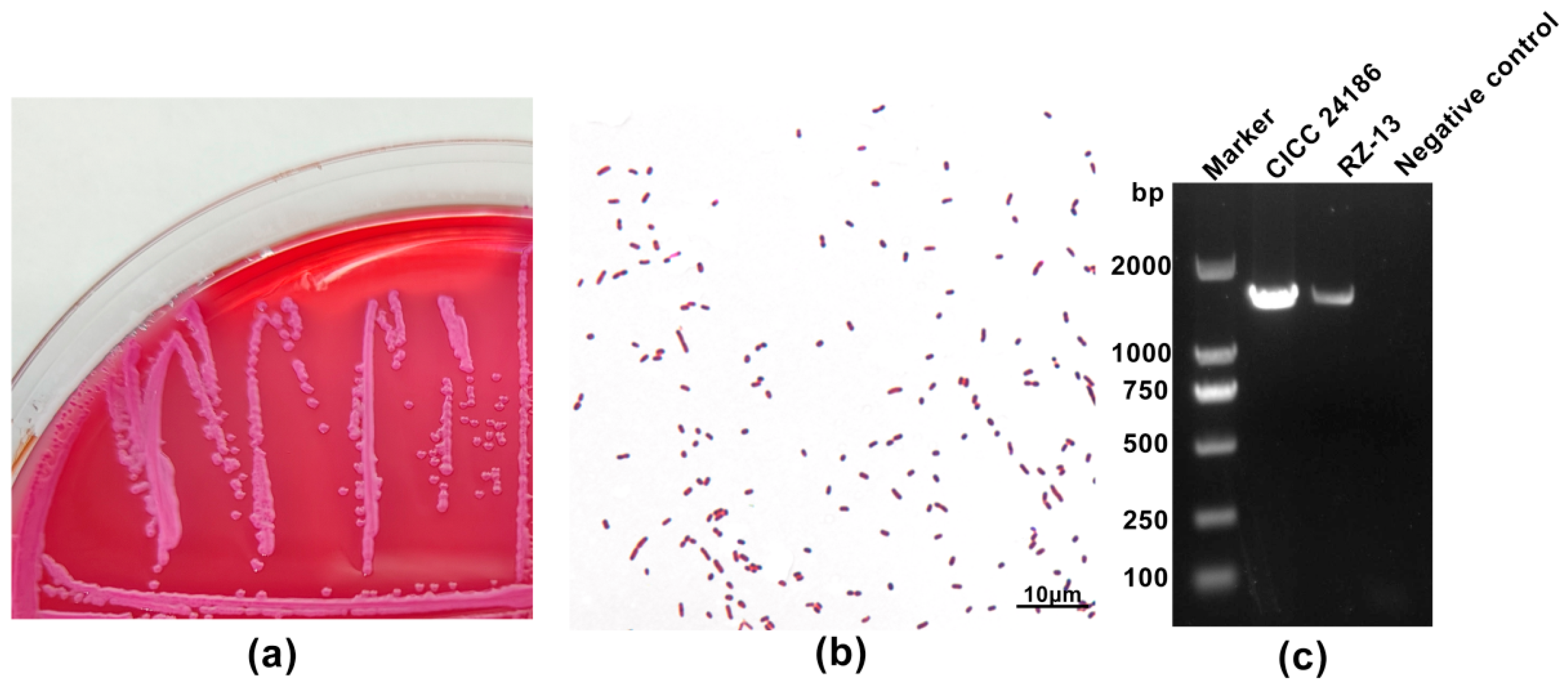
4.1. Molecular and Phenotypic Characteristics of the RZ-13 Strain

The same isolate strain, named RZ-13, was detected in all 66 samples from 11 cattle. It exhibited red colonies on MacConkey agar plates (Figure 1a), and its Gram-staining characteristics were consistent with the Escherichia genus, showing Gram-negative, short rods (Figure 1b). The 16S rRNA gene sequencing was compared with the NCBI database, identifying the RZ-13 strain as E. coli with an identification rate of 99.86%. The results of the PCR assay showed that the amplification of the RZ-13 strain using uidA gene amplification primers could amplify a band of the same size as that of the standard strain CICC 24186, with a band size of 1487 bp (Figure 1c). The toxin-typing results showed that no toxin genes in the national standard were detected, indicating that RZ-13 is an atypical pathogenic E. coli. The HEp-2 cell adhesion assay results showed that the RZ-13 strain exhibits a less obvious pattern of “stacked bricks”, similar to the Enteroaggregative Escherichia coli (EAEC) reference strain CICC 24186, on HEp-2 cells (Figure 2).

4.2. Antibiotic Resistance of RZ-13 Strain

The results of the drug sensitivity test showed that the RZ-13 strain was only susceptible to cefoxitin among the 14 antibiotics used, and it showed resistance to 13 antibiotics, including fluoroquinolones (norfloxacin, ofloxacin, enrofloxacin), sulfonamides (sulfamethoxazole), chloramphenicols (chloramphenicol), aminoglycosides (gentamicin, kanamycin), tetracyclines (doxycycline), cephalosporins (ceftriaxone, ceftazidime, cephalexin), and penicillins (ampicillin, amoxicillin).

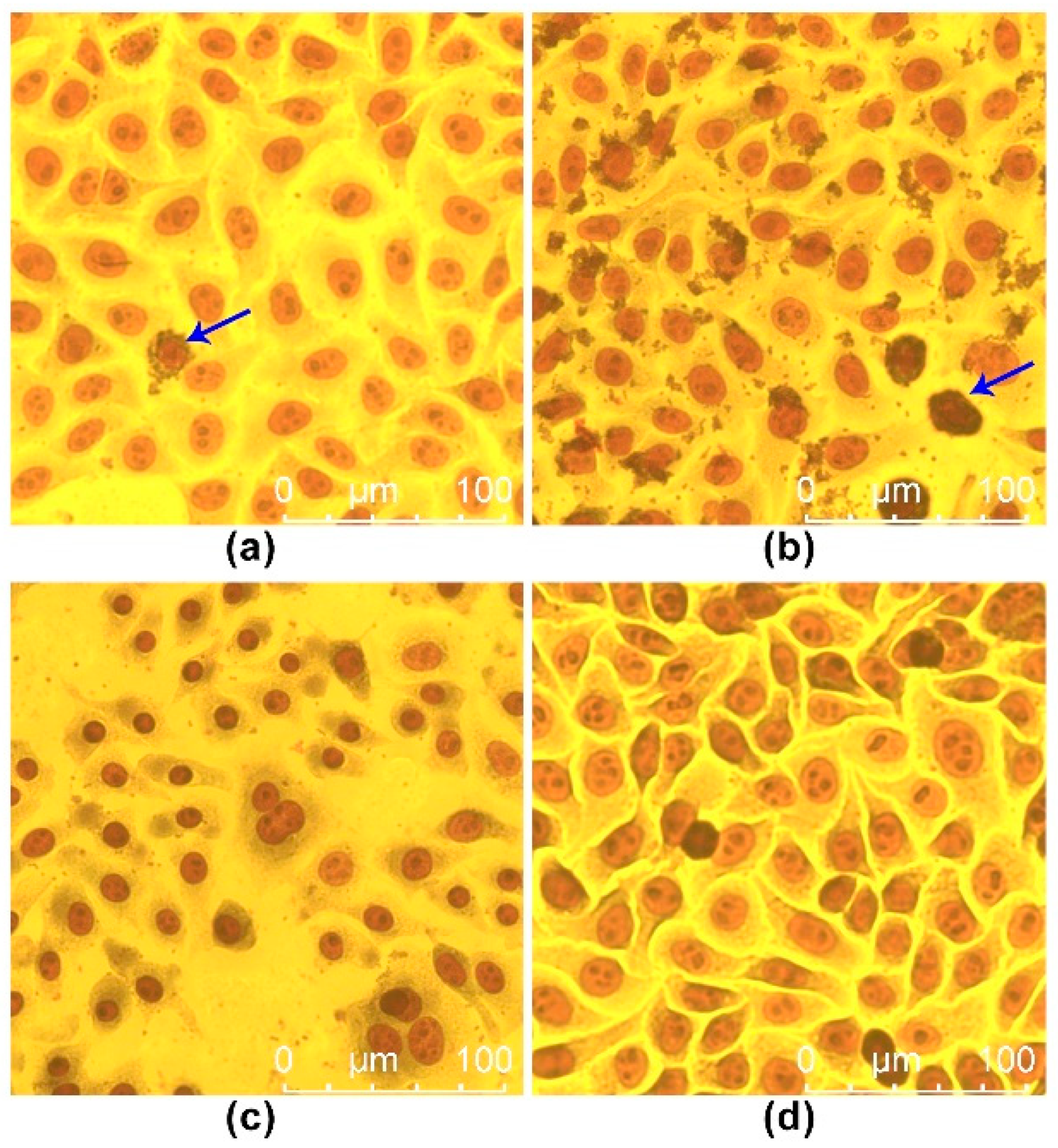
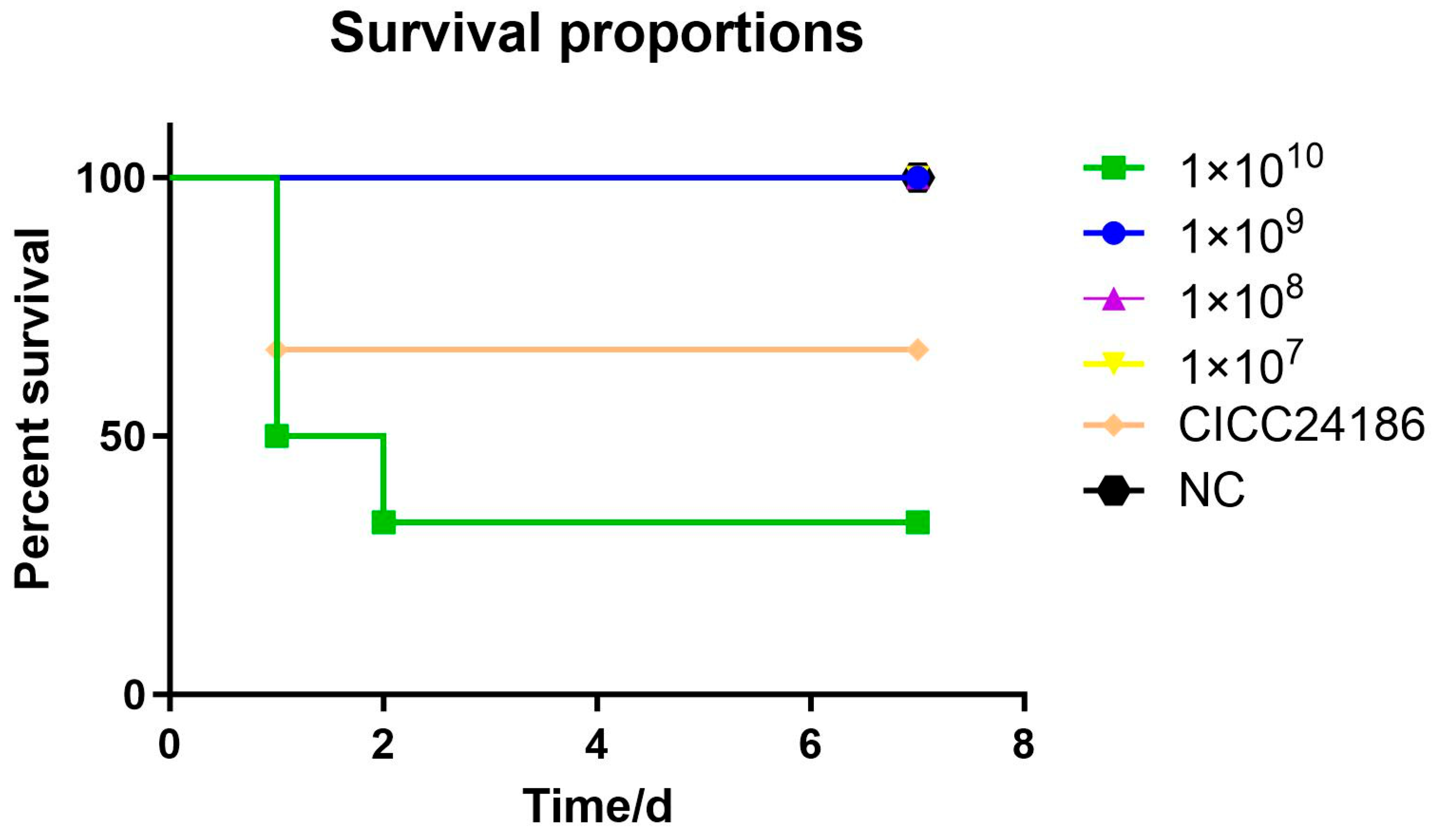
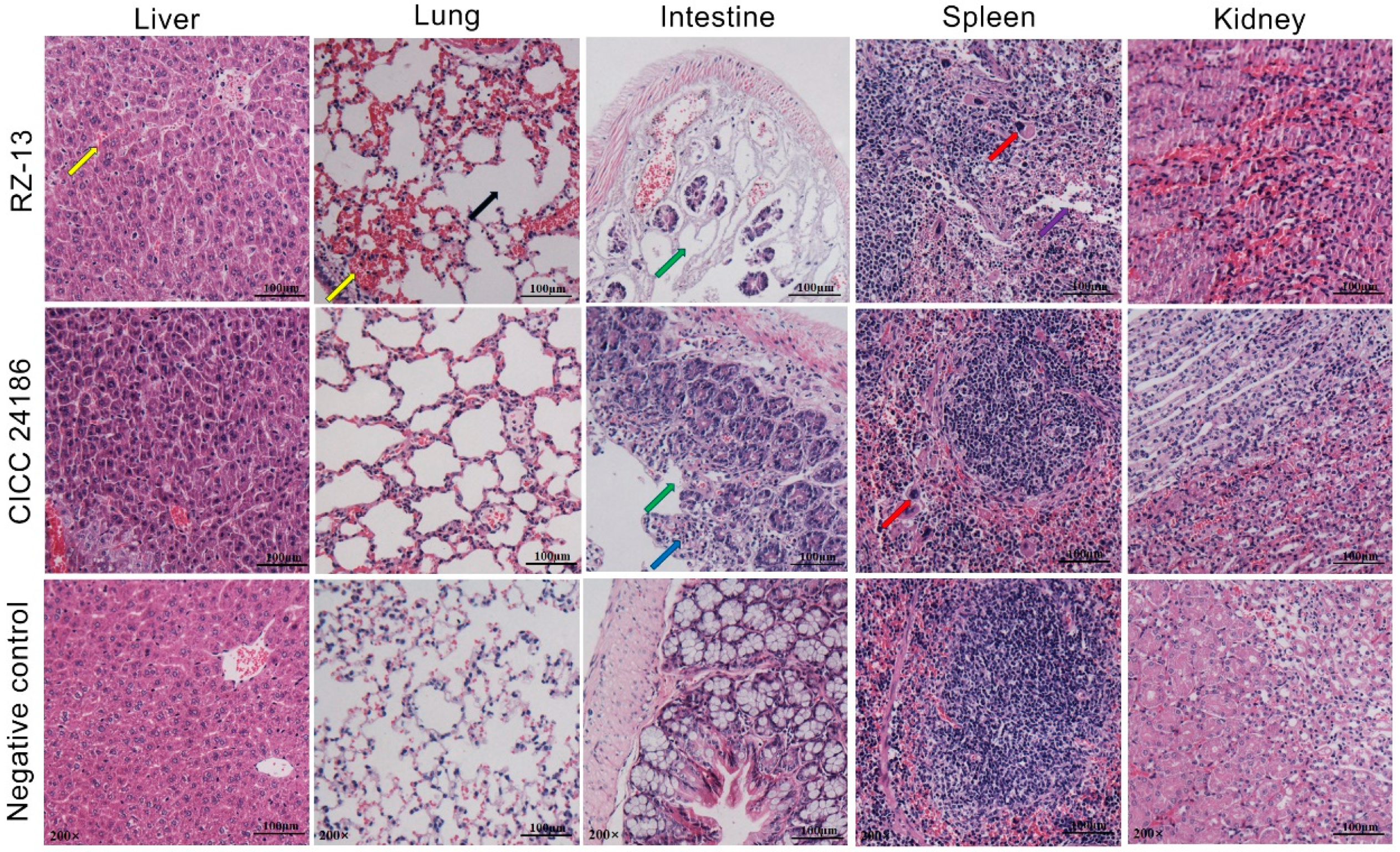
4.3. Analysis of Pathogenicity and Pathological Characteristics of RZ-13 Strain Infection in Mice

Mice in the 1010 RZ-13 group displayed symptoms such as disheveled hair and mental distress after 12 h of infection. Following injection with a 1010 dose of the RZ-13 strain, three mice perished within 24 h and one within 48 h (LD50 = 109.75 cfu/mL) (Figure 3). Mice in the RZ-13 group and CICC 24186 group experienced weight loss after infection with the 1010 dose compared to the control group. The fecal bacterial load in the RZ-13 group was higher in the dead mice than in the euthanized mice from the same group. We extracted bacteria from the livers and spleens of the infected dead mice in the CICC 24186 group and the 1010 RZ-13 group, with no bacteria isolated from the euthanized mice. After the intraperitoneal injection of RZ-13, there was a small amount of hemorrhage in the livers of the mice, with a large number of inflammatory cells infiltrated, and some hepatocytes underwent nuclear condensation, nuclear fragmentation, and nuclear lysis; there was intestinal villus detachment in the intestines, and the intestinal structure was missing; there was extensive hemorrhage in the lungs, and the alveolar wall was ruptured; there was inflammatory edema in the spleen, and the boundaries between the red medulla and white medulla were blurred, with a large number of macrophages infiltrated; there was obvious hemorrhage in the kidneys, and the boundaries between the cortex and medulla were blurred. After the intraperitoneal injection of CICC 24186, the mice had no obvious pathological changes in the livers, lungs, or kidneys; there was large inflammatory cell infiltration in the intestines, and the intestinal villi were detached; and there was macrophage infiltration in the spleen. The mice injected with saline had no pathological changes in any of the tissues (Figure 4).

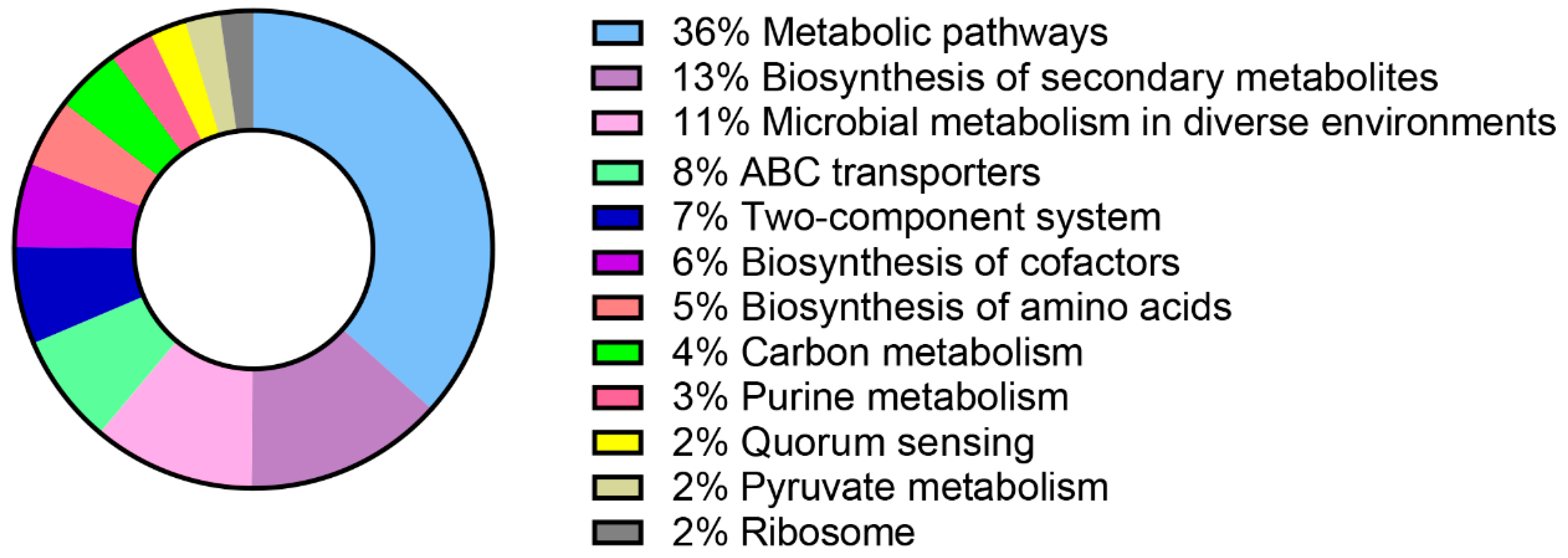
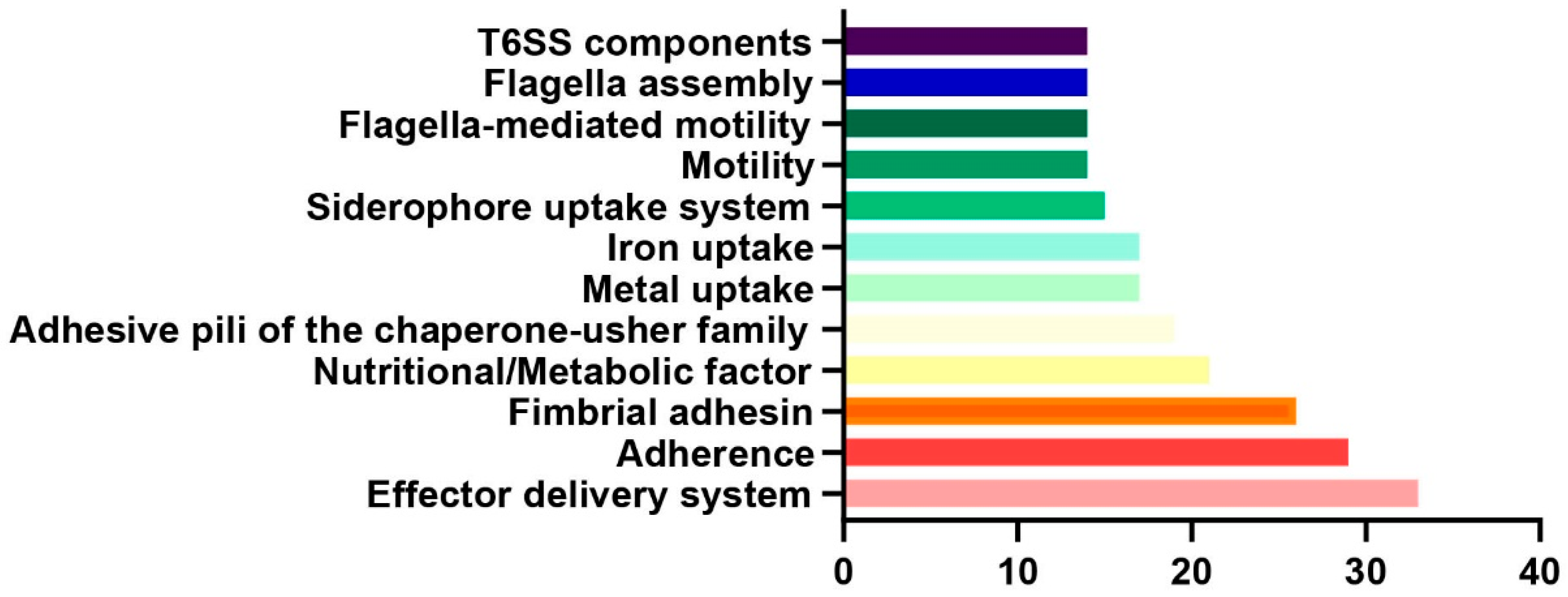
4.4. Comprehensive Bioinformatics Analysis of RZ-13 Strain

Results of the bioinformatics analysis showed that RZ-13 belonged to ST345, and its serotype was O134:H21. In terms of resistance genes, RZ-13 contained more resistance genes, including the quinolone resistance gene qepA1, the sulfonamide resistance genes sul2 and sul3, the lincomycin resistance gene lnu(F), the macrolide resistance gene mph(A), the rifamycin resistance gene arr-2, the tetracycline resistance gene tet(A), and another 17 resistance genes of 10 antibiotics. RZ-13 also carries the resistance genes mdtM, acrF, and emrD associated with efflux pumps, the tellurium resistance genes terD, terZ, and terW, the arsenic resistance genes arsC and arsR, the metal resistance gene fieF, and the multiple antibiotic resistance proteins marA and marR, and the presence of the resistance genes is consistent with the results of the drug resistance tests (Table 1). Regarding virulence factors, 86 pathogenic genes in RZ-13 involve various aspects such as adhesion, invasion, biofilm formation, regulatory factors, metabolic regulation, iron/heme acquisition, lipopolysaccharide synthesis, and efflux pumps. Each type is composed of several specific virulence factors with defined functions and activities (Supplementary Table S1). The KEGG pathway analysis showed that more than 60% of the codon genes in the RZ-13 strain were related to biological metabolism, of which 914 were related to metabolic pathways (914/2492), 337 were related to the biosynthesis of secondary metabolites (337/2492), and 273 differential genes were associated with microbial metabolism in different environments (273/2492) (Figure 5). An enrichment analysis of the codon genes by the GO database showed that 33 genes were related to the effector delivery system, 29 genes were related to adherence, and 26 genes were related to fimbrial adhesins (Figure 6).

5. Discussion

In 2019, the World Health Organization listed antibiotic resistance as one of the top ten global public health threats [48]. Bacteria are gradually showing resistance to one or multiple traditional antibiotics [49]. In this study, we first reported a case of MDR ExPEC leading to severe mortality in calf herds in China, and we isolated an MDR strain, RZ-13, in dairy cattle. In the early stages of clinical treatment, veterinarians used penicillin, streptomycin, cefotaxime, kanamycin, and gentamicin, with no obvious improvements. A global analysis of antibiotic resistance in ExPEC revealed that the highest resistance levels were to sulfonamides (35%) and fluoroquinolones (32%), followed by 9% resistance to extended-spectrum third- and fourth-generation cephalosporins. The resistances to the last-resort antibiotics colistin and carbapenems were relatively low at 0.7% and 0.1–0.2%, respectively [50]. In China, isolates exhibited high resistance rates to tetracycline (95.5%), ampicillin (95.5%), piperacillin (90.1%), sulfamethoxazole–trimethoprim (89.9%), chloramphenicol (85.2%), gentamicin (70%), and fluoroquinolones (65%) [5]. Additionally, 51.8%, 13.4%, and 44.7% of isolates were resistant to cefotaxime, ceftazidime, and cefepime, respectively, which are third- and fourth-generation cephalosporins. The antibiotic susceptibility test proved that RZ-13 presented an MDR ExPEC with wider antibiotic resistance than that of most others. The WGS results show that RZ-13 includes resistance genes to 10 types of antibiotics, such as quinolones, sulfonamides, lincosamides, macrolides, rifamycins, and tetracyclines. RZ-13 also carries resistance genes related to efflux pumps, including mdtM, acrF, and emrD, the tellurium resistance genes terD, terZ, and terW, the arsenic resistance genes arsC and arsR, the metal resistance gene fieF, and the multiple antibiotic resistance proteins marA and marR. The presence of these resistance genes indicates that RZ-13 strains exhibit a broad resistance spectrum. This finding underscores the necessity for further investigation into the distribution and transfer capabilities of the ARGs in the RZ-13 strain. The veterinarians on the farm tended to apply antibiotics based on their limited clinical experience rather than follow reasonable guidance, resulting in the enrichment of the resistance genes in RZ-13.
Although we intervened scientifically at the earliest opportunity upon receiving the consultation, 78 calves died and 30 infectious calves survived in this outbreak, illustrating the contagiousness and virulence of RZ-13. The experimental infection of the mice presented similar pathological changes to those of the clinical pathology, including symptoms such as multiple-organ hemorrhage. The histopathological investigation showed emerging inflammatory cell infiltration, increased neutrophils, and hemorrhage, indicating that death was caused by infection and not by improper manual operation. At a titer of 1010, the RZ-13 strain exhibited a higher mortality rate in the mice compared to that of the CICC 24186 strain. It seemed that RZ-13 had a stronger virulence and pathogenicity than the CICC 24186 strain. E. coli possesses a variety of virulence genes that contribute to its ability to cause illness [11]. When these toxins are secreted, they interact with intestinal cells, activating cyclic nucleotide production, which leads to a disruption in normal ion and fluid transport, culminating in secretory diarrhea [51]. Despite no classical toxin genes found in the RZ-13 genome except the truncated pic gene, the clinical manifestation of strong pathogenicity by the RZ-13 strain suggests the possible presence of additional toxins or alternative infection mechanisms, such as three type VI secretion system factors, 7 capsule genes, and 17 iron depletion-related genes in RZ-13. Iron acquisition genes [52], like the iutA gene, are vital for ensuring the survival and virulence of E. coli in iron-limited environments [53]. This could explain its heightened virulence compared to other strains, indicating the need for further investigation into its specific virulence factors and infection pathways. Although RZ-13 lacks the aggR gene, the presence of two adhesin fimbriae genes, eight type 1 fimbriae genes, six pili synthesis genes, and twelve biofilm formation-related genes collectively contribute to RZ-13, endowing it with strong adhesive capabilities. This allows it to form structures on cell surfaces similar to those of EAEC. The curli fimbriae are involved in biofilm formation, which helps the bacteria adhere to surfaces and resist environmental stresses [54]. By adhering firmly to host cell surfaces, E. coli can evade the host’s natural clearance mechanisms and establish a foothold for further infection. A total of 14 flagellum-related genes are involved in bacterial adhesion and invasion processes [55]. Flagellar genes enhance motility, aiding in reaching and colonizing different host tissues [52], and secretion systems facilitate the transport of effector molecules into host cells and mediate interactions between the bacterium and the host [56]. The complex and powerful flagellar system enables the RZ-13 strain to move outside the intestine and cause disease in other organs. The presence and expression of these virulence genes are undeniably key factors in the pathogenicity of E. coli. They appear in different combinations in different E. coli pathotypes, leading to a vast array of diseases caused by E. coli. Studying these genes not only deepens our understanding of the pathogenic mechanisms of E. coli but also provides a solid scientific basis for devising effective strategies for preventing and treating related diseases.
We propose implementing the following measures after this diarrhea outbreak: Firstly, we suggest reasonable feeding management for the dairy farm. All cows should be regularly tested for mastitis to avoid infection. Since the immune systems of calves have not fully developed, they tend to present various clinical symptoms, including diarrhea, pneumonia, fever, and even death, once they have ingested infected colostrum. As soon as an outbreak occurs, the infectious cows should be isolated from the calves. Colostrum and mature milk should be sterilized by pasteurization and the supply should be ensured. Secondly, veterinarians should identify the specific pathogen before deciding on the use of antibiotics to ensure targeted and effective treatment. To address the potential emergence of MDR pathogens in the future, we suggest conducting a thorough clinical diagnosis and drug resistance test prior to treatment. Thirdly, farms should implement strict sterilization measures to prevent the spread of pathogens, especially in contaminated areas affected by infectious cattle. Finally, given the widespread misuse of antibiotics, effective antibiotic alternatives should be applied in clinical treatment. Traditional Chinese medicine, antibiotic peptides, phages, and probiotics have broad applications for MDR E. coli. Emodin in rhubarb extract not only has efficient antibacterial effects against E. coli but also enhances animal immunity [57]. Bacteriophages have low toxicity, high specificity, and short development cycles and are an efficient treatment method against drug resistance in the post-antibiotic era [58,59]. Tianshi Xiao et al. isolated a bacteriophage with lytic activity against colistin-resistant E. coli strains that can achieve maximum bactericidal activity within 2 h [60]. Although there is some controversy, many studies have found that microecological agents can significantly alleviate diarrhea caused by E. coli [61,62]. Comprehensive prevention and control measures, including addressing the pathogen source, environmental control, diagnosis, and treatment, can help farms to promptly control and prevent the occurrence and development of similar epidemics. Upon our recommendation, the farm has already implemented the isolation of the diseased cattle, environmental disinfection, and the harmless treatment of the dead cows. Subsequently, we will continue to monitor the farm’s environment and diarrheic calves for MDR strains.

6. Conclusions

We report an outbreak of infection in calves accompanied by a high incidence, a high fatality rate, and multiple drug resistance. One ExPEC strain, designated as RZ-13, was isolated from deceased calves in Shandong Province of China. This strain exhibited strong pathogenicity and multiple antibiotic resistance, highlighting the need for careful monitoring and appropriate antibiotic use in veterinary practice. Given its potential role in causing calf diarrhea, pneumonia, and mass mortality, increased vigilance and surveillance are recommended to prevent further spread and to develop effective control strategies.

Supplementary Materials

The following supporting information can be downloaded at: https://www.mdpi.com/article/10.3390/microbiolres16030059/s1, Table S1: Virulence factor of RZ-13.

Author Contributions

L.Z. and H.-J.Y. conceived and designed the experiments. D.-D.Z., J.C., J.-J.Z., M.-M.Q., J.-Q.C. and X.-R.L. performed the experiments. C.-H.G. analyzed the data. L.Z. and D.-D.Z. analyzed the data and participated in the article writing. T.-F.M. directed and revised the article. S.-H.Y. and H.-J.Y. contributed reagents, materials, and analysis tools. X.-R.L. wrote the first draft of the paper, L.Z. critically revised the manuscript, and W.Z. and H.-J.Y. supervised all the work. All authors have read and agreed to the published version of the manuscript.

Funding

This research was supported by projects including the Key Research and Development Program of Ningxia Hui Autonomous Region (2024BBF02014), National Modern Agricultural Industry Technology System (CARS-36), Key Research and Development Program of Shandong Province Project (Rural Revitalization Science and Technology Innovation Boosting Action Plan) “Innovation and Application of Intelligent Production Technology for the Whole Chain of Precooked Meals” (No. 2022TZXD0021), Natural Science Foundation of Shandong Province (ZR2024QC275), and Key Research and Development Program of Shandong Province Project (Competitive Innovation Platform, 2022CXPT010).

Institutional Review Board Statement

The study was conducted in accordance with the Declaration of Helsinki, and approved by the Animal Experimental Ethical Inspection Committee of the Institute of Animal Sciences and Veterinary Medicine, the Shandong Academy of Agricultural Sciences (No.: IASVM-2024-018). Written informed consent was obtained from the owners for the participation of their livestock in this study. The goals, methods, possible dangers, and advantages of the research were all clearly stated on the consent form. In addition, we carefully followed the guidelines for animal welfare and took the necessary steps to reduce the pain and suffering that the animals went through.

Conflicts of Interest

The authors declare no competing interests regarding the publication of this article. The founding sponsors had no role in the design of the study, in the collection, analysis, or interpretation of the data, in the writing of the manuscript, or in the decision to publish the results.

References

  1. Foster, D.M.; Smith, G.W. Pathophysiology of diarrhea in calves. Vet. Clin. N. Am. Food Anim. Pract. 2009, 25, 13–36. [Google Scholar] [CrossRef] [PubMed]
  2. Garcia, J.; Pempek, J.; Hengy, M.; Hinds, A.; Diaz-Campos, D.; Habing, G. Prevalence and predictors of bacteremia in dairy calves with diarrhea. J. Dairy Sci. 2022, 105, 807–817. [Google Scholar] [CrossRef] [PubMed]
  3. Cho, Y.I.; Yoon, K.J. An overview of calf diarrhea—Infectious etiology, diagnosis, and intervention. J. Vet. Sci. 2014, 15, 1–17. [Google Scholar] [CrossRef] [PubMed]
  4. Arowska, J.; Futoma-Koloch, B.; Jama-Kmiecik, A.; Frej-Madrzak, M.; Ksiazczyk, M.; Bugla-Ploskonska, G.; Choroszy-Krol, I. Virulence factors, prevalence and potential transmission of extraintestinal pathogenic Escherichia coli isolated from different sources: Recent reports. Gut Pathog. 2019, 21, 10. [Google Scholar] [CrossRef]
  5. Li, X.; Hu, H.; Zhu, Y.; Wang, T.; Lu, Y.; Wang, X.; Peng, Z.; Sun, M.; Chen, H.; Zheng, J.; et al. Population structure and antibiotic resistance of swine extraintestinal pathogenic Escherichia coli from China. Nat. Commun. 2024, 15, 5811. [Google Scholar] [CrossRef]
  6. Karwowska, E. Antibiotic Resistance in the Farming Environment. Appl. Sci. 2024, 14, 5776. [Google Scholar] [CrossRef]
  7. Li, H.L.; Zhang, L. Molecular Mechanisms of Antibiotic Resistance and Antibacterial Strategies. Chin. J. Biochem. Mol. Biol. 2024, 40, 759–769. (In Chinese) [Google Scholar] [CrossRef]
  8. Sabino, Y.N.V.; Santana, M.F.; Oyama, L.B.; Santos, F.G.; Moreira, A.J.S.; Huws, S.A.; Mantovani, H.C. Characterization of antibiotic resistance genes in the species of the rumen microbiota. Nat. Commun. 2019, 10, 5252. [Google Scholar] [CrossRef]
  9. Nguyet, L.T.Y.; Keeratikunakorn, K.; Kaeoket, K.; Ngamwongsatit, N. Antibiotic resistant Escherichia coli from diarrheic piglets from pig farms in Thailand that harbor colistin-resistant mcr genes. Sci. Rep. 2022, 12, 9083. [Google Scholar] [CrossRef]
  10. Riley, D.S.; Barber, M.S.; Kienle, G.S.; Aronson, J.K.; von Schoen-Angerer, T.; Tugwell, P.; Kiene, H.; Helfand, M.; Altman, D.G.; Sox, H.; et al. CARE guidelines for case reports: Explanation and elaboration document. J. Clin. Epidemiol. 2017, 89, 218–235. [Google Scholar] [CrossRef]
  11. Vrenna, G.; Agosta, M.; Fox, V.; Rossitto, M.; Cortazzo, V.; Raimondi, S.; Lucignano, B.; Onori, M.; Mancinelli, L.; Pereyra Boza, M.D.C.; et al. Integrating Diagnostic Approaches in Infant Bacterial Meningitis Caused by a Non-K1 Escherichia coli: A Case Report. Antibiotics 2024, 13, 1144. [Google Scholar] [CrossRef] [PubMed]
  12. Caballero, M.; Rivera, I.; Jara, L.M.; Ulloa-Stanojlovic, F.M.; Shiva, C. Isolation and molecular identification of potentially pathogenic Escherichia coli and Campylobacter jejuni in feral pigeons from an urban area in the city of lima, Peru. Rev. Inst. Med. Trop. Sao Paulo 2015, 57, 393–396. [Google Scholar] [CrossRef] [PubMed]
  13. GB 4789.6-2016; National Standard for Food Safety Food Microbiological Testing for Diarrheal Escherichia coli. National Health and Family Planning Commission of the People’s Republic of China, China Food and Drug Administration: Beijing, China, 2016.
  14. Camacho, C.; Coulouris, G.; Avagyan, V.; Ma, N.; Papadopoulos, J.; Bealer, K.; Madden, T.L. BLAST+: Architecture and applications. BMC Bioinform. 2009, 10, 421. [Google Scholar] [CrossRef] [PubMed]
  15. Jønsson, R.; Struve, C.; Boisen, N.; Mateiu, R.V.; Santiago, A.E.; Jenssen, H.; Nataro, J.P.; Krogfelt, K.A. Novel aggregative adherence fimbria variant of enteroaggregative Escherichia coli. Infect. Immun. 2015, 83, 1396–1405. [Google Scholar] [CrossRef]
  16. Hackel, M.A.; Tsuji, M.; Yamano, Y.; Echols, R.; Karlowsky, J.A.; Sahm, D.F. Reproducibility of broth microdilution MICs for the novel siderophore cephalosporin, cefiderocol, determined using iron-depleted cation-adjusted Mueller-Hinton broth. Diagn. Microbiol. Infect. Dis. 2019, 94, 321–325. [Google Scholar] [CrossRef]
  17. CLSI Standard M02; Performance Standards for Antimicrobial Disk Susceptibility Tests, 14th ed. Clinical and Laboratory Standards Institute: Wayne, PA, USA, 2024.
  18. CLSI Quick Guide M02-Ed14-QG; CLSI. M02. Disk Diffusion Reading Guide, 2nd ed. Clinical and Laboratory Standards Institute: Wayne, PA, USA, 2024.
  19. Tamma, P.D.; Heil, E.L.; Justo, J.A.; Mathers, A.J.; Satlin, M.J.; Bonomo, R.A. Infectious Diseases Society of America 2024 Guidance on the Treatment of Antimicrobial-Resistant Gram-Negative Infections. Clin. Infect. Dis. 2024, ciae403. [Google Scholar] [CrossRef]
  20. Perrott, J.; Mabasa, V.H.; Ensom, M.H.H. Comparing outcomes of meropenem administration strategies based on pharmacokinetic and pharmacodynamic principles: A qualitative systematic review. Ann. Pharmacother. 2010, 44, 557–564. [Google Scholar] [CrossRef]
  21. Cirillo, I.; Vaccaro, N.; Turner, K.; Solanki, B.; Natarajan, J.; Redman, R. Pharmacokinetics, safety, and tolerability of doripenem after 0.5-, 1-, and 4-hour infusions in healthy volunteers. J. Clin. Pharmacol. 2009, 49, 798–806. [Google Scholar] [CrossRef]
  22. Sakka, S.G.; Glauner, A.K.; Bulitta, J.B.; Kinzig-Schippers, M.; Pfister, W.; Drusano, G.L.; Sörgel, F. Population pharmacokinetics and pharmacodynamics of continuous versus short-term infusion of imipenem-cilastatin in critically ill patients in a randomized, controlled trial. Antimicrob. Agents Chemother. 2007, 51, 3304–3310. [Google Scholar] [CrossRef]
  23. Peleg, A.Y.; Hooper, D.C. Hospital-acquired infections due to gram-negative bacteria. N. Engl. J. Med. 2010, 362, 1804–1813. [Google Scholar] [CrossRef]
  24. Tsuji, B.T.; Pogue, J.M.; Zavascki, A.P.; Paul, M.; Daikos, G.L.; Forrest, A.; Giacobbe, D.R.; Viscoli, C.; Giamarellou, H.; Karaiskos, I.; et al. International consensus guidelines for the optimal use of the polymyxins: Endorsed by the American College of Clinical Pharmacy (ACCP), European Society of Clinical Microbiology and Infectious Diseases (ESCMID), Infectious Diseases Society of America (IDSA), International Society for Anti-Infective Pharmacology (ISAP), Society of Critical Care Medicine (SCCM), and Society of Infectious Diseases Pharmacists (SIDP). Pharmacotherapy 2019, 39, 10–39. [Google Scholar] [CrossRef] [PubMed]
  25. Long, X.Q.; Zhao, W.X.; Li, X.Z.; Hu, W.J.; Chen, Z.L.; Xing, J.K.; Wang, Z.; Zhao, L.; Sao, Y.B.; Luo, Y. Isolation and identification of a pathogenic Escherichia coli strain from bovine milk and the antibacterial effect of pomegranate peel aqueous extract. Chin. J. Anim. Husb. 2025. (In Chinese) [Google Scholar] [CrossRef]
  26. Larsen, M.V.; Cosentino, S.; Rasmussen, S.; Friis, C.; Hasman, H.; Marvig, R.L.; Jelsbak, L.; Sicheritz-Pontén, T.; Ussery, D.W.; Aarestrup, F.M.; et al. Multilocus sequence typing of total-genome-sequenced bacteria. J. Clin. Microbiol. 2012, 50, 1355–1361. [Google Scholar] [CrossRef]
  27. Bartual, S.G.; Seifert, H.; Hippler, C.; Luzon, M.A.; Wisplinghoff, H.; Rodríguez-Valera, F. Development of a multilocus sequence typing scheme for characterization of clinical isolates of Acinetobacter baumannii. J. Clin. Microbiol. 2005, 43, 4382–4390. [Google Scholar] [CrossRef]
  28. Griffiths, D.; Fawley, W.; Kachrimanidou, M.; Bowden, R.; Crook, D.W.; Fung, R.; Golubchik, T.; Harding, R.M.; Jeffery, K.J.; Jolley, K.A.; et al. Multilocus sequence typing of Clostridium difficile. J. Clin. Microbiol. 2010, 48, 770–778. [Google Scholar] [CrossRef]
  29. Lemee, L.; Dhalluin, A.; Pestel-Caron, M.; Lemeland, J.F.; Pons, J.L. Multilocus sequence typing analysis of human and animal Clostridium difficile isolates of various toxigenic types. J. Clin. Microbiol. 2004, 42, 2609–2617. [Google Scholar] [CrossRef]
  30. Wirth, T.; Falush, D.; Lan, R.; Colles, F.; Mensa, P.; Wieler, L.H.; Karch, H.; Reeves, P.R.; Maiden, M.C.; Ochman, H.; et al. Sex and virulence in Escherichia coli: An evolutionary perspective. Mol. Microbiol. 2006, 60, 1136–1151. [Google Scholar] [CrossRef]
  31. Jaureguy, F.; Landraud, L.; Passet, V.; Diancourt, L.; Frapy, E.; Guigon, G.; Carbonnelle, E.; Lortholary, O.; Clermont, O.; Denamur, E.; et al. Phylogenetic and genomic diversity of human bacteremic Escherichia coli strains. BMC Genom. 2008, 9, 560. [Google Scholar] [CrossRef]
  32. Joensen, K.G.; Tetzschner, A.M.; Iguchi, A.; Aarestrup, F.M.; Scheutz, F. Rapid and Easy In Silico Serotyping of Escherichia coli Isolates by Use of Whole-Genome Sequencing Data. J. Clin. Microbiol. 2015, 53, 2410–2426. [Google Scholar] [CrossRef]
  33. Brandis, G.; Gockel, J.; Garoff, L.; Guy, L.; Hughes, D. Expression of the qepA1 gene is induced under antibiotic exposure. J. Antimicrob. Chemother. 2021, 76, 1433–1440. [Google Scholar] [CrossRef]
  34. Sköld, O. Resistance to trimethoprim and sulfonamides. Vet. Res. 2001, 32, 261–273. [Google Scholar] [CrossRef] [PubMed]
  35. Mukhopadhyay, R.; Rosen, B.P.; Phung, L.T.; Silver, S. Microbial arsenic: From geocycles to genes and enzymes. FEMS Microbiol. Rev. 2002, 26, 311–325. [Google Scholar] [CrossRef] [PubMed]
  36. Rosen, B.P. Resistance mechanisms to arsenicals and antimonials. J. Basic. Clin. Physiol. Pharmacol. 1995, 6, 251–263. [Google Scholar] [CrossRef]
  37. Du, D.; Wang-Kan, X.; Neuberger, A.; van Veen, H.W.; Pos, K.M.; Piddock, L.J.V.; Luisi, B.F. Multidrug efflux pumps: Structure, function and regulation. Nat. Rev. Microbiol. 2018, 16, 523–539. [Google Scholar] [CrossRef]
  38. Gomes, C.; Martínez-Puchol, S.; Palma, N.; Horna, G.; Ruiz-Roldán, L.; Pons, M.J.; Ruiz, J. Macrolide resistance mechanisms in Enterobacteriaceae: Focus on azithromycin. Crit. Rev. Microbiol. 2017, 43, 1–30. [Google Scholar] [CrossRef]
  39. Galimand, M.; Courvalin, P.; Lambert, T. Plasmid-mediated high-level resistance to aminoglycosides in Enterobacteriaceae due to 16S rRNA methylation. Antimicrob. Agents Chemother. 2003, 47, 2565–2571. [Google Scholar] [CrossRef]
  40. Chopra, I.; Roberts, M. Tetracycline antibiotics: Mode of action, applications, molecular biology, and epidemiology of bacterial resistance. Microbiol. Mol. Biol. Rev. 2001, 65, 232–260. [Google Scholar] [CrossRef]
  41. De Angelis, G.; Del Giacomo, P.; Posteraro, B.; Sanguinetti, M.; Tumbarello, M. Molecular Mechanisms, Epidemiology, and Clinical Importance of β-Lactam Resistance in Enterobacteriaceae. Int. J. Mol. Sci. 2020, 21, 5090. [Google Scholar] [CrossRef]
  42. Walter, E.G.; Taylor, D.E. Plasmid-mediated resistance to tellurite: Expressed and cryptic. Plasmid 1992, 27, 52–64. [Google Scholar] [CrossRef]
  43. Schwarz, S.; Kehrenberg, C.; Doublet, B.; Cloeckaert, A. Molecular basis of bacterial resistance to chloramphenicol and florfenicol. FEMS Microbiol. Rev. 2004, 28, 519–542. [Google Scholar] [CrossRef]
  44. Tang, M.J.; He, Z.L.; Pu, W.X. The Research Progress of Acquired 16S rRNA Methyltransferases. Acta Vet. Zootech. Sin. 2021, 52, 2369–2383. [Google Scholar] [CrossRef]
  45. Krause, K.M.; Serio, A.W.; Kane, T.R.; Connolly, L.E. Aminoglycosides: An Overview. Cold Spring Harb. Perspect. Med. 2016, 6, a027029. [Google Scholar] [CrossRef] [PubMed]
  46. Miranda, A.; Ávila, B.; Díaz, P.; Rivas, L.; Bravo, K.; Astudillo, J.; Bueno, C.; Ulloa, M.T.; Hermosilla, G.; Del Canto, F.; et al. Emergence of Plasmid-Borne dfrA14 Trimethoprim Resistance Gene in Shigella sonnei. Front. Cell. Infect. Microbiol. 2016, 6, 77. [Google Scholar] [CrossRef] [PubMed]
  47. Beggs, G.A.; Brennan, R.G.; Arshad, M. MarR family proteins are important regulators of clinically relevant antibiotic resistance. Protein Sci. 2020, 29, 647–653. [Google Scholar] [CrossRef]
  48. Zhang, H.; Zhao, D.; Quan, J.; Hua, X.; Yu, Y. mcr-1 facilitated selection of high-level colistin-resistant mutants in Escherichia coli. Clin. Microbiol. Infect. 2019, 25, 517.e1–517.e4. [Google Scholar] [CrossRef]
  49. Bernal-Córdoba, C.; Branco-Lopes, R.; Latorre-Segura, L.; de Barros-Abreu, M.; Fausak, E.D.; Silva-Del-Río, N. Use of antimicrobials in the treatment of calf diarrhea: A systematic review. Anim. Health Res. Rev. 2022, 23, 101–112. [Google Scholar] [CrossRef]
  50. Weerdenburg, E.; Davies, T.; Morrow, B.; Zomer, A.L.; Hermans, P.; Go, O.; Spiessens, B.; van den Hoven, T.; van Geet, G.; Aitabi, M.; et al. Global Distribution of O Serotypes and Antibiotic Resistance in Extraintestinal Pathogenic Escherichia coli Collected From the Blood of Patients With Bacteremia Across Multiple Surveillance Studies. Clin. Infect. Dis. 2023, 76, e1236–e1243. [Google Scholar] [CrossRef]
  51. Laohachai, K.N.; Bahadi, R.; Hardo, M.B.; Hardo, P.G.; Kourie, J.I. The role of bacterial and non-bacterial toxins in the induction of changes in membrane transport: Implications for diarrhea. Toxicon 2003, 42, 687–707. [Google Scholar] [CrossRef]
  52. Croxen, M.A.; Finlay, B.B. Molecular mechanisms of Escherichia coli pathogenicity. Nat. Rev. Microbiol. 2010, 8, 26–38. [Google Scholar] [CrossRef]
  53. Karbalaei, M.; Esmailpour, M.; Oksenych, V.; Eslami, M. Comprehensive Analysis of blaCTX-M1 Gene Expression Alongside iutA, csgA, and kpsMII Virulence Genes in Septicemic Escherichia coli Using Real-Time PCR. Microorganisms 2025, 13, 95. [Google Scholar] [CrossRef]
  54. Iyer, M.S.; Abhinand, P.A.; Hemalatha, C.R. Protein interaction studies of curli fimbriae in Escherichia coli biofilms. Bioinformation 2019, 15, 918–921. [Google Scholar] [CrossRef] [PubMed]
  55. Soria-Bustos, J.; Saldaña-Ahuactzi, Z.; Samadder, P.; Yañez-Santos, J.A.; Laguna, Y.M.; Cedillo-Ramírez, M.L.; Girón, J.A. The Assembly of Flagella in Enteropathogenic Escherichia coli Requires the Presence of a Functional Type III Secretion System. Int. J. Mol. Sci. 2022, 23, 13705. [Google Scholar] [CrossRef] [PubMed]
  56. Alvarez-Martinez, C.E.; Christie, P.J. Biological diversity of prokaryotic type IV secretion systems. Microbiol. Mol. Biol. Rev. 2009, 73, 775–808. [Google Scholar] [CrossRef] [PubMed]
  57. Gao, R.; Wang, C.; Han, A.; Tian, Y.; Ren, S.; Lv, W.; Chen, A.; Zhang, J. Emodin Improves Intestinal Health and Immunity through Modulation of Gut Microbiota in Mice Infected by Pathogenic Escherichia coli O1. Animals 2021, 11, 3314. [Google Scholar] [CrossRef]
  58. Alexyuk, P.; Bogoyavlenskiy, A.; Alexyuk, M.; Akanova, K.; Moldakhanov, Y.; Berezin, V. Isolation and Characterization of Lytic Bacteriophages Active against Clinical Strains of E. coli and Development of a Phage Antimicrobial Cocktail. Viruses 2022, 14, 2381. [Google Scholar] [CrossRef]
  59. Cui, J.Q.; Liu, W.H.; Zhang, C.; Zou, L.; Ren, H.Y. Genomic characterization of three bacteriophages infecting donkey-derived Escherichia coli. Virus Genes 2023, 59, 752–762. [Google Scholar] [CrossRef]
  60. Xiao, T.; Zhu, X.; Wang, W.; Jia, X.; Guo, C.; Wang, X.; Hao, Z. A novel lytic bacteriophage against colistin-resistant Escherichia coli isolated from different animals. Virus Res. 2023, 329, 199090. [Google Scholar] [CrossRef]
  61. Li, W.; Kai, L.; Jiang, Z.; He, H.; Yang, M.; Su, W.; Wang, Y.; Jin, M.; Lu, Z. Bifidobacterium longum, Lactobacillus plantarum and Pediococcus acidilactici Reversed ETEC-Inducing Intestinal Inflammation in Mice. Microorganisms 2022, 10, 2350. [Google Scholar] [CrossRef]
  62. Li, K.; Yang, M.; Jia, L.; Tian, M.; Du, J.; Wu, Y.; Yuan, L.; Li, L.; Ma, Y. The Prevention Effect of Lactobacillus plantarum 17-5 on Escherichia coli-Induced Mastitis in Mice. Probiotics Antimicrob. Proteins 2023, 15, 1644–1652. [Google Scholar] [CrossRef]
Figure 1. Isolation and identification of RZ-13 strain. (a) Represents the colony morphology of RZ-13 on MacConkey agar plates; (b) represents the Gram-staining result of RZ-13; the numbers in the lower right corner indicate the magnification of the images to 100×; (c) PCR results of RZ-13.
Microbiolres 16 00059 g001
Figure 2. Results of HEp-2 cell adhesion test. (a) Aggregative adherence pattern of RZ-13 on HEp-2 cells; (b) aggregative adherence pattern of CICC 24186 on HEp-2 cells; (c) aggregative adherence pattern of ATCC 25922 on HEp-2 cells; (d) blank control. Arrows indicate the adhesion of bacteria to HEp-2 cells. Scale bar is 100 µm.
Microbiolres 16 00059 g002
Figure 3. Survival curve of RZ-13-infected mice.
Microbiolres 16 00059 g003
Figure 4. Pathologic changes in various organs of mice. The numbers in the lower right corner indicate the magnification of the images to 200×. Yellow arrows indicate hemorrhage. Blue arrow indicates exudation of inflammatory cells. Green arrows indicate detachment of intestinal villi. Black arrow indicates alveolar rupture. Red arrows indicate macrophage exudation. Purple arrow indicates inflammatory edema. Scale bar is 100 µm.
Microbiolres 16 00059 g004
Figure 5. KEGG pathway analysis of codon genes in RZ-13 genome.
Microbiolres 16 00059 g005
Figure 6. GO enrichment analysis of codon genes in RZ-13 genome.
Microbiolres 16 00059 g006
Table 1. Antibiotic resistance genes (ARGs) in isolated strain, RZ-13.
CategoryAntibiotic-Resistant GenesFunction
QuinoloneqepA1Expels fluoroquinolone antibiotics from bacterial cells [33]
Sulfonamidesul2A variant that encodes dihydropteric acid synthase, making it less susceptible to sulfonamide antibiotics [34]
sul3
ArsenicarsCEliminates arsenic from the cell [35]
arsRRegulates the expression of related arsenic tolerance genes [36]
Lincosamidelnu(F)Protects ribosomes from the effects of lincoamide antibiotics
EffluxmdtMEncodes efflux pump proteins that remove a variety of antibiotics from cells [37]
acrF
emrD
Macrolidemph(A)Catalyzes the phosphorylation of macrolide antibiotics, conferring bacterial resistance to macrolides [38]
Rifamycinarr-2Encodes adenylate cyclase ribosyltransferase, which reduces antibiotic activity [39]
Tetracyclinetet(A)Codes tetracycline efflux pump [40]
Beta-lactamblaECCodes for beta-lactamase, which inactivates antibiotics [41]
blaCTX-M-55
blaTEM
TelluriumterDInvolved in the expulsion or isolation of tellurides [42]
terZ
terW
PhenicolfloREncodes a membrane transporter that excretes amidoalcohol antibiotics [43]
AminoglycosidermtB1Aminoglycoside antibiotics fail to bind to ribosomes [44,45]
aac(3)-IIdAcetylated aminoglycoside antibiotics [44,45]
aadA22Adenosine aminoglycoside antibiotics [44,45]
aph(6)-IdPhosphorylated aminoglycoside antibiotics [44,45]
aph(3′)-IaPhosphorylated aminoglycoside antibiotics [44,45]
TrimethoprimdfrA14Synthesizes a variant of dihydrofolate reductase, thereby reducing the sensitivity of sulfonamides to its effects [46]
Ferrous-iron efflux pump (FieF)fieFExpels iron ions from the cell
Multiple antibiotic resistance protein (MarA)marAMultiple-antibiotic-resistant proteins [47]
Multiple antibiotic resistance protein (MarR)marRRegulates the expression of MarA and other drug resistance genes [47]
Disclaimer/Publisher’s Note: The statements, opinions and data contained in all publications are solely those of the individual author(s) and contributor(s) and not of MDPI and/or the editor(s). MDPI and/or the editor(s) disclaim responsibility for any injury to people or property resulting from any ideas, methods, instructions or products referred to in the content.

