Article

Vimentin Methylation as a Potential Screening Biomarker for Colorectal Cancer in HIV-Helminth Co-Infected Individuals

by
Botle Precious Damane
1,2,*,
Shakeel Kader
3,
Mohammed Alaouna
4,
Pragalathan Naidoo
2,5,
Zodwa Dlamini
4,6 and
Zilungile Lynette Mkhize-Kwitshana
2,5,7,*
1
Department of Surgery, Steve Biko Academic Hospital, University of Pretoria, Hatfield 0028, South Africa
2
Department of Medical Microbiology, School of Medicine, College of Health Sciences, University of KwaZulu-Natal, Nelson R. Mandela Medical School Campus, Durban 4041, South Africa
3
Department of Surgery, University of KwaZulu-Natal, Congella, Durban 4001, South Africa
4
SAMRC Precision Oncology Research Unit (PORU), DSTI/NRF SARChI Chair in Precision Oncology and Cancer Prevention (POCP), Pan Africa Cancer Research Institute (PACRI), University of Pretoria, Hatfield 0028, South Africa
5
Division of Research Capacity Development, South African Medical Research Council (SAMRC), Cape Town 7505, South Africa
6
Wolfson Wohl Cancer Research Centre, School of Cancer Sciences, University of Glasgow, Garscube Estate, Glasgow G61 1QH, UK
7
Biomedical Sciences Department, School of Life and Consumer Sciences, College of Agriculture and Environmental Sciences, University of South Africa, Florida Campus, Johannesburg 1710, South Africa
*
Authors to whom correspondence should be addressed.
Microbiol. Res. 2025, 16(11), 236; https://doi.org/10.3390/microbiolres16110236
Submission received: 12 September 2025 / Revised: 15 October 2025 / Accepted: 3 November 2025 / Published: 11 November 2025
(This article belongs to the Special Issue Host–Microbe Interactions in Health and Disease)

Abstract

Colonoscopy remains the gold standard for colorectal cancer (CRC) screening, but its invasiveness, cost, and limited availability in resource-constrained settings pose major barriers. Stool-based methylated DNA biomarkers, such as vimentin, offer sensitive, non-invasive alternatives. Given the high burden of HIV and helminth co-infections in sub-Saharan Africa and their potential contribution to cancer susceptibility, this study investigated whether stool-derived vimentin methylation could detect early oncogenic changes in these high-risk groups. In this retrospective cross-sectional study, archived stool samples from 62 South African adults were stratified into five groups: uninfected controls, HIV-infected only, helminth-infected only, HIV-helminth co-infected, and CRC-confirmed patients. DNA was extracted, bisulfite-converted, and analyzed for vimentin methylation using a high-resolution melt assay. Fecal occult blood testing (FOBT) was also performed. Vimentin methylation differed significantly across groups (p < 0.0001). CRC cases showed 90% methylation, confirming its role as a CRC biomarker. Interestingly, vimentin methylation frequencies were also observed in HIV-only (92.9%, p < 0.0001 vs. controls), helminth-only (93.3%, p < 0.0001), and HIV-helminth co-infected (77.9%, p < 0.0001) individuals without diagnosed cancer, compared to 10% in controls. Methylation levels in infected groups were not significantly different from CRC patients (all p > 0.05), suggesting infection-induced epigenetic changes of comparable magnitude to malignancy. To support these results, DNMT1–RG108 molecular docking (PDB 4WXX, Maestro 2025-3) demonstrated stable binding (GlideScore −6.285 kcal/mol; ΔG_bind −49.61 kcal/mol) via hydrogen bonding with Glu1266 and Asn1578 and π–π stacking with Phe1145, providing a mechanistic explanation for infection-driven vimentin methylation. No significant differences were found between infected groups. FOBT was positive in 83.3% of CRC cases, with only sporadic positives in infected groups. These findings provide novel evidence that chronic HIV and helminth infections are associated with vimentin promoter methylation at levels indistinguishable from CRC. This supports the hypothesis that persistent infection-driven inflammation promotes early epigenetic reprogramming toward oncogenesis. In high-burden African settings, stool-based methylation assays could serve as early diagnostic tools to identify at-risk individuals long before clinical disease manifests, enabling targeted surveillance and prevention.

1. Introduction

Several helminth species, especially schistosomes, have been implicated in the initiation and progression of cancer. The association between schistosomiasis and cancer was first described in the early 1900s, with Schistosoma haematobium strongly linked to bladder cancer and Schistosoma mansoni to hepatic carcinoma [1]. More recent studies have expanded this view, showing that additional helminths, including blood flukes (Schistosoma haematobium, Schistosoma japonicum, Schistosoma mansoni) and liver flukes (Clonorchis sinensis, Opisthorchis viverrini), contribute to human cancers [2]. However, current evidence supports but also complicates this association. Experimental studies indicate that Schistosoma and Trichuris infections promote CRC progression, whereas Taenia species may exert a protective role [3]. Schistosoma japonicum induces immunosuppressive responses that drive CRC and worsen outcomes. Interestingly, other parasites such as Eimeria granulosa can trigger anti-cancer immune activity at early stages of infection, but this effect diminishes as the infection advances, shifting immunity toward a Th2-dominant profile that promotes tumor growth and metastasis [3,4]. It is important to note that the protective effects of helminth infections may be context dependent. For example, in co-infections with Helicobacter pylori, helminths can skew immunity toward Th2 and regulatory pathways, mitigating the pro-inflammatory damage induced by H. pylori and thereby reducing the risk of gastric disease complications, including cancer [5]. However, in the setting of HIV, which is already well known for its immunosuppressive effects and modulation of genetic pathways that increase susceptibility to cancer and infections, including helminths [6], the dynamics shift dramatically. In this context, some helminths, particularly the intestinal groups, not only promote a cancer-favoring immunosuppressive microenvironment but also contribute to direct mechanical damage to the colon. Moreover, through the release of secretory molecules, helminths further modulate host immunity and tissue integrity, ultimately supporting carcinogenesis [7].
Further cancer-promoting mechanisms shared by helminth infections and the tumor microenvironment (TME) involve shifts in immune cell metabolism. Helminths modulate the metabolic programming of infiltrating immune cells, particularly macrophages. For example, pattern recognition receptor activation, such as toll-like receptor 4 (TLR4) signaling by lipopolysaccharide, enhances hypoxia-inducible factor 1-α (HIF1-α) expression in M1 macrophages, driving glycolysis for rapid energy acquisition. In contrast, helminth infections promote interleukin-4 (IL-4) production and polarization toward M2 macrophages. IL-4, a hallmark of type 2 immune responses, enhances oxidative phosphorylation (OXPHOS) and mitochondrial respiration in M2 macrophages surrounding parasites. This metabolic reprogramming, with reduced glycolysis, decreases the availability of intermediates required for nucleotide synthesis and limits proliferative capacity in macrophages. Notably, parallels can be drawn between helminth-infected tissue and the TME: in both contexts, M1/Th1 phenotypes are suppressed, impairing the energy-intensive demands of infiltrating immune cells and creating an immunosuppressive niche favorable for disease progression. This convergence highlights how helminth infections may reinforce tumor-associated metabolic programs, further promoting cancer initiation and progression [7].
Helminth infections contribute to genetic instability, a hallmark of CRC. Jusakul et al. [8] showed that in fluke-associated cholangiocarcinoma (CCA), widespread epigenetic disruption drives spontaneous 5-methylcytosine deamination and CpG > TpG transitions, whereas in fluke-negative CCA, intrinsic mutations occur first, followed by methylation changes. Integrative clustering revealed four CCA subgroups: fluke-positive tumors enriched in ERBB2 amplifications and TP53 mutations, and fluke-negative tumors marked by either high copy-number alterations with PD-1/PD-L2 expression or epigenetic mutations (IDH1/2, BAP1) with FGFR/PRKA rearrangements. Whole-genome analysis also identified FGFR2 3′ UTR deletions as a driver of FGFR2 upregulation, alongside noncoding promoter mutations altering H3K27me3 sites. Distinct DNA hypermethylation patterns further suggest divergent mutational pathways between clusters [8]. These findings highlight how helminth infections not only remodel immunity and metabolism but also fuel genetic and epigenetic instability, processes central to CRC initiation and progression. Another helminth-associated pro-cancer mechanism, similar to HIV immune suppression, includes the increased expression of immune checkpoints such as PD-1. This limits the cancer-immune surveillance.
CRC harbors oncogenic mutations that serve as potential biomarkers and treatment targets. Amid the global burden of cancer, which claimed approximately 10 million lives worldwide in 2022 [9], efforts in therapeutic research are increasingly focused on targeted therapies. However, access to these advanced treatments remains determined by socioeconomic disparities, particularly pronounced in developing regions. In South Africa, communities are starkly divided into rural, township, and suburban areas, with rural populations often comprising the most economically disadvantaged. Healthcare resources, including state-of-the-art facilities, are predominantly concentrated in suburban private hospitals, accessible to a minority with greater financial means [10]. Helminth infections are highly prevalent among people living in rural and low-resource peri-urban areas, where access to costly CRC screening methods such as colonoscopy is limited. Identifying predictive CRC biomarkers in these populations could enable the establishment of targeted screening programs for individuals with helminth infections, either alone or in combination with HIV co-infection [11,12].
Dysregulated methylated events were shown to be potentially effective, non-invasive, and easily accessible screening markers of CRC. In 2004, a stool DNA panel containing APC, TP53, KRAS, and BAT26 could detect CRC at 52% sensitivity and 94% specificity. The stool DNA panel detected 16 of 31 invasive cancers, and the fecal occult blood test (FOBT) detected 4 of 31 cancers with a sensitivity of 12.9%. There was a difference of 13 cancers that were not detected by FOBT but picked up by the stool DNA panel [13]. The FOBT has been used routinely over the years as a diagnostic marker for CRC [14]. The effectiveness of using a multitarget stool sample was further studied by this approach to a commercial fecal immunochemical test (FIT) in average-risk CRC patients. The DNA test showed higher sensitivity for detecting CRC (92.3%) and advanced precancerous lesions (42.4%), exceeding FIT by about 20 percentage points. [15]. Similarly, another study reported that stool DNA–Fecal Occult Blood Testing (sDNA-FOBT) exhibited significantly higher sensitivity for the detection of CRC and precancerous lesions compared with conventional FOBT (61.70% vs. 51.06%, p < 0.01). The authors concluded that sDNA-FOBT represents a promising non-invasive strategy for CRC screening. However, they also emphasized the necessity of validating these results in studies with larger cohorts to ensure broader applicability [16].
Vimentin methylation detected in the stool of CRC patients has been shown to have a specificity of 100%, a sensitivity of 60%, and a positive predictive value of 100% [17]. Vimentin methylation assay is now a validated test known as the ColoGuard assay, used for the early detection of CRC. Epi proColon® 2.0 CE is another blood-based, FDA-approved test for screening of CRC [18]. These methods have been shown to be more cost-effective than the current widespread use of colonoscopy, especially in less privileged areas such as sub-Saharan Africa. The use of stool-derived DNA-based assays allows collection of stool from patients at the comfort of their own home, which will later be dropped off at the nearest health facility for transport to relevant testing sites. Studies comparing cost effectiveness between the colonoscopy and methylation studies have confirmed the cost effectiveness of the latter. For instance, a study evaluating CRC screening strategies in Alaska Native populations compared multitarget stool DNA (MT-sDNA) testing, colonoscopy, and FIT. All three approaches reduced healthcare costs and improved quality-adjusted life years (QALYs) relative to no screening. Among the strategies, MT-sDNA provided the greatest gain in QALYs. Using a Markov model, the analysis demonstrated that MT-sDNA screening was cost-effective compared with both colonoscopy and FIT across a broad range of adherence scenarios [19]. A study by Rui et al. (2025) found that all noninvasive CRC screening methods they investigated decreased cancer incidence and mortality compared with no screening. Under perfect adherence, colonoscopy every 10 years was the most cost-effective, but in real-world settings, every-3-year multitarget stool RNA testing (mt-sRNA) was preferred. Cost, test performance, and adherence were key determinants. Thus, this indicated that noninvasive methods can be cost-effective alongside all guideline-recommended strategies [20]. Similarly, another study showed that a minimum Centers for Medicare & Medicaid Services’ thresholds (CMS-minimum) blood-based test reduced cancer burden but was less effective than multitarget stool DNA testing, FIT, or colonoscopy, and would only rival FIT if participation rates were markedly higher. Together, these findings reinforce the value of DNA-based stool assays as cost-effective, noninvasive strategies, particularly when adherence to traditional screening methods is suboptimal [21]. Therefore, this study used vimentin methylation and FOBT to detect potential CRC risk in HIV and helminth infected individuals. According to our knowledge, no study has looked at CRC predictive molecular markers in HIV alone and HIV-helminths co-infected patients in Sub-Saharan Africa. Thus, the overarching goal of this study is to assess the association of helminth infections with oncogenesis concerning immunomodulation, particularly in HIV-helminth co-infected individuals.

2. Materials and Methods

2.1. Study Design and Setting

This was a retrospective cross-sectional sub-study nested within a larger ethically approved project (BE351/19). Additional approval for this sub-study was granted by the Biomedical Research Ethics Committee of the University of KwaZulu-Natal (BREC/00005458/2023). The main study was conducted in a peri-urban area south of Durban, KwaZulu-Natal, South Africa, between March 2020 and May 2021. A detailed account of the study design and setting has been published [22].

2.2. Study Population and Sampling

This sub-study utilized stored stool samples collected from consenting participants enrolled in the main study. During the collection process, unpreserved stool samples were aliquoted immediately after collection and stored at −80 °C until DNA extraction. The interval between collection and analysis ranged from approximately 2 to 3 years. Although stool without preservative may be more prone to DNA degradation at room temperature, prior studies have shown that nucleic acids remain stable when promptly frozen and maintained at −80 °C. Sixty-two (62) participants were purposively selected based on their infection status and the availability of complete datasets for subsequent analyses. Participants were stratified into five groups: HIV-negative and helminth-negative uninfected controls (n = 10), CRC-positive controls (n = 10), HIV-infected only (n = 14), helminth-infected only (n = 15), and HIV-helminth co-infected (n = 13). HIV infection was confirmed using standard serological assays in line with national guidelines, while helminth infections were identified through stool microscopy and antigen-based tests, as described in our related publication [23] and unpublished data. Ambiguous or discordant cases were excluded from downstream analyses. These combined diagnostic and stratification approaches ensured accurate classification and minimized bias from mixed or uncertain infections. The CRC positive controls were confirmed cancer patients due for surgery. Control reactions for methylation-specific PCR were prepared using the EpiTect control DNA set (QIAGEN, Hilden, Germany) and used to verify primer specificity. A 50% methylated control was generated by mixing equal volumes of 100% methylated and 0% methylated bisulfite-converted DNA.

2.3. Experimental Procedures

DNA Extraction from Stool Samples
DNA was extracted from stool samples using the ZymoBIOMICS™ DNA Miniprep Kit (Zymo Research, Irvine, CA, USA), optimized for microbiome and metagenome analyses. Approximately 200 mg of stool was added to a ZR BashingBead™ Lysis Tube, followed by the addition of 750 μL ZymoBIOMICS™ lysis solution. The tube was capped tightly and subjected to mechanical lysis using an Omni Bead Ruptor 4 Bead Mill homogenizer (Lasec, Johannesburg, South Africa) operated under optimized speed and duration settings suitable for 2 mL tube holders. Following lysis, tubes were centrifuged at 10,000× g for 1 min to pellet the debris. Up to 400 μL of the resulting supernatant was transferred to a Zymo-Spin™ III-F filter placed in a collection tube and centrifuged at 8000× g for 1 min before the filter was discarded.
A volume of 1200 μL of ZymoBIOMICS™ DNA binding buffer was added to the filtrate and mixed thoroughly, and 800 μL of the mixture was applied to a Zymo-Spin™ IICR column in a collection tube before centrifuging at 10,000× g for 1 min. This step was repeated to ensure maximum DNA capture. The bound DNA was then washed by sequential addition of 400 μL of ZymoBIOMICS™ DNA wash buffer 1 and two washes with ZymoBIOMICS™ DNA wash buffer 2 (700 μL followed by 200 μL), with centrifugation at 10,000× g for 1 min after each wash. The spin column was then transferred to a clean 1.5 mL microcentrifuge tube, and DNA was eluted by adding 100 μL of ZymoBIOMICS™ DNase/RNase-free water directly to the column matrix. After incubating for 1 min, the column was centrifuged at 10,000× g for 1 min to collect the purified DNA. To remove potential PCR inhibitors, 600 μL of ZymoBIOMICS™ HRC prep solution was added to a Zymo-Spin™ III-HRC filter in a fresh collection tube and centrifuged at 8000× g for 3 min. The previously eluted DNA was then passed through the prepared HRC Filter by centrifugation at exactly 16,000× g for 3 min. The resulting DNA was free of inhibitors and suitable for PCR and other downstream applications.
Bisulfite Conversion of Genomic DNA
Bisulfite modification of the eluted DNA was performed using the EpiJET Bisulfite Conversion Kit (Thermo Fisher Scientific, Waltham, MA, USA). Prior to use, the wash buffer, desulfonation buffer, and modification reagent were prepared according to the manufacturer’s instructions. For each reaction, 20 μL of 400 ng genomic DNA was transferred to a PCR tube. A total of 120 μL of freshly prepared modification reagent was added to each sample, mixed by pipetting, and centrifuged briefly. Bisulfite conversion was carried out using protocol A on the manufacturer’s instruction manual. The protocol consists of incubation at 98 °C for 10 min followed by 60 °C for 150 min in a QuantStudio™ 5 Real-Time PCR System (Thermo Fisher Scientific, Waltham, MA, USA). Following conversion, 400 μL of binding buffer was added to a DNA purification microcolumn placed in a collection tube. The converted DNA sample was then added to the column and mixed thoroughly before centrifugation at 12,000 rpm for 30 s, and the flow-through discarded. The column was washed with 200 μL of wash buffer, centrifuged again, and treated with 200 μL of desulfonation buffer for 20 min at room temperature. After another centrifugation at 12,000 rpm for 30 s, two additional washes with 200 μL of wash buffer were performed, with the final wash centrifuged for 60 s. Purified DNA was eluted by adding 10 μL of elution buffer directly to the column, followed by centrifugation at 12,000 rpm for 60 s. We did not amplify the converted DNA but instead stored it at −20 °C until further use.
Amplifying bisulfite-converted DNA extracted from stool samples presents several technical challenges that can compromise the accurate detection of methylated genes. Firstly, bisulfite treatment chemically modifies unmethylated cytosines to uracil, a process known to cause substantial DNA fragmentation and degradation [24,25]. When combined with the naturally fragmented and low-quality DNA typically recovered from stool, due to enzymatic digestion, bacterial overgrowth, and environmental exposure, the resulting DNA may be too degraded for successful amplification [26]. In addition, bisulfite conversion requires multiple purification steps, each of which contributes to significant DNA loss, particularly problematic in samples with limited input material [27]. Finally, primer design for bisulfite-converted DNA is complicated due to the non-complementary nature of converted strands and the loss of cytosine-guanine pairing. Without carefully optimized, short amplicon primers, amplification is often inefficient or fails altogether [28]. An in-house optimization was performed using previously established primer sets, as shown in Table 1.
High-Resolution Melting Analysis
High-Resolution Melting (HRM) analysis enhances post-PCR analysis by enabling precise genotyping and mutation scanning through the detection of subtle DNA sequence variations. It shortens analysis time by auto-calling genotypes, excluding no-template controls, and reducing subjectivity via automated clustering of unknown variants. The platform also supports streamlined data visualization and multi-target analysis on a single plate, improving workflow efficiency.
High-resolution melt analysis was performed using the MeltDoctor™ HRM Reagents (Thermo Fisher Scientific, Waltham, MA, USA) on a QuantStudio™ 5 Real-Time PCR System (Thermo Fisher Scientific, Waltham, MA, USA), in combination with the high-resolution Melt Software v3.0 for data analysis. Each HRM reaction was carried out in a final volume of 10 μL using the MeltDoctor™ HRM Master Mix, with 10 ng of bisulfite-converted template DNA as input. To validate HRM performance and ensure accurate methylation discrimination, we included the EpiTect PCR Control DNA Set (Qiagen, Hilden, Germany) in each experiment. This human DNA control set contains both bisulfite-converted methylated and unmethylated DNA, as well as unconverted unmethylated DNA. These were used to generate standard curves and validate primer efficiency for methylation-sensitive discrimination. Reactions were assembled on ice and included 1x MeltDoctor™ HRM Master Mix, 0.2 μM each of forward and reverse primers specific for the target region, and DNA template. Negative controls (no-template controls) and positive controls from the EpiTect DNA set were included in all runs. Thermal cycling parameters, including denaturation, annealing/extension, and HRM steps, were optimized. HRM analysis was performed with a continuous ramping mode selected and a ramp rate of 0.1 °C/s. Data acquisition occurred continuously throughout the melting phase. Instrument filters were optimized using the HRM-specific Filter-1 setting (Table 2).
Following amplification, the melt curves were normalized, temperature-shifted, and analyzed using the high-resolution Melt Software v3.0 (Thermo Fisher Scientific, Waltham, MA, USA), which enables genotype and methylation calling based on curve shape and melting temperature differences. All assays were run in triplicate, and only samples with consistent amplification and clear melting profiles were included in the final analysis.
Validation of methylation cutoffs
To confirm assay specificity and ensure reliable interpretation of methylation status, commercially available DNA standards, EpiTect PCR Control DNA Set (Qiagen, JHB, SA, Cat. No. 59695), representing 0%, 50%, 75%, and 100% methylation, were used. The 0% and 100% standards served as negative and positive controls, respectively, while intermediate ratios (50% and 75%) were generated by mixing defined amounts of fully methylated and unmethylated DNA as per the manufacturer’s instructions. This gradient provided reference points to validate the assay’s ability to detect both full and partial methylation.
Computational Structural Modeling
The molecular docking and binding-free-energy analyses were performed using the Maestro v14.5.131 and MMshare v7.1.131 (Schrödinger Suite Release 2025-3, Windows x64). The modules used, Glide in SP (Standard Precision) mode and Prime MM-GBSA re-scoring, are integral components of this release. The crystal structure of human DNA methyltransferase 1 (DNMT1; PDB ID: 4WXX) was imported into Maestro and prepared with the Protein Preparation Wizard. Chain A, which contains the catalytic domain and the co-crystallized S-adenosyl-homocysteine (SAH) cofactor, was retained, whereas chain B and non-essential ions or solvent molecules were removed. Bond orders were reassigned, hydrogens were added, and missing side chains were rebuilt. The structure was optimized with the OPLS4 force field at physiological pH 7.4 ± 0.5 and heavy-atom restraints (RMSD 0.30 Å).
RG108 (N-[4-(hydroxycarbamoyl) benzoyl] acetamide; PubChem CID 6626651), a non-nucleoside DNMT1 inhibitor, was prepared using Lig-Prep (OPLS4, Epik pH 7.4 ± 0.5). A receptor grid was generated around the centroid of the SAH binding pocket with inner and outer boxes of 10 Å and 24 Å, respectively, encompassing residues Cys1226, Pro1225, Gly1223, Glu1266, Arg1310, and Asn1578. Docking was executed with Glide SP (enhanced sampling and post-minimization), followed by Glide XP refinement of the five best SP poses. Binding-free energy (ΔG_bind) was estimated with Prime MM-GBSA (OPLS4/VSGB 2.0).
Fecal Occult Blood Test
The fecal occult blood test (FOBT) was performed on stool samples from the same cohort of 62 participants using the OnSite FOB-Hi Rapid Test-Cassette (CTK Biotech, Poway, CA, USA) according to the manufacturer’s instructions. This test is a lateral flow immunoassay designed to qualitatively detect human hemoglobin in stool samples with a sensitivity threshold of 25 ng/mL. A small amount of stool sample was transferred into the sample collection tube containing the extraction buffer. After shaking the tube to ensure homogenization of the stool sample, 3 drops of the diluted specimen were added to the sample well of the test cassette. Results were interpreted visually within 5–10 min, following the appearance of pink to red-colored lines with a control line (C) indicating a valid test function and a test line (T) indicating the presence of occult blood. A test was considered positive if both C and T lines appeared, regardless of T line intensity, negative if only the C line appeared, and invalid if the C line was absent.
Statistical analysis
The correlation between infected and uninfected VIM methylation status was analyzed using the Chi-squared (χ2) test and Fisher’s exact test. A p-value of <0.05 was considered statistically significant. GraphPad Prism 5 (GraphPad Software, Inc., San Diego, CA, USA) statistical software package was used for analysis.

3. Results

3.1. Assessment of Vimentin Methylation Across CRC and Infection-Related Groups

Figure 1 below represents a melt curve analysis of standard and reference genes showing distinct peaks corresponding to 0%, 50%, 75%, and 100% methylation levels, prepared from mixtures of fully methylated and unmethylated control DNA. During optimization, the reference gene (ACTB) consistently aligned with the 0% methylated standard, confirming assay specificity. These standards were therefore used to calibrate the HRM assay. The clear separation of curves confirms the assay’s ability to discriminate between different methylation levels, validating the experimental conditions.
Validation of the assay using methylated DNA standards at 0%, 50%, 75%, and 100%. The primers were methylation-specific, targeting CpG sites within the region of interest. The reference gene (ACTB), not subject to methylation at these sites, served as a control and aligned with the 0% methylated standard.
Vimentin methylation was consistent across all infected groups, with 90% methylation observed in the CRC-confirmed group, supporting its established role as a sensitive stool-based biomarker for CRC. Notably, methylation frequencies were also observed in infected groups: 92.9% in HIV-infected only, 93.3% in helminth-infected only, and 77.9% in HIV-helminth co-infected individuals, compared to only 10% in HIV- and helminth-uninfected controls (Figure 2).
This distribution revealed significant variation in methylation patterns across uninfected, singly infected, co-infected, and CRC patient groups (p < 0.0001). While the uninfected controls exhibited markedly lower methylation compared to CRC patients (p = 0.0011), methylation frequencies in CRC patients were not significantly different from those in HIV-infected, helminth-infected, or co-infected individuals. These findings suggest that infection status may drive methylation changes of comparable magnitude to those seen in malignancy. When compared to uninfected controls, significantly higher vimentin methylation frequencies were observed in all infected groups, HIV-infected (p < 0.0001), helminth-infected (p < 0.0001), and coinfected individuals (p < 0.0001). In contrast, no significant differences in methylation levels were detected between the infected groups themselves (HIV versus helminth, coinfected versus HIV, and coinfected versus helminth) (Table 3). This indicates that while infection, whether by HIV, helminths, or both, drives elevated vimentin methylation, the type of infection does not appear to substantially influence the extent of methylation once it has occurred.
The amplification of vimentin in CRC-confirmed samples is consistent with previous reports highlighting vimentin methylation as a potential early biomarker of colorectal oncogenesis [17].

3.2. Computational Docking and Binding-Energy Results

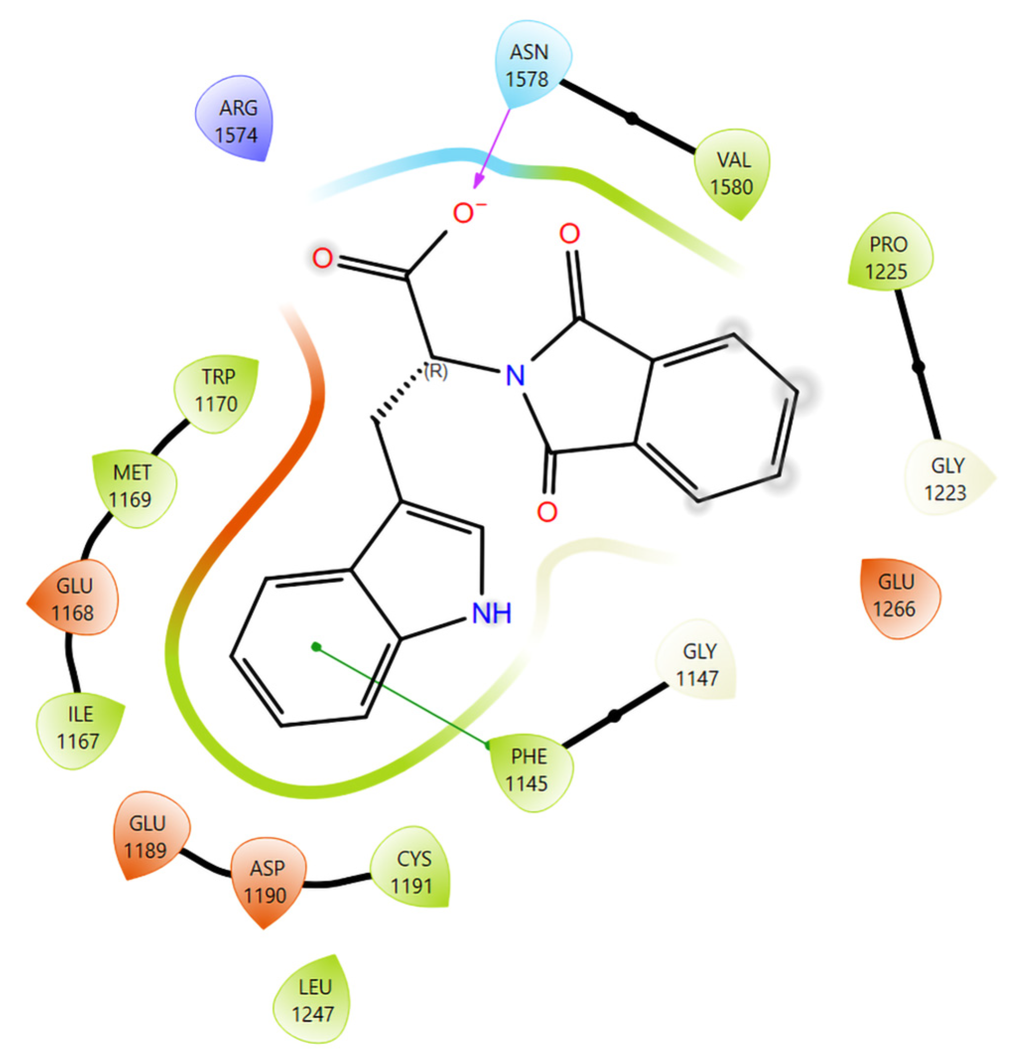
Docking confirmed that RG108 binds stably within the DNMT1 catalytic pocket that normally accommodates the SAM/SAH cofactor. In Glide SP mode, RG108 achieved a GlideScore of −6.285 kcal mol−1 with an Emodel of −59.3, indicating strong complementarity between ligand and receptor. XP redocking maintained the same orientation (GlideScore −5.293 kcal mol−1), validating pose convergence and conformational stability. The 3D model (Figure 3) revealed that the hydroxamate and amide moieties of RG108 insert between Cys1226 and Glu1266, forming hydrogen bonds with Glu1266 (OE1, 2.4 Å) and Asn1578 (OD1, 2.8 Å). A π–π stacking interaction was observed between the benzoyl ring of RG108 and Phe1145, while Pro1225 and Gly1223 contributed additional van der Waals stabilization. Trp1170, Val1144, and Leu1247 reinforced hydrophobic contacts, and Glu1168 and Arg1574 maintained solvent-exposed hydrogen bonds at the pocket entrance.
The 2D interaction diagram (Figure 4) confirmed these contacts: green lines mark hydrogen bonds to Glu1266 and Asn1578; a violet line indicates π–π stacking with Phe1145; and shaded regions denote solvent exposure around Glu1168, Glu1189, and Asp1190. This configuration shows a partially buried aromatic core with a solvent-accessible hydroxamate terminus. The Prime MM-GBSA calculation produced a ΔG_bind of −49.61 kcal mol−1, confirming a strong and energetically favorable complex. Major residue contributions were Glu1266 (−5.84), Asn1578 (−4.19), Phe1145 (−3.71), Pro1225 (−3.26), Cys1226 (−2.88), Trp1170 (−2.33), and Arg1574 (−1.95) kcal mol−1 (Table 4).
Per-residue energies confirm that Glu1266, Asn1578, and Phe1145 dominate RG108 stabilization within the DNMT1 catalytic pocket (Figure 3).

3.3. Assessment of Fecal Occult Blood Test Across CRC and Infection-Related Groups

Among the CRC confirmed group, 10 out of 12 samples (83.3%) tested positive for occult blood. In contrast, only 1 sample each from the HIV-only infected group (1/13; 7.7%) and the HIV-helminth co-infected group (1/12; 8.3%) showed a positive FOBT result. All other participants, including those in the helminth-only group (0/15) and HIV and helminth-uninfected controls (0/10), tested negative. Integrating methylation profiling with FOBT enhances the accuracy of CRC screening by compensating for FOBT’s limited sensitivity in early lesions. In our cohort, cases with positive FOBT results, particularly those with HIV or HIV/helminth co-infection, also showed altered vimentin methylation, suggesting a potential complementary diagnostic value between the two approaches.

4. Discussion

This study aimed to investigate the association between HIV-helminth co-infections and predictive biomarkers of CRC, focusing on the fecal occult blood test and vimentin methylated DNA marker. Given the high burden of infectious diseases in sub-Saharan Africa and their potential role in chronic inflammation and cancer susceptibility [30], we sought to explore whether individuals with HIV infection only, helminth infection only, or HIV-helminth co-infected individuals exhibit molecular signatures that may reflect susceptibility to CRC development. Understanding such associations is critical for improving non-invasive, accessible CRC screening strategies tailored to high-risk populations in resource-limited settings.

4.1. Vimentin Methylation

Methylation analysis of the vimentin gene in stool samples showed a marked difference between HIV and helminth uninfected controls and all infection groups, supporting our hypothesis that chronic infectious co-states may elevate CRC risk through epigenetic mechanisms. In HIV-helminth uninfected controls, vimentin methylation was rarely detected (1/10, 10%), in contrast to the CRC-confirmed group (9/10, 90%). Inflammation-associated infectious states mirrored this elevation: HIV-only (13/14, 92.9%), helminth-only (14/15, 93.3%), and HIV-helminth co-infected individuals (10/13, 76.9%). Vimentin methylation is a well-established stool-based biomarker for CRC, with high specificity in distinguishing cancerous lesions from healthy tissue [17,31]. Moreover, infectious agents are increasingly implicated in oncogenic epigenetic modulation. Infection-driven DNA methylation changes are recognized as mechanisms through which pathogens may promote tumorigenesis [32].
The present study findings demonstrate that vimentin gene methylation is not exclusive to CRC but is also significantly elevated in individuals with HIV or helminth infections, irrespective of coinfection status. The comparable methylation levels between infected groups and CRC patients suggest that chronic infection-driven inflammation may induce epigenetic alterations of a magnitude similar to those associated with malignancy. This aligns with evidence linking persistent immune activation and inflammatory signaling to aberrant DNA methylation patterns, which may facilitate tumorigenic pathways [33]. Importantly, in this case, the absence of differences between HIV-only, helminth-only, and HIV-helminth coinfected groups indicates that the presence of infection, rather than its specific etiology, is the dominant driver of this methylation change. Dysregulated DNA methylation is associated with HIV infection, with the potential for developing genetic therapies targeting methylated HIV viral genes [34]. Such infection-associated epigenetic modulation could represent an early carcinogenic event, particularly in populations with high burdens of infectious diseases. These findings show the potential of vimentin methylation as both a biomarker of CRC and a marker of infection-related epigenetic reprogramming, with implications for screening strategies in endemic regions.

4.2. Mechanistic and Translational Insights

The computational modeling provides a structural and energetic context linking DNMT1 inhibition by RG108 to vimentin (VIM) promoter hypermethylation observed experimentally. DNMT1 is essential for maintenance methylation during DNA replication; chronic activation causes aberrant CpG methylation of tumor-suppressor genes, including VIM. Docking and MM-GBSA analyses reveal that RG108 occupies the SAM/SAH-binding channel, establishing strong hydrogen bonds with Glu1266 and Asn1578, and π–π stacking with Phe1145, key residues that govern methyl-transfer catalysis. These contacts obstruct the correct alignment of the methyl donor and acceptor, effectively blocking enzymatic turnover. The top SP pose (GlideScore −6.285 kcal mol−1; Emodel −59.3) and MM-GBSA energy (ΔG_bind −49.61 kcal mol−1) agree with reported affinities for validated DNMT1 inhibitors [35,36]. Electrostatic stabilization dominated by Glu1266, followed by Asn1578 and Phe1145, underscores the role of polar and aromatic interactions in ligand affinity [35].
The structural overlap between RG108 and SAH indicates competitive inhibition, particularly relevant under inflammatory conditions with elevated S-adenosylmethionine (SAM) levels [36,37]. These mechanistic insights extend the biological observations from stool-based methylation analyses. In chronic infections such as HIV and helminthiasis, oxidative and cytokine stress upregulate DNMT1, promoting aberrant VIM methylation. By binding to the catalytic loop (Glu1266-Asn1578-Pro1225), RG108 may suppress infection-driven epigenetic silencing and restore tumor-suppressor gene expression [36,38]. The favorable MM-GBSA profile and stable hydrogen-bond/π–π network confirm RG108’s demethylating potential and provide a structural rationale for DNMT1 inhibition in infection-associated colorectal carcinogenesis.
Overall, this computational component strengthens the translational narrative by linking experimental methylation data with a molecular-mechanistic explanation. The integration of in silico docking, MM-GBSA energetics, and experimental methylation profiles establishes RG108 as a mechanistically plausible DNMT1 inhibitor capable of reversing infection-induced VIM silencing and supporting the design of next-generation DNMT1-targeted therapeutics.
With further evaluation of potential markers for CRC susceptibility, the FOBT demonstrated relatively strong diagnostic alignment with CRC, with 83.3% (10/12) in the CRC confirmed group. This supports existing evidence that FOBT, despite its simplicity, remains a useful initial screening tool for CRC, especially in low-resource settings where more advanced molecular testing may be unavailable. However, the detection of positive FOBT results in one participant in the HIV-infected only group and another in the HIV-helminth co-infection group raises concerns about the specificity of FOBT in populations burdened with chronic infections. The detection of blood in these cases may be correlated with previous studies that demonstrated a significantly higher false-positive rate associated with infections [39] or altered mucosal integrity [40]. While FOBT remains cost-effective and non-invasive, its lack of molecular specificity [41] emphasizes the need to complement it with additional validated biomarkers to improve diagnostic accuracy. These findings suggest that combining FOBT with methylation markers, or integrating them into a multi-modal screening algorithm, may enhance early CRC detection, particularly in high-risk populations with co-infections.

5. Challenges and Limitations

It is important to note that genetic variability plays an important role in methylation studies. Thus, genetic and epigenetic variability in African populations may influence assay performance. African populations have the greatest genomic diversity globally [42], including polymorphisms that may occur within primer binding sites, potentially affecting amplification efficiency or specificity. In their study on genetic variability in helminth infections, Papaiakovou et al. [43] investigated single-nucleotide variants within nuclear repeat regions, with a specific focus on primer binding sites, to assess their impact on the performance of diagnostic qPCR assays. The authors found that genetic variation within these target regions can significantly influence qPCR assay sensitivity and specificity. Such variability may lead to reduced amplification efficiency or complete failure of detection, thereby increasing the risk of false-negative results. These findings underscore the importance of continuously monitoring genetic diversity in diagnostic targets, particularly in endemic regions, to ensure the reliability and accuracy of qPCR-based diagnostics [43].
Additionally, while the frequency of methylation in infection groups approached that of CRC cases, it is important to consider that its elevation in non-cancer infectious disease cohorts likely reflects background epithelial turnover, chronic inflammation, or infection-associated epigenetic remodeling hence future studies need to also consider the use of blood-based analysis, as indicated in a study by Gopal et al., 2023 to ascertain which route would be best for use as a susceptibility marker [44]. Stool-derived DNA, while advantageous due to its non-invasive collection and acceptability in low-resource settings, can be more variable in quality and prone to degradation over time [45]. Furthermore, sample-to-sample variability, particularly in stool-derived DNA, which contains a complex mixture of human, microbial, dietary, and inhibitory substances, should be taken into consideration when performing this kind of assay. Variability in epithelial cell shedding, the degree of microbial contamination, and PCR inhibitors can all affect DNA quality and downstream assay performance [15].
Optimization of stool-derived DNA methylation markers remains a challenge, and this can be further compounded by variable sample quality [46]. In this study, archived stool samples collected several years prior were utilized. Stool samples are inherently more prone to variability: they contain mixed DNA populations (host, microbial, dietary) and are technically more challenging to preserve than blood or plasma. Studies demonstrate that stool DNA integrity and microbial community composition significantly deteriorate if not frozen within 15 min of defecation, aliquoted immediately, and stored at −80 °C, with degradation increasing with daily freeze–thaw cycles [47]. While some microbiome data remain stable in domestic freezers at −20 °C for up to six months [48], high-sensitivity assays like methylation-specific PCR are likely affected by even small losses in DNA integrity.
This study did not assay IL-6, TNF-α, or similar inflammatory mediators in the same stool samples used for vimentin methylation profiling. Although related work in the same cohort has demonstrated elevated Th1/Th17 cytokine expression in HIV-helminth co-infection [49], the causal relationship between chronic inflammation and methylation remains hypothetical and warrants further targeted investigation. Lastly, we acknowledge that the group sizes in our study are relatively small, which limits the statistical power to detect subtle differences in methylation frequency. However, we used appropriate statistical methods for small samples (Fisher’s exact test) and reported exact p-values, effect sizes (odds ratio), and 95% confidence intervals as evident in Table 3. We therefore recommend validation in larger, independent cohorts to confirm these findings.

6. Future Directions

Helminth infections are well known to induce a dominant Th2 immune profile characterized by elevated secretion of IL-4, IL-5, IL-10, IL-13, and TGF-β, accompanied by the activation of alternatively activated macrophages (M2 phenotype) and regulatory T cells. These immune signatures create an anti-inflammatory, tissue-repairing milieu that mirrors tumor-promoting environments, favoring immune suppression, angiogenesis, and epithelial proliferation [7]. Chronic exposure to Th2 cytokines such as IL-4 and IL-13 has been shown to enhance cellular proliferation, modulate DNA methyltransferase activity, and promote tumorigenesis through persistent epigenetic reprogramming [50]. To strengthen causal inference, future studies should integrate cytokine profiling with DNA methylation analyses to assess whether specific inflammatory pathways are correlated with methylation at cancer-related loci such as the VIM promoter. Correlative and mediation analyses between Th2 cytokine concentrations and stool-based methylation markers could delineate the contribution of immune dysregulation to epigenetic alterations that precede tumor initiation. Given the ethical limitations of obtaining colon biopsies from asymptomatic HIV- or helminth-infected individuals, future research should prioritize non-invasive sampling approaches. These include fecal cytokine profiling [51], which offers a promising strategy to assess intestinal immune activity alongside stool-based methylation and microbial analyses. Combining these complementary non-invasive biomarkers may enable longitudinal monitoring of infection-associated inflammation and its epigenetic consequences, providing an opportunity to identify early molecular changes that precede CRC development.
Moreover, integrated multi-omics studies that simultaneously assess the gut microbiome, well established for its role in cancer initiation and progression, yet notably altered in the presence of helminth infection [52], together with host cytokine networks and DNA methylation patterns, could elucidate how infectious co-states reconfigure the intestinal microenvironment and contribute to carcinogenesis. Experimental validation in cell-based and animal models should investigate whether parasite-induced cytokines or helminth excretory/secretory molecules (E-S products) drive DNMT expression or direct methylation of oncogenic targets [53]. Such mechanistic insights may open translational avenues for early detection and intervention, potentially reversing or halting precancerous epigenetic changes following infection clearance or immune modulation. Finally, contextualizing these investigations within Sub-Saharan African settings is essential. In this region, HIV and helminth infections remain prevalent, and access to invasive diagnostic modalities is limited. Validating stool-based DNA methylation and fecal cytokine markers in these populations could enable low-cost, community-based screening for individuals at elevated CRC risk. Ultimately, this integrated approach may inform precision prevention strategies aimed at reducing the infection-associated cancer burden in Africa.

7. Conclusions

This study provides the first evidence linking HIV-helminth co-infections to molecular signatures consistent with increased CRC susceptibility in a Sub-Saharan African context. We demonstrated that stool-based vimentin promoter methylation, a well-established CRC biomarker, was markedly elevated not only in CRC-confirmed patients but also in individuals with HIV infection, helminth infection, and HIV-helminth co-infection, compared to uninfected controls. These findings strongly support our hypothesis that chronic infectious co-states may drive epigenetic remodeling, mimic oncogenic methylation patterns, and potentially predispose to malignancy. However, the clinical utility of infection-associated methylation as a screening tool remains underexplored in the absence of longitudinal follow-up linking these changes to actual cancer development. Thus, this study provides a foundation for future large-scale, long-term studies to validate these observations and establish translational relevance. The FOBT showed substantial concordance with CRC diagnosis, attesting to its ongoing relevance as a low-cost screening tool in resource-limited settings. However, positive FOBT results in the 2 individuals in the non-cancer infection groups highlight the risk of false positives in populations with chronic inflammatory or mucosal pathology, reinforcing the need for molecular adjuncts to enhance specificity. Overall, our findings suggest that integrating stool-based DNA methylation assays might improve early CRC detection in high-risk, infection-burdened populations. Longitudinal studies are now warranted to determine whether infection-associated methylation changes precede neoplastic transformation, thereby offering predictive value for targeted prevention strategies, Figure 5.

Author Contributions

Conceptualization, B.P.D. and Z.L.M.-K.; methodology, B.P.D.; validation, P.N., Z.D. and Z.L.M.-K.; formal analysis, B.P.D. and P.N.; investigation, B.P.D.; writing, original draft preparation, B.P.D. and Z.L.M.-K.; writing, review and editing, B.P.D., S.K., M.A., P.N., Z.D. and Z.L.M.-K.; supervision, P.N., Z.D. and Z.L.M.-K.; project administration, B.P.D. and Z.L.M.-K.; funding acquisition, B.P.D., Z.D. and Z.L.M.-K.; CRC sample collection, S.K. All authors have read and agreed to the published version of the manuscript.

Funding

This research was funded by the South African Medical Research Council (SAMRC) (ZLMK grant number: HDID5149/KR/202) through its Division of Research Capacity Development under the Research Capacity Development Initiative from funding received from the South African National Treasury. The research was also supported by the SAMRC Researcher Development Award (BPD), the University Capacity Development Programme (UCDP), University of Pretoria (BPD), South African Medical Research Council (SAMRC) Grant Number 23108, and the National Research Foundation (NRF) Grant Number 138139 (ZD). The content and findings reported/illustrated are the sole deduction, view, and responsibility of the researchers and do not reflect the official position and sentiments of the SAMRC.

Institutional Review Board Statement

The study was conducted in accordance with the Declaration of Helsinki and approved by the Biomedical Research Ethics Committee (BREC) of the University of KwaZulu-Natal (protocol code BREC/00005458/2023 on 18 May 2023).

Informed Consent Statement

Informed consent was obtained from all subjects involved in the study.

Data Availability Statement

Data generated from this study can be accessed from the corresponding author upon reasonable request.

Acknowledgments

The authors would like to thank Bongekile Duma and her team for assisting with the study participants, the staff at the HCT clinic, and the eThekwini District and KwaZulu-Natal Provincial Departments of Health for their support throughout the research During the preparation of this manuscript, the authors used ChatGPT (version GPT-4.5, OpenAI, San Francisco, CA, USA, 2025) to improve the grammar, phrasing, and readability of the manuscript. No content was generated or interpreted by AI, and all the authors have reviewed and edited the output and take full responsibility for the content of this publication.

Conflicts of Interest

The authors declare no conflicts of interest.

Abbreviations

The following abbreviations are used in this manuscript:
CRCColorectal cancer
CpGCytosine phosphate guanine dinucleotide
DNADeoxyribonucleic acid
HRMHigh-resolution melt
HIVHuman immunodeficiency virus
qPCRQuantitative polymerase chain reaction

References

  1. Mostafa, M.H.; Sheweita, S.A.; O’Connor, P.J. Relationship between schistosomiasis and bladder cancer. Clin. Microbiol. Rev. 1999, 12, 97–111. [Google Scholar] [CrossRef] [PubMed] [PubMed Central]
  2. Scholte, L.L.S.; Pascoal-Xavier, M.A.; Nahum, L.A. Helminths and Cancers from the Evolutionary Perspective. Front. Med. 2018, 5, 90. [Google Scholar] [CrossRef] [PubMed] [PubMed Central]
  3. Sánchez-Barrera, C.Á.; Fernandez-Muñoz, K.V.; Mendoza-Rodríguez, M.G.; Ortiz-Melo, M.T.; Carrillo-Pérez, J.A.; Rodríguez-Sosa, M.; Terrazas, L.I. The Impact of Helminths on Colorectal Cancer: From Infections to the Isolation of Biotherapeutics. Pathogens 2025, 14, 949. [Google Scholar] [CrossRef] [PubMed] [PubMed Central]
  4. Jain, S.; Rana, M. From the discovery of helminths to the discovery of their carcinogenic potential. Parasitol. Res. 2024, 123, 47. [Google Scholar] [CrossRef]
  5. Li, H.; Shan, C.; Zhu, Y.; Yao, X.; Lin, L.; Zhang, X.; Qian, Y.; Wang, Y.; Xu, J.; Zhang, Y.; et al. Helminth-induced immune modulation in colorectal cancer: Exploring therapeutic applications. Front. Immunol. 2025, 16, 1484686. [Google Scholar] [CrossRef] [PubMed] [PubMed Central]
  6. Brown, M.; Mawa, P.A.; Kaleebu, P.; Elliott, A.M. Helminths and HIV infection: Epidemiological observations on immunological hypotheses. Parasite Immunol. 2006, 28, 613–623. [Google Scholar] [CrossRef] [PubMed] [PubMed Central]
  7. Esperante, D.; Gutiérrez, M.I.M.; Issa, M.E.; Schcolnik-Cabrera, A.; Mendlovic, F. Similarities and divergences in the metabolism of immune cells in cancer and helminthic infections. Front. Oncol. 2023, 13, 1251355. [Google Scholar] [CrossRef] [PubMed] [PubMed Central]
  8. Jusakul, A.; Cutcutache, I.; Yong, C.H.; Lim, J.Q.; Huang, M.N.; Padmanabhan, N.; Nellore, V.; Kongpetch, S.; Ng, A.W.T.; Ng, L.M.; et al. Whole-Genome and Epigenomic Landscapes of Etiologically Distinct Subtypes of Cholangiocarcinoma. Cancer Discov. 2017, 7, 1116–1135. [Google Scholar] [CrossRef] [PubMed] [PubMed Central]
  9. Bray, F.; Laversanne, M.; Sung, H.; Ferlay, J.; Siegel, R.L.; Soerjomataram, I.; Jemal, A. Global cancer statistics 2022: Globocan estimates of incidence and mortality worldwide for 36 cancers in 185 countries. CA Cancer J. Clin. 2024, 74, 229–263. [Google Scholar] [CrossRef] [PubMed]
  10. Mapanga, W.; Norris, S.A.; Chen, W.C.; Blanchard, C.; Graham, A.; Baldwin-Ragaven, L.; Boyles, T.; Donde, B.; Greef, L.; Huddle, K.; et al. Consensus study on the health system and patient-related barriers for lung cancer management in South Africa. PLoS ONE 2021, 16, e0246716. [Google Scholar] [CrossRef] [PubMed]
  11. Sepassi, A.; Li, M.; Zell, J.A.; Chan, A.; Saunders, I.M.; Mukamel, D.B. Rural-urban disparities in colorectal cancer screening, diagnosis, treatment, and survivorship care: A systematic review and meta-analysis. Oncologist 2024, 29, e431–e446. [Google Scholar] [CrossRef]
  12. Lala, V.G.; Mohamed, A.; Tate, D.J.; Seabi, N.M.; Mokgoko, D. Colorectal cancer screening: An update and South African perspective. Wits J. Clin. Med. 2024, 6, 95–102. [Google Scholar] [CrossRef]
  13. Imperiale, T.F.; Ransohoff, D.F.; Itzkowitz, S.H.; Turnbull, B.A.; Ross, M.E. Fecal DNA versus fecal occult blood for colorectal-cancer screening in an average-risk population. N. Engl. J. Med. 2004, 351, 2704–2714. [Google Scholar] [CrossRef]
  14. Khakimov, N.; Khasanova, G.; Ershova, K.; Gibadullina, L.; Vetkina, T.; Lobisheva, G.; Chumakova, A. Screening for colon cancer: A test for occult blood. Int. J. Risk Saf. Med. 2015, 27 (Suppl. S1), S110–S111. [Google Scholar] [CrossRef] [PubMed]
  15. Imperiale, T.F.; Ransohoff, D.F.; Itzkowitz, S.H.; Levin, T.R.; Lavin, P.; Lidgard, G.P.; Ahlquist, D.A.; Berger, B.M. Multitarget stool DNA testing for colorectal-cancer screening. N. Engl. J. Med. 2014, 370, 1287–1297. [Google Scholar] [CrossRef] [PubMed]
  16. Wang, D.Y.; He, K.X.; Huang, Y.; Lou, Q.Q.; He, T.; Xu, X. A New Method for the Detection of Colorectal Cancer and the Precancerous Lesions: Occult Blood Testing Combination with Promoter Methylation in the Fecal Sample. J. Cancer 2021, 12, 335–342. [Google Scholar] [CrossRef] [PubMed]
  17. Pakbaz, B.; Jabinin, R.; Soltani, N.; Ayatollahi, H.; Farzanehfar, M.R. Quantitative study of vimentin gene methylation in stool samples for colorectal cancer screening. J. Adv. Pharm. Technol. Res. 2019, 10, 121–125. [Google Scholar] [CrossRef]
  18. Lamb, Y.N.; Dhillon, S. Epi proColon® 2.0 CE: A blood-based screening test for colorectal cancer. Mol. Diagn. Ther. 2017, 21, 225–232. [Google Scholar] [CrossRef]
  19. Redwood, D.G.; Dinh, T.A.; Kisiel, J.B.; Borah, B.J.; Moriarty, J.P.; Provost, E.M.; Sacco, F.D.; Tiesinga, J.J.; Ahlquist, D.A. Cost-Effectiveness of Multitarget Stool DNA Testing vs Colonoscopy or Fecal Immunochemical Testing for Colorectal Cancer Screening in Alaska Native People. Mayo Clin. Proc. 2021, 96, 1203–1217. [Google Scholar] [CrossRef]
  20. Rui, M.; Wang, Y.; You, J.H.S. Novel Noninvasive Tests for Colorectal Cancer Screening—A Cost-Effectiveness Analysis. Cancer Epidemiol. Biomark. Prev. 2025, 34, 1111–1121. [Google Scholar] [CrossRef]
  21. Ladabaum, U.; Mannalithara, A.; Weng, Y.; Schoen, R.E.; Dominitz, J.A.; Desai, M.; Lieberman, D. Comparative Effectiveness and Cost-Effectiveness of Colorectal Cancer Screening with Blood-Based Biomarkers (Liquid Biopsy) vs. Fecal Tests or Colonoscopy. Gastroenterology 2024, 167, 378–391. [Google Scholar] [CrossRef]
  22. Mpaka-Mbatha, M.N.; Naidoo, P.; Islam, M.; Singh, R.; Mkhize-Kwitshana, Z.L. Demographic profile of HIV and helminth-coinfected adults in KwaZulu-Natal, South Africa. S. Afr. J. Infect. Dis. 2023, 38, 466. [Google Scholar] [CrossRef]
  23. Damane, B.P.; Mulaudzi, T.V.; Kader, S.S.; Naidoo, P.; Dlamini, Z.; Mkhize-Kwitshana, Z.L. HIV-Helminth Co-Infections and Immune Checkpoints: Implications for Cancer Risk in South Africa. Viruses 2025, 17, 451. [Google Scholar] [CrossRef]
  24. Tanaka, K.; Okamoto, A. Degradation of DNA by bisulfite treatment. Bioorg. Med. Chem. Lett. 2007, 17, 1912–1915. [Google Scholar] [CrossRef]
  25. Hong, S.R.; Shin, K.J. Bisulfite-converted DNA quantity evaluation: A multiplex quantitative real-time PCR system for evaluation of bisulfite conversion. Front. Genet. 2021, 12, 618955. [Google Scholar] [CrossRef] [PubMed]
  26. Srirungruang, S.; Mahajindawong, B.; Nimitpanya, P.; Bunkasem, U.; Ayuyoe, P.; Nuchprayoon, S.; Sanprasert, V. Comparative study of DNA extraction methods for the PCR detection of intestinal parasites in human stool samples. Diagnostics 2022, 12, 2588. [Google Scholar] [CrossRef] [PubMed]
  27. Kresse, S.H.; Brandt-Winge, S.; Pharo, H.; Flatin, B.T.B.; Jeanmougin, M.; Vedeld, H.M.; Lind, G.E. Evaluation of commercial kits for isolation and bisulfite conversion of circulating cell-free tumor DNA from blood. Clin. Epigenetics 2023, 15, 151. [Google Scholar] [CrossRef] [PubMed]
  28. Davidović, R.S.; Božović, A.M.; Mandušić, V.L.; Krajnović, M.M. Methylation-specific PCR: Four steps in primer design. Cent. Eur. J. Biol. 2014, 9, 1127–1139. [Google Scholar] [CrossRef]
  29. Chen, J.; Sun, H.; Tang, W.; Zhou, L.; Xie, X.; Qu, Z.; Chen, M.; Wang, S.; Yang, T.; Dai, Y.; et al. DNA methylation biomarkers in stool for early screening of colorectal cancer. J. Cancer 2019, 10, 5264–5271. [Google Scholar] [CrossRef]
  30. Diakite, M.; Shaw-Saliba, K.; Lau, C.Y. Malignancy and viral infections in sub-Saharan Africa: A review. Front. Virol. 2023, 3, 1103737. [Google Scholar] [CrossRef]
  31. Chen, W.D.; Han, Z.J.; Skoletsky, J.; Olson, J.; Sah, J.; Myeroff, L.; Platzer, P.; Lu, S.; Dawson, D.; Willis, J.; et al. Detection in fecal DNA of colon cancer–specific methylation of the nonexpressed vimentin gene. J. Natl. Cancer Inst. 2005, 97, 1124–1132. [Google Scholar] [CrossRef]
  32. Tristan-Flores, F.E.; de la Rocha, C.; Pliego-Arreaga, R.; Cervantes-Montelongo, J.A.; Silva-Martínez, G.A. Epigenetic changes induced by infectious agents in cancer. In Pathogens Associated with the Development of Cancer in Humans: Omics, Immunological, and Pathophysiological Studies; Velázquez-Márquez, N., Paredes-Juárez, G.A., Vallejo-Ruiz, V., Eds.; Springer Nature: Cham, Switzerland, 2024; pp. 411–457. [Google Scholar]
  33. Das, D.; Karthik, N.; Taneja, R. Crosstalk Between Inflammatory Signaling and Methylation in Cancer. Front. Cell Dev. Biol. 2021, 9, 756458. [Google Scholar] [CrossRef]
  34. Arumugam, T.; Ramphal, U.; Adimulam, T.; Chinniah, R.; Ramsuran, V. Deciphering DNA Methylation in HIV Infection. Front. Immunol. 2021, 12, 795121. [Google Scholar] [CrossRef] [PubMed]
  35. Mendonca, A.; Sánchez, O.F.; Zhao, H.; Lin, L.; Min, A.; Yuan, C. Development and application of novel BiFC probes for cell sorting based on epigenetic modification. Cytom. Part A 2022, 101, 339–350. [Google Scholar] [CrossRef]
  36. Ou, Y.; Zhang, Q.; Tang, Y.; Lu, Z.; Lu, X.; Zhou, X.; Liu, C. DNA methylation enzyme inhibitor rg108 suppresses the radioresistance of esophageal cancer. Oncol. Rep. 2018, 39, 993–1002. [Google Scholar] [CrossRef]
  37. Lascano, S.; Lopez, M.; Arimondo, P.B. Natural products and chemical biology tools: Alternatives to target epigenetic mechanisms in cancers. Chem. Rec. 2018, 18, 1854–1876. [Google Scholar] [CrossRef] [PubMed]
  38. Assis, R.I.; Wiench, M.; Silvério, K.G.; Silva, R.A.D.; Feltran, G.D.S.; Sallum, E.A.; Casati, M.Z.; Nociti, F.H.; Andia, D.C. Rg108 increases nanog and oct4 in bone marrow-derived mesenchymal cells through global changes in DNA modifications and epigenetic activation. PLoS ONE 2018, 13, e0207873. [Google Scholar] [CrossRef]
  39. Launoy, G.D.; Bertrand, H.J.; Berchi, C.; Talbourdet, V.Y.; Guizard, A.V.; Bouvier, V.M.; Caces, E.R. Evaluation of an immunochemical fecal occult blood test with automated reading in screening for colorectal cancer in a general average-risk population. Int. J. Cancer 2005, 115, 493–496. [Google Scholar] [CrossRef] [PubMed]
  40. Doshi, P.; Sievers, C. Understanding the utility of fecal occult blood testing in hospitalized patients with suspected GI bleeding. Cureus 2024, 16, e57406. [Google Scholar] [CrossRef]
  41. Gómez-Molina, R.; Suárez, M.; Martínez, R.; Chilet, M.; Bauça, J.M.; Mateo, J. Utility of stool-based tests for colorectal cancer detection: A comprehensive review. Healthcare 2024, 12, 1645. [Google Scholar] [CrossRef]
  42. Gomez, F.; Hirbo, J.; Tishkoff, S.A. Genetic variation and adaptation in Africa: Implications for human evolution and disease. Cold Spring Harb. Perspect. Biol. 2014, 6, a008524. [Google Scholar] [CrossRef] [PubMed]
  43. Papaiakovou, M.; Waeschenbach, A.; Ajibola, O.; Ajjampur, S.S.; Anderson, R.M.; Bailey, R.; Benjamin-Chung, J.; Cambra-Pellejà, M.; Caro, N.R.; Chaima, D.; et al. Global diversity of soil-transmitted helminths reveals population-biased genetic variation that impacts diagnostic targets. Nat. Commun. 2025, 16, 6374. [Google Scholar] [CrossRef] [PubMed]
  44. Gopal, P.; Ahmed, Z.; Kant, V.V.R.; Rao, G.V.; Rebala, P. Circulating tumor DNA for monitoring colorectal cancer: A prospective observational study to assess the presence of methylated SEPT9 and VIM promoter genes and its role as a biomarker in colorectal cancer management. Turk. J. Surg. 2023, 39, 107–114. [Google Scholar] [CrossRef]
  45. Mpaka-Mbatha, M.N.; Naidoo, P.; Bhengu, K.N.; Islam, M.M.; Singh, R.; Nembe-Mafa, N.; Mkhize-Kwitshana, Z.L. Cytokine Gene Expression Profiles during HIV and Helminth Coinfection in Underprivileged Peri-Urban South African Adults. Diagnostics 2023, 13, 2475. [Google Scholar] [CrossRef]
  46. Itzkowitz, S.H.; Jandorf, L.; Brand, R.; Rabeneck, L.; Schroy, P.C., III; Sontag, S.; Johnson, D.; Skoletsky, J.; Durkee, K.; Markowitz, S.; et al. Improved fecal DNA test for colorectal cancer screening. Clin. Gastroenterol. Hepatol. 2007, 5, 111–117. [Google Scholar] [CrossRef]
  47. Porcaro, F.; Voccola, S.; Cardinale, G.; Porcaro, P.; Vito, P. DNA methylation biomarkers in stool samples: Enhancing colorectal cancer screening strategies. Oncol. Rev. 2024, 18, 1408529. [Google Scholar] [CrossRef]
  48. Su, W.; Du, Y.; Lian, F.; Wu, H.; Zhang, X.; Yang, W.; Duan, Y.; Pan, Y.; Liu, W.; Wu, A.; et al. Standards for collection, preservation, and transportation of fecal samples in TCM clinical trials. Front. Cell Infect. Microbiol. 2022, 12, 783682. [Google Scholar] [CrossRef]
  49. Momo Cabrera, P.; Bokulich, N.A.; Zimmermann, P. Evaluating stool microbiome integrity after domestic freezer storage using whole-metagenome sequencing, genome assembly, and antimicrobial resistance gene analysis. Microbiol. Spectr. 2025, 13, e0227824. [Google Scholar] [CrossRef]
  50. Finkelman, F.D.; Wynn, T.A.; Donaldson, D.D.; Urban, J.F. The role of IL-13 in helminth-induced inflammation and protective immunity against nematode infections. Curr. Opin. Immunol. 1999, 11, 420–426. [Google Scholar] [CrossRef] [PubMed]
  51. Riva, A.; Gray, E.H.; Azarian, S.; Zamalloa, A.; McPhail, M.J.W.; Vincent, R.P.; Williams, R.; Chokshi, S.; Patel, V.C.; Edwards, L.A. Faecal cytokine profiling as a marker of intestinal inflammation in acutely decompensated cirrhosis. JHEP Rep. 2020, 2, 100151. [Google Scholar] [CrossRef]
  52. Walusimbi, B.; Lawson, M.A.E.; Nassuuna, J.; Kateete, D.P.; Webb, E.L.; Grencis, R.K.; Elliott, A.M. The effects of helminth infections on the human gut microbiome: A systematic review and meta-analysis. Front. Microbiomes 2023, 2, 1174034. [Google Scholar] [CrossRef]
  53. Harnett, W.; Harnett, M.M. Epigenetic changes induced by parasitic worms and their excretory-secretory products. Biochem. Soc. Trans. 2024, 52, 55–63. [Google Scholar] [CrossRef] [PubMed]
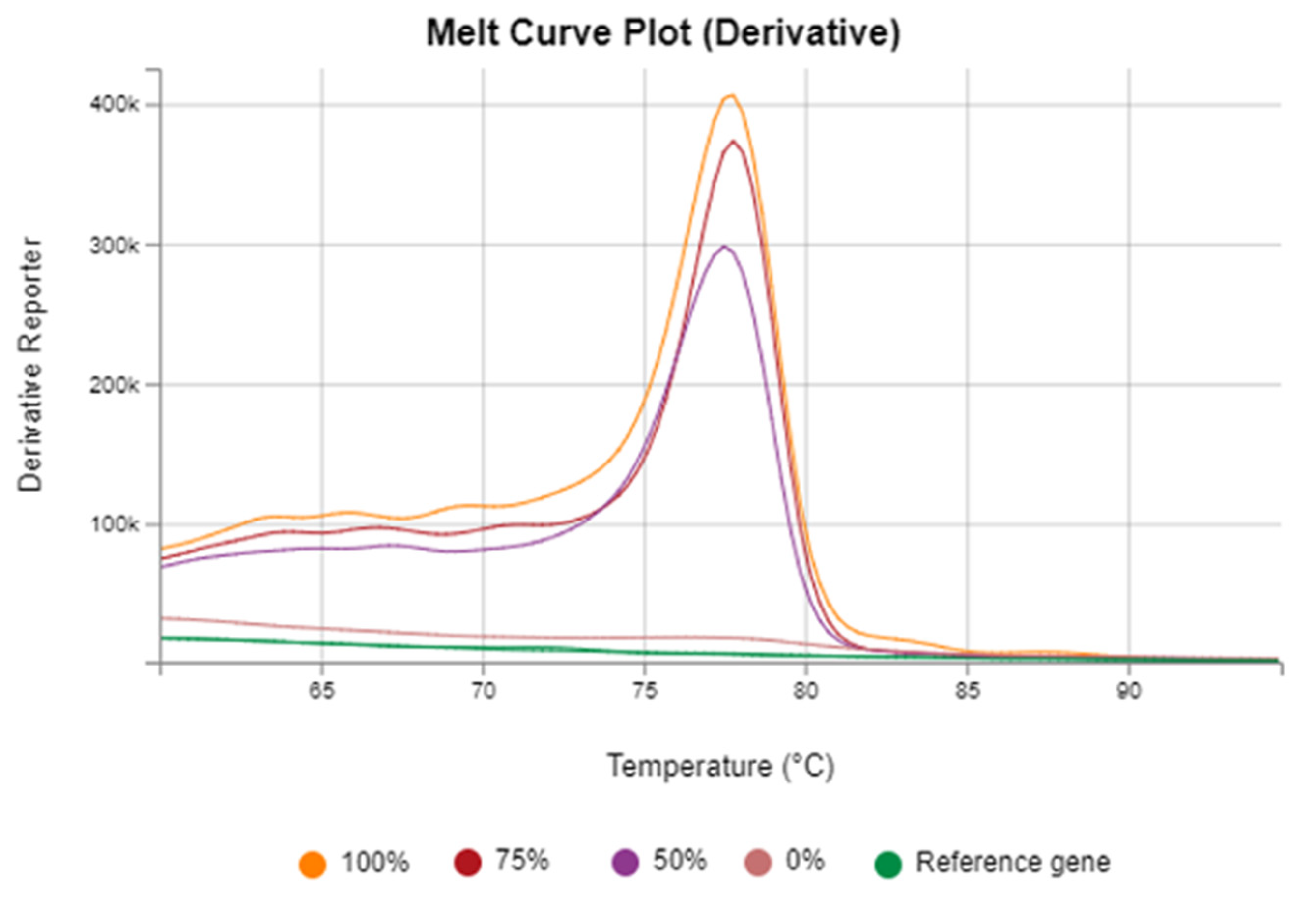
Figure 1. DNA standards Melt Curve plot.
Microbiolres 16 00236 g001
Figure 2. Correlation of Vimentin Methylation Patterns with Colorectal Cancer and Chronic Infections. (A) Stool vimentin methylation was observed in HIV-, helminth-, and co-infected individuals, at frequencies comparable to colorectal cancer patients. Although further studies are needed to confirm this, it is important to note that these observations suggest that chronic infection-associated methylation signatures might overlap with those previously described in colorectal cancer, posing challenges for cancer screening in regions endemic for infection. (B) This infographic summarizes how chronic infections like HIV and helminths cause inflammation and immune disruption, leading to vimentin gene methylation patterns similar to those in colorectal cancer patients. It highlights the potential link between infection-driven epigenetic changes and cancer risk, emphasizing the need for combined infection and cancer prevention efforts.
Microbiolres 16 00236 g002
Figure 3. DNMT1–RG108 3D binding pose. Three-dimensional representation of RG108 (green sticks) bound within the DNMT1 catalytic pocket (PDB 4WXX). Hydrogen bonds (yellow dashed lines) involve Glu1266, Asn1578, and Pro1225; hydrophobic contacts include Phe1145, Trp1170, and Val1144.
Microbiolres 16 00236 g003
Figure 4. DNMT1–RG108 2D interaction diagram. Two-dimensional schematic generated in Maestro. Green lines show hydrogen bonds with Glu1266 and Asn1578; a violet line indicates π–π stacking with Phe1145; shaded regions depict solvent-exposed polar residues (Glu1168, Glu1189, Asp1190) surrounding the hydroxamate terminus.
Microbiolres 16 00236 g004
Figure 5. Concluding Figure. Conceptual framework for CRC screening in populations with high HIV and helminth prevalence based on vimentin methylation status. The schematic illustrates different approaches for interpreting stool-based vimentin methylation results. (A) Using vimentin methylation alone may result in false positives due to infection-induced methylation in HIV- and helminth-infected individuals. (B) Combining vimentin methylation with other CRC-specific biomarkers enhances screening accuracy by distinguishing infection-induced epigenetic changes from malignant alterations. (C) Integration with infection diagnosis and treatment helps reduce false positives, ensuring appropriate resource allocation. (D) Conducting longitudinal studies in infected populations allows assessment of long-term CRC risk associated with infection-driven methylation, guiding targeted surveillance strategies. This framework emphasizes the need for a multifaceted approach to early CRC detection in high-risk, infection-burdened regions.
Microbiolres 16 00236 g005
Table 1. Primers and probes for bisulfite-converted DNA.
GenePrimer/ProbePrimer SequencesReferences
Vimentin Forward5′TCGTTTCGAGGTTTTCGCGTTAGAGAC-3′[17]
Reverse5′CGACTAAAACTCGACCG ACTCGCGA-3′
ACTBForwardGAAAGGGTGTAGTTTTGGGAGGTTAG[29]
ReverseAATAACCCAAATAAATAACCCACTACCTC
Table 2. Thermal cycling conditions.
StepTemperature (°C)TimeNotes
Initial denaturation9510 minPolymerase activation
Denaturation9515 s
Annealing/Extension6060 sData collected during this step
Steps 2–3 were repeated 40 cycles
HRM preconditioning9510 s
HRM step60Ramp at 0.1 °CContinuous ramp with data acquisition
Table 3. Frequency of stool vimentin gene methylation in HIV and helminth uninfected, single-infected, and coinfected individuals, and colorectal cancer patients.
Group, (n)Methylation, n (%)Non-Methylation, n (%)p ValueOdds Ratio (95% CI)
Uninfected controls
(n = 10)
1 (10)9 (90)<0.0001 *
HIV-infected
(n = 14)
13 (92.9)1 (7.1)
Helminth-infected
(n = 15)
14 (93.3)1 (6.7)
Coinfection
(n = 13)
10 (76.9)3 (20)
CRC confirmed patients
(n = 10)
9 (90)1 (10)
HIV and helminth uninfected, single-infected and coinfected individuals
versus
CRC confirmed patients (Positive Control)
Uninfected controls versus CRC confirmed patients 0.00110.01
(0.00–0.23)
HIV-infected versus CRC confirmed patients1.00001.44
(0.08–26.25)
Helminth-infected versus CRC confirmed patients1.00001.56
(0.09–28.17)
Coinfection versus CRC confirmed patients0.60360.37
(0.03–4.23)
HIV and helminth single and coinfected groups
Versus
Uninfected controls (Negative Control)
HIV-infected versus Uninfected controls <0.0001117
(6.44–2126)
Helminth-infected versus Uninfected controls<0.0001126
(6.96–2281)
Coinfection versus Uninfected controls<0.000130
(2.62–342.9)
Comparison between infected groups
HIV-infected versus Helminth-infected 1.00000.93
(0.05–16.43)
Coinfection versus HIV-infected0.32590.26
(0.02–2.85)
Coinfection versus Helminth-infected0.31110.24
(0.02–2.64)
Footnote: * Denotes p-value was derived using the Chi-squared (χ2) test. All other p-values were derived using Fisher’s exact test.
Table 4. Prime MM-GBSA binding-energy decomposition for the DNMT1–RG108 complex.
ResidueΔG (kcal mol−1)Interaction Type
Glu1266−5.84Hydrogen bond/electrostatic
Asn1578−4.19Hydrogen bond
Phe1145−3.71π–π stacking/hydrophobic
Pro1225−3.26Hydrophobic/backbone contact
Cys1226−2.88Catalytic loop stabilization
Trp1170−2.33Hydrophobic
Arg1574−1.95Polar/solvent-exposed
Total ΔG_bind−49.61
Disclaimer/Publisher’s Note: The statements, opinions and data contained in all publications are solely those of the individual author(s) and contributor(s) and not of MDPI and/or the editor(s). MDPI and/or the editor(s) disclaim responsibility for any injury to people or property resulting from any ideas, methods, instructions or products referred to in the content.