Article

Isolation and Characterization of Lactic Acid Bacteria from Cocoa Mucilage and Meat: Exploring Their Potential as Biopreservatives for Beef

1
Programa de Doctorado en Ciencias Agroalimentarias y Medioambiente, Universidad de La Frontera, Temuco 4780000, Chile
2
Faculty of Agricultural and Forestry Sciences, State Technical University of Quevedo, Quevedo 120310, Ecuador
3
One Health Research Group, Universidad de las Américas, Quito 170521, Ecuador
4
UWA Institute of Agriculture, The University of Western Australia, Perth 6009, Australia
5
Facultad de Ciencias Agropecuarias y Medioambiente, Universidad de la Frontera, Temuco 4780000, Chile
*
Authors to whom correspondence should be addressed.
Microbiol. Res. 2023, 14(3), 1150-1167; https://doi.org/10.3390/microbiolres14030077
Submission received: 6 July 2023 / Revised: 7 August 2023 / Accepted: 8 August 2023 / Published: 18 August 2023
(This article belongs to the Collection Microbiology and Technology of Fermented Foods)

Abstract

:
The aim of this study was to characterize lactic acid bacteria (LAB) isolated from cocoa mucilage and beef and evaluate their inhibitory effect in vitro against pathogenic bacteria, as well as determine their effect on beef quality. For the antagonist assay, 11 strains of LAB were selected and tested against pathogenic strains of Escherichia coli and Salmonella sp. The pathogenic bacteria were cultured in a medium, and a previously reactivated LAB bacterial pellet was added. After incubation, halos were observed around the bacterial colonies of the pathogenic strains, indicating inhibition by the LAB. It was identified that the LAB strains used belonged to the genus Lactobacillus, and the CCN-5 strain showed high percentages of inhibition against Salmonella sp. (58.33%) and E. coli (59%). The effectiveness of LAB application methods (immersion, injection, and spraying) did not present statistical differences. Furthermore, no significant changes in the physicochemical characteristics of beef were observed after the application of LAB. The results obtained demonstrate the potential of cocoa mucilage, as a biological control agent through LAB application, for beef biopreservation due to its ability to inhibit the growth of pathogenic bacteria.

1. Introduction

From the early days of plant cultivation and animal domestication, humans have faced the persistent dilemma of food preservation [1,2]. Most fresh foods have the problem that their shelf life is restricted, primarily due to the growth of food spoilage microorganisms and oxidative reactions [3]. In this context, meat is one of the most perishable products; thus, conservation measures must be applied immediately after the death of the animal, with the aim of delaying or preventing certain changes that make it unsuitable for consumption or that degrade some quality characteristic. The modes of alteration are multiple and can be physical, chemical, or microbiological and other sensory characteristics perceived by consumers [4].
The growing concern for food safety worldwide has led to the study of different alternatives for obtaining compounds that favor its conservation [5,6]. Despite the great variety of microbiota present in meat, most of the microorganisms that alter fresh meat are aerobic psychrotrophic, facultative anaerobic, and Gram-positive bacteria [7]. However, some foodborne toxi-infections can be caused by pathogenic mesophilic bacteria found in meat, including Salmonella sp., Staphylococcus aureus, Yersinia enterocolitica, Clostridium botulinum, C. perfringes, Campylobacter sp., Escherichia coli, and Listeria monocytogenes. These bacteria pose a significant health risk and can lead to changes in the organoleptic properties of meat [8,9,10]. The chemical composition of fresh meat and its biological characteristics provide an environment for the development of deteriorating microorganisms and pathogens. Certainly, this reduction in shelf life can lead to potential poisoning in humans caused by these organisms. For these reasons, complementary preservation procedures have been developed, which, when combined with refrigeration, effectively can increase the sanitary quality of fresh meat [8,11].
The presence of microorganisms in food does not always pose a threat of spoilage, but they play different roles in food [6]. The lactic acid bacteria (LAB) are composed of a group of Gram-positive bacteria, usually immobile, non-sporulated cocci or bacilli. These microorganisms are capable of fermenting carbohydrates, primarily producing lactic acid. They are able to grow at temperatures below 5 °C and others at temperatures as high as 45 °C [12]. The genera of LAB most used to slow spoilage and preserve food naturally are Lactococcus, Streptococcus, Pediococcus, Leuconostoc, Lactobacillus, and Carnobacterium [13].
LAB strains exhibit beneficial characteristics, including the production of inhibitory substances, fewer chemical preservatives in the food industry, prolonging the shelf life of meat products, and contributing to the reduction in cases of foodborne diseases [10,14]. LAB are capable of inhibiting the proliferation of pathogenic bacteria through various mechanisms of action [15].
Some genera of LAB can bind to specific carbohydrates of Enterobacteriaceae and inhibit their adhesion by antimicrobial agents and the production of low-molecular-weight substances. Additionally, these LAB can produce toxic effects on other bacteria by attaching to receptors on their surfaces. By adhering to the surfaces of competing bacteria, LAB can release antimicrobial substances. For this reason, they are very useful in human health to prevent foodborne and toxi-infections. These microorganisms, by competitive exclusion, decrease the probability of transmission of pathogens and improve the sensory characteristics of various food products [16]. Bacteriocins are the most interesting antimicrobial substances produced by LAB, since they have several advantages; they can be degraded by proteolytic enzymes in the gastrointestinal tract, making them nontoxic [17]. For a bacteriocin to be used in food as a preservative, it must be nontoxic, stable, and highly active and possess a broad spectrum of activity while not affecting the sensory attributes of food. It also has to be cost-effective and easy to use [18,19].
With the aim of extending the shelf life of some foods, including beef, lactic acid bacteria (LAB) from cocoa mucilage have been used in biopreservation, functioning as bioprotective cultures that counteract the growth of unwanted bacteria in beef [20]. Cocoa mucilage is obtained from the process of fermentation of the cocoa bean (Theobroma cacao). This component is predominantly discarded, causing great environmental problems, due to the foul odors that are generated during the decomposition. This results in substantial economic losses in cocoa activity, because it focuses solely on marketing a single component, instead of a proper use of all the byproducts generated from cocoa production [21]. In addition, mucilage offers an ideal environment (with 82–87% water, sugars 12%, citric acid 1–2%) for the growth of some microorganisms that play an important role in fermentation. These microorganisms convert sugars into ethyl alcohol, subsequently transformed into acetic acid by the action of lactic and acetic bacteria [22].
Currently, consumer pressure to obtain food free of residues that threaten human health is present throughout the world with changes in agricultural production models. Although knowledge of chemical residuality in food is scarce, efforts are being undertaken by the governmental, academic, and research sectors. In light of this, the present study aims to characterize lactic acid bacteria (LAB) isolated from cocoa mucilage and beef, assess their inhibitory effect in vitro against pathogenic bacteria, and determine their effect on beef quality.

2. Materials and Methods

2.1. Sampling and Isolation of Lactic Acid Bacteria (LAB)

Samples of beef (Longissimus muscle) were taken from different meat supplies in the cities of Quevedo (Los Ríos province) and La Maná (Cotopaxi province). The samples were placed in sterile Ziploc® bags (S. C. Johnson & Son, Inc., Racine, WI, USA), each sample was labeled and encoded, and later they were preserved at 4 °C and were taken for processing to the Laboratory of Biotechnology and Microbiology of the State Technical University of Quevedo, located at Km 7 1/2 of the Quevedo road, El Empalme, province of Los Ríos. For isolation, the meat samples were washed with 500 mL of sterile distilled water, 1 g of meat and 10 mL of water were taken, and then it was constantly macerated until homogenized. Finally, serial dilutions were made up to −10 and inoculated in Petri dishes containing Man, Rogosa, Sharpe agar culture medium (MRS) [23], and Solid King B [24].
CCN 51 and Nacional cocoa cobs (Theobroma cacao) were collected in the Pajarito and Faita enclosure belonging to the Mocache canton, Los Ríos province, Ecuador. Harvesting was done manually 24 h before processing. They were then washed with chlorinated water (100 ppm chlorine) and rinsed with tap water. The fruits were chopped making 2 longitudinal and 2 transverse cuts, and the shell of the almonds was manually separated. For the extraction of the exudate, a canvas of fabric was used with a special framework of 100 to 200 μm that facilitated the passage of the liquid. The measurement of the lattice was 0.75 × 0.75 m, which allowed 3 L of mucilage to be filtered by pressure.
The mucilage obtained was left to ferment during 24, 48, and 72 h, and a sample of 10 mL was taken. Serial dilutions ranging from 10−1 to 10−6 were prepared and subsequently plated in duplicate on MRS agar culture medium [23] using the plate-emptying technique. The plates were incubated under microaerophilic conditions for 48 h at 35 °C. The bacteria were isolated by successive reseedings by the cross-striation method. The selected colonies were purified three times to obtain a pure culture. The purified bacteria were stored at −4 °C in 50% glycerol (v/v) with MRS broth. For the subsequent tests, the bacteria were reactivated in Petri dishes by the microdroplet reseeding technique [25].

2.2. Morphological Characterization

2.2.1. Morphological Analysis

For morphological characterization, a bacterial colony was cultured on MRS solid culture medium (bacteriological agar 10 g/L, bacteriological peptone 10 g/L, dextrose 20 g/L, dipotassium phosphate 2 g/L, magnesium sulfate 0.2 g/L, manganese sulfate 0.05 g/L, meat extract 8 g/L, sodium acetate 5 g/L, Tween 80 1 g/L, yeast extract 4 g/L, ammonium citrate 2 g/L) and allowed to incubate for 24 h at 31 °C [26]. From the bacterial growth obtained, one colony was selected for each sample, based on its circular and irregular morphology, wavy and smooth edges, convex surfaces, opaqueness, and color (white or creamy). Additionally, the Gram-positive stain and negative reaction to catalase and peroxidase tests were used to identify lactic acid bacteria based on their morphological and biochemical characteristics. Subsequently, a cluster analysis was performed for all isolated strains.

2.2.2. Biochemical Characterization of Isolated Bacteria

For the detection of the ureolytic capacity of the bacteria, we followed Phang’s methodology [27], using Christensen medium or urea agar (1.0 g/L peptone, 1.0 g/L C₆H₁₂O₆, 5.0 g/L NaCl, 2 g/L KH₂PO₄, 0.012 g/L phenol red, and 15.0 g/L agar). The medium was supplemented with sterilized urea with 2% filtrate (Sigma, U5378). A single colony was selected and inoculated into the medium and then incubated at 30 °C for 48 h. Ureolytic production was determined when the medium changed from pale yellow to pinkish-red [28].

2.2.3. Detection of Protease in the Bacteria under Study

For the detection of protease activity, we prepared a medium containing 2 g/L yeast extract, 3 g/L casein, 5 g/L gelatin, and 15 g/L agar powder. Subsequently, 15 mL of the culture was transferred to Petri dishes, and a pure colony was striated onto the plates. The plates were then incubated at 15 °C for 72 h [29]. The presence of the hydrolysis halo around the puncture was considered positive activity, as described by Linares [30].

2.2.4. Detection of CO2 in the Study Bacteria

For the detection of CO2, we followed the methodology of Jensen et al. [31]). First, a Blood Agar Base medium was prepared (protease peptone 15.0 g/L, liver digestion 2.5 g/L, yeast extract 5.0 g/L, sodium chloride 5.0 g/L, agar 15.0 g/L) with a pH of 7.4 ± 0.2 at 25 °C. The medium was then transferred to Petri dishes, and a colony was inoculated in the center of each dish at room temperature. The production of carbon dioxide (CO2) was established with qualitative values, where (-) absence, (+) presence, (++) greater presence.

2.2.5. Catalase Test by Slippage (Drop) Method

We followed MacFaddin’s methodology [32] to test for catalase activity. A single bacterial colony was transferred to a slide, and then a drop of 3% hydrogen peroxide was added on the microorganism. Using a 40× magnifying lens microscope (Euromex, Amsterdam, The Netherlands) the reaction was visualized given the following values: the absence of bubble formation (without catalase enzyme to hydrolyze hydrogen peroxide) represents a negative catalase reaction, and a positive catalase when forming a bubble [33].

2.2.6. Gram Stain

We followed Beveridge’s methodology to differentiate Gram-positive and Gram-negative bacteria [34]. A colony of bacteria was spread on a microscope slide and fixed by heating for 5 s. Then, 500 μL of violet crystal (1.24 g in 100 mL water) was added and allowed to stand for 30 s. A total of 500 μL of Gram (a mixture of 0.33 g of iodine and 0.67 g of potassium iodide in 100 mL of water) was added to the violet crystal for 30 s. The violet–Gram crystal iodine mixture was poured over a container, and the mixture was quickly washed with distilled water and 1 mL of liquid chemical bleach (95% v/v ethanol in water) for 20 s. The slide was washed with distilled water, and 500 μL of safranin was added to the sample for 1 min. The slide was rinsed with distilled water and dried with filter paper. Finally, the sample was visualized under microscopy.

2.2.7. Growth Kinetics

Growth kinetics was analyzed using the methodology proposed by Rodriguez [35]. First, the pre-inoculum was cultivated using the MRS culture medium, where 20 μL were carefully deposited. Then, it was incubated in an Erlenmeyer flask containing 50 mL of medium and placed in a Benchmark incushaker® shaker (Benchmark Scientific, Inc., Sayreville, NJ, USA) at 150 rpm, maintaining a temperature of 26 °C for 48 h.
After 48 h, the inoculum was obtained by transferring 1 mL of the pre-inoculum from each bacterium. Individual Erlenmeyer flasks were prepared for each bacterium, each of them containing 99 mL of MRS culture medium. The flasks were then incubated at 26 °C and 150 rpm in a Benchmark incushaker shaker® until reaching the desired OD (optical density) and cell concentration or cells per mL (CFU—colony-forming units/mL) measured at 600 nm using a Unico® bw-54 brand spectrophotometer (Honeywell, Charlotte, NC, USA). To measure the OD, 3 mL of the inoculum sample was placed in quartz cuvettes at 12 h intervals for a total of 72 h.
The count of the CFU/mL was determined by the method of plate count by seeding drops on the surface described by Vargas [36], for which a series of dilutions was prepared. In total, 270 μL of sterile H2O and 30 μL of the culture incubated in Eppendorf tubes were placed, and this procedure was repeated until the dilution factor was −6. The plates were left in incubation for 24 h, and the colony count was carried out in the dilutions that formed between 30 and 300 colonies. To determine the number of viable cells per 1 mL, the following formula was used:
U F C m l = l i s t e d c o l o n i e s m l p l a n t e d × d i l u t i o n f a c t o r
Statistical Six Sigma Software V.10. was used in conjunction with the use of the Gompertz equation model to determine essential kinetic parameters, including the specific growth rate (μ max), generation time (G), and duration of the latency phase (λ). Additionally, we analyzed the cell concentration during the stationary phase.

2.2.8. Supernatant Antibiosis—Bacteria

For the antibiosis test, we followed the methodology of Singh [37]. First, we prepared Escherichia coli and Salmonella sp. cultures each with a cellular concentration of 1.0 × 10⁻⁸ grown in specific commercial media such as BD differentials Salmonella, Shigella Agar (SS agar) and Luria–Bertani (LB) medium. Next, we placed filter paper with a diameter of 6 mm in four compartments on the MRS agar medium. Finally, 10 μL of supernatant extracted from the strains under study was inoculated onto the filter paper. The plates were then incubated for 72 h at 35 °C. A completely randomized design (DCA) was performed with four replications and an experimental unit.

2.2.9. Bacteria–Bacteria Antagonism

The methodology of Rivera [38] for strains with inhibitory capacity was followed. The process involved reactivating the strains in MRS broth at 35 °C and 170 rpm for 48 h. Then, the culture was centrifuged at 13,000 rpm for 5 min, and the supernatant was discarded and the bacterial pellet removed. The pathogenic strains were provided by the Laboratory of Biotechnology and Microbiology of the State Technical University of Quevedo. Salmonella sp. and Escherichia coli were cultivated in LB culture medium, and 20 μL of bacterial culture was spread on King B agar medium with a concentration of 1 × 10⁸ CFU mL−1. Then 10 μL of lactic acid bacteria previously grown in the center of the Petri dish was added, and the plates were incubated for 48 h at 35 °C. The test was considered positive when areas of inhibition corresponding to a halo equal to or greater than 2 mm were observed. We selected those strains that showed greater antimicrobial capacity simultaneously for both pathogens.

2.2.10. LAB Antagonism—Beef

The collected beef was cleaned with sterile water according to the methodology of Katikou et al. [39]. Subsequently, the beef was cut into 10 g blocks and deposited in Petri dishes. The experiment comprised three types of treatments, each with three repetitions:
T1: Immersion—The beef was immersed in 2 μL of LAB bacteria + Pathogens (500 μL) for 5 min. After the immersion, the bacteria from the Petri dish were removed and then sealed in a transparent Ziploc bag. The sealed dish was then incubated in a controlled medium at 10 °C for seven days.
T2: Injection—LAB bacteria + Pathogens (500 µL) were applied to the beef using a micropipette. The Petri dish was sealed in a transparent Ziploc bag and incubated in a controlled medium at 10 °C for seven days.
T3: Spraying—LAB bacteria + Pathogens (500 µL) were applied to the beef using a sterilized atomizer. The sealed Petri dish was then wrapped in a transparent Ziploc bag and incubated in a controlled medium at 10 °C for seven days.

2.3. Physicochemical Analysis

After the study of antagonism in beef, we proceeded to analyze the physicochemical parameters in meat treated with LAB; parameters such as pH, moisture, protein, ash, fat, and fiber were studied.

2.3.1. Humidity

In order to determine the net moisture content of a beef sample [40], it is necessary to analyze the weight loss experienced by the sample when dried in an oven at constant temperatures of 100 °C until the dry residue maintains a constant weight. The following formula was used:
%   H u m i d i t y = W e i g h t l o s s o f t h e s a m p l e T o t a l s a m p l e w e i g h t × 100
The dry matter content is obtained by subtracting 100 from the moisture percentage of the sample by the following formula:
% D M = 100 H u m i d i t y %

2.3.2. Protein

Crude protein includes all the nitrogenous substances contained in the food, i.e., true protein and other non-protein nitrogenous compounds [41]. The protein content of raw meat varies inversely proportional to fat and due to moisture losses [42]. The following formula was used:
%   P r o t e i n r a w m e a t = 99 %   F a t %   H u m i d i t y

2.3.3. Ash

The ash fraction of the analysis represents the inorganic constituents of the animal material. This determination was made by placing 1 g sample in a porcelain capsule [43]. The samples were incinerated at 500 °C until obtaining ash and then allowed to cool at room temperature. The percentage of ash is obtained by the following formula:
% A s h = P 3 P 1 P 2 P 1 × 100
where:
P1: Empty capsule weight
P2: Capsule weight with sample
P3: weight of capsule with ashes.

2.3.4. Fat

To obtain the percentage of fat from a meat sample, Reyes and Mendieta [40] propose the use of Soxlet extractor equipment. The petroleum ether (100 mL) passes through the sample (500 g) for approximately 6 h at 105 °C. Subsequently, by heating, the ether is recovered, leaving only the lipid extract. The percentage of fat was obtained by the formula:
%   E E = a b × 100 c
where:
a: Weight of the Soxlet ball plus EE
b: Weight of empty ball
c: Weight of the initial sample in grams.

2.3.5. Fiber

The fibers are all those non-nitrogenous organic substances that do not dissolve by boiling with diluted acids or alkalis. The total weight of these substances is subtracted from the ashes to determine the fiber content. In order to obtain the net fiber content, [44] recommends weighing 1 g of degreased sample, placing it in an Erlenmeyer flask, adding 5 drops of defoamer, placing the container on the heating iron, attaching the digester, and allowing it to boil for 30 min. Then, remove the condenser and add to the container (without removing it from the plate) 10 cc of 24% sodium hydroxide while it is coupled in the equipment and continues boiling for 30 more min. The hot solution was filtered through a crucible with glass wool, washing the residue successively with boiling water, 1.25% sulfuric acid, acetone, and ethanol. Finally, the crucible was placed in the oven at 130 °C for 2 h and then cooled in a desiccator and weighed (Weight A); then, it was placed in the muffle at 600 °C for 30 min and cooled in desiccator and weighed (Weight B).
%   C r u d e f i b e r = F i b e r w e i g h t S a m p l e w e i g h t × 100 F i b e r = W e i g h t A W e i g h t B

3. Results and Discussion

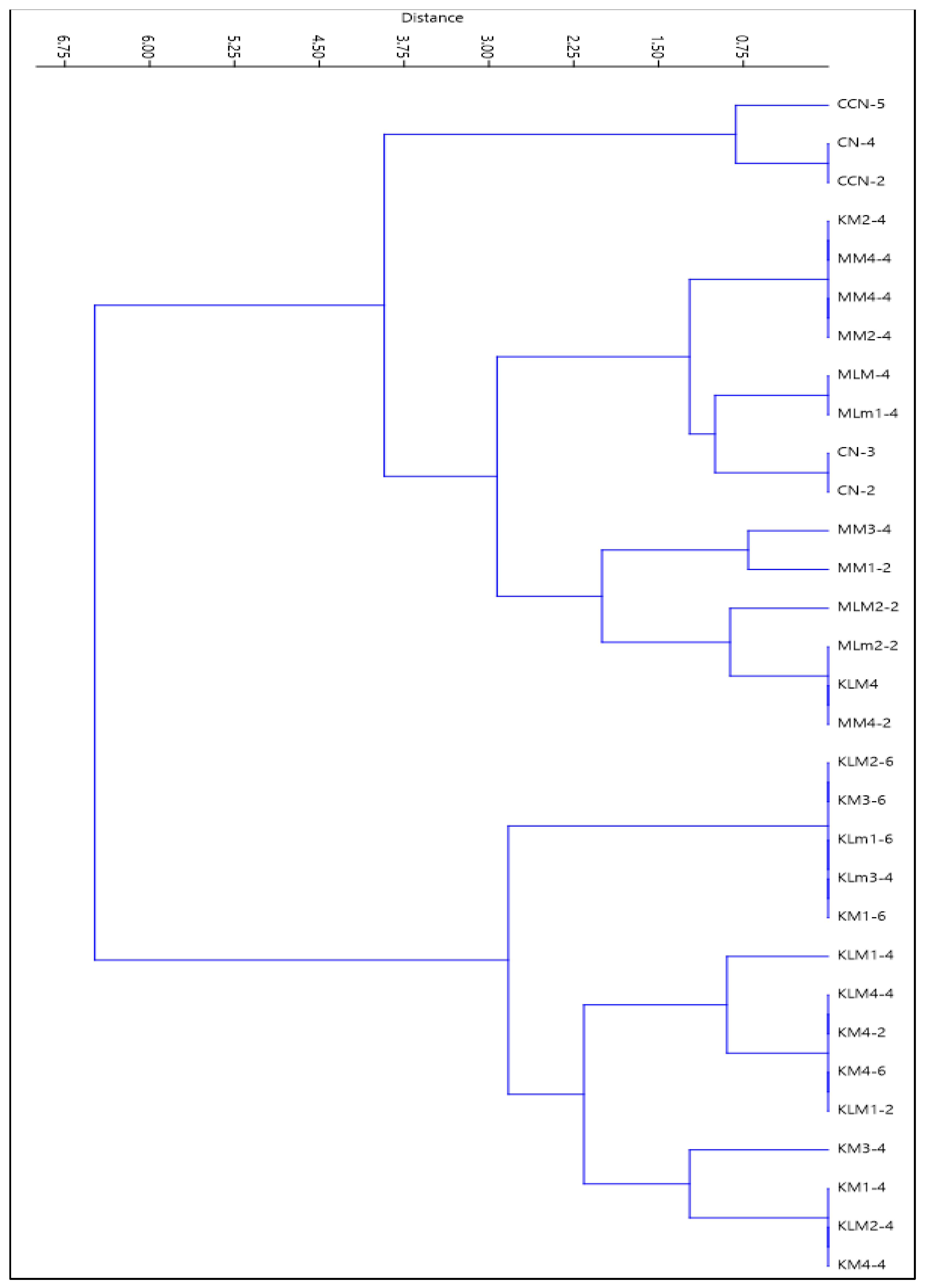
3.1. Isolation of Lactic Acid Bacteria (LAB)

A bank of 31 bacterial strains was obtained that were isolated from cocoa mucilage (CCN-51 and National) and beef (Table 1); the isolated strains were characterized morphologically and biochemically, resulting in 11 bacteria with characteristics of the genus Lactobacillus. These characteristics included the morphology of the colonies, positive Gram stain, ability to grow in the absence of O2, and negative catalase reaction. There is currently a growing demand for fourth-generation feeds, which implement natural additives. This has led to the use of bacteriocins produced by LAB and their antimicrobial potential, since they are presented as natural preservatives [5] and biopreservatives [45]. The application of biopreservative strains, as well as the extracts and metabolites produced by them, have been shown to have control over various unwanted microorganisms, extending the shelf life of food and providing security against bacteria that may affect the health of the consumer [11,46].
From 31 isolated strains, a phylogenetic analysis was performed (Figure 1) [47]. The phylogenetic tree was found to be shared by two groups; among them are A and B. Group A maintained gender characteristics of Lactobacillus and is differentiated by three subgroups, where the CCN, KM, MM, KLM, and MLM strains are grouped. Group B subdivides into three subgroups where KM and KLm bacteria predominate. These results are consistent with those presented by Papalexandratou et al. [48], a study in which 529 LAB from cocoa mucilage were grouped in a similar dendrogram that illustrated the diversity of LAB species. Through the use of phylogenetic trees, Wong et al. [49] in their study determined that different cheese isolates were related to L. brevis and L. plantarum.
Vallejo et al. [12] aimed to identify and characterize lactic acid bacteria (Lactococcus spp.) of the mucilage of two varieties of cocoa, Nacional EET-103 and Trinitario CCN-51, for use as natural preservation precursors in minimally processed and fresh food products. Bacteria Lactococcus spp. extracted from the Nacional cocoa presented greater acidification capacity against antibiotics compared to the bacteria extracted from the Trinitario cocoa. De Vuyst and Weckx [50] expressed that as fermentation continues and the cocoa mucilage is drained, more air enters the fermenting cocoa bean and pulp mass, creating ideal conditions for LAB growth between 24 and 72 h of fermentation. Our results showed intensely increasing LAB counts during the first 30 h and decrease after 40 h [48]. In the fermentation process of cocoa mucilage, the LAB are microaerophilic, tolerant to acids, and tolerant to ethanol and fructophiles (citrate converters or not), particularly Leuconostoc pseudomesenteroides, Fructobacillus pseudoficulneus, F. tropeaoli, L. cacaonum, L. fabifermentans, and L. plantarum, followed by L. fermentum [48,50,51,52,53,54,55,56,57,58,59,60,61,62,63,64].

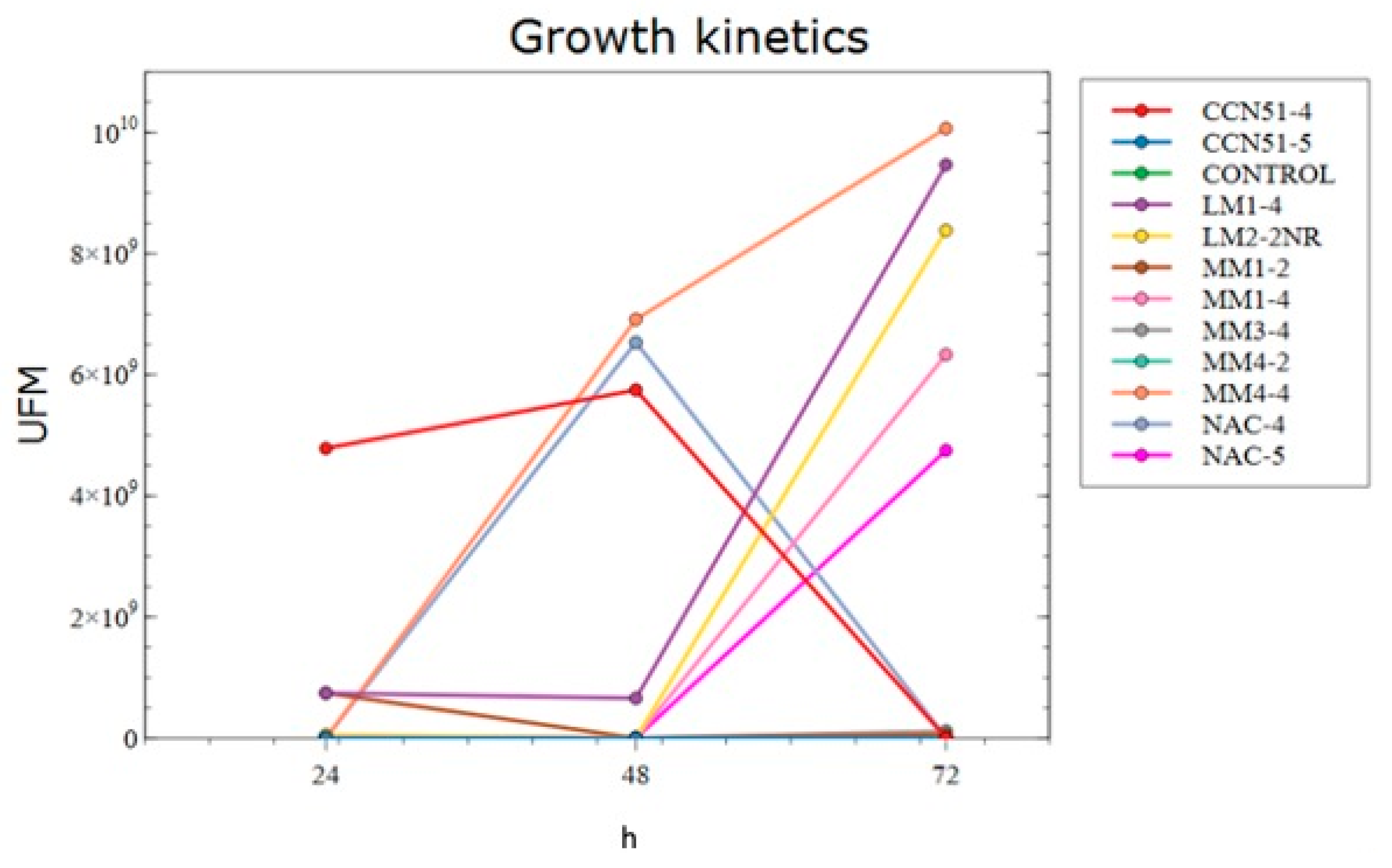
3.2. Growth Kinetics

The study of the growth kinetics obtained the exponential growth time, the seasonal phase, the ideal state, and the death of the colonies of LAB at 24, 48, and 72 h of life in selective medium (MRS broth). Twelve strains were studied, from which data such as pH and CFU counts were collected. These data played a crucial role facilitating the analysis for the proper management of the studied bacteria (Figure 2).
Mesa et al. [65] found results similar to those obtained in the present work where the control treatment without previous inoculation presents the least significant average compared to the rest of the treatments, registering 7.13 CFU/mL and a pH of 4.10, contrasting with the average of 10.48 CFU/mL and a pH of 3.80. It was observed that lower microbial growth corresponded to a higher pH level. Jurado et al. [66] showed a growth kinetics with an exponential phase of L. plantarum at 4 P.M. where the PRO medium (sugar base, soy milk, powdered milk, and wheat bran) reached a value of 3 × 10¹³ CFU/150 μL, which is higher than the MRS medium, where 1.2 × 10¹² CFU/150 μL was found. The effect of culture medium on plate microorganisms for L. plantarum, expressed in CFU/150 μL, showed statistically significant differences.
In the growth stage, it is possible to notice that during the first 12 h of the application of the inoculum, the microorganism adapts to the conditions of the environment and begins to consume the substrate [67]. For a microorganism to be optimal for bioconservation, it must have capacities adaptable to the different food matrices that it must protect [68].
Calderón [67] demonstrated that at 84 h of growth, when the microorganism would be expected to enter the stationary phase, the concentration of microorganisms at 96 h exceeded that observed at 72 and 84 h. It was determined that during fermentation, L. acidophilus produces a primary metabolite lactic acid, which is closely associated with microbial growth. The production of this metabolite increases as the population grows. Therefore, when the exponential growth stage concludes, the production of lactic acid will have reached its maximum peak, and it will not be possible to achieve higher concentrations than those already obtained. On the other hand, lactic fermentation could be evidenced, the result of comparing microbial growth with substrate consumption. It was possible to notice that as microbial growth occurred, the concentration of substrate in the culture medium decreased progressively.
Of the twelve treatments studied, eight showed similarities with the growth curve proposed by Calderón [67], who observed that LAB populations presented an exponential curve of bacterial growth after 72 h. This characteristic is attributable to the fact that lactic acid is a metabolite associated with primary growth, which causes an increase in the pH of the medium This increase in pH acts as a precursor for the hydrolysis of non-reducing sugars present in the culture medium, converting them into reducing sugars. This conversion results in elevated levels in the readings. The ability of certain lactic acid bacteria to be tolerant of acidic pH is one of the most relevant selection criteria for implementing biopreservatives in animal-derived products. LAB are exposed to a naturally acidic environment, which serves as a significant barrier for numerous other microorganisms.
The pH level is an important parameter related to the susceptibility of meat to spoilage, and it plays a pivotal role in determining the appropriate processing method for the meat’s intended use [69]. LAB are considered acid-tolerant bacteria because they can grow at pH values as low as 3.2 and, in some cases, as high as 9.6. Their unique metabolism primarily generates lactic acid, leading to the acidification of the medium. As demonstrated earlier, the inclusion of LAB in the meat production process causes sensory and physicochemical alterations, including a reduction in pH [70]. Acting as leavening agents in meat products, LAB bacteria significantly influence color and texture. Through acid production, LAB lowers the pH, affecting the water retention capacity of myogenic fibrin [71].

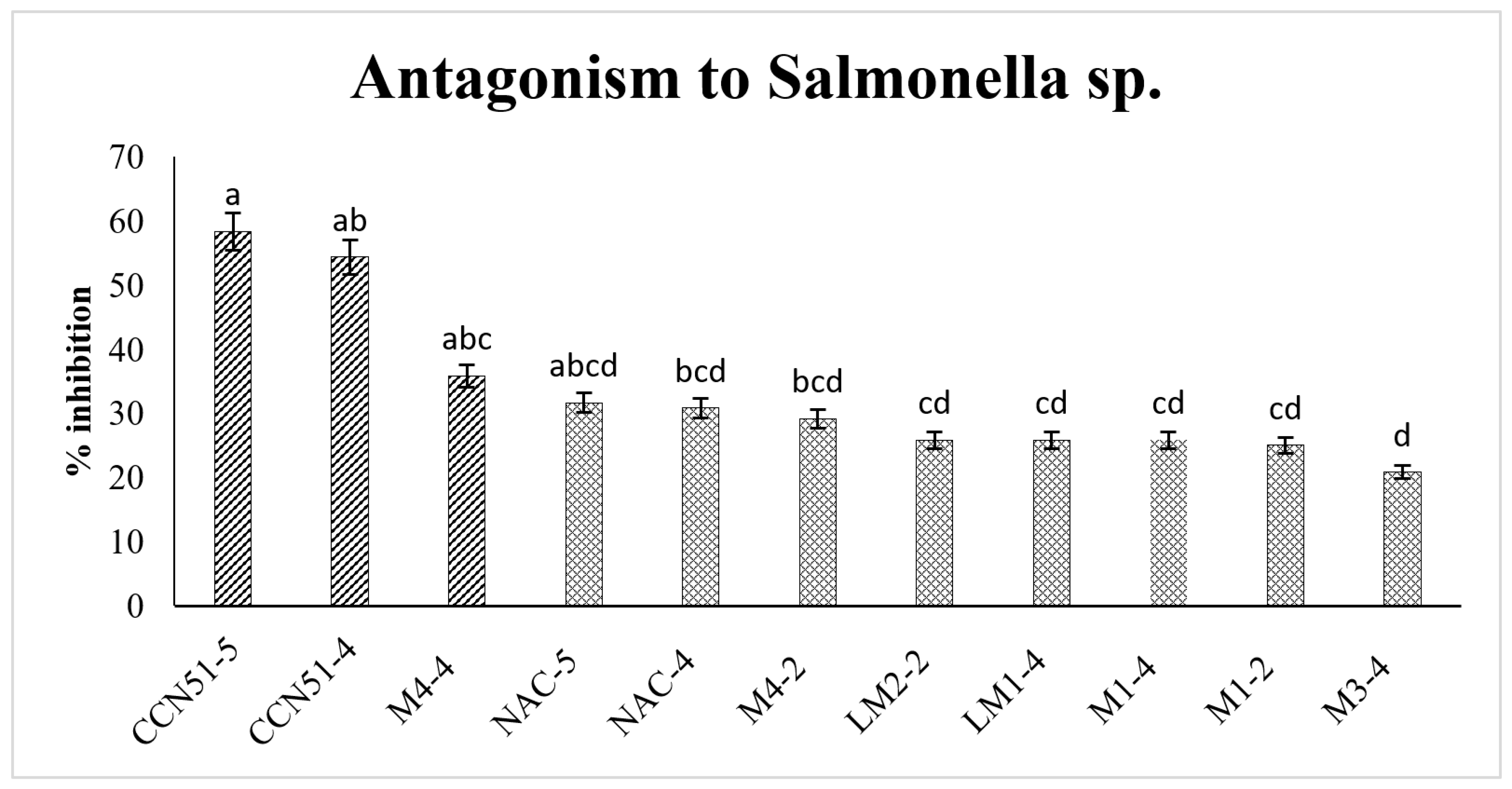
3.3. Antagonism of Lactic Acid Bacteria on Salmonella sp.

Eleven LAB strains were selected, and the antagonistic capacity of lactic acid bacteria against Salmonella sp. was determined. Notably, the CCN51-5 and CCN51-4 strains exhibited the most significant inhibition percentages among all the treatments, with values of 58.33 and 54.33%, respectively (Figure 3). Results are consistent with Borrás et al. [72], who used a microbial preparation using L. delbrueckii ssp. bulgaricus and Streptococcust hermophilus from a commercial lyophilized culture (Liofast Y452B, SACCO®, Sakho, Italy). The study analyzed the growth of pathogenic bacteria, revealing the absence of Clostridium spores and Salmonella. These results strongly support the safety of using this inoculum.
Rivera [38] identified 11 strains of LAB with antagonistic activity,; the species L. lactis subspecies Cremoris and Leuconostoc spp. had a greater inhibition effect. The presence of LAB in artisanal cheese shows antagonistic activity against pathogens such as Salmonella; evidence has shown that lactic acid fermentation is an alternative to pasteurization. Mendoza [73] analyzed the presence of E. coli and Salmonella in cream cheese fermented with LAB from fine aroma cocoa mucilage, where it was determined that there was no presence of Gram-negative bacteria. These results are consistent with those observed in the present investigation. Mirkovic et al. [74] analyzed the antagonistic capacity of L. lactis against Salmonella and E. coli from beef. The isolated LAB demonstrated favorable behavior, presenting a moderately rapid growth and a limited maximum population. Regarding the activity of its cell-free supernatant, it displayed greater effectiveness against Salmonella compared to E. coli.
The use of biopreservation should be considered only as an additional measure to complement good manufacturing practices during the processing, storage, and distribution of meats. In addition, bacteriocins should not be considered to be inhibitors by themselves since they act synergistically as an additional barrier with other preservation methods, in which the combined effects of pH, temperature, and oxygen availability simultaneously serve to preserve the food [75]. The microbiological quality for raw products is extremely critical for both meat and fat. It should maintain low microbiological counts, typically ranging between 102 and 105 CFU/g for mesophilic aerobic count. Additionally, an acceptable range for E. coli is 103 CFU/g, which serves as the standard for most manufacturing companies [76].

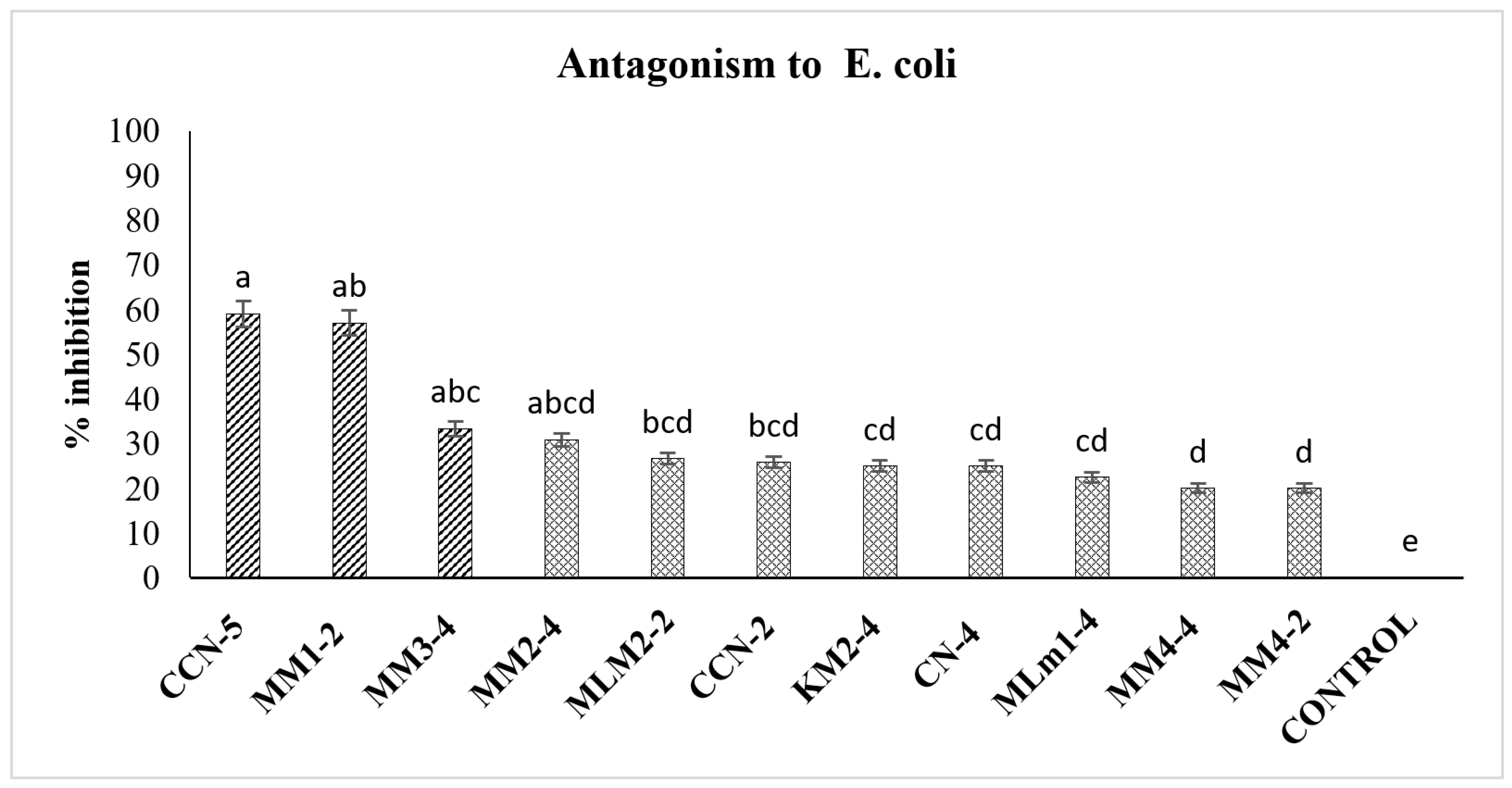
3.4. Antagonism of Lactic Acid Bacteria on E. coli

While assessing the antagonistic capacity of lactic acid bacteria against E. coli, the CCN-5 and MM1-2 strains demonstrated the highest percentages of inhibition compared to the other treatments, with values of 59 and 57% (Figure 4). These results are consistent with findings reported by Moscoso [77], where E. coli counts decreased during the 15-day shelf life of the product under the application of a protective culture composed of L. plantarum and Staplylococcus carnosus. Although there were no significant differences in the final counts of E. coli, better outcomes were observed in T2, using a concentration of 0.03% of the crop. Some studies indicate that moderate concentrations of L. delbrueckii (5.3 log UFC g−1) [78] and L. dairy [79] manage to reduce the population of E. coli to three logarithmic units in a period of three days without altering the organoleptic characteristics.
Gutierrez et al. [80] indicated that the spectrum of inhibition of bacteriocins or their extracts vary according to the treatment to which they are subjected, such as lyophilization, supernatant concentration, purification, and neutralization, among others. Bacteriocin extracts exhibit intensified activity when concentrated. Suarez et al. [81] showed that the initial count in raw fish fillets from mesophiles (4.8 log CFU)−1) indicates a good initial state of quality for the fillets. After 30 days of storage, the fillets inoculated with the crude extract of LAB bacteriocins reached a mesophile count of 5.7 log cycles. This count was lower than the fillets treated with lactic acid (6.3 log cycles) and the control fillets (6.6 log cycles). Notably, the fillets treated with the crude extract of bacteriocins exhibited a reduction of 0.9 log cycles in mesophile count compared to the control sample.
Purified or partially purified bacteriocins have diverse applications, serving as feed additives or being incorporated into active packages. Additionally, bacteriocin-producing cells can be used as starter or protective cultures in the production of fermented meats [82].
Hernandez [83] analyzed lactic acid bacteria (LAB) native to meat capable of inhibiting the growth of pathogenic bacteria. From the ground beef samples, 115 LAB isolates were obtained, with 44 of them demonstrating antagonistic effects against reference bacteria such as Salmonella and E. coli. Five isolates of LAB as well as its cell-free supernatant (CFS) showed antagonistic capacity in the population of ground beef bacteria at refrigeration temperature, concluding that the strains of L. delbrueckii and L. lactis have potential to be used as biopreservatives reducing the population of Salmonella and E. coli contaminant in ground beef, and its CFS can be used as an alternative or complementary disinfection treatment [84].
Alvarado et al. [85] conducted an evaluation of 27 unbranded Mexican artisanal foods, resulting in the isolation of 94 LAB strains. Out of these, 25 strains showed antimicrobial activity. The inhibitory activity shown by the LAB isolated strains was attributed mainly to the reduction in pH by the production of organic acids. Cell-free cultures Leuconostoc mesenteroides (isolated from chorizo and pulque) managed to reduce the number of viable cells of E. coli enteropathogenic (EPEC), whereas L. plantarum (isolated from vinegar mother) significantly inhibited S. aureus 8943. E. faecium isolated from panela cheese produced a bacteriocin with broad activity against L. monocytogenes, demonstrating that some LAB isolated from traditional Mexican foods have potential as bioconservatives.
Although the deterioration of animal products under anaerobic conditions is usually attributed to lactic acid bacteria [81], the results obtained in this study show that LAB strains from cocoa mucilage effectively reduced the population of pathogenic bacteria Salmonella and E. coli in beef. According to Simova et al. [86], the antagonistic effect observed in L. delbrueckii against psychrophilic and coliform bacteria is mainly due to the action of hydrogen peroxide produced under aerobic growth conditions. However, although several authors [86,87] indicate that hydrogen peroxide is effective in reducing the growth of E. coli and Salmonella, some authors indicate that under special conditions these bacteria can produce enough catalase that protects them from the peroxide-reducing effect [88,89,90]. This could explain the low population reduction of E. coli and Salmonella observed [84,91].
Most bacteriocins act on the membrane of sensitive cells, destabilizing and permeabilizing them by forming ionic channels or pores [92], which will give output to compounds such as phosphate, potassium, amino acids, and ATP, decreasing the synthesis of macromolecules, and consequently cell death [93]. In addition, the amphipathic nature of bacteriocins enhances their distribution along the surface of the bacterial cell membrane [10]. The consequences of the above reflect the decrease in the membrane potential and the scarce availability of the energy reserves of the cell, which leads to a decrease in the synthesis of DNA, RNA, and proteins, which ultimately triggers the death of the cell [10,11,94,95].
In the studies conducted by Suarez et al. [81], it was determined that Lactobacillus exhibits an antagonistic effect on Gram-positive microorganisms and, in some cases, on Gram-negative microorganisms. The same authors point out that plantaricin F, produced by the Lactobacillus, is effective on Lactobacillus, Lactococcus, Listeria, Micrococcus, Pediococcus, Staphylococcus, Streptococcus, Salmonella, and Pseudomonas.
For proper human consumption, beef must maintain certain physical–chemical parameters to ensure its shelf life, without presenting any structural damage and preserving its organoleptic parameters, among which the pH, humidity, protein, ashes, fat, and fibers stand out—the same ones that ensure a range of appetizing qualities for the consumer. To safeguard the safety of the consumer, the analysis of the physical–chemical parameters of the meat inoculated with LAB was carried out, through the evaluation of three different methods of application: immersion, injection, and spraying. The results are shown in Figure 5.
It was determined that there were no statistical differences between the treatments evaluated—that is, the form of application of lactic acid bacteria does not affect the physical and chemical characteristics of the meat; in the same way, it was determined that the LAB do not alter the content of fat, ash, fiber, moisture, and proteins, which is a relevant result since it seeks to preserve the shelf life of meats and not change their nutritional content. Despite not presenting statistical differences, the different treatments presented a wide average range: among which, the humidity stands out with an average range of 71.28–74.18%, the protein with a range of 20.05–24.96% of the total of the loin meat studied, the ash with a percentage range of 3–4.90% of the beef analyzed, the total fat distributed with a range of 1.77–4.77%, and muscle fiber with a range of 0.007–0.040% distributed in the meat of loin studied.
Nogales et al. [96] indicate that beef presents significant differences among 24 h production systems postmortem, with an average humidity of 76.11% in the extensive production system and 71.98% in the intensive production system, results similar to those obtained by León et al. [41] in which beef presented an average moisture percentage of 71% compared to chicken and sheep meat, which exhibited a moisture percentage of 77%. Farfan et al. [97] reported varying moisture content percentages in raw cuts of Creole cattle. The moisture content ranged from 75.80 to 72.16% in cuts such as buttock (located in the femoral region), empty (located in the abdominal region), wide steak (located in the dorsal region), and palette (located at the angle formed between the caudal edges of the humerus and scapula).
Several studies [78,98,99,100] have examined the efficacy of CFS produced by selected strains of LAB as antimicrobials in vitro, targeting bacteria such as E. coli, S. aureus [100], Shigella sonnei, Pseudomonas fluorescens, S. Typhimurium, and L. monocytogenes. The results indicate that when used as a marinade for meat, the supernatant successfully reduces pathogenic bacteria such as those mentioned. In addition, the raw meat displays notable color changes; however, it does not adversely affect the meat’s organoleptic characteristics, as no color changes were detected in grilled meat [101].
Katikou et al. [39] inoculated L. sakei CECT 4808 and L. curvatus CECT 904(T) in slices of beef for a storage period of 28 days, stored at 4 ± 1, where it was possible to observe averages in the percentages of protein, moisture, fat, and ash of 21.57, 74.23, 2.64, and 0.99, respectively, under the application of lactic acid bacteria.
The fatty acid composition is strongly determined by the breeds of animals and the production systems adopted in each different region. For instance, under the North American system, a piece of “select” loin may contain more than 5% fat. In comparison, the fat content in the loin (longissimus muscle) of the Angus steers category “choice” was found to be 9.3%, while in the muscle of Wagyu steers raised according to Japanese standards, it reached 20.4%. A loin of Wagyu animals of the highest infiltration categories can have almost 40% fat. in a previous experiment conducted at the Pontificia Universidad Católica de Chile, the fat content of 37 fattened steers and bulls, categorized under the national standard with fat coverage grades 1 and 2, was evaluated. The study revealed an average fat content of 1.34% in the smooth loin [102].

4. Conclusions

In this study, we were able to isolate and characterize 31 bacterial strains from cocoa and meat. Among the strains evaluated, an important group corresponded to lactic acid bacteria, which showed antagonistic activity against pathogens such as Salmonella sp. and Escherichia coli. In particular, bacteria CCN51-5 and CCN51-4 showed the highest antagonism against Salmonella sp., while CCN-5 and MM1-2 exhibited strong antagonism against Escherichia coli. The application of LAB using methods such as dipping, injection, and spraying did not show statistically significant differences in their efficacy for inhibiting the growth of pathogens in beef. The application of LAB to beef had no significant impact on the physicochemical characteristics, suggesting that these biopreservatives have the potential to maintain meat quality during storage and transport. The use of cocoa mucilage as a biological control agent through the application of LAB proved to be an effective strategy for the biopreservation of beef due to its ability to inhibit the growth of pathogenic bacteria.
Taken together, these results highlight the potential of LAB isolated from cocoa mucilage and beef as biopreservatives for beef, which could have a positive impact on food safety and beef shelf-life extension. The use of natural agents such as LAB for the control of pathogens in food is a promising alternative to synthetic chemical preservatives, opening up new opportunities for the development of more sustainable and healthier strategies in meat preservation. However, further research in this field is needed to fully understand the mechanisms of action of LAB and to optimize their application in different types of meat and meat products.

Author Contributions

Conceptualization, L.M.-C., C.B.-C. and F.A.; methodology, L.M.-C., C.B.-C. and E.A.P.; validation, R.D., N.S. and J.Q.; formal analysis, R.H. and N.S.; investigation, L.M.-C. and J.Q.; resources, F.A. and J.Q.; data curation, R.H. and M.V.; writing—original draft preparation, L.M.-C., D.C. and J.Q.; writing—review and editing, L.M.-C., F.A., J.Q., R.D. and M.V.; supervision, J.Q., F.A. and D.C. All authors have read and agreed to the published version of the manuscript.

Funding

This research was funded by PhD Scholarship UFRO.

Acknowledgments

The author thanks PhD Scholarship UFRO and the State Technical University of Quevedo (UTEQ) and the Department of the Research, Innovation, and Technology Transfer Unit of the UTEQ—Biotechnology Laboratory in the areas of Plant Tissue Culture, Microbiology, and Molecular Biology, located on the University Campus “La María”, for the support for the realization of this project.

Conflicts of Interest

The authors declare no conflict of interest.

References

  1. Conejo, Á. Evaluation of New Technologies in the Development of Healthy Meat Products; Universidad Politécnica de Madrid: Madrid, Spain, 2019. [Google Scholar]
  2. Morata, A. New Food Preservation Technologies, 2nd ed.; Editorial Cima Express: Madrid, Spain, 2010. [Google Scholar]
  3. Roncalés, P. Optimization of Packaging Systems and Food Preservation; Elsevier: Zaragoza, Spain, 2010. [Google Scholar]
  4. Sánchez Escalante, A.; Torrescano Urrutia, G.; Camou Arriola, J.; González Méndez, N.; Hernández Watanabe, G. Combined preservation systems to extend the shelf life of meat and meat products. Nacameh 2008, 2, 124–159. [Google Scholar] [CrossRef]
  5. Agudelo, N.; Torres, M.; Alvarez, C.; Vélez, L. Bacteriocins Produced by Lactic Acid Bacteria and Their Application in the Food Industry. Aliment. Hoy 2015, 23, 63–72. [Google Scholar]
  6. Altamarino, D.; Chavarría, M. Microbial Consortium Dose and Temperature Degree in the Useful Life of an Artisanal Sausage; Escuela Superior Politécnica Agropecuaria de Manabí Manuel Féliz López: Calceta, Ecuador, 2019. [Google Scholar]
  7. Fadda, S.; Chambon, C.; Champomier-Vergès, M.C.; Talon, R.; Vignolo, G. Lactobacillus role during conditioning of refrigerated and vacuum-packaged Argentinean meat. Meat Sci. 2018, 79, 603–610. [Google Scholar] [CrossRef] [PubMed]
  8. García Lacarra, T.; Martín de Santos, M.d.R.; Sanz Pérez, B.; Hernández, P. Extension of the shelf life of fresh meat I: Packaging in modified atmospheres and use of lactic acid bacteria and bacteriocins. Rev. Española Cienc. Tecnol. Aliment. 1995, 35, 1–18. [Google Scholar]
  9. Vásquez, M.S.M.; Suárez, M.H.; Montoya, O.I. Evaluation of bacteriocins as a protective medium for the biopreservation of meat under refrigeration. Rev. Chil. Nutr. 2009, 36, 228–238. [Google Scholar]
  10. Heredia, P.; Hernández, A.; González, A.; Vallejo, B. Bacteriocins of lactic acid bacteria: Mechanisms of action and antimicrobial activity against pathogens in cheeses. Interciencia 2017, 42, 340–346. [Google Scholar]
  11. Vásquez, M.S.M.; Suárez, M.H.; Zapata, B.S. Use of antimicrobial substances produced by lactic acid bacteria in meat preservation. Rev. Chil. Nutr. 2009, 36, 64–71. [Google Scholar]
  12. Vallejo, C.; Vera, J.; Quintana, J.; Verdezoto, D.; Cajas, L.; Mendoza, T. Lactic acid bacteria present in the cocoa (Theobroma cacao L.) mucilage of two varieties. Rev. Investig. Talent. V 2018, 1, 59–68. [Google Scholar]
  13. Fiorentini, Â.M.; Sant’Anna, E.S.; Porto, A.C.S.; Mazo, J.Z.; Franco, B.D.G.M. Influence of bacteriocins produced by Lactobacillus plantarum BN in the shelf-life of refrigerated bovine meat. Braz. J. Microbiol. 2001, 32, 42–46. [Google Scholar] [CrossRef]
  14. Vanegas, M.; Ossa, J.; Gardeazábal, P.; Coral Durango, A. Lactic acid bacteria (LAB) as an alternative for the conservation of beef packed in modified atmosphere. Aliment. Hoy 2011, 20, 18–27. [Google Scholar]
  15. Singh, V.P. Recent approaches in food bio-preservation—A review. Open Vet. J. 2018, 8, 104–111. [Google Scholar] [CrossRef]
  16. Villa, K.; Chanci, I.; Wilches, L.; Cardona, J. Characterization of lactic acid bacteria metabolites and inhibitory effect of bacteriocins on pathogenic microorganisms in food: Systematic review of the literature, 2008–2012. Rev. Biosalud 2014, 13, 45–61. [Google Scholar]
  17. Guerrero, I.; Mendiolea, R.; Ponce, E.; Prado, A. Inoculation of lactic acid bacteria on meat surfaces as a means of decontamination in semitropical conditions. Meat Sci. 1995, 40, 397–411. [Google Scholar] [CrossRef]
  18. Daeschel, M.A. Applications of Bacteriocins in Food Systems. In Biotechnology and Food Safety; Butterworth-Heinemann: Oxford, UK, 1990; pp. 91–104. [Google Scholar]
  19. Monroy, D.; Castro, T.; Fernández, F.; Mayorga, L. Bacteriocins produced by probiotic bacteria. ContactoS 2009, 73, 63–72. [Google Scholar]
  20. Chen, H.; Hoover, D.G. Bacteriocins and Their Food Applications. Compr. Rev. Food Sci. Food Saf. 2003, 2, 82–100. [Google Scholar]
  21. Luzuriaga Peña, D.L. Extraccion y Aprovechamieno del Mucilago de Cacao (Theobroma cacao) Como Materia Prima en la Elaboracion de Vino. Undergraduate Thesis, Universidad Tecnológica Equinoccial, Quito, Ecuador, 2012. [Google Scholar]
  22. Albán, D. Use of Lactic Acid Bacteria from the Mucilage of National Cocoa (Theobroma cacao L.) for the Preservation of Beef. Ph.D. Thesis, Universidad Técnica Estatal de Quevedo, Quevedo, Ecuador, 2017. [Google Scholar]
  23. De Man, J.; Rogosa, D.; Sharpe, M. A medium for the cultivation of lactobacilli. J. Appl. Bacteriol. 1960, 23, 130–135. [Google Scholar] [CrossRef]
  24. King, E.O.; Ward, M.K.; Raney, D.E. Two simple media for the demonstration of pyocyanin and fluorescin. J. Lab. Clin. Med. 1954, 44, 301–307. [Google Scholar]
  25. Atlas, R. Handbook of Microbiological Media, 4th ed.; Taylor & Francis Group, LLC.: Washington, DC, USA, 2010. [Google Scholar]
  26. Vázquez, R.; Salvador, M.; Adriano, L.; DeGyves, G.; Vázquez, A. Use of starter culture of native lactic acid bacteria for producing an artisanal Mexican cheese safe and sensory acceptable. CyTA-J. Food 2018, 16, 460–468. [Google Scholar] [CrossRef]
  27. Phang, I.; San Chan, Y.; Wong, K.; Lau, S.Y. Isolation and characterization of urease-producing bacteria from tropical peat. Biocatal. Agric. Biotechnol. 2018, 13, 168–175. [Google Scholar] [CrossRef]
  28. Liu, Z.; Li, Y.C.; Zhang, S.; Fu, Y.; Fan, X.; Patel, J.S.; Zhang, M. Characterization of phosphate-solubilizing bacteria isolated from calcareous soils. Appl. Soil Ecol. 2015, 96, 217–224. [Google Scholar] [CrossRef]
  29. Zhou, X.; Wang, Y.; Li, W. Effect of probiotic on larvae shrimp (Penaeus vannamei) based on water quality, survival rate and digestive enzyme activities. Aquaculture 2009, 287, 349–353. [Google Scholar] [CrossRef]
  30. Linares, J. Selection of Lactic Acid Bacteria Isolated from Plant Sources for Their Antimicrobial Capacity and Their Use in Food Biopreservation. Ph.D. Thesis, Autonomous University of Chihuahua, Chihuahua, México, 2021. [Google Scholar]
  31. Jensen, J.; Jeppesen, E.; Olrik, K.; Kristensen, P. Impact of nutrients and physical factors on the shift from cyanobacterial to chlorophyte dominance in shallow Danish lakes. Can. J. Fish. Aquat. Sci. 1994, 51, 1692–1699. [Google Scholar] [CrossRef]
  32. MacFaddin, J. Biochemical Tests for the Identification of Clinically Important Bacteria; Editorial Médica Panamericana: Madrid, Spain, 2003. [Google Scholar]
  33. Liu, W.; Zhang, L.; Yi, H.; Shi, J.; Xue, C.; Li, H.; Jiao, Y.; Shigwedha, N.; Du, M.; Han, X. Qualitative detection of class IIa bacteriocinogenic lactic acid bacteria from traditional Chinese fermented food using a YGNGV-motif-based assay. J. Microbiol. Methods 2014, 100, 121–127. [Google Scholar] [CrossRef]
  34. Beveridge, T. Use of the Gram stain in microbiology. Biotech. Histochem. 2001, 76, 111–118. [Google Scholar] [CrossRef] [PubMed]
  35. Rodríguez, A.J. Evaluation of the Growth Kinetics of PGPR and Its Antagonistic Activity Towards Meloidogyne Incognita “In Vitro”. Ph.D. Thesis, Universidad Técnica Estatal de Quevedo, Quevedo, Ecuador, 2018. [Google Scholar]
  36. Vargas Cedeño, G.E. Biodegradation of Polyethylene through the Use of Microorganisms. Bachelor’s Thesis, Universidad Técnica Estatal de Quevedo, Quevedo, Ecuador, 2020. [Google Scholar]
  37. Singh, A.; Maurya, S.; Singh, R.; Singh, U.P. Antibiotic potential of plant growth promoting rhizobacteria (PGPR) against Sclerotium rolfsii. Arch. Phytopathol. Plant Prot. 2012, 45, 1655–1662. [Google Scholar] [CrossRef]
  38. Rivera, J.; Villegas, A.; Miranda, L.; García, J. Identification of antagonistic acidolactic bacteria of Salmonella enterica var. Typhimurium isolated from artisanal cheese. Rev. Mex. Cienc. Agrícolas 2017, 8, 785–797. [Google Scholar] [CrossRef]
  39. Katikou, P.; Ambrosiadis, I.; Georgantelis, D.; Koidis, P.; Georgakis, S.A. Effect of Lactobacillus-protective cultures with bacteriocin-like inhibitory substances’ producing ability on microbiological, chemical and sensory changes during storage of refrigerated vacuum-packaged sliced beef. J. Appl. Microbiol. 2005, 99, 1303–1313. [Google Scholar] [CrossRef]
  40. Reyes, N.; Mendieta, B. Determination of the Nutritional Value of Food; Universidad Nacional Agraria: Managua, Nicaragua, 2000. [Google Scholar]
  41. León, M.; Orduz, A.; Velandia, M. Physicochemical composition of sheep, chicken, beef and pork meat. @Limentech Food Technol. 2017, 15, 62–75. [Google Scholar] [CrossRef]
  42. Braña Varela, D.; Ramírez, E.; Rubio, M.d.l.S.; Sánchez, A.; Torrescano, G.; Arenas, M.; Partida, J.; Ponce, E.; Ríos, F. Manual of Quality Qnalysis in Meat Samples, 1st ed.; Instituto Nacional de Investigaciones Forestales Agrícolas y Pecuarias: Ciudad de México, México, 2011. [Google Scholar]
  43. Niño de Polanía, L.; López, D.; Malagón, M. Meat: Manual of Analysis, 1st ed.; Hernández, C., Ed.; Ministerio de Salud: Bogotá, Colombia, 1995. [Google Scholar]
  44. Apráez, J. Chemical Analysis of Animal Feed, 1st ed.; Editorial Universidad de Nariño: Nariño, Columbia, 2020. [Google Scholar]
  45. Dikeman, M.; Devine, C. Encyclopedia of Meat Sciences, 2nd ed.; Elsevier: Amsterdam, The Netherlands, 2014. [Google Scholar]
  46. Ibrahim, S.A.; Ayivi, R.D.; Zimmerman, T.; Siddiqui, S.A.; Altemimi, A.B.; Fidan, H.; Esatbeyoglu, T.; Bakhshayesh, R.V. Lactic Acid Bacteria as Antimicrobial Agents: Food Safety and Microbial Food Spoilage Prevention. Foods 2021, 10, 3131. [Google Scholar] [CrossRef]
  47. Morrison, D.A. Phylogenetic tree-building. Int. J. Parasitol. 1996, 26, 589–617. [Google Scholar] [CrossRef]
  48. Papalexandratou, Z.; Falony, G.; Romanens, E.; Jimenez, J.C.; Amores, F.; Daniel, H.M.; De Vuyst, L. Species diversity, community dynamics, and metabolite kinetics of the microbiota associated with traditional Ecuadorian spontaneous cocoa bean fermentations. Appl. Environ. Microbiol. 2011, 77, 7698–7714. [Google Scholar] [CrossRef] [PubMed]
  49. Wong, A.; Corzo, H.; Hernández, E.; González, A.; Giacomán, G. Characterization of lactic acid bacteria with antimicrobial activity isolated from cream cheese from Chiapas, Mexico. CienciaUAT 2021, 15, 144–155. [Google Scholar]
  50. De Vuyst, L.; Weckx, S. The cocoa bean fermentation process: From ecosystem analysis to starter culture development. J. Appl. Microbiol. 2016, 121, 5–17. [Google Scholar] [CrossRef] [PubMed]
  51. Crafack, M.; Mikkelsen, M.B.; Saerens, S.; Knudsen, M.; Blennow, A.; Lowor, S.; Takrama, J.; Swieg-ers, J.H.; Petersen, G.B.; Heimdal, H.; et al. Influencing cocoa flavour using Pichia kluyveri and Kluyveromyces marxianus in a defined mixed starter culture for cocoa fermentation. Int. J. Food Microbiol. 2013, 167, 103–116. [Google Scholar] [CrossRef] [PubMed]
  52. Meersman, E.; Steensels, J.; Mathawan, M.; Pieter-Jan, W.; Saels, V. Detailed Analysis of the Microbial Population in Malaysian Spontaneous Cocoa Pulp Fermentations Reveals a Core and Variable Microbiota. PLoS ONE 2013, 8, e81559. [Google Scholar] [CrossRef]
  53. Scarpari, L.M.; Meinhardt, L.W.; Maizzafera, P.; Pomella, A.W.V.; Schiavinato, M.A.; Cascardo, J.C.M.; Pereira, G.A.G. Biochemical changes during the development of witches’ broom: The most important disease of cocoa in Brazil caused by Crinipellis perniciosa. J. Exp. Bot. 2005, 56, 865–877. [Google Scholar] [CrossRef]
  54. de Melo Pereira, G.V.; Magalhães, K.T.; de Almeida, E.G.; da Silva Coelho, I.; Schwan, R.F. Spontaneous cocoa bean fermentation carried out in a novel-design stainless steel tank: Influence on the dynamics of microbial populations and physical-chemical properties. Int. J. Food Microbiol. 2013, 161, 121–133. [Google Scholar] [CrossRef]
  55. de Melo Pereira, G.V.; Da Cruz Pedrozo Miguel, M.G.; Lacerda Ramos, C.; Freitas Schwan, R. Microbiological and physicochemical characterization of small-scale cocoa fermentations and screening of yeast and bacterial strains to develop a defined starter culture. Appl. Environ. Microbiol. 2012, 78, 5395–5405. [Google Scholar] [CrossRef]
  56. Snauwaert, I.; Papalexandratou, Z.; De Vuyst, L.; Vandamme, P. Characterization of strains of Weissella fabalis sp. nov. and Fructobacillus tropaeoli from spontaneous cocoa bean fermentations. Int. J. Syst. Evol. Microbiol. 2013, 63, 1709–1716. [Google Scholar]
  57. Ardhana, M.M.; Fleet, G.H. The microbial ecology of cocoa bean fermentations in Indonesia. Int. J. Food Microbiol. 2003, 86, 87–99. [Google Scholar] [CrossRef]
  58. Camu, N.; De Winter, T.; Verbrugghe, K.; Cleenwerck, I.; Vandamme, P.; Takrama, J.S.; Vancanneyt, M.; De Vuyst, L. Dynamics and Biodiversity of Populations of Lactic Acid Bacteria and Acetic Acid Bacteria Involved in Spontaneous Heap Fermentation of Cocoa Beans in Ghana. Appl. Environ. Microbiol. 2007, 73, 1824. [Google Scholar] [CrossRef] [PubMed]
  59. Lagunes, S.; Loiseau, G.; Paredes, J.; Barel, M.; Guiraud, J. Study on the microflora and biochemistry of cocoa fermentation in the Dominican Republic. Int. J. Food Microbiol. 2007, 114, 124–130. [Google Scholar] [CrossRef] [PubMed]
  60. Nielsen, D.S.; Teniola, O.D.; Ban-Koffi, L.; Owusu, M.; Andersson, T.S.; Holzapfel, W.H. The microbiology of Ghanaian cocoa fermentations analysed using culture-dependent and culture-independent methods. Int. J. Food Microbiol. 2007, 114, 168–186. [Google Scholar] [CrossRef] [PubMed]
  61. Kostinek, M.; Ban-Koffi, L.; Ottah-Atikpo, M.; Teniola, D.; Schillinger, U.; Holzapfel, W.H.; Franz, C.M.A.P. Diversity of predominant lactic acid bacteria associated with cocoa fermentation in Nigeria. Curr. Microbiol. 2008, 56, 306–314. [Google Scholar] [CrossRef] [PubMed]
  62. Lefeber, T.; Janssens, M.; Camu, N.; De Vuyst, L. Kinetic analysis of strains of lactic acid bacteria and acetic acid bacteria in cocoa pulp simulation media toward development of a starter culture for cocoa bean fermentation. Appl. Environ. Microbiol. 2010, 76, 7708–7716. [Google Scholar] [CrossRef]
  63. Lozano, R.; Orellana, Z. Unidad Académica de Ciencias Administrativas y Proyecto de Grado Previo a la Obtención del Título de: Ingeniero en Contaduría Pública y Auditoría—CPA Título del Proyecto: “Estudio de Factibilidad Para la Instalación de una Fábrica de Plástico Dedicad”; Universidad Estatal de Milagro: Milagro, Ecuador, 2013. [Google Scholar]
  64. Papalexandratou, Z.; Camu, N.; Falony, G.; De Vuyst, L. Comparison of the bacterial species diversity of spontaneous cocoa bean fermentations carried out at selected farms in Ivory Coast and Brazil. Food Microbiol. 2011, 28, 964–973. [Google Scholar] [CrossRef]
  65. Mesa, Y.; Mas, S.; Anaya, M.; Cobo, H.; Díaz, M. Study of the behavior of Lactic Acid Bacteria (LAB) of the Bioyogur culture at different doses of magnetic treatment. Tecnol. Química 2016, 36, 439–456. [Google Scholar]
  66. Jurado, H.; Martínez, J.; Morillo, J.; Romero, D.; Orbes, A.; Mesías, L. Determination of fermentation kinetics in two probiotic media, in vitro performance tests and inhibition effect of Lactobacillus plantarum. Vet. Zootecnía 2011, 10, 23–41. [Google Scholar]
  67. Calderón, J. Adjustment of a Kinetic Model for the Growth of Lactobacillus acidophilus in the Fermentation of a Complex Substrate. Bachelor’s Thesis, Fundación Universidad de América, Bogotá, Columbia, 2017. [Google Scholar]
  68. Zamora Rodríguez, L. Isolation, Identification and Conservation of Cultures of Lactic Bacteria Antagonistic to the Contaminating Microbiota of Slaughterhouse Blood. Ph.D. Thesis, Universidad de Girona, Girona, Spain, 2003. [Google Scholar]
  69. Wirth, F. The pH is an important parameter related to the susceptibility of meat to deterioration and is used to decide on the type of processing to which the meat is going to be used. Fleischwirtsch. Español 1987, 1, 22–28. [Google Scholar]
  70. Fernández Yanza, M.A.; Morocho Quichimbo, B.J. Determination of Lactic Acid Bacteria and Identification of Sources of Contamination in Finished Meat Products Vacuum-Packed. Master’s Thesis, Universidad de Cuenca, Cuenca, Ecuador, 2020. [Google Scholar]
  71. Flores, M. Understanding the implications of current health trends on the aroma of wet and dry cured meat products. Meat Sci. 2018, 144, 53–61. [Google Scholar] [CrossRef]
  72. Borrás, L.; Valiño, E.; Rodríguez, C. Microbial preparation with lactic acid activity as a biological accelerator in fermentation processes for animal feed. Cienc. Agric. 2017, 14, 7–13. [Google Scholar]
  73. Mendoza, T. Elaboration of Cream Cheese with Lactic Bacteria from Cocoa Mucilage (Theobroma cacao L.) Fine Aroma. Bachelor’s Thesis, Universidad Técnica Estatal de Quevedo, Quevedo, Ecuador, 2017. [Google Scholar]
  74. Mirkovic, N.; Polovic, N.; Vukotic, G.; Jovcic, B.; Miljkovic, M.; Radulovic, Z.; Diep, D.; Kojic, M. Lactococcus lactis LMG2081 Produces Two Bacteriocins, a Nonlantibiotic and a Novel Lantibiotic. Appl. Environ. Microbiol. 2016, 82, 2555–2562. [Google Scholar] [CrossRef] [PubMed]
  75. Castellano, P.; Belfiore, C.; Fadda, S.; Vignolo, G. A review of bacteriocinogenic lactic acid bacteria used as bioprotective cultures in fresh meat produced in Argentina. Meat Sci. 2008, 79, 483–499. [Google Scholar] [CrossRef] [PubMed]
  76. Feiner, G. Chapter 39—The microbiology of specific bacteria. In Meat Products Handbook; Elsevier: Amsterdam, The Netherlands, 2006; pp. 670–672. [Google Scholar]
  77. Moscoso, J. Biopreservation of Raw Sausages through the Use of Staphylococcus carnosus and Lactobacillus plantarum as Protective Cultures. Master’s Thesis, Universidad del Azuay, Cuenca, Spain, 2017. [Google Scholar]
  78. Hovhannisyan, H.; Goodarzi, A.; Barseghyan, A. Antimicrobial substances production at refrigeration temperatures by Lactobacillus 52 delbrueckii MH10: A Candidate for food biopreservation. Int. J. Nutr. Food Sci. 2016, 5, 184. [Google Scholar]
  79. Jones, R.J.; Hussein, H.M.; Zagorec, M.; Brightwell, G.; Tagg, J.R. Isolation of lactic acid bacteria with inhibitory activity against pathogens and spoilage organisms associated with fresh meat. Food Microbiol. 2008, 25, 228–234. [Google Scholar] [CrossRef]
  80. Gutierrez Ramírez, L.A.; Campuzano Montoya, O.I.; Villadiego Ruiz, O.S. Evaluation of the bactericidal potential of extracts of lactic acid bacteria extracts on the in vitro growth of E. coli, Salmonella sp. and Listeria monocytogenes. Rev. CENIC Cienc. Biológicas 2005, 36, 1–25. [Google Scholar]
  81. Suárez, H.; Francisco, A.; Beirão, L. Influence of Bacteriocins produced by Lactobacillus plantarum LPBM10 on the shelf life of Cachama Piaractus brachypomus × Colossoma macropomum hybrid fillets vacuum packed. VITAE 2008, 15, 32–40. [Google Scholar]
  82. Bhattacharya, D.; Nanda, P.K.; Pateiro, M.; Lorenzo, J.M.; Dhar, P.; Das, A.K. Lactic acid bacteria and bacteriocins: Novel biotechnological approach for biopreservation of meat and meat products. Microorganisms 2022, 10, 2058. [Google Scholar] [CrossRef]
  83. Hernández, S. Lactic Acid Bacteria as Biopreservatives for Beef; Universidad Autónoma de Chapingo: Chapingo, Mexico, 2019. [Google Scholar]
  84. Hernández, S.; Miranda, L.; Maldonado, E.; Alarcón, B. Inhibition of Salmonella and E. coli on ground beef by lactic acid bacteria. Univ. Autónoma Chapingo 2019, 1, 39–52. [Google Scholar]
  85. Alvarado, C.; García, B.; Martin, S.; Regalado, C. Food-associated lactic acid bacteria with antimicrobial potential from traditional Mexican foods. Rev. Latinoam. Microbiol. 2006, 48, 260–268. [Google Scholar]
  86. Simova, E.D.; Beshkova, D.M.; Angelov, M.P.; Dimitrov, Z.P. Bacteriocin production by strain Lactobacillus delbrueckii ssp. bulgaricus BB18 during continuous prefermentation of yogurt starter culture and subsequent batch coagulation of milk. J. Ind. Microbiol. Biotechnol. 2008, 35, 559–567. [Google Scholar] [PubMed]
  87. Senne, M.M.; Gilliland, S.E. Antagonistic action of cells of Lactobacillus delbrueckii subsp. lactis against pathogenic and spoilage microorganisms in fresh meat systems. J. Food Prot. 2003, 66, 418–425. [Google Scholar] [PubMed]
  88. Watson, J.A.; Schubert, J. Action of hydrogen peroxide on growth inhibition of Salmonella typhimurium. J. Gen. Microbiol. 1969, 57, 25–34. [Google Scholar] [CrossRef]
  89. Finn, G.J.; Condon, S. Regulation of catalase synthesis in Salmonella typhimurium. J. Bacteriol. 1975, 123, 570–579. [Google Scholar] [CrossRef] [PubMed]
  90. Humphrey, T.J.; Slater, E.; McAlpine, K.; Rowbury, R.J.; Gilbert, R.J. Salmonella enteritidis phage type 4 isolates more tolerant of heat, acid, or hydrogen peroxide also survive longer on surfaces. Appl. Environ. Microbiol. 1995, 61, 3161–3164. [Google Scholar] [CrossRef] [PubMed]
  91. Lindqvist, R.; Lindblad, M. Inactivation of Escherichia coli, Listeria monocytogenes and Yersinia enterocolitica in fermented sausages during maturation/storage. Int. J. Food Microbiol. 2009, 129, 59–67. [Google Scholar] [CrossRef]
  92. Grande, M.J.; Lucas, R.; Abriouel, H.; Ben Omar, N.; Maqueda, M.; Martínez, M.; Martínez, M.; Valdivia, E.; Gálvez, A. Control of Alicyclobacillus acidoterrestris in fruit juices by enterocin AS-48. Int. J. Food Microbiol. 2005, 104, 289–297. [Google Scholar] [CrossRef]
  93. González, B.E.; Gómez, M.; Jiménez, Z. Bacteriocinas de probióticos. RESPYN Rev. Salud Pública Nutr. 2003, 4, 1–9. [Google Scholar]
  94. Klaenhammer, T.R. Genetics of bacteriocins produced by lactic acid bacteria. FEMS Microbiol. Rev. 1993, 12, 39–85. [Google Scholar] [CrossRef]
  95. Bemena, L.D.; Mohamed, L.A.; Fernandes, A.M.; Lee, B. Applications of bacteriocins in food, livestock health and medicine. Int. J. Curr. Microbiol. Appl. Sci. 2014, 3, 924–949. [Google Scholar]
  96. Nogales, S.; Bressan, M.C.; Vaz, A.P.; Delgado, J.V.; Camacho, M.E. Physical-chemical study of the meat of the Marismeña bovine breed in different finishing systems. Arch. Zootec. 2011, 60, 453–456. [Google Scholar]
  97. Farfán, N.; Juarez, D.; Rossi, A.; Sammán, N. Chemical composition of Creole cattle meat. Arch. Latinoam. Nutr. 2000, 50, 400–404. [Google Scholar] [PubMed]
  98. Barcenilla, C.; Ducic, M.; López, M.; Prieto, M.; Álvarez, A. Application of lactic acid bacteria for the biopreservation of meat products: A systematic review. Meat Sci. 2022, 183, 108661. [Google Scholar] [CrossRef] [PubMed]
  99. Ghanbari, M.; Jami, M.; Kneifel, W.; Domig, K.J. Antimicrobial activity and partial characterization of bacteriocins produced by lactobacilli isolated from Sturgeon fish. Food Control 2013, 2, 379–385. [Google Scholar] [CrossRef]
  100. Vatanyoopaisarn, S.; Prapatsornwattana, K.; Kuhakongkeat, T.; Phalakornkule, C. Potential use of lactic acid bacteria with bacteriocin-like activity against Staphylococcus aureus as dual starter cultures in Thai fermented sausage “Sai Krok Prew”. Int. Food Res. J. 2011, 18, 697–704. [Google Scholar]
  101. Arrioja, D.; Mani, E.; Palou, E.; López, A. Antimicrobial activity and storage stability of cell-free supernatants from lactic acid bacteria and their applications with fresh beef. Food Control 2020, 115, 107286. [Google Scholar] [CrossRef]
  102. Larraín Prieto, R.; Bello Pérez, E.V. Composition of National Bovine Meat; Fundación para la Innovación Agraria (FIA): Santiago de Chile, Chile, 2013. [Google Scholar]
Figure 1. Dendrogram of strains from beef and cocoa mucilage.
Microbiolres 14 00077 g001
Figure 2. Growth kinetics of lactic acid bacteria isolated from cocoa mucilage and beef.
Microbiolres 14 00077 g002
Figure 3. Antagonism of lactic acid bacteria on Salmonella sp. Bars indicate mean ± standard error; different letters indicate significant differences between the averages at p < 0.05 (Tukey’s test).
Microbiolres 14 00077 g003
Figure 4. Antagonism of lactic acid bacteria on E. coli. Error bars indicate ±standard error; different letters indicate significant differences between the averages at p < 0.05 (Tukey’s test).
Microbiolres 14 00077 g004
Figure 5. Physical–chemical parameters of beef under the application of lactic acid bacteria by three inoculation methods. (A) Percentage of fat. (B) Percentage of humidity. (C) Percentage of fiber. (D) Percentage of ashes. (E) Percentage of proteins. (F) pH.
Microbiolres 14 00077 g005
Table 1. Colonial morphology and biochemical tests to bacterial strains from beef and cocoa mucilage.
StrainsBiochemical TestsColonial Morphology
CatalaseUreaseProteaseCO2Gram StainFormElevationMargin
KLM2-4+++CircularHighWhole
KLM4 ++++
KLM1-4+++
MM4-2++++
KLM4-4++++
MM3-4 +++
MM1-2 ++
KM3-4 ++
KM4-4 +++
KM2-4 +++
KLm3-4++++
KM1-6 ++++
MM4-4 +++
KLM2-6++++
MM4-4 +++
KM4-2 ++++
CN-4 ++
CN-3 +++
KM4-6 ++++
MM2-4 +++
KLM1-2++++
CCN-2 ++
CCN-5 +
KM1-4 +++
MLm2-2++++
KM3-6 ++++
MLM-4 +++
MLM2-2+++
MLm1-4++++
CN-2 +++++
KLm1-6+++++
High presence (++); Positive reaction (+); Negative reaction (−).
Disclaimer/Publisher’s Note: The statements, opinions and data contained in all publications are solely those of the individual author(s) and contributor(s) and not of MDPI and/or the editor(s). MDPI and/or the editor(s) disclaim responsibility for any injury to people or property resulting from any ideas, methods, instructions or products referred to in the content.

