Abstract
This study compared the minimal inhibition concentrations (MICs) and their effects on the growth kinetics of seven different types of zinc (Zn) compounds and Na2EDTA in the case of three typical commensal beneficial microorganisms (Bacillus subtilis, Lactococcus lactis, and Saccharomyces cerevisiae). The seven Zn compounds included ZnSO4, four Zn–amino acid chelates, and two Zn–EDTA complexes. Both MICs and growth kinetic parameters indicated that different microorganisms show different sensitivities; for example, B. subtilis, L. lactis, and S. cerevisiae were most sensitive to ZnSO4, Na2EDTA, and Zn(NH3)2(Gly)2, respectively. Both ZnEDTA and Zn(NH3)2(Lys)2 improved the growth rate of all beneficial commensal intestinal microorganisms at low concentrations (5–10 mg/L) and showed low toxicity towards all tested strains. At higher concentrations (100–500 mg/L), all compounds decreased the growth rate and increased the lag phase. In conclusion, both growth kinetic parameters and MICs tested effectively measured the inhibitory effects of the test materials; however, growth kinetics provides a more detailed picture of the concentration-dependent effects and those on the mechanisms of microbial growth inhibition.
1. Introduction
The gastrointestinal tract (GIT) is one of the most complex ecosystems known in nature, containing a diverse microbiota [1]. Within the microbiota, many microorganisms play essential roles in metabolic processes and in the modulation of the immune system [2,3]. Estimates suggest that the gut microbiome contains 500–1000 different species of microorganisms and that it outnumbers the host’s total number of genes and cells by an estimated 10 fold [4,5]. The microecosystem of the GIT, which is a direct consequence of the mutualism between the host and its microbiota, is fundamental to maintaining a healthy individual [6]. The intestinal tract of humans and animals hosts a high and diverse number of different microorganisms, including bacteria, fungi, protozoa, and viruses [7]. The relationship between the gut microbiota and host can be commensal, pathogenic, a harmless co-existence, and mutualistic [8]. Intestinal commensal microorganisms can supply essential nutrients, synthesize vitamins, aid in the digestion, and promote angiogenesis and enteric nerve function [9,10,11]. In exchange, the host provides the microorganisms with nutrients and a stable environment [6]. Both host and indigenous microorganisms adapted to each other in a particular microevolution case to maintain the benefits this mutualism confers [12]. It has been reported that Bacillus subtilis, Lactococcus lactis, and Saccharomyces cerevisiae improved the microbial balance in the GIT by immune stimulation and by competitive exclusion, and thus can be considered as beneficial microorganisms [13,14,15]. B. subtilis, L. lactis, and S. cerevisiae are present in the GIT of animals as well and are considered generally-recognized-as-safe (GRAS) organisms by the Food and Drug Administration (FDA) [16,17]. Both B. subtilis and S. cerevisiae have broad anti-competitive activity against animal enteric pathogens (e.g., Clostridium spp.), and were found to improve the immune status, modulate the intestinal microflora, and promote growth performance in animals [18,19]. Kobierecka et al. [20] determined that Lactococcus lactis significantly reduced enteric pathogen (e.g., Campylobacter jejune) colonization in animals.
Zinc (Zn) is an important trace element for humans and animals and a required dietary supplement for livestock (e.g., poultry and pigs) as a premix form. Zn is essential for microorganisms as well due to its role in their metabolism as part of many enzymes, such as alcohol dehydrogenase, Zn-dependent proteinases, DNA and RNA polymerases, phospholipase C, endopeptidases, and aminopeptidases [21,22,23]. Zinc deficiency in microorganisms manifests itself in metabolic disturbances and growth depression [24]. Conversely, the antimicrobial effect of excess Zn is well-known; high dietary ZnO levels (i.e., 2000–3000 mg Zn/kg) have been shown to reduce diarrhea and improve growth performance in weaning pigs [25,26]. The susceptibility to Zn, especially to ZnO and ZnSO4, among microorganisms and even within strains of individual species can be highly variable [27,28,29]. Consequently, all microorganisms must precisely regulate intracellular Zn levels.
Currently, standard antibiotic efficacy testing relies on off-line, endpoint measurements, such as minimal inhibitory concentration (MIC) determination or the agar diffusion method [30]. However, these test methods do not allow for tracking changes in microbial growth profiles. Growth kinetics is a useful tool for designing and controlling biotechnological processes [31]. Four phases are distinguished in microbial growth: lag, exponential, stationary, and decay. Microbial growth is influenced by many factors, such as temperature, oxygen availability, light, and medium composition including micro- and macroelement concentrations such as Zn [32,33]. Sigmoidal mathematical models can describe the first three phases of the microbial growth curve and identify growth parameters [34]. Microbial growth may be followed by online optical density (OD) measurement, which gives real-time information about the number of cells [35]. While MIC measurements give precise information about the effect of a given molecule in a singular media, variations in growth kinetics may produce differential outcomes in a competitive microbial community, such as the intestine of humans, monogastric animals, or ruminants.
Previous studies investigated the growth kinetics of B. subtilis, L. lactis, and S. cerevisiae [31,36,37]. However, few in vitro studies are available on the impacts of Zn compounds on beneficial intestinal microorganisms. Therefore, in the present study, we investigated the effects of various formulations and concentrations of Zn (ZnSO4, ZnNa2EDTA, ZnEDTA, Zn(Gly)2, Zn(NH3)2(Gly)2, Zn(Lys)2, and Zn(NH3)2(Lys)2) on the growth of three commensal beneficial microorganisms (B. subtilis, L. lactis, and S. cerevisiae). As ZnNa2EDTA and ZnEDTA are non-regular formulations of nutritional Zn, we sought to dissect the effects of metal and ligands on microbial growth by including Na2EDTA in the study. In order to contrast the two techniques and completely understand the impact of the different forms of nutritional Zn on the commensal beneficial microorganisms, changes in growth kinetics were compared to the MIC values of the same compounds.
2. Materials and Methods
2.1. Microorganisms
Bacillus subtilis ATCC 6633, Lactococcus lactis ATCC 19435, and Saccharomyces cerevisiae ATCC 2341 were obtained from the American Type Culture Collection (ATCC) and were grown on Nutrient Agar (Merck, Darmstadt, Germany), MRS Agar (VWR Chemical, Radnor, PA, USA), and Malt Extract Agar (Merck, Darmstadt, Germany), respectively. Microorganisms were cultivated under aerobic conditions at 37 °C.
2.2. Test Compounds
The test compounds ZnSO4·7H2O and Na2EDTA·2H2O were purchased from Merck (Darmstadt, Germany). The test compound Zn(Gly)2 was purchased from BASF, Animal Nutrition (Mannheim, Germany). The starting materials ZnO, Urea, NH4OH, Lysine, Glycine, and EDTA were purchased from Merck, (Darmstadt, Germany), and the test compounds Zn(NH3)2(Gly)2, Zn(Lys)2, Zn(NH3)2(Lys)2, and ZnEDTA were produced according to the procedures described by EP2866799B1. The test compound ZnNa2EDTA was synthetized by mixing ZnO and Na2EDTA in equimolar amounts in an aqueous solution.
2.3. MIC
The determination of the MIC was performed in 48-well suspension culture plates (Greiner bio-one, Frickenhausen, Germany) using the European Committee’s standard method for antimicrobial susceptibility testing [38]. The wells were filled with 500 µL of liquid culture medium. The compounds were tested in a two-fold dilution at a concentration in the range of 63–16,000 mg/L. The wells were inoculated with 10 µL of cell suspensions of each microorganism, containing 5 × 10⁷ colony-forming units (CFU)/mL, resulting in a starting CFU number of 106/mL for all microorganisms. The plates were incubated at 37 °C for 24 h with linear shaking. All compounds were tested in duplicate. After 24 h, the OD (λ = 600 nm) of each well was determined with a microplate reader (Synergy H1, BioTek, Winooski, VT, USA). The highest concentration where microbial growth was absent (OD600 = OD600 negative control) determined the MIC of the given test compound. Trials were performed in triplicate; however, there was no difference between the replicates.
2.4. Correction between CFU and OD
For each microorganism, the OD600 adsorption values were correlated with CFU numbers by the culturing method. A cell suspension was created for each microorganism from the agar plates by suspending two inoculum loops in 1 mL of liquid culture medium. Serial dilutions were created in the growth media in the same 48-well format plate as used for the growth kinetics measurements. The OD values were read at 600 nm using a microplate reader (Synergy H1, BioTek, Winooski, VT, USA). The serial dilutions were further diluted for culturing purposes using the corresponding medium detailed in Section 2.1. Cells were counted after 24 h of culturing at 37 °C. The OD values were correlated with the cell counts. The fitted parameters and the graphical representations of the results are shown in Figure 1. A linear correlation was found between the OD values and the viable cell numbers, as shown by the R2 values above 0.99. These results are in agreement with previous reports for all three microorganisms: S. cerevisiae [39], L. lactis [40], and B. subtilis [41].
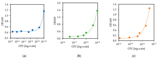
Figure 1.
Correlation between OD600 and cell number for (a) L. lactis, for which the regression equation is OD600 = 7.257 × 10¹¹ CFU + 0.436, where the R2 is 0.995; (b) B. subtilis, for which the regression equation is OD600 = 1.622 × 10⁹ CFU + 0.104, where the R2 is 0.997; (c) and S. cerevisiae, for which the regression equation is OD600 = 5.629 × 10⁸ CFU + 0.082, where the R2 is 0.991.
2.5. Growth Kinetics
Growth kinetic measurements were performed in 48-well suspension culture plates. Wells were filled with 500 µL of liquid culture medium containing the test compounds in 5–500 mg/L concentrations in duplicates. Each trial was repeated three times. Three positive control wells without test compounds and three blank wells were included in each test. Wells were inoculated with 10 µL of cell suspensions of each microorganism, containing 5 × 10⁷ CFU/mL cells, resulting in a starting CFU number of 106/mL for all microorganisms. Microplates were incubated with linear shaking at 37 °C inside the chamber of the microplate reader, and OD was read every 15 min at λ = 600 nm.
The OD was converted to cell numbers using the relationships determined prior. The growth parameters were determined by fitting the Gompertz equation [34] as follows:
where N is the cell number, N0 is the starting cell number, and t is for time. The advantage of using the Gompertz equation for the description of microbial growth kinetics is that biological meaning is attributed to the parameters of the equation. The parameters A, μm, and λ represent the maximal growth, the maximal growth rate, and the lag phase length. A is proportionate to the number of cells at saturation: the maximal cell density reached given the space and nutrient limitation [34]. The μm parameter of the Gompertz equation is the slope of the growth curve that corresponds to the maximum specific growth rate of the microorganism, which is reciprocal to the doubling time of the organism [34]. The λ parameter of the equation shows the lag time of the growth curve, the time needed for the microorganism to become adapted to the conditions and for the exponential phase to begin [34]. All primary data analysis and equation fitting was carried out using R (R version 3.3.1, R Foundation for Statistical Computing, Vienna, Austria). In some cases, growth was absent at the highest tested concentration of 500 mg/L during the 24 h of the test; therefore, equation fitting was hindered, and the parameters could not be determined. In these cases, A and μm were assumed to be 0 and λ 24 h, the duration of the experiment.
2.6. Statistical Analysis
The effects of different formulations of nutritional Zn and Na2EDTA on the growth parameters were compared to the untreated control to obtain a complete picture of the effect of the treatments on the individual growth parameters; therefore, relative values compared to the respective untreated controls were calculated for each parameter. The relative maximal growth (Rel. A), relative growth rate (Rel. μm), and relative lag phase (Rel. λ) for each test compound (dependent factor) in each concentration (categorical predictor) in each microorganism (categorical predictor) were subjected to one-way analysis of variance as a completely randomized design using the Statistica 13.3 (TIBCO Software Inc., Palo Alto, CA, USA). Dunnett’s multiple range test determined significant differences (p < 0.05) among the means. Data were statistically analyzed using the General Linear Models (GLM) procedure (PC-SAS® ver. 9.2, SAS Institute Inc. Cary, NC, USA) following two factorial treatments at the same concentration. Treatment groups consisted of 3 microorganisms (B. subtilis, L. lactis, and S. cerevisiae) × 8 compounds.
3. Results
3.1. MIC
All tested beneficial commensal intestinal microorganisms were less susceptible to ZnEDTA than other Zn compounds or Na2EDTA (Figure 2). In addition, all tested microorganisms were more sensitive to ZnNa2EDTA than ZnEDTA except S. cerevisiae. The MICs of all tested compounds for the three microorganisms were 500 mg/L or higher, except for Na2EDTA for L. lactis (125 mg/L) and ZnSO4 for B. subtilis (250 mg/L). B. subtilis, L. lactis, and S. cerevisiae were most sensitive to ZnSO4, Na2EDTA, and Zn(NH3)2(Gly)2, respectively. The MICs of Zn(Gly)2 and Zn(Lys)2 were both lower compared to the MICs for Zn(NH3)2(Gly)2 and Zn(NH3)2(Lys)2 in B. subtilis. In contrast, the MICs of Zn(Gly)2 and Zn(Lys)2 were both higher than the MICs of Zn(NH3)2(Gly)2 and Zn(NH3)2(Lys)2 in S. cerevisiae. B. subtilis proved to be the most susceptible to all tested compounds (except for Na2EDTA and Zn(NH3)2(Gly)2) with MIC values between 250 and 2000 mg/L. S. cerevisiae tolerated the highest concentration of all the tested compounds except for ZnEDTA and Zn(NH3)2(Lys)2 with MIC values of 1000–4000 mg/L.
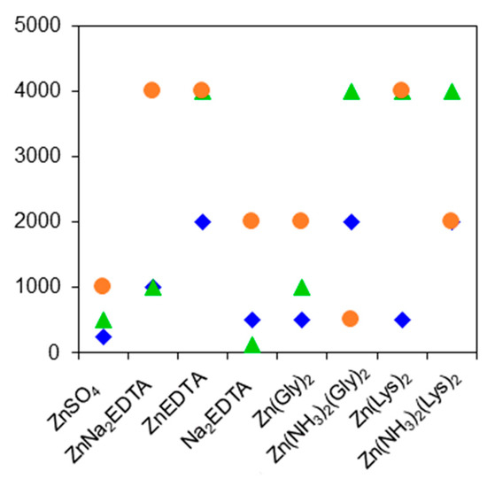
Figure 2.
Minimal inhibition concentrations (MICs) of the eight investigated compounds on the three beneficial microorganism strains. MIC values are shown by green triangles for L. lactis (▲), blue rhombi for B. subtilis (♦), and orange dots for S. cerevisiae (●).
3.2. Growth Kinetic Parameters
The effects of increasing concentrations of different Zn compounds and Na2EDTA on the growth parameters of B. subtilis, L. lactis, and S. cerevisiae are shown in Figure 3, Figure 4 and Figure 5, respectively. In general, the Rel. A and Rel. μm decreased with increasing concentrations of most tested compounds in B. subtilis, L. lactis, and S. cerevisiae. In contrast, the Rel. λ increased as the concentrations of most test compounds increased in three tested beneficial microbes. In the case of B. subtilis (Figure 3), the Rel. A, the Rel. μm, and the Rel. λ were not affected by the increasing concentration of Zn(NH3)2(Lys)2. Furthermore, the Rel. λ was not affected by the increasing concentrations of ZnNa2EDTA, ZnEDTA, or Zn(NH3)2(Gly)2. B. subtilis was most sensitive to the increasing concentrations of ZnSO4. In the case of L. lactis (Figure 4), the Rel. A was not affected by any concentrations of ZnEDTA, Zn(Gly)2, or ZnLys2Zn(NH3)2Lys2. The Rel. λ of L. lactis was not affected by the increasing concentrations of ZnNa2EDTA, ZnEDTA, or Zn(NH3)2Gly2. L. lactis was most sensitive to Na2EDTA. In the case of S. cerevisiae (Figure 5), the Rel. A and Rel. μm were not affected by any concentration of Zn(Lys)2 or Zn(NH3)2Lys2. Furthermore, the Rel. A and Rel. λ of S. cerevisiae were not affected by the concentration changes in the ZnEDTA treatment. S. cerevisiae was most sensitive to Zn(NH3)2(Gly)2. It is noteworthy that the small concentrations of almost all test materials had growth-enhancing effects compared to the untreated control for all three microorganisms, as shown by the Rel A and Rel μm values above 100%.
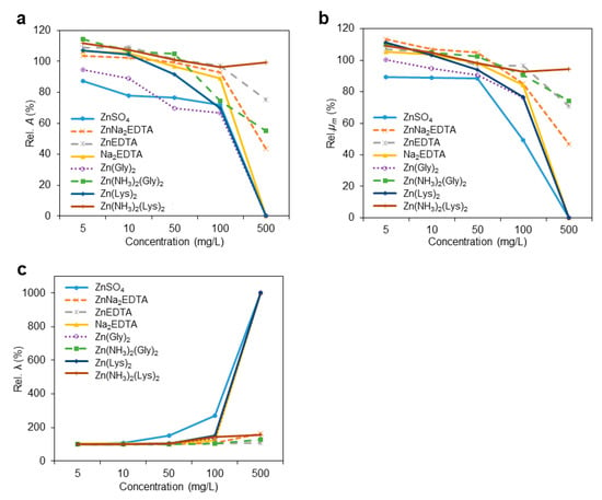
Figure 3.
Effect of increasing doses of test compounds on the growth parameters of B. subtilis (a) relative maximal growth (Rel. A), (b) relative growth rate (Rel. μm), and (c) relative lag phase (Rel. λ). The growth parameters are shown relative to the test-compound-free B. subtilis growth parameters. When no growth was observed, A and μm were considered to be 0, while λ, the lag phase, was considered to be the duration of the experiment, 24 h.
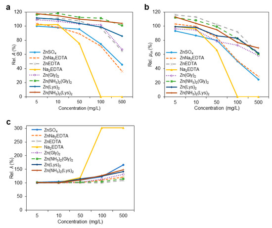
Figure 4.
Effect of increasing doses of test compounds on the growth parameters of L. lactis, (a) relative maximal growth (Rel. A), (b) relative growth rate (Rel. μm), and (c) relative lag phase (Rel. λ). The growth parameters are shown relative to the test-compound-free L. lactis growth parameters. When no growth was observed, A and μm were considered to be 0, while λ, the lag phase, was considered to be the duration of the experiment, 24 h.
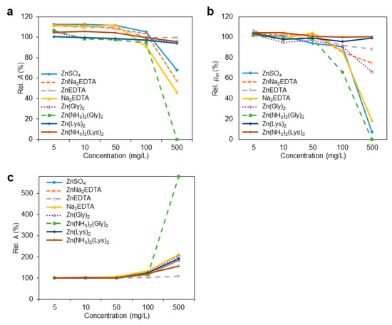
Figure 5.
Effect of increasing doses of test compounds on the growth parameters of S. cerevisiae, (a) relative maximal growth (Rel. A), (b) relative growth rate (Rel. μm), and (c) relative lag phase (Rel. λ). The growth parameters are shown relative to the test-compound-free S. cerevisiae growth parameters. When no growth was observed, A and μm were considered to be 0, while λ, the lag phase, was considered to be the duration of the experiment, 24 h.
The effects of seven Zn compounds and Na2EDTA on the growth parameters of three commensal intestinal microorganisms are shown in Table 1. Compared to Figure 3, Figure 4 and Figure 5, the results given in Table 1 compare whether there was an interaction between different microorganisms and different compounds at the same concentration. The Rel. A, Rel. μm, and Rel. λ were all significantly affected by the species at all tested concentrations (p < 0.05), except for 10 mg/L. The type of compound had a significant effect on the Rel. A, Rel. μm, and Rel. λ with all tested concentrations (p < 0.05). There was a significant interaction between the tested species and compound types (p < 0.0001) on the Rel. A, Rel. μm, and Rel. λ. From the lowest concentration (5 mg/L) to the highest concentration (500 mg/L), the highest Rel. A was observed in L. lactis with the Zn(NH3)2(Gly)2 treatment. In contrast, B. subtilis treated with ZnSO4 had the lowest Rel. A in all tested concentrations from all tested compounds. In addition, the Rel. A and Rel. μm of B. subtilis were significantly decreased by ZnSO4 in all tested concentrations (p < 0.05), corresponding to an inhibition of growth. The low and moderate concentrations (5–50 mg/L) of ZnEDTA had the most positive effects on the Rel. A of all tested microorganisms (p < 0.05). Furthermore, the complete inhibition of microbial growth was observed for all three microorganisms (Rel. A and Rel. μm indicated zero) for some tested compounds in the highest concentrations of 500 mg/L, such as B. subtilis treated with ZnSO4, Na2EDTA, Zn(Gly)2, and Zn(Lys)2, L. lactis treated with Na2EDTA, and S. cerevisiae treated with Zn(NH3)2(Gly)2. In the case of Na2EDTA, growth was also inhibited at the 100 mg/L concentration for L. lactis. The Rel. A of all tested microorganisms significantly decreased (p < 0.05) when any of the tested compounds was present at the highest concentration in the culture medium, except for B. subtilis treated with Zn(NH3)2(Lys)2; L. lactis treated with Zn(NH3)2(Gly)2 and Zn(NH3)2(Lys)2; and S. cerevisiae treated with ZnEDTA. L. lactis had the highest Rel. A and Rel. μm at 5–100 mg/L of Zn(NH3)2(Gly)2 and Zn(NH3)2Lys2, which indicates a beneficial effect on its growth. Regarding the Rel. λ, no significant difference was observed among the three microorganisms treated with the same concentrations of ZnEDTA (p > 0.05). The highest Rel. λ of B. subtilis, L. lactis, and S. cerevisiae was presented in the growth medium containing ZnSO4, Na2EDTA, and Zn(NH3)2(Lys)2 at 100 and 500 mg/L, respectively, which is indicative of longer lag phase.

Table 1.
Effects of different Zn-compounds and Na2EDTA on growth parameters of B. subitis, L. lactis, and S.cerevisiae.
4. Discussion
Zinc is an essential nutrient for the physiological processes of microorganisms, including DNA replication and protein synthesis [42]. Various studies investigated the bioavailability of Zn in both animal [43] and human nutrition [44]. A high dietary ZnO concentration used to be a common method for the prevention of diarrhea in weaning piglets [45,46]. One substantial effect of high concentrations of ZnO in the nutrition of weaning piglets is the modulation of the GIT microbiota composition [47]. It was shown that free Zn ions and protein-bound Zn correlated with various and partially different parameters of intestinal microbiota in the colon of pigs fed high dietary ZnO concentrations [48]. The major effect is likely exerted by bacteriostatic or toxic effects of Zn in the intestine [47,48]. However, little is known about the effects of different forms and quantities of Zn on the intestinal microbiota. In the present study, measurements regarding growth kinetics and MIC values of water-soluble organic and inorganic Zn sources were performed to better understand which Zn compounds or concentrations could stimulate or inhibit the intestinal microbial growth.
The MIC is a parameter that is widely used to assess the susceptibility of microorganisms to antibiotics and other compounds. It is defined as the lowest antimicrobial drug concentration that prevents the visible growth of the microorganism after overnight incubation. An advantage of the MIC is that it is quantitative, and if standardized procedures are used [38], the values obtained by different laboratories can be compared [33]. Our results confirm that the sensitivities of the three microorganisms to Zn compounds, in the MIC test and in the growth kinetics tests, are in agreement. This result is similar to previous results for essential oils or antibiotics against pathogenic bacteria [49,50]. It is worth noting that the growth kinetic parameters, besides showing the inhibitory effects, can also detect potential beneficial effects at lower concentrations than the MIC; thus, the growth kinetics test can reveal more subtle changes and mechanisms in microbial growth. Researchers may observe subtle dose-dependent effects with changing lag phase, growth rate, and maximal growth density. These details would be missed by only using the endpoint readings of the MIC.
The growth kinetics revealed that ZnEDTA and Zn(NH3)2(Lys)2 stimulated the growth of all tested microorganisms, which also tolerated these two compounds most in the MIC tests. It is worth noting that the maximal tested concentration (500 mg/L) of ZnEDTA still enhanced the growth rate of S. cerevisiae. Furthermore, the highest MIC value for each tested microorganism was obtained with ZnEDTA. It indicates that ZnEDTA is the least toxic formulation of Zn amongst those tested and has the potential to promote the growth of beneficial commensal intestinal microorganisms. In contrast, both the MIC test and growth kinetics indicated that B. subtilis, L. lactis, and S. cerevisiae were the most sensitive to ZnSO4, Na2EDTA, and Zn(NH3)2(Gly)2 compounds, respectively. Results show that the tested microorganisms were most sensitive to ZnSO4; thus, this compound has the highest potential to inhibit the growth of the beneficial commensal intestinal microorganisms. Results also correlate with previous studies, indicating that Zn from inorganic Zn sources is toxic to microorganisms [47,48]. The high concentrations of all tested Zn compounds also elongated the lag phase of commensal microorganisms, similar to the effect of antibiotics [24], resulting in differences in the growth kinetic profiles of these microorganisms. Our results also found the lag phase to be the least sensitive growth kinetic parameter, while the growth rate was the most sensitive indicator of the effect of the test compounds.
Spears et al. [51] reported that Zn bioavailability in Zn–amino chelates is higher than in ZnSO4 in animals. Many in vitro studies also indicated that Zn–glycinate and Zn–lysinate are more stable than ZnSO4 [52,53]; therefore, Zn(Gly)2 and Zn(Lys)2 chelates may be more available for absorption than ZnSO4 in commensal intestinal microorganisms. In addition to bioavailability, Pieper et al. [54] showed that Zn(Lys)2, as an organic Zn source, promoted the formation of a differentially composed microbiome that was enriched in genes capable of promoting glycan foraging and maintaining the formation of beneficial metabolites. The present study indicated that Zn(Gly)2 and Zn(Lys)2 had equal effects on the growth kinetic parameters of L. lactis and S. cerevisiae. Thus, in terms of the overall microbiota, the bioavailability between Zn(Gly)2 and Zn(Lys)2 is not expected to be different. Lin et al. [55] detected no significant differences in growth performance and immune response between Zn(Gly)2 and Zn(Lys)2 in shrimp; this result is consistent with our observations. The growth of B. subtilis and L. lactis with diamine–zinc–amino acid chelates was increased compared to the use of simple Zn–amino chelates at the same concentrations. The diamine–zinc–amino acid chelates and Zn–amino acid chelates showed similar MIC results; B. subtilis tolerated higher concentrations of diamine–zinc–amino acid chelates than Zn chelates. Ammonia complex formation is known to improve the stability and solubility of Zn chelates (US4312815A); thus, it is possible that diamine–zinc–amino acid chelates enhanced Zn bioavailability for the commensal intestinal microorganisms and, as a consequence, stimulated their growth at low concentrations.
The tested microorganisms are beneficial commensal intestinal microorganisms that improve the absorption of nutrients and produce a healthier intestinal system [56]. The interaction of the host and its GIT microbiome is an important and direct factor in gastrointestinal functionality and health condition, including effective digestion, nutrition absorption, and immune status [57]. The growth of B. subtilis is easily impaired by the excessively high concentration of Zn in a diet [58]. Damaskos and Kolios [59] also proposed B. subtilis could enhance the lactic acid bacterial counts in the GIT of many animals and exert a positive effect on protection against intestinal pathophysiology. The intestinal microbiome limits the growth of pathogens such as Salmonella, a mechanism referred to as colonization resistance [60]. Based on the results of previous studies and the current research, it is clear that low concentrations of ZnEDTA and Zn(NH3)2(Lys)2 simulate the growth of beneficial intestinal microorganisms, which can improve nutrient availability and promote intestinal health.
5. Conclusions
MICs have been widely used to assess the susceptibility of microorganisms to antimicrobial agents. To our best knowledge, this study is the first research to compare the effects on growth kinetics and MIC values of different members of the commensal beneficial microbiome for various Zn compounds. Our results indicate that growth kinetics provides more sophisticated data in terms of microbial behaviors and reactions toward different Zn-containing compounds and concentrations than MIC evaluations. We quantitatively evaluated the growth of microorganisms in the presence of different Zn compounds by growth kinetics in order to quantify the promotion or the inhibition of growth. Low concentrations of ZnEDTA and Zn(NH3)2(Lys)2 promoted the growth of all tested microorganisms; thus, prebiotic effects in vivo may be expected of these two compounds. With increasing concentrations, all test compounds inhibited the growth of these beneficial microorganisms, which were most sensitive to ZnSO4, Na2EDTA, and Zn(NH3)2(Gly)2 for B. subtilis, L. lactis, and S. cerevisiae, respectively. In the future, researchers are encouraged to use growth kinetics studies to investigate the modulatory effects on other microorganisms (such as pathogen and neutral microorganisms) of different Zn compounds.
Author Contributions
V.M.-N. and Z.B.: Conceptualization, Methodology, and Data analysis. K.-H.T.: Visualization, Investigation, and Data curation. V.M.-N., Z.B., K.-H.T., J.W.H. and S.L.: Supervision, Writing—original draft preparation. J.W.H., S.L., G.T.-I. and X.H.-V.: Reviewing and Editing. All authors have read and agreed to the published version of the manuscript.
Funding
This research was supported by the National Research, Development and Innovation Office, Hungary, project number: 2018-1.1.2-KFI-2018-00111.
Institutional Review Board Statement
Not applicable.
Informed Consent Statement
Not applicable.
Data Availability Statement
Data available in a publicly accessible repository.
Acknowledgments
The authors would like to thank Sándor Kemény and Éva Pusztai at the Budapest University of Technology and Economics for the insightful discussions about the statistical analysis of the data.
Conflicts of Interest
Dr. Bata Ltd. employs V.M.-N., K.-H.T. and Z.B.; BV Science employs J.W.H. and S.L. The remaining authors declare that the research was conducted in the absence of any commercial or financial relationships that could be construed as a potential conflict of interest.
References
- Anders, J.L.; Moustafa, M.A.M.; Mohamed, W.M.A.; Hayakawa, T.; Nakao, R.; Koizumi, I. Comparing the gut microbiome along the gastrointestinal tract of three sympatric species of wild rodents. Sci. Rep. 2021, 11, 19929. [Google Scholar] [CrossRef] [PubMed]
- Hooper, L.V.; Gordon, J.I. Commensal host-bacterial relationships in the gut. Science 2001, 292, 1115–1118. [Google Scholar] [CrossRef]
- Tellez-Isaias, G.; Latorre, J.D. Editorial: Alternatives to Antimicrobial Growth Promoters and Their Impact in Gut Microbiota, Health and Disease: Volume II. Front. Vet. Sci. 2022, 9, 857583. [Google Scholar] [CrossRef] [PubMed]
- Neish, A.S. Microbes in gastrointestinal health and disease. Gastroenterology 2009, 136, 65–80. [Google Scholar] [CrossRef] [Green Version]
- Qin, J.; Li, R.; Raes, J.; Arumugam, M.; Burgdorf, K.S.; Manichanh, C.; Nielsen, T.; Pons, N.; Levenez, F.; Yamada, T.; et al. A human gut microbial gene catalogue established by metagenomic sequencing. Nature 2010, 464, 59–65. [Google Scholar] [CrossRef] [Green Version]
- Leser, T.D.; Mølbak, L. Better living through microbial action: The benefits of the mammalian gastrointestinal microbiota on the host. Environ. Microbiol. 2009, 11, 2194–2206. [Google Scholar] [CrossRef]
- Ryan, M.; Schloter, M.; Berg, G.; Kostic, T.; Kinkel, L.; Eversole, K.; Macklin, J.; Schelkle, B.; Kazou, M.; Sarand, I.; et al. Development of Microbiome Biobanks-Challenges and Opportunities. Trends Microbiol. 2021, 29, 89–92. [Google Scholar] [CrossRef]
- Dahiya, D.; Nigam, P.S. The Gut Microbiota Influenced by the Intake of Probiotics and Functional Foods with Prebiotics Can Sustain Wellness and Alleviate Certain Ailments like Gut-Inflammation and Colon-Cancer. Microorganisms 2022, 10, 665. [Google Scholar] [CrossRef]
- Blandino, G.; Inturri, R.; Lazzara, F.; Di Rosa, M.; Malaguarnera, L. Impact of gut microbiota on diabetes mellitus. Diabetes Metab. 2016, 42, 303–315. [Google Scholar] [CrossRef] [PubMed]
- Schneiderhan, J.; Master-Hunter, T.; Locke, A. Targeting gut flora to treat and prevent disease. J. Fam. Pract. 2016, 65, 34–38. [Google Scholar] [PubMed]
- Ganatsios, V.; Nigam, P.; Plessas, S.; Terpou, A. Kefir as a Functional Beverage Gaining Momentum towards Its Health Promoting Attributes. Beverages 2021, 7, 48. [Google Scholar] [CrossRef]
- Gareau, M.G.; Sherman, P.M.; Walker, W.A. Probiotics and the gut microbiota in intestinal health and disease. Nat. Rev. Gastroenterol. Hepatol. 2010, 7, 503–514. [Google Scholar] [CrossRef] [PubMed] [Green Version]
- Chen, K.-L.; Kho, W.-L.; You, S.-H.; Yeh, R.-H.; Tang, S.-W.; Hsieh, C.-W. Effects of Bacillus subtilis var. natto and Saccharomyces cerevisiae mixed fermented feed on the enhanced growth performance of broilers. Poult. Sci. 2009, 88, 309–315. [Google Scholar] [CrossRef] [PubMed]
- Iwashita, M.K.P.; Nakandakare, I.B.; Terhune, J.S.; Wood, T.; Paiva, M.J.R. Dietary supplementation with Bacillus subtilis, Saccharomyces cerevisiae and Aspergillus oryzae enhance immunity and disease resistance against Aeromonas hydrophila and Streptococcus iniae infection in juvenile tilapia Oreochromis niloticus. Fish Shellfish Immunol. 2015, 43, 60–66. [Google Scholar] [CrossRef] [PubMed]
- Darby, T.M.; Owens, J.; Saeedi, B.; Luo, L.; Matthews, J.D.; Robinson, B.S.; Naudin, C.; Jones, R.M. Lactococcus Lactis Subsp. cremoris Is an Efficacious Beneficial Bacterium that Limits Tissue Injury in the Intestine. iScience 2019, 12, 356–367. [Google Scholar] [CrossRef] [PubMed] [Green Version]
- De Lacerda, J.R.M.; Da Silva, T.F.; Vollú, R.E.; Marques, J.M.; Seldin, L. Generally recognized as safe (GRAS) Lactococcus lactis strains associated with Lippia sidoides Cham. are able to solubilize/mineralize phosphate. Springerplus 2016, 5, 828. [Google Scholar] [CrossRef] [Green Version]
- Elshaghabee, E.M.F.; Rokana, N.; Gulhane, R.D.; Sharma, C.; Panwar, H. Bacillus as Potential Probiotics: Status, Concerns, and Future Perspectives. Front. Microbiol. 2017, 8, 1490. [Google Scholar] [CrossRef] [Green Version]
- Li, Z.; Wang, W.; Lv, Z.; Liu, D.; Guo, Y. Bacillus subtilis and yeast cell wall improve the intestinal health of broilers challenged by Clostridium perfringens. Br. Poult. Sci. 2017, 58, 635–643. [Google Scholar] [CrossRef]
- Granstad, S.; Kristoffersen, A.B.; Benestad, S.L.; Sjurseth, S.K.; David, B.; Sørensen, L.; Fjermedal, A.; Edvardsen, D.H.; Sanson, G.; Løvland, A.; et al. Effect of Feed Additives as Alternatives to In-feed Antimicrobials on Production Performance and Intestinal Clostridium perfringens Counts in Broiler Chickens. Animals 2020, 10, 240. [Google Scholar] [CrossRef] [PubMed] [Green Version]
- Kobierecka, P.A.; Eolech, B.; eKsiążek, M.; Ederlatka, K.; Adamska, I.; Majewski, P.; Jagusztyn-Krynicka, E.; Wyszyńska, A.K. Cell Wall Anchoring of the Campylobacter Antigens to Lactococcus lactis. Front. Microbiol. 2016, 7, 165. [Google Scholar] [CrossRef] [Green Version]
- Jozic, D.; Bourenkow, G.; Bartunik, H.; Scholze, H.; Dive, V.; Henrich, B.; Huber, R.; Bode, W.; Maskos, K. Crystal structure of the dinuclear zinc aminopeptidase PepV from Lactobacillus delbrueckii unravels its preference for dipeptides. Structure 2002, 10, 1097–1106. [Google Scholar] [CrossRef] [Green Version]
- Chen, Y.-S.; Christensen, J.E.; Broadbent, J.R.; Steele, J.L. Identification and characterization of Lactobacillus helveticus PepO2, an endopeptidase with post-proline specificity. Appl. Environ. Microbiol. 2003, 69, 1276–1282. [Google Scholar] [CrossRef] [Green Version]
- Maret, W. Zinc biochemistry: From a single zinc enzyme to a key element of life. Adv. Nutr. Int. Rev. J. 2013, 4, 82–91. [Google Scholar] [CrossRef] [Green Version]
- Capdevila, D.A.; Wang, J.; Giedroc, D.P. Bacterial Strategies to Maintain Zinc Metallostasis at the Host-Pathogen Interface. J. Biol. Chem. 2016, 291, 20858–20868. [Google Scholar] [CrossRef] [PubMed] [Green Version]
- Yang, S.-X.; Tian, Q.-J.; Liang, S.-C.; Zhou, Y.-Y.; Zou, H.-C. Bioaccumulation of heavy metals by the dominant plants growing in Huayuan manganese and lead/zinc mineland, Xiangxi. Huan Jing Ke Xue 2012, 33, 2038–2045. [Google Scholar]
- Milani, N.; Sbardella, M.; Ikeda, N.; Arno, A.; Mascarenhas, B.; Miyada, V. Dietary zinc oxide nanoparticles as growth promoter for weanling pigs. Anim. Feed Sci. Technol. 2017, 227, 13–23. [Google Scholar] [CrossRef]
- Fontecha-Umaña, F.; Ríos-Castillo, A.G.; Ripolles-Avila, C.; Rodríguez-Jerez, J.J. Antimicrobial Activity and Prevention of Bacterial Biofilm Formation of Silver and Zinc Oxide Nanoparticle-Containing Polyester Surfaces at Various Concentrations for Use. Foods 2020, 9, 442. [Google Scholar] [CrossRef] [PubMed] [Green Version]
- Surjawidjaja, J.E.; Hidayat, A.; Lesmana, M. Growth inhibition of enteric pathogens by zinc sulfate: An in vitro study. Med. Princ. Pract. 2004, 13, 286–289. [Google Scholar] [CrossRef] [PubMed]
- Osinaga, P.W.; Grande, R.H.M.; Ballester, R.Y.; Simionato, M.R.L.; Rodrigues, C.R.M.D.; Muench, A. Zinc sulfate addition to glass-ionomer-based cements: Influence on physical and antibacterial properties, zinc and fluoride release. Dent. Mater. 2003, 19, 212–217. [Google Scholar] [CrossRef]
- Theophel, K.; Schacht, V.J.; Schlã¼Ter, M.; Schnell, S.; Stingu, C.-S.; Schaumann, R.; Bunge, M. The importance of growth kinetic analysis in determining bacterial susceptibility against antibiotics and silver nanoparticles. Front. Microbiol. 2014, 5, 544. [Google Scholar] [CrossRef] [PubMed]
- Rezvani, F.; Ardestani, F.; Najafpour, G. Growth kinetic models of five species of Lactobacilli and lactose consumption in batch submerged culture. Braz. J. Microbiol. 2017, 48, 251–258. [Google Scholar] [CrossRef] [PubMed]
- Rolfe, M.D.; Rice, C.J.; Lucchini, S.; Pin, C.; Thompson, A.; Cameron, A.; Alston, M.; Stringer, M.F.; Betts, R.P.; Baranyi, J.; et al. Lag Phase is a Distinct Growth Phase that Prepares Bacteria for Exponential Growth and Involves Transient Metal Accumulation. J. Bacteriol. 2012, 194, 686–701. [Google Scholar] [CrossRef] [Green Version]
- Rudilla, H.; Merlos, A.; Sans-Serramitjana, E.; Fuste, E.; Sierra, J.M.; Zalacain, A.; Vinuesa, T.; Vinas, M. New and old tools to evaluate new antimicrobial peptides. AIMS Microbiol. 2018, 4, 522–540. [Google Scholar] [CrossRef] [PubMed]
- Zwietering, M.H.; Jongenburger, I.; Rombouts, F.M.; Van’t Riet, K. Modeling of the bacterial growth curve. Appl. Environ. Microbiol. 1990, 56, 1875–1881. [Google Scholar] [CrossRef] [PubMed] [Green Version]
- Dalgaard, P.; Koutsoumanis, K. Comparison of maximum specific growth rates and lag times estimated from absorbance and viable count data by different mathematical models. J. Microbiol. Methods 2001, 43, 183–196. [Google Scholar] [CrossRef]
- Burdett, I.D.; Kirkwood, T.B.; Whalley, J.B. Growth kinetics of individual Bacillus subtilis cells and correlation with nucleoid extension. J. Bacteriol. 1986, 167, 219–230. [Google Scholar] [CrossRef] [PubMed] [Green Version]
- Olivares-Marin, I.K.; González-Hernández, J.C.; Regalado-Gonzalez, C.; Madrigal-Perez, L.A. Saccharomyces cerevisiae Exponential Growth Kinetics in Batch Culture to Analyze Respiratory and Fermentative Metabolism. J. Vis. Exp. 2018, 139, e58192. [Google Scholar] [CrossRef] [PubMed] [Green Version]
- European Committee for Antimicrobial Susceptibility Testing of the European Society of Clinical, Microbiology; Infectious Diseases (ESCMID). EUCAST Definitive Document E.DEF 3.1, June 2000: Determination of minimum inhibitory concentrations (MICs) of antibacterial agents by agar dilution. Clin. Microbiol. Infect. 2000, 6, 509–515. [Google Scholar] [CrossRef] [PubMed] [Green Version]
- Howland, J.L. Biochemical Education. Short Protocols in Molecular Biology, Third Edition; Ausubel, F., Brent, E., Kingston, R.E., Moore, D.D., Seidman, J.G., Smith, J.A., Struhl, K., Eds.; John Wiley & Sons: New York, NY, USA, 1996; Volume 24, pp. 68, 836. [Google Scholar] [CrossRef]
- Larsen, N.; Boye, M.; Siegumfeldt, H.; Jakobsen, M. Differential expression of proteins and genes in the lag phase of Lactococcus lactis subsp. lactis grown in synthetic medium and reconstituted skim milk. Appl. Environ. Microbiol. 2006, 72, 1173–1179. [Google Scholar] [CrossRef] [Green Version]
- Gilpin, R.W.; Patterson, S.K.; A Knight, R. Quantitation of bacillus subtilis L-form growth parameters in batch culture. J. Bacteriol. 1981, 145, 651–653. [Google Scholar] [CrossRef] [PubMed] [Green Version]
- Velasco, E.; Wang, S.; Sanet, M.; Fernández-Vázquez, J.; Jové, D.; Glaría, E.; Valledor, A.F.; O’Halloran, T.V.; Balsalobre, C. A new role for Zinc limitation in bacterial pathogenicity: Modulation of α-hemolysin from uropathogenic Escherichia coli. Sci. Rep. 2018, 8, 6535. [Google Scholar] [CrossRef] [Green Version]
- Brugger, D.; Windisch, W.M. Strategies and challenges to increase the precision in feeding zinc to monogastric livestock. Anim. Nutr. 2017, 3, 103–108. [Google Scholar] [CrossRef]
- Maares, M.; Haase, H. A Guide to Human Zinc Absorption: General Overview and Recent Advances of In Vitro Intestinal Models. Nutrients 2020, 12, 762. [Google Scholar] [CrossRef] [PubMed] [Green Version]
- Sales, J. Effects of pharmacological concentrations of dietary zinc oxide on growth of post-weaning pigs: A meta-analysis. Biol. Trace Elem. Res. 2013, 152, 343–349. [Google Scholar] [CrossRef]
- Davin, R.; Manzanilla, E.; Klasing, K.; Pérez, J. Effect of weaning and in-feed high doses of zinc oxide on zinc levels in different body compartments of piglets. J. Anim. Physiol. Anim. Nutr. 2013, 97 (Suppl. S1), 6–12. [Google Scholar] [CrossRef] [PubMed]
- Liedtke, J.; Vahjen, W. In vitro antibacterial activity of zinc oxide on a broad range of reference strains of intestinal origin. Vet. Microbiol. 2012, 160, 251–255. [Google Scholar] [CrossRef]
- Starke, I.C.; Pieper, R.; Neumann, K.; Zentek, J.; Vahjen, W. The impact of high dietary zinc oxide on the development of the intestinal microbiota in weaned piglets. FEMS Microbiol. Ecol. 2014, 87, 416–427. [Google Scholar] [CrossRef] [PubMed] [Green Version]
- Kaya, E.G.; Albayrak, S. Determination of the effect of gentamicin against staphylococcus aureus by using microbroth kinetic system. Biology 2009, 23, 110–114. [Google Scholar]
- Hulankova, R. The Influence of Liquid Medium Choice in Determination of Minimum Inhibitory Concentration of Essential Oils against Pathogenic Bacteria. Antibiotics 2022, 11, 150. [Google Scholar] [CrossRef] [PubMed]
- Spears, J.W.; Schlegel, P.; Seal, M.C.; Lloyd, K.E. Bioavailability of zinc from zinc sulfate and different organic zinc sources and their effects on ruminal volatile fatty acid proportions. Livest. Prod. Sci. 2004, 90, 211–217. [Google Scholar] [CrossRef]
- Alimohamady, R.; Aliarabi, H.; Bruckmaier, R.M.; Christensen, R.G. Effect of Different Sources of Supplemental Zinc on Performance, Nutrient Digestibility, and Antioxidant Enzyme Activities in Lambs. Biol. Trace Elem. Res. 2019, 189, 75–84. [Google Scholar] [CrossRef] [PubMed]
- Van Heugten, E.; Spears, J.W.; Kegley, E.B.; Ward, J.D.; Qureshi, M.A. Effects of organic forms of zinc on growth performance, tissue zinc distribution, and immune response of weanling pigs. J. Anim. Sci. 2003, 81, 2063–2071. [Google Scholar] [CrossRef] [PubMed] [Green Version]
- Pieper, R.; Dadi, T.H.; Pieper, L.; Vahjen, W.; Franke, A.; Reinert, K.; Zentek, J. Concentration and chemical form of dietary zinc shape the porcine colon microbiome, its functional capacity and antibiotic resistance gene repertoire. ISME J. 2020, 14, 2783–2793. [Google Scholar] [CrossRef]
- Lin, S.; Lin, X.; Yang, Y.; Li, F.; Luo, L. Comparison of chelated zinc and zinc sulfate as zinc sources for growth and immune response of shrimp (Litopenaeus vannamei). Aquaculture 2013, 406–407, 79–84. [Google Scholar] [CrossRef] [Green Version]
- Morowitz, M.J.; Carlisle, E.; Alverdy, J.C. Contributions of intestinal bacteria to nutrition and metabolism in the critically Ill. Surg. Clin. North Am. 2011, 91, 771–785. [Google Scholar] [CrossRef] [PubMed] [Green Version]
- Celi, P.; Cowieson, A.; Fru-Nji, F.; Steinert, R.; Kluenter, A.-M.; Verlhac, V. Gastrointestinal functionality in animal nutrition and health: New opportunities for sustainable animal production. Anim. Feed Sci. Technol. 2017, 234, 88–100. [Google Scholar] [CrossRef]
- Hsueh, Y.-H.; Ke, W.-J.; Hsieh, C.-T.; Lin, K.-S.; Tzou, D.-Y.; Chiang, C.-L. ZnO Nanoparticles Affect Bacillus subtilis Cell Growth and Biofilm Formation. PLoS ONE 2015, 10, e0128457. [Google Scholar] [CrossRef] [Green Version]
- Damaskos, D.; Kolios, G. Probiotics and prebiotics in inflammatory bowel disease: Microflora ‘on the scope’. Br. J. Clin. Pharmacol. 2008, 65, 453–467. [Google Scholar] [CrossRef] [Green Version]
- Wijburg, O.L.; Uren, T.K.; Simpfendorfer, K.; Johansen, F.-E.; Brandtzaeg, P.; Strugnell, R. Innate secretory antibodies protect against natural Salmonella typhimurium infection. J. Exp. Med. 2006, 203, 21–26. [Google Scholar] [CrossRef] [PubMed]
Publisher’s Note: MDPI stays neutral with regard to jurisdictional claims in published maps and institutional affiliations. |
© 2022 by the authors. Licensee MDPI, Basel, Switzerland. This article is an open access article distributed under the terms and conditions of the Creative Commons Attribution (CC BY) license (https://creativecommons.org/licenses/by/4.0/).