
The Resistance to Traction Forces Differs Substantially Between Intestinal Parts, but Not Between In- and Outbred Strains of Mice

 ,
,  and
and
Abstract
1. Introduction
2. Materials and Methods
2.1. Mice and Husbandry
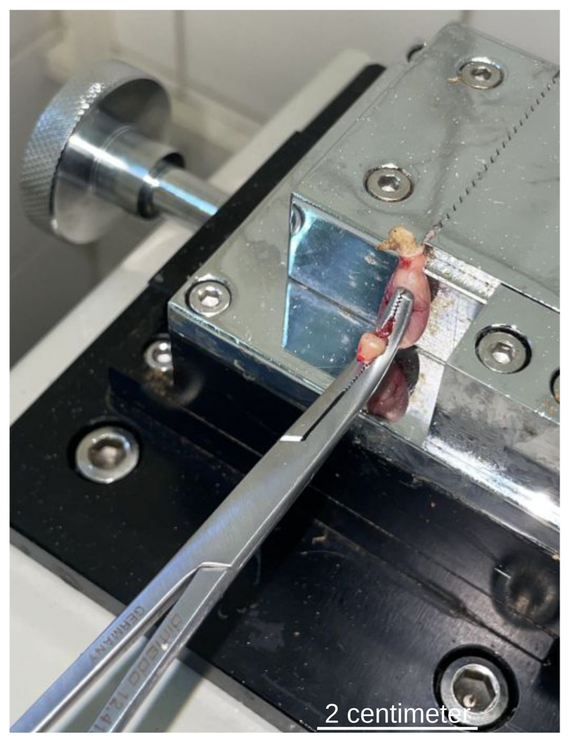
2.2. Experimental Approach
2.3. Power Analysis
2.4. Statistical Analysis
2.5. Ethical Approval
3. Results
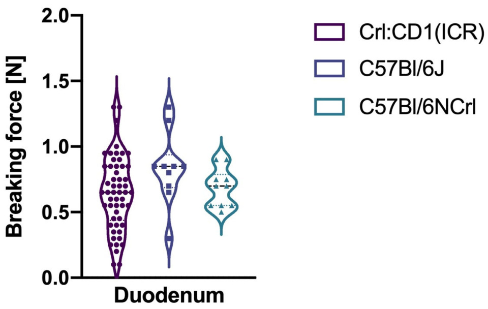
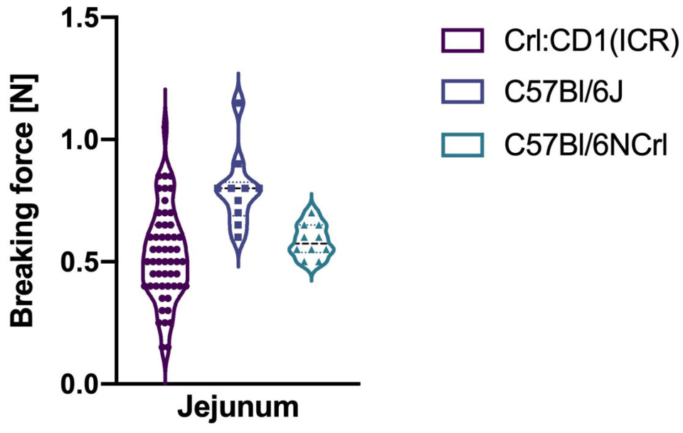
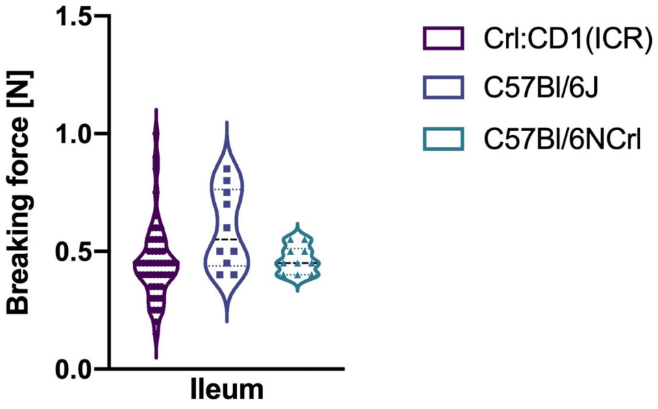
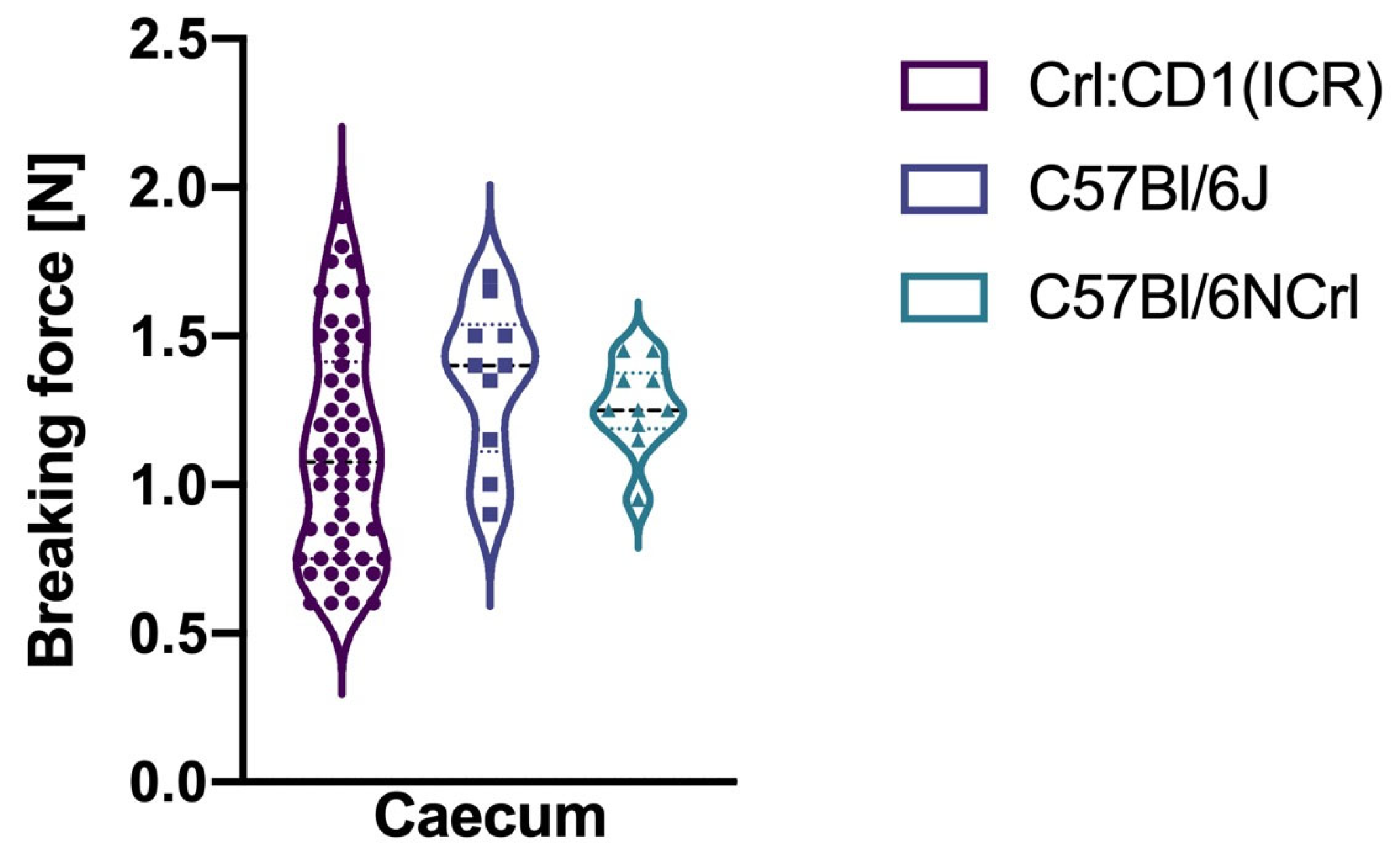
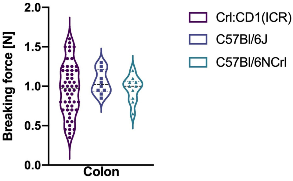
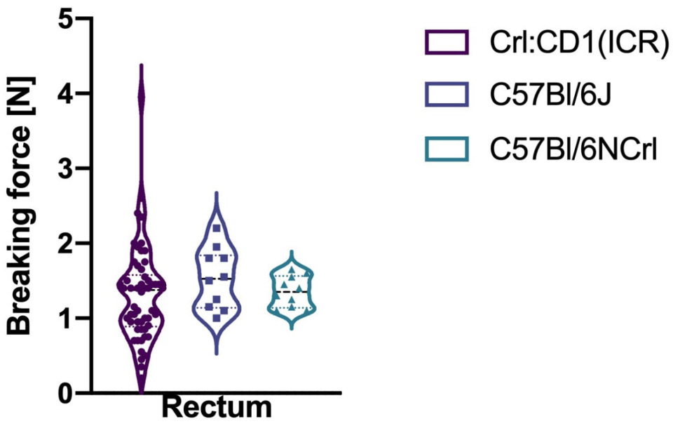
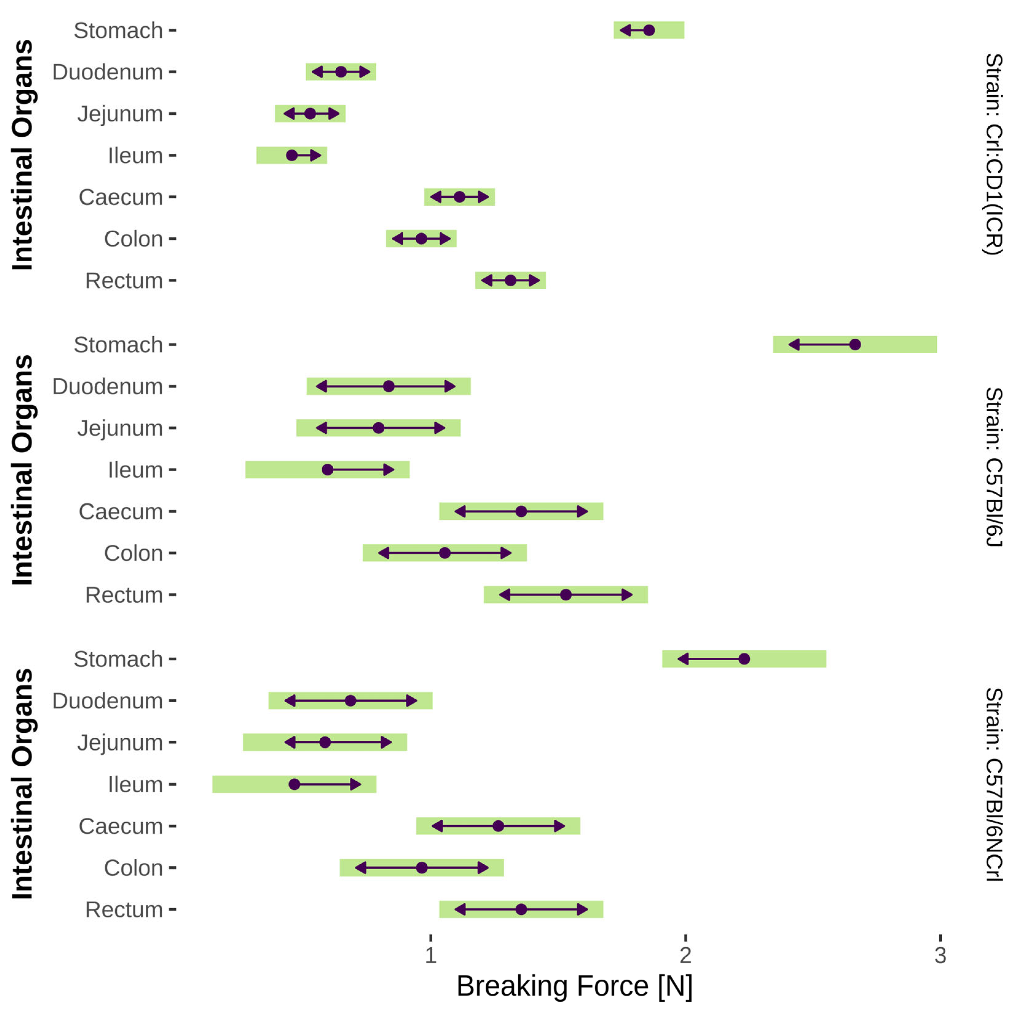
3.1. Breaking Forces of the Organs of the Gastrointestinal Tract in In- and Outbred Mice
3.2. Comparison of Breaking Forces Within and Between the Different Mouse Strains
4. Discussion
5. Conclusions
Supplementary Materials
Author Contributions
Funding
Institutional Review Board Statement
Informed Consent Statement
Data Availability Statement
Acknowledgments
Conflicts of Interest
References
- Hamel, J.; Alves, A.; Beyer-Bergot, L.; Zerbib, P.; Bridoux, V.; Manceau, G.; Panis, Y.; Buscail, E.; Khaoudy, I.; Gaillard, M.; et al. Stenosis of the Colorectal Anastomosis after Surgery for Diverticulitis: A National Retrospective Cohort Study. Color. Dis. 2024, 26, 1437–1446. [Google Scholar] [CrossRef] [PubMed]
- Wells, C.I.; Baraza, W.; O’Grady, G.; Bissett, I.P. ‘Failure to Rescue’ from Anastomotic Leak Following Colorectal Cancer Resection: An Observational Study from a Binational Registry. Color. Dis. 2025, 27, e70262. [Google Scholar] [CrossRef] [PubMed]
- Rencuzogullari, A.; Benlice, C.; Valente, M.; Abbas, M.A.; Remzi, F.H.; Gorgun, E. Predictors of Anastomotic Leak in Elderly Patients After Colectomy: Nomogram-Based Assessment from the American College of Surgeons National Surgical Quality Program Procedure-Targeted Cohort. Dis. Colon. Rectum 2017, 60, 527–536. [Google Scholar] [CrossRef] [PubMed]
- Moojen, T.B.; Vlug, M.S.; Visser, E.; Reijntjes, M.A.; Lange, J.F.M.; Bislenghi, G.; Carvello, M.; Warusavitarne, J.; Hompes, R.; Stassen, L.P.S.; et al. Anastomotic Leakage after Ileoanal Pouch Surgery: Risk Factors and Salvage Rate. BJS Open 2025, 9, zraf110. [Google Scholar] [CrossRef]
- Bao, X.; Yi, K.; Cheng, J.; Shen, Y.; Cao, S.; Hu, B.; Wang, X.; Su, P.; Li, Y.; Xu, Q.; et al. Risk and Protective Factors for Postoperative Anastomotic Leakage in Esophageal and Gastrointestinal Surgery: An Umbrella Review of Meta-Analyses and Systematic Reviews. Int. J. Surg. 2025, 112, 1722–1736. [Google Scholar] [CrossRef]
- Morse, B.C.; Simpson, J.P.; Jones, Y.R.; Johnson, B.L.; Knott, B.M.; Kotrady, J.A. Determination of Independent Predictive Factors for Anastomotic Leak: Analysis of 682 Intestinal Anastomoses. Am. J. Surg. 2013, 206, 950–956. [Google Scholar] [CrossRef]
- Ito, R.; Matsubara, H.; Shimizu, R.; Maehata, T.; Miura, Y.; Uji, M.; Mokuno, Y. Anastomotic Tension “Bridging”: A Risk Factor for Anastomotic Leakage Following Low Anterior Resection. Surg. Endosc. 2024, 38, 4916–4925. [Google Scholar] [CrossRef]
- Wu, X.-R.; Kirat, H.T.; Xhaja, X.; Hammel, J.P.; Kiran, R.P.; Church, J.M. The Impact of Mesenteric Tension on Pouch Outcome and Quality of Life in Patients Undergoing Restorative Proctocolectomy. Color. Dis. 2014, 16, 986–994. [Google Scholar] [CrossRef]
- Man, J.; Hrabe, J. Anastomotic Technique—How to Optimize Success and Minimize Leak Rates. Clin. Colon. Rectal Surg. 2021, 34, 371–378. [Google Scholar] [CrossRef]
- Vargas, H.D. Gaining Mesenteric Length Following Colorectal Resection: Essential Maneuvers to Avoid Anastomotic Tension. Clin. Colon. Rectal Surg. 2023, 36, 37–46. [Google Scholar] [CrossRef]
- Dalmau, M.; Marti-Gallostra, M.; Pellino, G.; Espin-Basany, E.; Armengol, M. The Colon Does Not Reach! A Technical Note with Tricks to Avoid Colorectal Anastomoses under Tension. Color. Dis. 2024, 26, 564–569. [Google Scholar] [CrossRef] [PubMed]
- Khalid, M.U.; Ali, D.; Wu, J.Y.; Lee, H.; Khan, A. Impact and Measurement of Mechanical Tension in Bowel Anastomosis: A Scoping Review of the Current Literature. J. Surg. Res. 2025, 308, 161–173. [Google Scholar] [CrossRef] [PubMed]
- Herron, R.; Abbas, G. Techniques of Esophageal Anastomoses for Esophagectomy. Surg. Clin. North. Am. 2021, 101, 511–524. [Google Scholar] [CrossRef] [PubMed]
- Chen, H.; Chen, Z.-X.; Shi, G.-Q. Risk Factors and Prevention and Treatment Methods of Anastomotic Stricture after Esophageal Atresia Repair: A Literature Review. Pediatr. Surg. Int. 2025, 41, 99. [Google Scholar] [CrossRef]
- Hermreck, A.S.; Crawlord, D.G. The Esophageal Anastomotic Leak. Am. J. Surg. 1976, 132, 794–798. [Google Scholar] [CrossRef]
- Mahoney, J.L.; Condon, R.E. Adenocarcinoma of the Esophagus. Ann. Surg. 1987, 205, 557–562. [Google Scholar] [CrossRef]
- Kitayama, J.; Kaisaki, S.; Ishigami, H.; Hidemura, A.; Nagawa, H. Angleplasty in Gastric Tube Reconstruction after Esophagectomy. Dis. Esophagus 2009, 22, 418–421. [Google Scholar] [CrossRef]
- Hölscher, A.H.; Vallböhmer, D.; Brabender, J. The Prevention and Management of Perioperative Complications. Best. Pract. Res. Clin. Gastroenterol. 2006, 20, 907–923. [Google Scholar] [CrossRef]
- Schneider, A.; Blanc, S.; Bonnard, A.; Khen-Dunlop, N.; Auber, F.; Breton, A.; Podevin, G.; Sfeir, R.; Fouquet, V.; Jacquier, C.; et al. Results from the French National Esophageal Atresia Register: One-Year Outcome. Orphanet J. Rare Dis. 2014, 9, 206. [Google Scholar] [CrossRef]
- Aumar, M.; Sfeir, R.; Pierache, A.; Turck, D.; Gottrand, F. Predictors of Anastomotic Strictures Following Œsophageal Atresia Repair. Arch. Dis. Child. Fetal Neonatal Ed. 2022, 107, 545–550. [Google Scholar] [CrossRef]
- Serhal, L.; Gottrand, F.; Sfeir, R.; Guimber, D.; Devos, P.; Bonnevalle, M.; Storme, L.; Turck, D.; Michaud, L. Anastomotic Stricture after Surgical Repair of Esophageal Atresia: Frequency, Risk Factors, and Efficacy of Esophageal Bougie Dilatations. J. Pediatr. Surg. 2010, 45, 1459–1462. [Google Scholar] [CrossRef]
- Donoso, F.; Lilja, H. Risk Factors for Anastomotic Strictures after Esophageal Atresia Repair: Prophylactic Proton Pump Inhibitors Do Not Reduce the Incidence of Strictures. Eur. J. Pediatr. Surg. 2016, 27, 50–55. [Google Scholar] [CrossRef]
- Campos, J.; Tan Tanny, S.P.; Kuyruk, S.; Sekaran, P.; Hawley, A.; Brooks, J.-A.; Bekhit, E.; Hutson, J.M.; Crameri, J.; McLeod, E.; et al. The Burden of Esophageal Dilatations Following Repair of Esophageal Atresia. J. Pediatr. Surg. 2020, 55, 2329–2334. [Google Scholar] [CrossRef] [PubMed]
- Nelsen, T.S. Dynamic Aspects of Small Intestinal Rupture with Special Consideration of Anastomotic Strength. Arch. Surg. 1966, 93, 309. [Google Scholar] [CrossRef] [PubMed]
- Postlethwait, R.W.; Weinberg, M.; Jenkins, L.B.; Brockington, W.S. Mechanical Strength of Esophageal Anastomoses. Ann. Surg. 1951, 133, 472–476. [Google Scholar] [CrossRef] [PubMed]
- Livaditis, A.; Okmian, L.; Björck, G.; Ivemark, B. Esophageal Suture Anastomosis: An Experimental Study in Piglets. Scand. J. Thorac. Cardiovasc. Surg. 1969, 3, 163–173. [Google Scholar] [CrossRef]
- Cui, Y.; Urschel, J.D. Comparison of Anastomotic Suturing Techniques in the Rat Esophagus. J. Cardiovasc. Surg. 1999, 40, 613–614. [Google Scholar]
- Cui, Y.; Urschel, J.D. Esophagogastric Anastomotic Wound Healing in Rats. Dis. Esophagus 1999, 12, 149–151. [Google Scholar] [CrossRef]
- Drescher, D.G.; Vogt, J.; Gabriel, M.; Baumgart, J.; Schimanski, C.C.; Lang, H.; Gockel, I. Model of Wound Healing for Esophagogastric Anastomoses in Rats. Eur. Surg. Res. 2012, 48, 194–199. [Google Scholar] [CrossRef]
- Landes, L.C.; Drescher, D.; Tagkalos, E.; Grimminger, P.P.; Thieme, R.; Jansen-Winkeln, B.; Lang, H.; Gockel, I. Upregulation of VEGFR1 in a Rat Model of Esophagogastric Anastomotic Healing. Acta Chir. Belg. 2018, 118, 161–166. [Google Scholar] [CrossRef]
- Chung, R.S. Blood Flow in Colonic Anastomoses. Effect of Stapling and Suturing. Ann. Surg. 1987, 206, 335–339. [Google Scholar] [CrossRef] [PubMed]
- Wojtyczka, A.; Górka, Z.; Bierzynska-Macyszyn, G.; Rümenapf, G.; Jonderko, K.; Schwille, P.O. Gastrectomy in the Rat Using Two Modifications of Esophagojejunal Anastomosis. Eur. Surg. Res. 1999, 31, 497–507. [Google Scholar] [CrossRef] [PubMed]
- Ekmektzoglou, K.A.; Xanthos, T.; Dontas, I.A.; Zografos, G.C.; Giannopoulos, P.; Pantopoulou, A.; Papanicolopulos, S.A.; Kourkoulis, S.K.; Perrea, D.N. A Research Model of Measuring the Tensile Strength of Colonic Anastomosis in Wistar Rats. Scand. J. Lab. Anim. Sci. 2008, 35, 313–320. [Google Scholar]
- Holzner, P.; Kulemann, B.; Seifert, G.; Glatz, T.; Chikhladze, S.; Höppner, J.; Hopt, U.; Timme, S.; Bronsert, P.; Sick, O.; et al. Double 90 Degrees Counterrotated End-to-End-Anastomosis: An Experimental Study of an Intestinal Anastomosis Technique. Eur. J. Pediatr. Surg. 2015, 25, 269–276. [Google Scholar] [CrossRef] [PubMed]
- Khoorjestan, S.M.; Rouhi, G.; Toolabi, K. Experimental Investigations on Intestinal Anastomosis—A Comparison between Automatic and Hand Suturing Techniques. J. Mech. Med. Biol. 2016, 16, 1650056. [Google Scholar] [CrossRef]
- Khoorjestan, S.M.; Rouhi, G.; Toolabi, K. An Investigation of the Effects of Suture Patterns on Mechanical Strength of Intestinal Anastomosis: An Experimental Study. Biomed. Eng./Biomed. Tech. 2017, 62, 429–437. [Google Scholar] [CrossRef]
- Watters, D.A.; Smith, A.N.; Eastwood, M.A.; Anderson, K.C.; Elton, R.A.; Mugerwa, J.W. Mechanical Properties of the Colon: Comparison of the Features of the African and European Colon in Vitro. Gut 1985, 26, 384–392. [Google Scholar] [CrossRef]
- Bourgouin, S.; Bège, T.; Masson, C.; Arnoux, P.-J.; Mancini, J.; Garcia, S.; Brunet, C.; Berdah, S.V. Biomechanical Characterisation of Fresh and Cadaverous Human Small Intestine: Applications for Abdominal Trauma. Med. Biol. Eng. Comput. 2012, 50, 1279–1288. [Google Scholar] [CrossRef]
- Egorov, V.I.; Schastlivtsev, I.V.; Prut, E.V.; Baranov, A.O.; Turusov, R.A. Mechanical Properties of the Human Gastrointestinal Tract. J. Biomech. 2002, 35, 1417–1425. [Google Scholar] [CrossRef]
- Ogurtan, Z.; Gezici, M.; Kul, M.; Ceylan, C.; Alkan, F. Compararative Study of Bursting and Tensile Strengths of Digestive Tract in the Dog. Application to Esophago-Intestinal Sutures. Rev. Méd. Vet. 2001, 152, 491–494. [Google Scholar]
- Kratz, T.; Dauvergne, J.; Kronberg, A.-S.; Katzer, D.; Ganschow, R.; Bernhardt, M.; Westeppe, S.; Bierbach, B.; Strohm, J.; Oetzmann Von Sochaczewski, C. Not all Porcine Intestinal Segments are Equal in Terms of Breaking Force, but None were Associated to Allometric Parameters. Gastroenterol. Insights 2023, 14, 475–490. [Google Scholar] [CrossRef]
- Hendriks, T.; Mastboom, W.J.B. Healing of Experimental Intestinal Anastomoses: Parameters for Repair. Dis. Colon. Rectum 1990, 33, 891–901. [Google Scholar] [CrossRef]
- Tera, H.; Aberg, C. Tissue Holding Power to a Single Suture in Different Parts of the Alimentary Tract. Acta Chir. Scand. 1976, 142, 343–348. [Google Scholar] [PubMed]
- Graffner, H.; Andersson, L.; Löwenhielm, P.; Walther, B. The Healing Process of Anastomoses of the Colon: A Comparative Study Using Single, Double-Layer, or Stapled Anastomosis. Dis. Colon Rectum 1984, 27, 767–771. [Google Scholar] [CrossRef] [PubMed]
- Walther, B.; Löwenhielm, P.; Strand, S.E.; Ståhlberg, F.; Uvelius, B.; Oscarson, J.; Evander, A. Healing of Esophagojejunal Anastomoses after Experimental Total Gastrectomy. A Comparative Study Using Manually Sutured or Stapled Anastomoses. Ann. Surg. 1986, 203, 439–446. [Google Scholar] [CrossRef] [PubMed]
- Waninger, J.; Kauffmann, G.W.; Shah, I.A.; Farthmann, E.H. Influence of the Distance between Interrupted Sutures and the Tension of Sutures on the Healing of Experimental Colonic Anastomoses. Am. J. Surg. 1992, 163, 319–323. [Google Scholar] [CrossRef]
- Jansson, O.K.; Zilling, T.L.; Walther, B.S. Healing of Colonic Anastomoses: Comparative Experimental Study of Glued, Manually Sutured, and Stapled Anastomoses. Dis. Colon. Rectum 1991, 34, 557–562. [Google Scholar] [CrossRef]
- Schädel-Höpfner, M.; Windolf, J.; Lögters, T.T.; Hakimi, M.; Celik, I. Flexor Tendon Repair Using a New Suture Technique: A Comparative In Vitro Biomechanical Study. Eur. J. Trauma. Emerg. Surg. 2011, 37, 79–84. [Google Scholar] [CrossRef]
- Andrzejewski, T.; Czarnecki, P.; Dąbrowski, M.; Spławski, R.; Rogala, P.; Romanowski, L. Biomechanical Properties of Alternative Suture Technique for Flexor Tendon Repair. Acta Bioeng. Biomech. 2017, 19, 167–172. [Google Scholar] [CrossRef]
- Behrend, M.; Kluge, E.; Schüttler, W.; Klempnauer, J. Breaking Strength of Native and Sutured Trachea. An Experimental Study on Sheep Trachea. Eur. Surg. Res. 2001, 33, 255–263. [Google Scholar] [CrossRef]
- Oetzmann von Sochaczewski, C.; Tagkalos, E.; Lindner, A.; Lang, H.; Heimann, A.; Schröder, A.; Grimminger, P.P.; Muensterer, O.J. Esophageal Biomechanics Revisited: A Tale of Tenacity, Anastomoses, and Suture Bite Lengths in Swine. Ann. Thorac. Surg. 2019, 107, 1670–1677. [Google Scholar] [CrossRef] [PubMed]
- Tagkalos, E.; Lindner, A.; Gruber, G.; Lang, H.; Heimann, A.; Grimminger, P.P.; Muensterer, O.J.; Oetzmann von Sochaczewski, C. Using Simple Interrupted Suture Anastomoses May Impair Translatability of Experimental Rodent Oesophageal Surgery. Acta Chir. Belg. 2020, 120, 310–314. [Google Scholar] [CrossRef] [PubMed]
- Varga, A.; Matrai, A.A.; Fazekas, L.A.; Al-Khafaji, M.Q.M.; Vanyolos, E.; Deak, A.; Szentkereszty, Z.; Peto, K.; Nemeth, N. Changes in Microcirculation of Small Intestine End-to-End Anastomoses in an Experimental Model. Microvasc. Res. 2024, 156, 104731. [Google Scholar] [CrossRef] [PubMed]
- Kapoor, H.; Lohani, K.R.; Lee, T.H.; Agrawal, D.K.; Mittal, S.K. Animal Models of Barrett’s Esophagus and Esophageal Adenocarcinoma-Past, Present, and Future: Animal Models of Barrett’s Carcinogenesis. Clin. Transl. Sci. 2015, 8, 841–847. [Google Scholar] [CrossRef]
- Pommergaard, H.C.; Rosenberg, J.; Schumacher-Petersen, C.; Achiam, M.P. Choosing the Best Animal Species to Mimic Clinical Colon Anastomotic Leakage in Humans: A Qualitative Systematic Review. Eur. Surg. Res. 2011, 47, 173–181. [Google Scholar] [CrossRef]
- McCarthy, C.K.; McGaha, P.K.; Rozich, N.S.; Yokell, N.A.; Lees, J.S.; Berry, W.L. Creation of Colonic Anastomosis in Mice. JoVE 2019, e58742. [Google Scholar] [CrossRef]
- Ågren, M.S.; Andersen, L.; Heegaard, A.M.; Jorgensen, L.N. Effect of Parenteral Zinc Sulfate on Colon Anastomosis Repair in the Rat. Int. J. Color. Dis. 2008, 23, 857–861. [Google Scholar] [CrossRef]
- Pommergaard, H.-C.; Achiam, M.P.; Burcharth, J.; Rosenberg, J. Impaired Blood Supply in the Colonic Anastomosis in Mice Compromises Healing. Int. Surg. 2015, 100, 70–76. [Google Scholar] [CrossRef]
- Festing, M.F.W. Evidence Should Trump Intuition by Preferring Inbred Strains to Outbred Stocks in Preclinical Research. ILAR J. 2014, 55, 399–404. [Google Scholar] [CrossRef]
- Tuttle, A.H.; Philip, V.M.; Chesler, E.J.; Mogil, J.S. Comparing Phenotypic Variation between Inbred and Outbred Mice. Nat. Methods 2018, 15, 994–996. [Google Scholar] [CrossRef]
- Von Stumberg, M.; Akinci, E.; Ertim, B.; Oetzmann Von Sochaczewski, C. Shortcoming of the Mouse Model of Postoperative Ileus: Small Intestinal Lengths Have Similar Variations in In- and Outbred Mice and Cannot Be Predicted by Allometric Parameters. Biomedicines 2025, 13, 2948. [Google Scholar] [CrossRef]
- Von Kortzfleisch, V.T.; Karp, N.A.; Palme, R.; Kaiser, S.; Sachser, N.; Richter, S.H. Improving Reproducibility in Animal Research by Splitting the Study Population into Several ‘Mini-Experiments’. Sci. Rep. 2020, 10, 16579. [Google Scholar] [CrossRef] [PubMed]
- Treuting, P.M.; Snyder, J.M. Mouse Necropsy. CP Mouse Biol. 2015, 5, 223–233. [Google Scholar] [CrossRef] [PubMed]
- Block, C.L.; Childers, L.; Cortez, A.L.; Sakers, K.; Lewis, T.R.; Levic, D.S.; Frazer, L.C.; Jania, C.M.; Arshavsky, V.Y.; Good, M.; et al. The Mouse Neonatal Small Intestine Is Regionally Specialized for Protein Absorption and Transepithelial Transport. Development 2025, 152, dev205127. [Google Scholar] [CrossRef] [PubMed]
- Oetzmann von Sochaczewski, C.; Tagkalos, E.; Lindner, A.; Lang, H.; Heimann, A.; Muensterer, O.J. Technical Aspects in Esophageal Lengthening: An Investigation of Traction Procedures and Suturing Techniques in Swine. Eur. J. Pediatr. Surg. 2019, 29, 481–484. [Google Scholar] [CrossRef]
- Oetzmann von Sochaczewski, C.; Tagkalos, E.; Lindner, A.; Lang, H.; Heimann, A.; Muensterer, O.J. A Continuous Suture Anastomosis Outperforms a Simple Interrupted Suture Anastomosis in Esophageal Elongation. Eur. J. Pediatr. Surg. 2021, 31, 177–181. [Google Scholar] [CrossRef]
- Oetzmann von Sochaczewski, C.; Tagkalos, E.; Lindner, A.; Baumgart, N.; Gruber, G.; Baumgart, J.; Lang, H.; Heimann, A.; Muensterer, O.J. Bodyweight, Not Age, Determines Oesophageal Length and Breaking Strength in Rats. J. Pediatr. Surg. 2019, 54, 297–302. [Google Scholar] [CrossRef]
- Ginghina, R.C.; Kronberg, A.-S.; Dauvergne, J.; Kratz, T.; Katzer, D.; Ganschow, R.; Bernhardt, M.; Westeppe, S.; Vilz, T.O.; Bierbach, B.; et al. Anatomical Parameters Do Not Determine Linear Breaking Strength or Dimensions of the Porcine Biliary System. Bull. Natl. Res. Cent. 2024, 48, 94. [Google Scholar] [CrossRef]
- Bhattarai, A.; May, C.A.; Staat, M.; Kowalczyk, W.; Tran, T.N. Layer-Specific Damage Modeling of Porcine Large Intestine under Biaxial Tension. Bioengineering 2022, 9, 528. [Google Scholar] [CrossRef]
- Saxena, A.K.; Biro, E.; Sommer, G.; Holzapfel, G.A. Esophagus Stretch Tests: Biomechanics for Tissue Engineering and Possible Implications on the Outcome of Esophageal Atresia Repairs Performed under Excessive Tension. Esophagus 2021, 18, 346–352. [Google Scholar] [CrossRef]
- Faul, F.; Erdfelder, E.; Lang, A.-G.; Buchner, A. G*Power 3: A Flexible Statistical Power Analysis Program for the Social, Behavioral, and Biomedical Sciences. Bev. Res. Method. 2007, 39, 175–191. [Google Scholar] [CrossRef] [PubMed]
- Benjamin, D.J.; Berger, J.O.; Johannesson, M.; Nosek, B.A.; Wagenmakers, E.-J.; Berk, R.; Bollen, K.A.; Brembs, B.; Brown, L.; Camerer, C.; et al. Redefine Statistical Significance. Nat. Hum. Behav. 2017, 2, 6–10. [Google Scholar] [CrossRef] [PubMed]
- Anderson, S.F.; Kelley, K.; Maxwell, S.E. Sample-Size Planning for More Accurate Statistical Power: A Method Adjusting Sample Effect Sizes for Publication Bias and Uncertainty. Psychol. Sci. 2017, 28, 1547–1562. [Google Scholar] [CrossRef] [PubMed]
- Anderson, S.F.; Kelley, K. BUCSS: Bias and Uncertainty Corrected Sample Size; The Comprehensive R Archive Network, CRAN: Vienna, Austria, 2020. [Google Scholar]
- R Core Team. R: A Language and Environment for Statistical Computing; R Foundation for Statistical Computing: Vienna, Austria, 2019. [Google Scholar]
- Heydweiller, A.; Kurz, R.; Schröder, A.; Oetzmann von Sochaczewski, C. Inguinal Hernia Repair in Inpatient Children: A Nationwide Analysis of German Administrative Data. BMC Surg. 2021, 21, 372. [Google Scholar] [CrossRef]
- Oetzmann von Sochaczewski, C.; Gödeke, J. Pilonidal Sinus Disease on the Rise: A One-Third Incidence Increase in Inpatients in 13 Years with Substantial Regional Variation in Germany. Int. J. Colorectal Dis. 2021, 36, 2135–2145. [Google Scholar] [CrossRef]
- Heydweiller, A.; Oetzmann von Sochaczewski, C. The Epidemiology of Funnel Chest Repairs in Germany: Monitoring the Success of Nuss’ Procedure. Cardiothorac. Surg. 2022, 30, 17. [Google Scholar] [CrossRef]
- Singmann, H.; Bolker, B.; Westfall, J.; Aust, F.; Ben-Shachar, M.S.; Højsgaard, S.; Fox, J.; Lawrence, M.A.; Mertens, U.; Love, J.; et al. Afex: Analysis of Factorial Experiments; The Comprehensive R Archive Network, CRAN: Vienna, Austria, 2021. [Google Scholar]
- Lenth, R.V.; Buerkner, P.; Herve, M.; Love, J.; Miguez, F.; Riebl, H.; Singmann, H. Emmeans: Estimated Marginal Means, Aka Least-Squares Means; The Comprehensive R Archive Network, CRAN: Vienna, Austria, 2021. [Google Scholar]
- Oetzmann von Sochaczewski, C.; Riedesel, A.; Lindner, A.; Heimann, A.; Schröder, A.; Muensterer, O.J. A Novel Piglet Model of Esophageal Stricture Following Variable Segmental Esophageal Resection and Re-anastomosis. Anim. Models Exp. Med. 2024, 7, 936–943. [Google Scholar] [CrossRef]
- Oetzmann von Sochaczewski, C.; Muensterer, O.J. Pediatric Surgical Research Output in Germany in the Last 30 Years—An Assessment and International Comparison of Three Dedicated Paediatric Surgical Journals. Front. Pediatr. 2020, 8, 152. [Google Scholar] [CrossRef]
- Oetzmann von Sochaczewski, C.; Deigendesch, N.; Lindner, A.; Baumgart, J.; Schröder, A.; Heimann, A.; Muensterer, O.J. Comparing Aachen Minipigs and Pietrain Piglets as Models of Experimental Pediatric Urology to Human Reference Data. Eur. Surg. Res. 2020, 61, 95–100. [Google Scholar] [CrossRef]
- Baumgart, J.; Deigendesch, N.; Lindner, A.; Muensterer, O.J.; Schröder, A.; Heimann, A.; Oetzmann von Sochaczewski, C. Using Multidimensional Scaling in Model Choice for Congenital Oesophageal Atresia: Similarity Analysis of Human Autopsy Organ Weights with Those from a Comparative Assessment of Aachen Minipig and Pietrain Piglets. Lab. Anim. 2020, 54, 576–587. [Google Scholar] [CrossRef]
- Nuber, M.; Lindner, A.; Baumgart, J.; Baumgart, N.; Heimann, A.; Schröder, A.; Muensterer, O.J.; Oetzmann von Sochaczewski, C. Sex Represents a Relevant Interaction in Sprague–Dawley Rats: The Example of Oesophageal Length*. All Life 2020, 13, 448–455. [Google Scholar] [CrossRef]
- Lindner, A.; Tagkalos, E.; Heimann, A.; Nuber, M.; Baumgart, J.; Baumgart, N.; Muensterer, O.J.; Oetzmann von Sochaczewski, C. Tracheal Bifurcation Located at Proximal Third of Oesophageal Length in Sprague Dawley Rats of All Ages. Scand. J. Lab. Anim. Sci. 2020, 46, 25–30. [Google Scholar]
- Montedonico, S.; Diez-Pardo, J.A.; Possögel, A.K.; Tovar, J.A. Effects of Esophageal Shortening on the Gastroesophageal Barrier: An Experimental Study on the Causes of Reflux in Esophageal Atresia. J. Pediatr. Surg. 1999, 34, 300–303. [Google Scholar] [CrossRef] [PubMed]
- Morozov, D.; Morozova, O.; Severgina, L.; Mokrushina, O.; Marchuk, T.; Budnik, I.; Özbey, H.; Morozov, D. Effects of Extensive Mobilization and Tension Anastomosis in Anorectal Reconstruction (Experimental Study). Pediatr. Surg. Int. 2022, 39, 10. [Google Scholar] [CrossRef]
- Zhang, M.; Ma, J.; Shi, A.; Gong, R.; Zhao, X.; Zhong, Q.; Shen, L.; Lyu, Y.; Yan, X. Effect of Tissue Tension on Magnetic Compression Anastomosis of Digestive Tract. Sci. Rep. 2024, 14, 14265. [Google Scholar] [CrossRef]
- Cui, Y.; Chen, H. The Effect of Tension on Esophagogastric Anastomotic Wound Healing in Rats. J. Cardiovasc. Surg. 2003, 44, 775–778. [Google Scholar]
- Shikata, J.; Shida, T. Effects of Tension on Local Blood Flow in Experimental Intestinal Anastomoses. J. Surg. Res. 1986, 40, 105–111. [Google Scholar] [CrossRef]
- Fujiwara, H.; Kuga, T.; Esato, K. High Submucosal Blood Flow and Low Anastomotic Tension Prevent Anastomotic Leakage in Rabbits. Surg. Today 1997, 27, 924–929. [Google Scholar] [CrossRef]
- Mun, S.-P.; Kim, S.-H.; Song, C.-H.; Min, Y.-D. The Effect of Anchoring Suture on the Anastomotic Blood Flow in a Rat Model of Human Esophagojejunostomy. J. Surg. Res. 2006, 134, 163–167. [Google Scholar] [CrossRef]
- Oetzmann von Sochaczewski, C.; Heimann, A.; Linder, A.; Kempski, O.; Muensterer, O.J. Esophageal Blood Flow May Not Be Directly Influenced by Anastomotic Tension: An Exploratory Laser Doppler Study in Swine. Eur. J. Pediatr. Surg. 2019, 29, 516–520. [Google Scholar] [CrossRef]
- Soyer, T.; Kalkışım, S.; Yalcin, S.; Müderrisoğlu, A.; Taş, S.T.; Tanyel, F.C.; Ertunç, M.; Sara, Y. The Effects of Acute Tension Increase on Rat Esophageal Muscle Contractions: An in Vitro Study. J. Pediatr. Surg. 2015, 50, 1691–1694. [Google Scholar] [CrossRef] [PubMed]
- Lam, A.; Fleischer, B.; Alverdy, J. The Biology of Anastomotic Healing—The Unknown Overwhelms the Known. J. Gastrointest. Surg. 2020, 24, 2160–2166. [Google Scholar] [CrossRef] [PubMed]
- Ikeuchi, D.; Onodera, H.; Aung, T.; Kan, S.; Kawamoto, K.; Imamura, M.; Maetani, S. Correlation of Tensile Strength with Bursting Pressure in the Evaluation of Intestinal Anastomosis. Digest Surg. 1999, 16, 478–485. [Google Scholar] [CrossRef] [PubMed]
- Muensterer, O.J.; Sterlin, A.; Oetzmann von Sochaczewski, C.; Lindner, A.; Heimann, A.; Balus, A.; Dickmann, J.; Nuber, M.; Patel, V.H.; Manfredi, M.A.; et al. An Experimental Study on Magnetic Esophageal Compression Anastomosis in Piglets. J. Pediatr. Surg. 2020, 55, 425–432. [Google Scholar] [CrossRef]
- Fahmy, Y.; Trabia, M.; Ward, B.; Gallup, L.; Elks, W. Ex Vivo and Simulation Comparison of Leakage in End-to-End Versus End-to-Side Anastomosed Porcine Large Intestine. Bioengineering 2025, 12, 676. [Google Scholar] [CrossRef]
- Pantelis, D.; Beissel, A.; Kahl, P.; Vilz, T.O.; Stoffels, B.; Wehner, S.; Kalff, J.C. Colonic Anastomotic Healing in the Context of Altered Macrophage Function and Endotoxemia. Int. J. Colorectal Dis. 2011, 26, 737–746. [Google Scholar] [CrossRef]
- Chlumsky, V. Experimentelle Untersuchungen Über Die Verschiedenen Methoden Der Darmvereinigung. Bruns Beitr. Klin. Chir. 1899, 25, 539–600. [Google Scholar]
- Howes, E.L.; Sooy, J.W.; Harvey, S.C. The Healing of Wounds as Determined by Their Tensile Strength. JAMA 1929, 92, 42–45. [Google Scholar] [CrossRef]
- Rimereit, J.E.; Lindgren, C.G.W.; Nerup, N.; Madsen, G.I.; Le, D.Q.S.; Möller, S.; Qvist, N.; Ellebaek, M.B. Incorporating a Poly-ε-Caprolactone Scaffold in a Stapled Small Intestinal Anastomosis with Induced Ischemia Significantly Increased Anastomotic Tensile Strength. An Experimental Study in Pigs. Scand. J. Gastroenterol. 2025, 60, 54–61. [Google Scholar] [CrossRef]
- Holland-Cunz, S.; Chmelnik, M.; Roll, M.; Günther, P.; Schäfer, K.-H. The Clipped Intestinal Non-Perforating Anastomosis of Small Bowel: A New Technique. Pediatr. Surg. Int. 2006, 23, 87–93. [Google Scholar] [CrossRef]
- Willems, M.C.M.; Van Der Vliet, J.A.; De Man, B.M.; Van Der Laak, J.A.W.M.; Lomme, R.M.L.M.; Hendriks, T. Persistent Effects of Everolimus on Strength of Experimental Wounds in Intestine and Fascia. Wound Repair. Regen. 2010, 18, 98–104. [Google Scholar] [CrossRef] [PubMed]
- Castilho, T.J.C.D.; Almeida, G.H.D.R.D.; Mello, E.V.D.S.L.; Campos, A.C.L. Effect of Supplementation with Probiotics on Clonic Anastomoses in Rats: Morphological and Tensiometric Study. Arq. Bras. Cir. Dig. 2021, 33, e1550. [Google Scholar] [CrossRef] [PubMed]
- Petersen, L.L.K.; Dursun, M.D.; Madsen, G.; Le, D.Q.S.; Möller, S.; Qvist, N.; Ellebæk, M.B. Poly-ϵ-Caprolactone Scaffold as Staple-Line Reinforcement of Rectal Anastomosis: An Experimental Piglet Study. BMC Gastroenterol. 2024, 24, 112. [Google Scholar] [CrossRef] [PubMed]
- Durães, L.D.C.; Durães, E.F.R.; Lobato, L.F.D.C.; Oliveira, P.G.D.; Sousa, J.B.D. Correlation between Bursting Pressure and Breaking Strength in Colonic Anastomosis. Acta Cir. Bras. 2013, 28, 447–452. [Google Scholar] [CrossRef]
- Juo, Y.-Y.; Dutson, E. Comment on: Improving the Side-to-Side Stapled Anastomosis: Comparison of Staplers for Robust Crotch Formation. Surg. Obes. Relat. Dis. 2018, 14, 21–22. [Google Scholar] [CrossRef]
- Stavropoulou, E.A.; Dafalias, Y.F.; Sokolis, D.P. Biomechanical and Histological Characteristics of Passive Esophagus: Experimental Investigation and Comparative Constitutive Modeling. J. Biomech. 2009, 42, 2654–2663. [Google Scholar] [CrossRef]
- Stavropoulou, E.A.; Dafalias, Y.F.; Sokolis, D.P. Biomechanical Behavior and Histological Organization of the Three-Layered Passive Esophagus as a Function of Topography. Proc. Inst. Mech. Eng. H. 2012, 226, 477–490. [Google Scholar] [CrossRef]
- Siri, S.; Maier, F.; Chen, L.; Santos, S.; Pierce, D.M.; Feng, B. Differential Biomechanical Properties of Mouse Distal Colon and Rectum Innervated by the Splanchnic and Pelvic Afferents. Am. J. Physiol. Gastrointest. Liver Physiol. 2019, 316, G473–G481. [Google Scholar] [CrossRef]
- Siri, S.; Zhao, Y.; Maier, F.; Pierce, D.M.; Feng, B. The Macro- and Micro-Mechanics of the Colon and Rectum I: Experimental Evidence. Bioengineering 2020, 7, 130. [Google Scholar] [CrossRef]
- Hosseini, H.S.; Dunn, J.C.Y. Biomechanical Force Prediction for Lengthening of Small Intestine during Distraction Enterogenesis. Bioengineering 2020, 7, 140. [Google Scholar] [CrossRef]
- Dou, Y.; Fan, Y.; Zhao, J.; Gregersen, H. Longitudinal Residual Strain and Stress-Strain Relationship in Rat Small Intestine. BioMed Eng. OnLine 2006, 5, 37. [Google Scholar] [CrossRef] [PubMed]
- Sokolis, D.P. Strain-Energy Function and Three-Dimensional Stress Distribution in Esophageal Biomechanics. J. Biomech. 2010, 43, 2753–2764. [Google Scholar] [CrossRef] [PubMed]
- Bonaldi, L.; Berardo, A.; Pirri, C.; Stecco, C.; Carniel, E.L.; Fontanella, C.G. Mechanical Characterization of Human Fascia Lata: Uniaxial Tensile Tests from Fresh-Frozen Cadaver Samples and Constitutive Modelling. Bioengineering 2023, 10, 226. [Google Scholar] [CrossRef] [PubMed]
- Yang, W.; Fung, T.C.; Chian, K.S.; Chong, C.K. Directional, Regional, and Layer Variations of Mechanical Properties of Esophageal Tissue and Its Interpretation Using a Structure-Based Constitutive Model. J. Biomech. Eng. 2006, 128, 409. [Google Scholar] [CrossRef]
- Jia, Z.G.; Li, W.; Zhou, Z.R. Mechanical Characterization of Stomach Tissue under Uniaxial Tensile Action. J. Biomech. 2015, 48, 651–658. [Google Scholar] [CrossRef]
- Standley, A.; Xie, J.; Lau, A.W.; Grote, L.; Gifford, A.J. Working with Miraculous Mice: Mus musculus as a Model Organism. Curr. Protoc. 2024, 4, e70021. [Google Scholar] [CrossRef]
- Usui, T.; Macleod, M.R.; McCann, S.K.; Senior, A.M.; Nakagawa, S. Meta-Analysis of Variation Suggests That Embracing Variability Improves Both Replicability and Generalizability in Preclinical Research. PLoS Biol. 2021, 19, e3001009. [Google Scholar] [CrossRef]
- Jensen, V.S.; Porsgaard, T.; Lykkesfeldt, J.; Hvid, H. Rodent Model Choice Has Major Impact on Variability of Standard Preclinical Readouts Associated with Diabetes and Obesity Research. Am. J. Transl. Res. 2016, 8, 3574–3584. [Google Scholar]
- Scholz, J.; LaLiberté, C.; Van Eede, M.; Lerch, J.P.; Henkelman, M. Variability of Brain Anatomy for Three Common Mouse Strains. NeuroImage 2016, 142, 656–662. [Google Scholar] [CrossRef]
- Hsieh, L.S.; Wen, J.H.; Miyares, L.; Lombroso, P.J.; Bordey, A. Outbred CD1 Mice Are as Suitable as Inbred C57BL/6J Mice in Performing Social Tasks. Neurosci. Lett. 2017, 637, 142–147. [Google Scholar] [CrossRef]
- Pohorec, V.; Križančić Bombek, L.; Skelin Klemen, M.; Dolenšek, J.; Stožer, A. Glucose-Stimulated Calcium Dynamics in Beta Cells from Male C57BL/6J, C57BL/6N, and NMRI Mice: A Comparison of Activation, Activity, and Deactivation Properties in Tissue Slices. Front. Endocrinol. 2022, 13, 867663. [Google Scholar] [CrossRef] [PubMed]
- Friis, S.J.; Hansen, T.S.; Poulsen, M.; Gregersen, H.; Brüel, A.; Vinge Nygaard, J. Biomechanical Properties of the Stomach: A Comprehensive Comparative Analysis of Human and Porcine Gastric Tissue. J. Mech. Behav. Biomed. Mater. 2023, 138, 105614. [Google Scholar] [CrossRef] [PubMed]
- Li, F.; Liu, J.; Liu, X.; Wu, Y.; Qian, L.; Huang, W.; Li, Y. Comparison of the Biomechanical Properties between Healthy and Whole Human and Porcine Stomachs. Bioengineering 2024, 11, 233. [Google Scholar] [CrossRef] [PubMed]
- Durcan, C.; Hossain, M.; Chagnon, G.; Perić, D.; Girard, E. Mechanical Experimentation of the Gastrointestinal Tract: A Systematic Review. Biomech. Model. Mechanobiol. 2024, 23, 23–59. [Google Scholar] [CrossRef]
- Flórez-Vargas, O.; Brass, A.; Karystianis, G.; Bramhall, M.; Stevens, R.; Cruickshank, S.; Nenadic, G. Bias in the Reporting of Sex and Age in Biomedical Research on Mouse Models. eLife 2016, 5, e13615. [Google Scholar] [CrossRef]
- Karp, N.A.; Reavey, N. Sex Bias in Preclinical Research and an Exploration of How to Change the Status Quo. Br. J. Pharmacol. 2019, 176, 4107–4118. [Google Scholar] [CrossRef]
- Gong, A.T.; Yau, S.-W.O.; Erickson, H.B.; Toepfer, R.J.; Zhang, J.; Deschmidt, A.M.; Parsey, C.J.; Norfleet, J.E.; Sweet, R.M. Characterizing the Suture Pullout Force for Human Small Bowel. J. Biomech. Eng. 2024, 146, 014502. [Google Scholar] [CrossRef]
- Massalou, D.; Masson, C.; Afquir, S.; Baqué, P.; Arnoux, P.-J.; Bège, T. Influence of Gender, Age, Shelf-Life, and Conservation Method on the Biomechanical Behavior of Colon Tissue under Dynamic Solicitation. Clin. Biomech. 2019, 65, 34–40. [Google Scholar] [CrossRef]
- Christensen, H.; Andreassen, T.T.; Oxlund, H. Age-Related Alterations in the Strength and Collagen Content of Left Colon in Rats. Int. J. Colorect Dis. 1992, 7, 85–88. [Google Scholar] [CrossRef]
- Zhao, J.; Gregersen, H. Morphometric and Biomechanical Remodeling of the Small Intestine during Aging in Rats. J. Biomech. 2015, 48, 4271–4278. [Google Scholar] [CrossRef]
- Eisen, E.J.; Lang, B.J.; Legates, J.E. Comparison of Growth Functions within and between Lines of Mice Selected for Large and Small Body Weight. Theoret Appl. Genet. 1969, 39, 251–260. [Google Scholar] [CrossRef]
- Eisen, E.J. Results of Growth Curve Analyses in Mice and Rats. J. Anim. Sci. 1976, 42, 1008–1023. [Google Scholar] [CrossRef]




| Contrast | ∆ [Newton] | t-Ratio | p-Value |
|---|---|---|---|
| Stomach–Duodenum | 1.21 | 16.66 | <0.001 |
| Stomach–Jejunum | 1.33 | 18.33 | <0.001 |
| Stomach–Ileum | 1.4 | 19.32 | <0.001 |
| Stomach–Caecum | 0.74 | 10.25 | <0.001 |
| Stomach–Colon | 0.89 | 12.32 | <0.001 |
| Stomach–Rectum | 0.54 | 7.49 | <0.001 |
| Duodenum–Jejunum | 0.12 | 1.66 | 0.102 |
| Duodenum–Ileum | 0.19 | 2.66 | 0.009 |
| Duodenum–Caecum | −0.47 | −6.41 | <0.001 |
| Duodenum–Colon | −0.32 | −4.35 | <0.001 |
| Duodenum–Rectum | −0.67 | −9.17 | <0.001 |
| Jejunum–Ileum | 0.07 | 1 | 0.32 |
| Jejunum–Caecum | −0.59 | −8.08 | <0.001 |
| Jejunum–Colon | −0.44 | 6.01 | <0.001 |
| Jejunum–Rectum | −0.79 | −10.84 | <0.001 |
| Ileum–Caecum | −0.66 | −9.07 | <0.001 |
| Ileum–Colon | −0.51 | −7.01 | <0.001 |
| Ileum–Rectum | −0.86 | −11.83 | <0.001 |
| Caecum–Colon | 0.15 | 2.07 | 0.043 |
| Caecum–Rectum | −0.2 | −2.76 | 0.008 |
| Colon–Rectum | −0.35 | −4.82 | <0.001 |
| Contrast | ∆ [Newton] | t-Ratio | p-Value |
|---|---|---|---|
| Stomach–Duodenum | 1.83 | −10.85 | <0.001 |
| Stomach–Jejunum | 1.87 | −11.09 | <0.001 |
| Stomach–Ileum | 2.07 | 12.28 | <0.001 |
| Stomach–Caecum | 1.31 | 7.77 | <0.001 |
| Stomach–Colon | 1.61 | 9.55 | <0.001 |
| Stomach–Rectum | 1.14 | 6.73 | <0.001 |
| Duodenum–Jejunum | 0.04 | 0.24 | 0.813 |
| Duodenum–Ileum | 0.24 | 1.42 | 0.192 |
| Duodenum–Caecum | −0.52 | −3.08 | 0.004 |
| Duodenum–Colon | −0.22 | −1.31 | 0.225 |
| Duodenum–Rectum | −0.7 | −4.12 | <0.001 |
| Jejunum–Ileum | 0.2 | 1.19 | 0.261 |
| Jejunum–Caecum | −0.56 | −3.32 | 0.002 |
| Jejunum–Colon | −0.26 | −1.54 | 0.162 |
| Jejunum–Rectum | −0.74 | −4.36 | <0.001 |
| Ileum–Caecum | −0.76 | −4.51 | <0.001 |
| Ileum–Colon | −0.46 | −2.73 | 0.01 |
| Ileum–Rectum | −0.94 | −5.55 | <0.001 |
| Caecum–Colon | 0.3 | 1.78 | 0.106 |
| Caecum–Rectum | −0.18 | −1.04 | 0.315 |
| Colon–Rectum | −0.48 | −2.82 | 0.008 |
| Contrast | ∆ [Newton] | t-Ratio | p-Value |
|---|---|---|---|
| Stomach–Duodenum | 1.55 | 9.16 | <0.001 |
| Stomach–Jejunum | 1.65 | 9.76 | <0.001 |
| Stomach–Ileum | 1.77 | 10.47 | <0.001 |
| Stomach–Caecum | 0.97 | 5.72 | <0.001 |
| Stomach–Colon | 1.27 | 7.5 | <0.001 |
| Stomach–Rectum | 0.88 | 5.19 | <0.001 |
| Duodenum–Jejunum | 0.1 | 0.59 | 0.581 |
| Duodenum–Ileum | 0.22 | 1.31 | 0.225 |
| Duodenum–Caecum | −0.58 | −3.44 | 0.001 |
| Duodenum–Colon | −0.28 | −1.66 | 0.12 |
| Duodenum–Rectum | −0.67 | −3.97 | <0.001 |
| Jejunum–Ileum | 0.12 | 0.71 | 0.572 |
| Jejunum–Caecum | −0.68 | −4.03 | <0.001 |
| Jejunum–Colon | −0.38 | −2.25 | 0.035 |
| Jejunum–Rectum | −0.77 | −4.57 | <0.001 |
| Ileum–Caecum | −0.8 | −4.75 | <0.001 |
| Ileum–Colon | −0.5 | −2.97 | 0.005 |
| Ileum–Rectum | −0.89 | −5.28 | <0.001 |
| Caecum–Colon | 0.3 | 1.78 | 0.1 |
| Caecum–Rectum | −0.09 | −0.53 | 0.594 |
| Colon–Rectum | −0.39 | −2.31 | 0.032 |
| Contrast | ∆ [Newton] | t-Ratio | p-Value |
|---|---|---|---|
| Stomach | |||
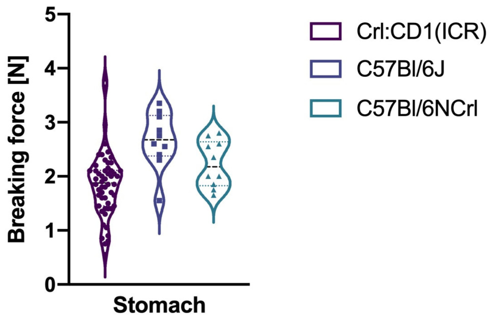
| Crl:CD1(ICR)–C57Bl/6J | −0.81 | −6.23 | <0.001 |
| Crl:CD1(ICR)–C57Bl/6NCrl | −0.37 | −2.88 | 0.006 |
| C57Bl/6J–C57Bl/6NCrl | 0.44 | 2.58 | 0.01 |
| Duodenum | |||
| Crl:CD1(ICR)–C57Bl/6J | −0.19 | −1.44 | 0.448 |
| Crl:CD1(ICR)–C57Bl/6NCrl | −0.04 | −0.29 | 0.773 |
| C57Bl/6J–C57Bl/6NCrl | 0.15 | 0.89 | 0.561 |
| Jejunum | |||
| Crl:CD1(ICR)–C57Bl/6J | −0.27 | −2.07 | 0.118 |
| Crl:CD1(ICR)–C57Bl/6NCrl | −0.06 | −0.45 | 0.654 |
| C57Bl/6J–C57Bl/6NCrl | 0.21 | 1.25 | 0.32 |
| Ileum | |||
| Crl:CD1(ICR)–C57Bl/6J | −0.14 | −1.08 | 0.662 |
| Crl:CD1(ICR)–C57Bl/6NCrl | −0.01 | −0.08 | 0.936 |
| C57Bl/6J–C57Bl/6NCrl | 0.13 | 0.77 | 0.662 |
| Caecum | |||
| Crl:CD1(ICR)–C57Bl/6J | −0.24 | −1.87 | 0.188 |
| Crl:CD1(ICR)–C57Bl/6NCrl | −0.15 | −1.17 | 0.363 |
| C57Bl/6J–C57Bl/6NCrl | 0.09 | 0.53 | 0.594 |
| Colon | |||
| Crl:CD1(ICR)–C57Bl/6J | −0.09 | −0.71 | 0.891 |
| Crl:CD1(ICR)–C57Bl/6NCrl | 0 | 0.02 | 0.988 |
| C57Bl/6J–C57Bl/6NCrl | 0.09 | 0.53 | 0.891 |
| Rectum | |||
| Crl:CD1(ICR)–C57Bl/6J | −0.22 | −1.67 | 0.285 |
| Crl:CD1(ICR)–C57Bl/6NCrl | −0.04 | −0.32 | 0.746 |
| C57Bl/6J–C57Bl/6NCrl | 0.18 | 1.04 | 0.5 |
Disclaimer/Publisher’s Note: The statements, opinions and data contained in all publications are solely those of the individual author(s) and contributor(s) and not of MDPI and/or the editor(s). MDPI and/or the editor(s) disclaim responsibility for any injury to people or property resulting from any ideas, methods, instructions or products referred to in the content. |
© 2026 by the authors. Licensee MDPI, Basel, Switzerland. This article is an open access article distributed under the terms and conditions of the Creative Commons Attribution (CC BY) license.
Share and Cite
Ertim, B.; Akinci, E.; von Stumberg, M.; Katzer, D.; Ganschow, R.; Vilz, T.O.; Oetzmann von Sochaczewski, C. The Resistance to Traction Forces Differs Substantially Between Intestinal Parts, but Not Between In- and Outbred Strains of Mice. Gastroenterol. Insights 2026, 17, 12. https://doi.org/10.3390/gastroent17010012
Ertim B, Akinci E, von Stumberg M, Katzer D, Ganschow R, Vilz TO, Oetzmann von Sochaczewski C. The Resistance to Traction Forces Differs Substantially Between Intestinal Parts, but Not Between In- and Outbred Strains of Mice. Gastroenterology Insights. 2026; 17(1):12. https://doi.org/10.3390/gastroent17010012
Chicago/Turabian StyleErtim, Berkan, Ejder Akinci, Maximiliane von Stumberg, David Katzer, Rainer Ganschow, Tim O. Vilz, and Christina Oetzmann von Sochaczewski. 2026. "The Resistance to Traction Forces Differs Substantially Between Intestinal Parts, but Not Between In- and Outbred Strains of Mice" Gastroenterology Insights 17, no. 1: 12. https://doi.org/10.3390/gastroent17010012
APA StyleErtim, B., Akinci, E., von Stumberg, M., Katzer, D., Ganschow, R., Vilz, T. O., & Oetzmann von Sochaczewski, C. (2026). The Resistance to Traction Forces Differs Substantially Between Intestinal Parts, but Not Between In- and Outbred Strains of Mice. Gastroenterology Insights, 17(1), 12. https://doi.org/10.3390/gastroent17010012