
Review

IL-6 and TNF-α in Ischemic Stroke: A Cardiogenetic Perspective on Inflammation and Clinical Outcomes

by
Maria-Gabriela Vlădoiu
1,2,* and
Claudiu Matei
3,4
1
Department of Neurology, Dr. Holhos Hospital, Calea Motilor 61, 510103 Alba Iulia, Romania
2
Department of Nursing, “1 Decembrie 1918” University of Alba Iulia, Gabriel Bethlen 5, 510009 Alba Iulia, Romania
3
MedLife Polisano Hospital, Izvorului 1A, 550234 Sibiu, Romania
4
Faculty of Medicine, “Lucian Blaga” University of Sibiu, 550024 Sibiu, Romania
*
Author to whom correspondence should be addressed.
Cardiogenetics 2026, 16(3), 18; https://doi.org/10.3390/cardiogenetics16030018
Submission received: 26 June 2026 / Revised: 27 August 2026 / Accepted: 10 September 2026 / Published: 11 September 2026
(This article belongs to the Section Molecular & Translational Genetics)

Abstract

Background: Ischemic stroke (IS) is a major cause of global disability, with outcomes increasingly dictated by post-ischemic neuroinflammation. Despite advances in reperfusion, many patients experience neurological decline due to divergent inflammatory responses. Objectives: This review synthesizes current evidence on inflammatory signaling, stroke genetics, and genetically informed precision medicine relevant to the interplay between interleukin-6 (IL-6), tumor necrosis factor-alpha (TNF-α), and ischemic stroke outcomes. Methods: We reviewed evidence from genome-wide association studies, Mendelian randomization analyses, experimental studies, observational biomarker cohorts, and relevant cardiovascular anti-inflammatory trials. Evidence from non-stroke populations is identified as indirect and is not interpreted as proof of efficacy in ischemic stroke. Results: IL-6 and TNF-α signaling involves receptor- and context-dependent mechanisms that may have both harmful and protective effects after cerebral ischemia. Multi-omic studies of the chromosome 9p21 locus and ANRIL suggest potential links among vascular-cell regulation, inflammation, and atherosclerotic risk, although stroke-specific mechanistic evidence remains limited. Elevated inflammatory-marker concentrations have been associated with greater stroke severity and unfavorable outcomes; however, their independent prognostic value and optimal sampling strategy require further validation. Selective anti-inflammatory approaches provide biologically plausible strategies for future study, but genotype-guided anti-cytokine treatment is not established for acute ischemic stroke or post-stroke recovery. Conclusions: Stroke genetics and multi-omic approaches may contribute to future precision-medicine strategies, but their clinical utility requires validation in prospective, stroke-specific studies. Current evidence does not support routine cytokine genotyping or genotype-guided anti-cytokine treatment in ischemic stroke.

1. Introduction

Ischemic stroke is a major cause of death and long-term disability worldwide. Although reperfusion therapies have improved outcomes for selected patients, disability remains common after successful recanalization, partly because of ischemia–reperfusion injury and secondary inflammatory processes [1]. These burden metrics are projected to escalate significantly as global populations continue to age. In contemporary clinical practice, modern therapeutic efforts heavily prioritize rapid vascular reperfusion, achieved primarily through mechanical thrombectomy and intravenous thrombolysis. However, a critical clinical discrepancy remains: a significant proportion of patients continue to suffer from progressive neurological decline despite achieving successful recanalization [1,2,3]. Post-ischemic inflammation contributes to secondary injury and repair processes, but its net effects depend on timing, cell type, infarct characteristics, and systemic comorbidity. Consequently, inflammation is no longer recognized merely as a secondary byproduct of vascular injury, but rather as a potentially modifiable driver of neurological outcomes. At the core of this response lies the “cytokine triad,” comprising Interleukin-6 (IL-6), Tumor Necrosis Factor-alpha (TNF-α), and Interleukin-1 beta (IL-1β), which act as the central regulatory nodes of the post-stroke cascade [4,5,6]. The emerging and highly specialized field of cardiogenetics seeks to elucidate why individuals presenting with identical clinical risk factors frequently exhibit divergent inflammatory trajectories and recovery profiles. By analyzing inherited genetic variations—specifically functional single-nucleotide polymorphisms (SNPs) and polygenic risk scores (PRSs)—researchers are now capable of mapping the underlying genetic program that dictates an individual’s baseline inflammatory state and overall vascular stability [7,8]. This comprehensive review systematically evaluates the intertwined roles of IL-6 and TNF-α in ischemic stroke, integrating compelling evidence from molecular biology, large-scale population genetics, and contemporary clinical trials to provide a comprehensive roadmap for precision stroke care.

2. Literature Search Strategy and Selection Criteria

This article is a narrative review with a structured literature search strategy. It was not designed as a systematic review and no PRISMA protocol or flow diagram was applied. The search was conducted to identify and prioritize relevant peer-reviewed evidence across molecular mechanisms, genetic association studies, Mendelian randomization analyses, biomarker studies, and translational therapeutic research.
Primary electronic databases, including PubMed/MEDLINE, Scopus, and the Web of Science Core Collection, were systematically queried for peer-reviewed articles published up to May 2026. The search strategy utilized a targeted combination of Medical Subject Headings (MeSH) and free-text keywords, structured by Boolean operators: (“ischemic stroke” OR “cerebral ischemia”) AND (“Interleukin-6” OR “IL-6” OR “Tumor Necrosis Factor-alpha” OR “TNF-α”) AND (“polymorphism” OR “cardiogenetics” OR “Mendelian Randomization” OR “heart-brain axis”).
Priority was given to original research, large genetic studies, systematic reviews, and well-characterized clinical trials. The final synthesis was narrative and interpretive; it should not be regarded as a systematic estimate of treatment effects or genetic associations.

3. Molecular Pathophysiology: The Bimodal Cytokine Networks

3.1. Interleukin-6: The Paradox of Classical vs. Trans-Signaling

Interleukin-6 is a pleiotropic cytokine involved in acute-phase responses, immune regulation, and tissue repair. In the physiological, uninjured brain, IL-6 expression is tightly maintained at low basal levels. However, immediately following an ischemic cerebral injury, affected neurons undergo necrosis and release Damage-Associated Molecular Patterns (DAMPs). Ischemic injury and damage-associated molecular patterns can stimulate IL-6 production by resident brain cells and infiltrating immune cells [4,6].
The pleiotropic nature of IL-6, exhibiting both neuroprotective and neurotoxic capacities, is intricately governed by its specific receptor interaction modes. In the classical signaling pathway, IL-6 binds to the cell-surface IL-6 receptor (mIL-6R), which is primarily localized on immune cells and hepatocytes, thereby facilitating anti-inflammatory and regenerative tissue responses. Conversely, trans-signaling occurs when IL-6 binds to the soluble form of the receptor (sIL-6R). This IL-6/sIL-6R complex subsequently activates the ubiquitously expressed gp130 signal transducer on cells that inherently lack mIL-6R, most notably vulnerable neurons. This trans-signaling pathway serves as the dominant driver of neurotoxicity post-stroke, actively promoting microglial M1 polarization and widespread blood–brain barrier (BBB) disruption. Furthermore, dendritic cells possess the capability to present IL-6 in trans via mIL-6R to naive T cells, thereby driving their differentiation into pro-inflammatory Th17 cells. Downstream of these receptor interactions, IL-6 heavily triggers the Janus kinase (JAK)/STAT3 pathway, where phosphorylated STAT3 (P-STAT3) translocates directly to the cell nucleus to drive the transcription of closely related genes to angiogenesis and functional recovery [4,7].
As systematically outlined in Table 1, the bimodal structural features of these networks clarify the paradoxical nature of cytokine signaling, explaining why non-selective pharmacological blockade can be ineffective or deleterious [4,7].

3.2. TNF-α and the Reciprocal Amplification Loop

TNF-α is rapidly induced after cerebral ischemia and may be produced by activated microglia, astrocytes, endothelial cells, infiltrating immune cells, and other cellular sources. TNF is initially synthesized as a transmembrane protein and can subsequently be cleaved to generate soluble TNF. These two forms may preferentially activate distinct receptor pathways and contribute differently to post-ischemic inflammation and tissue repair [8].
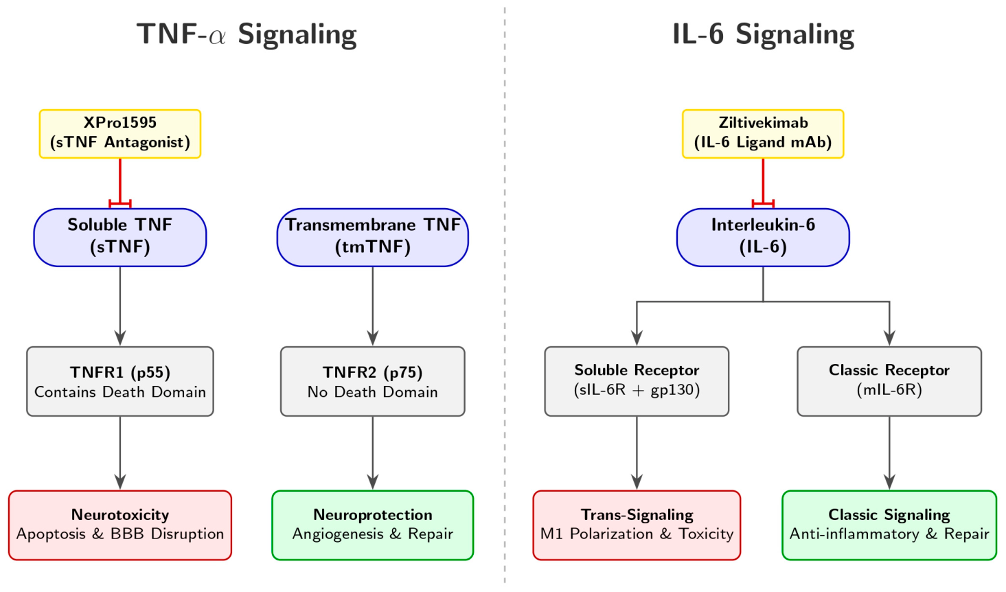
Signaling through TNFR1 (p55), which contains a cytoplasmic death domain and is preferentially activated by soluble TNF, has been associated with pro-inflammatory, apoptotic, and cytotoxic responses in experimental cerebral ischemia. TNFR1-dependent signaling can engage NF-κB and cell-death-related pathways and may contribute to blood–brain barrier dysfunction and neuronal injury. However, TNFR1-mediated effects are context-dependent; experimental evidence also indicates a role for TNFR1 in post-ischemic angiogenic responses [8,9].
TNFR2 (p75) lacks a death domain and is preferentially activated by transmembrane TNF. In selected experimental settings, TNFR2-associated signaling may contribute to immune regulation, neuronal survival, neurogenesis, and tissue-repair processes after ischemic injury. These potential reparative functions provide a biological rationale for selective approaches that inhibit soluble TNF while preserving transmembrane TNF signaling. However, this framework remains primarily experimental and does not establish clinical efficacy in human ischemic stroke [8,9].
TNF-α and IL-6 participate in interacting inflammatory networks. Experimental and systems-level studies suggest that TNF-α can promote IL-6 expression through NF-κB-associated pathways and that IL-6 may reinforce TNF-α-related inflammatory activity. The magnitude and clinical relevance of this potential positive feedback loop after ischemic stroke depend on timing, cellular source, receptor engagement, and the local tissue environment [4,6,7].

4. Genetic Architecture: Functional Polymorphisms and Environmental Synergies

4.1. IL-6 Promoter Variants and Ethnic Heterogeneity

The inherent genetic architecture of cytokine genes significantly and permanently influences an individual’s basal and injury-induced inflammatory responses. Located directly within the IL6 promoter region on chromosome 7p21, the most extensively characterized variant is the rs1800795 (−174G/C) polymorphism. This specific variant modulates transcriptional efficiency by directly altering transcription factor binding affinity. The G-allele is generally associated with generating higher circulating IL-6 levels in response to physiological stress; however, its precise role in stroke risk is highly ethnic-specific [10].
In Caucasian and Turkish populations, possessing the CC genotype has been significantly linked to a 4.3-fold increased risk of IS. Conversely, extensive meta-analyses conducted in large Chinese and general European cohorts have often yielded consistently null results regarding primary disease risk. This geographical divergence suggests that the rs1800795 SNP functions primarily as an important modulator of clinical stroke severity rather than as a primary pathological initiator. Other related variants, most notably rs1800796 (−572C/G), have been strongly and independently associated with IS in indigenous West African men and are clinically considered established genetic risk factors for both coronary artery disease and ischemic stroke across various Asian populations [10,11]. However, these risk estimates must be interpreted with caution. Early candidate-gene association studies in stroke genetics were frequently limited by small sample sizes, leading to wide confidence intervals and inconsistent replication across different ethnic cohorts. While recent meta-analyses have pooled data to increase statistical power, significant inter-study heterogeneity remains a limiting factor [10,11].
The association between IL6 rs1800795 (−174G/C) and ischemic stroke has been inconsistent across populations. Differences in ancestry, stroke subtype, environmental exposures, sample size, and study design may explain the observed heterogeneity. Accordingly, rs1800795 should currently be regarded as a candidate modifier of inflammatory phenotype rather than a validated clinical predictor of ischemic stroke risk. Other IL6 promoter variants, including rs1800796 (−572C/G), have also been investigated, but available evidence does not support their routine use for clinical risk stratification [10,11].

4.2. TNF-α Polymorphisms and Structural Vascular Determinants

The TNF gene, mapped to chromosome 6p21.3, resides within the highly polymorphic MHC class III genomic region. Within this locus, rs1800629 (−308G/A) represents a major functional variant driving inflammatory variability. The A-allele, frequently designated in the literature as TNF2, is consistently associated with significantly enhanced transcriptional activity. In vast Asian populations, possessing the −308A allele is associated with a significant two-fold increase in IS susceptibility. This genetic impact is synergistically amplified in individuals who present with concurrent environmental or metabolic risk factors, such as hypertension or hyperlipidemia [12].
Additional polymorphisms within this region, such as rs361525 (−238G/A), have also been significantly associated with heightened IS risk in adult Caucasian populations. Similar to rs1800629, the clinical impact of the −238G/A variant is highly potentiated by lifestyle and environmental factors, including smoking and chronic alcohol consumption [12].
Furthermore, while an individual’s systemic inflammatory profiles are largely genetically programmed by these specific cytokine SNPs, they inextricably interact with congenital structural determinants of the cerebral vasculature. Rare anatomical variations localizing in the posterior circulation—such as the persistence of fetal-type configurations of the Circle of Willis or the presence of the anomalous artery of Percheron—significantly impair collateral brain perfusion. These structural anomalies heighten an individual’s baseline physiological risk for atypical ischemic events, thereby complicating the clinical phenotype and substantially worsening the subsequent inflammatory response post-occlusion [13].
The TNF promoter variants rs1800629 (−308G/A) and rs361525 (−238G/A) have been investigated in relation to inflammatory phenotypes and ischemic stroke susceptibility. Although some case–control studies have reported positive associations, results remain heterogeneous across populations and stroke subtypes. These variants are not established markers for clinical stroke-risk prediction or treatment selection [11,12].
As systematically detailed in Table 2, the underlying genetic architecture clearly highlights how ethnicity and concurrent risk factors directly influence individual inflammatory responses [10,11,12].

5. Mendelian Randomization and Causal Inference: A Paradigm Shift

5.1. The MEGASTROKE and GIGASTROKE Consortia

Large-scale Mendelian randomization (MR) studies have recently revolutionized the landscape of translational neurology. By utilizing specific genetic variants as randomized instrumental variables, MR methodologies allow researchers to infer true biological causality, thereby circumventing the pervasive confounding variables inherent in traditional observational trial designs. Central to this emerging paradigm are the massive global initiatives spearheaded by the MEGASTROKE and GIGASTROKE consortia, both of which have effectively utilized the IL6R variant rs2228145 (Asp358Ala) as a robust, naturally occurring genetic proxy for pharmacological IL-6 inhibition [14,15,16,17].
The MEGASTROKE consortium, represented by its landmark 2018 study, marked a foundational turning point in the field by conducting the first large-scale genome-wide association study (GWAS) meta-analysis entirely dedicated to stroke. By meticulously analyzing the genomic data from approximately 60,000 ischemic stroke cases and over 400,000 controls—though predominantly of European ancestry—this initiative successfully identified over 30 distinct genetic loci associated with vascular risk. The major conceptual and clinical impact of MEGASTROKE lay in its unprecedented ability to demonstrate, with high statistical rigor, that the genetic architecture of stroke is dependent on its etiological subtype. It demonstrated that the polygenic mechanisms underlying large-artery atherosclerosis are biologically distinct from those governing small-vessel disease or acute cardioembolic events [16].
Building upon these established foundational metrics, the subsequent GIGASTROKE consortium (2022) expanded the genomic landscape of stroke to an unprecedented global scale. This initiative integrated over 110,000 stroke cases and more than 2.5 million control individuals. The revolutionary element of GIGASTROKE lies in its broadly multi-ancestry design. By actively incorporating large cohorts of East Asian, African, South Asian, and Latino populations, it successfully corrected the prominent Eurocentric bias of prior genomic research. This exceptional genetic diversity enabled the expansion of the global stroke risk map to over 89 independent loci. More importantly, it provided the highly sensitive fine-mapping resolution necessary for advanced MR investigations. Consequently, GIGASTROKE transformed specific inflammatory loci—such as those situated within the IL6R or TNF genes—into universal epidemiological instruments capable of validating clinical pharmacological targets with reliable, cross-ancestry predictability [17].

5.2. Causal Pathways and the Necessity of Sensitivity Analyses

Mendelian randomization (MR) studies use genetic variants as instrumental variables to investigate whether an exposure may have a causal relationship with a clinical outcome. In the context of ischemic stroke, variants in the IL6R locus have been used as proxies for altered IL-6 signaling. Available MR evidence suggests that genetically proxied reduction in IL-6 signaling may be associated with a lower risk of ischemic stroke, with potentially stronger associations for non-cardioembolic stroke subtypes. These findings are consistent with a possible contribution of IL-6 pathway activity to atherosclerotic and small-vessel mechanisms of cerebrovascular disease [14,15].
However, MR estimates must be interpreted cautiously. They reflect the consequences of lifelong genetically mediated differences in signaling and do not directly predict the effects of short-term pharmacological inhibition initiated during acute or subacute ischemic stroke. Lifelong exposure may be accompanied by developmental adaptation, compensatory biological pathways, and cumulative effects on vascular risk factors. Therefore, MR findings should be considered useful for biological target prioritization and hypothesis generation, rather than as evidence that pharmacological IL-6 inhibition improves clinical outcome after ischemic stroke [14,18].
The causal role of TNF-α signaling in ischemic stroke remains less clearly defined. This uncertainty partly reflects the biological complexity of the TNF system. Soluble TNF and transmembrane TNF may have different physiological effects, and TNFR1- and TNFR2-mediated signaling can activate distinct downstream pathways. TNFR1 signaling is frequently associated with inflammatory and cytotoxic responses in experimental models, whereas TNFR2 may contribute to immune regulation and tissue repair in selected contexts. Genetic instruments representing overall TNF signaling may not distinguish adequately between these ligand- and receptor-specific mechanisms [8,9].
Accordingly, null or weak MR findings based on broad TNF-related genetic proxies should not be interpreted as evidence against receptor-selective or ligand-selective therapeutic strategies. Instead, they emphasize the limitations of translating a complex, receptor-dependent cytokine system into a single genetic exposure variable. Experimental stroke studies of selective soluble TNF inhibition provide a mechanistic rationale for further investigation, but clinical efficacy in human ischemic stroke has not been established [19,20].
Sensitivity analyses are essential for strengthening the interpretation of MR studies. Methods such as MR-PRESSO may help identify potential horizontal pleiotropy by detecting outlier genetic variants that influence the outcome through pathways other than the exposure of interest. Steiger filtering can help assess whether the genetic instruments explain more variance in the proposed exposure than in the outcome, thereby supporting the assumed direction of effect. Nevertheless, these approaches cannot eliminate all potential sources of bias, including weak-instrument bias, residual pleiotropy, population stratification, selection bias, and survival bias [18,21].
Overall, MR provides an important complementary approach for evaluating inflammatory pathways in ischemic stroke, particularly when integrated with experimental studies, observational biomarker data, and randomized clinical trials. Its value lies in informing biological plausibility and therapeutic-target prioritization; stroke-specific interventional studies remain necessary to determine whether modulation of IL-6 or TNF-α signaling can improve clinical outcomes [3,14].

6. The Heart–Brain Axis: A Bidirectional Continuum of Injury

6.1. The Cerebrocardiac Syndrome and Catecholamine Storm

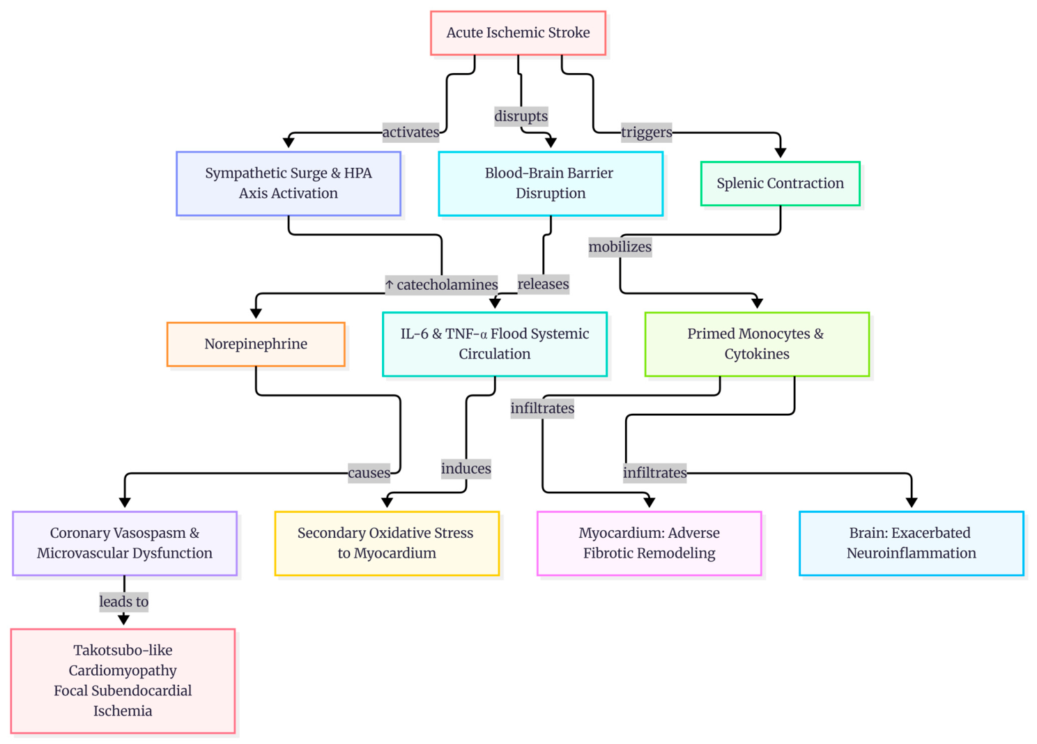
Acute ischemic stroke triggers an immediate and overwhelming systemic physiological response through the bidirectional heart–brain axis, a complex clinical phenomenon frequently termed the cerebrocardiac syndrome. The primary pathophysiological mechanism underlying this syndrome involves an intense, sudden sympathetic surge coupled with the concomitant activation of the hypothalamic–pituitary–adrenal (HPA) axis. This autonomic dysregulation culminates in a severe systemic catecholamine storm [21,22].
The abrupt and significant hypersecretion of norepinephrine, specifically from myocardial nerve endings, rapidly induces localized coronary vasospasm and severe microvascular dysfunction. At the cellular level, this neurogenic cardiac cascade leads to focal subendocardial ischemia, myocyte apoptosis, and the distinct histological finding of contraction band necrosis. Clinically, this acute injury profile effectively mimics Takotsubo-like cardiomyopathy, which is characteristically defined by reversible ventricular apical ballooning, prominent repolarization abnormalities on electrocardiography, and significantly elevated serum troponin levels in the absence of primary coronary occlusion. Furthermore, the extensive stroke-induced disruption of the blood–brain barrier allows large quantities of central pro-inflammatory cytokines, particularly IL-6 and TNF-α, to flood the systemic circulation. This cytokine efflux actively propagates secondary oxidative stress directly to the vulnerable myocardium, thereby highly predisposing stroke survivors to subsequent major adverse cardiovascular events (MACEs) (Figure 1) [22].

6.2. The Brain–Spleen Axis and Reciprocal Inflammatory Substrates

This severe neurocardiac interaction also intricately encompasses the brain–spleen axis. Acute stroke-induced sympathetic overactivation directly drives rapid splenic contraction within mere hours of the initial ischemic onset. This contraction mobilizes a substantial and highly concentrated surge of primed monocytes and pro-inflammatory cytokines into the systemic circulation. These aggressive, splenic-derived immune cells subsequently infiltrate both the ischemic brain parenchyma—where they severely exacerbate secondary neuroinflammation—and the compromised myocardium, where they accelerate pathological collagen deposition and drive adverse fibrotic remodeling [23].
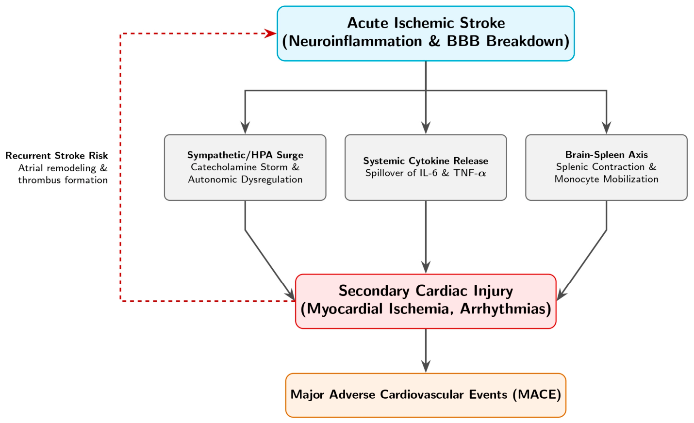
Conversely, this pathophysiological continuum is bidirectional; pre-existing cardiac substrates, such as atrial fibrillation (AF) and chronic heart failure (HF), actively sustain a persistent, systemic pro-inflammatory environment long before the ischemic event occurs. Within this context, persistently elevated levels of circulating IL-6 are independently and strongly linked to left atrial structural remodeling and endocardial thrombus formation. As mapped out visually in Figure 2, this interplay effectively establishes a reciprocal, self-perpetuating cycle of coupled cardiovascular and neurological decline that complicates secondary stroke prevention [22,23].

7. Multi-Omics and Integrative Bioinformatics

7.1. The Epigenetic ANRIL/YY1/IL-6 Signaling Axis

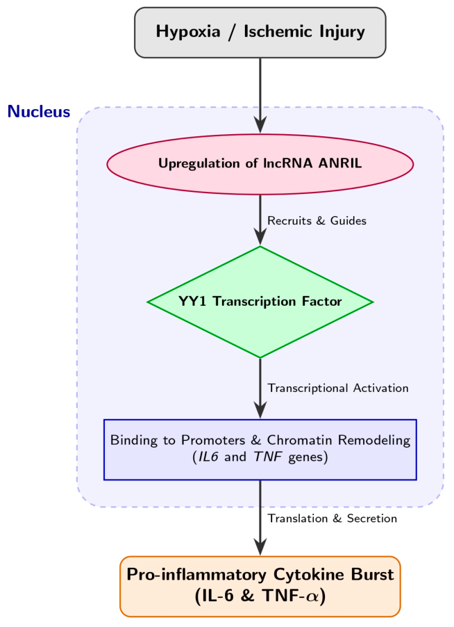
The modern integration of advanced multi-omics architectures, encompassing high-throughput transcriptomics, single-cell sequencing, and precise expression quantitative trait loci (eQTL) mapping, is significantly redefining our baseline understanding of systemic inflammatory regulatory networks. A prominent, highly researched paradigm of this complex cross-layer regulation is the ANRIL/YY1/IL-6 signaling axis, which is localized at the 9p21.3 genomic locus. Currently, this specific locus represents a major genetic risk factor for atherosclerosis identified in the human genome to date, a molecular cascade explicitly detailed in Figure 3 [24,25].
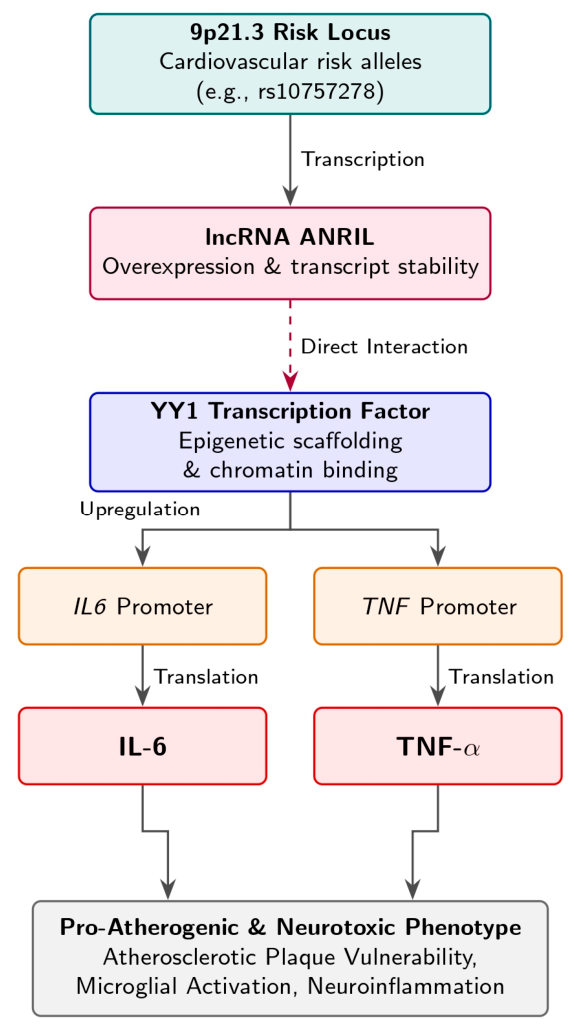
Under sustained pro-inflammatory biological conditions, systemic TNF-α directly induces the cellular expression of the long non-coding RNA (lncRNA) known as ANRIL via the canonical NF-κB pathway. Once expressed, ANRIL acts as a sophisticated molecular scaffold. Beyond its interaction with the transcription factor Yin Yang 1 (YY1), recent epigenetic profiling indicates that ANRIL physically recruits the Polycomb Repressive Complexes (PRC1 and PRC2). While the PRC complexes actively silence downstream anti-atherogenic and endothelial-protective genes, the ANRIL-YY1 complex acts in tandem to forcefully upregulate the direct transcription of both IL6 and IL8 within vulnerable vascular endothelial and smooth muscle cells (Figure 4) [25]. Furthermore, this dynamic chromatin remodeling actively skews the local microenvironment, promoting the proliferation of vascular smooth muscle cells and the polarization of macrophages toward the inflammatory M1 phenotype. Ultimately, this localized, epigenetically driven cytokine overproduction heavily drives acute plaque destabilization and precipitates chronic atherogenesis [24,25].

7.2. Single-Cell eQTL Mapping and Cellular Granularity

Beyond assessing global vascular tissue dynamics, contemporary integrative bioinformatic analyses have successfully uncovered cell-type-specific immune signals that are intrinsically shared between systemic inflammatory pathologies and severe ischemic cerebrovascular risk. Specifically, single-cell eQTL colocalization frameworks have conclusively demonstrated that key genetic variants mapping near the IL6R and TNF loci modulate downstream gene expression through highly divergent, functionally cell-specific profiles in circulating monocytes compared to T cells [26].
Emerging literature has particularly highlighted the concept of response eQTLs (reQTLs): genetic regulatory effects that remain entirely dormant under basal conditions but are strongly activated specifically under ischemic or infectious stress. For instance, high-resolution single-cell RNA sequencing (scRNA-seq) has revealed that certain protective IL6R variants exert their primary regulatory effects exclusively within CD14+ classical monocytes shortly after tissue hypoxia, altering monocyte–endothelial adhesion dynamics, while having null effects on CD4+ T-lymphocyte populations. This high cellular granularity is scientifically crucial, as it clearly explains the high degree of heterogeneous responses to targeted anti-inflammatory therapies frequently observed within diverse clinical trial cohorts. Consequently, this underscores the immense clinical potential of utilizing molecular inflammatory subtyping to meticulously guide precision secondary prevention and structure highly personalized post-stroke rehabilitation strategies [8,26].

8. Clinical Outcomes and Prognostic Biomarkers

8.1. Acute Phase Correlations and Functional Disability

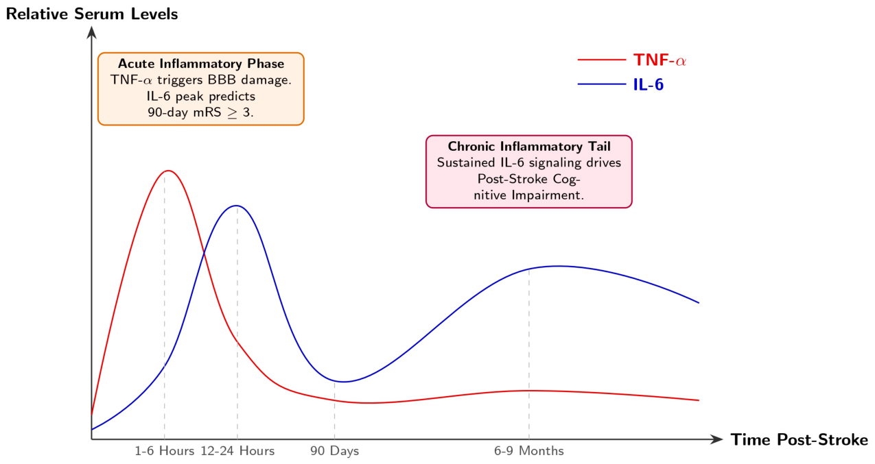
In the highly acute clinical setting, serum levels of IL-6 and TNF-α serve as important prognostic biomarkers for predicting both initial ischemic stroke severity and long-term functional neurological outcomes. The peak systemic concentrations of these cytokines typically manifest within a critical window between 6 and 24 h post-stroke [27].
Elevated admission levels of these cytokines demonstrate remarkably strong positive correlations with initial severity, as quantified by the National Institutes of Health Stroke Scale (NIHSS) scores. Specifically, serum IL-6 exhibits powerful correlation coefficients (r) ranging consistently from 0.68 to 0.745, while TNF-α shows an r value of 0.61. Furthermore, early elevated IL-6 levels exceeding 2.5 pg/mL on Day 1 serve as a tremendously powerful independent predictor of disability (Figure 5). Surpassing this threshold confers a substantial 4.84-fold increased risk of poor 90-day functional recovery, rigorously defined by a modified Rankin Scale (mRS) score of ≥3 [27,28].

8.2. The Chronic Inflammatory Tail and Cognitive Impairment

Beyond immediate physical disability and motor deficits, an emergent and highly pressing area of clinical concern is the established mechanistic link between sustained systemic inflammation and long-term post-stroke cognitive impairment (PSCI). Notably, recent longitudinal tracking has revealed that a progressive, sustained rise in systemic IL-6 levels between 6 and 9 months post-stroke acts as a far more potent predictor of debilitating cognitive decline than baseline acute admission levels. This finding explicitly underscores the deleterious and silent neurological impact of a persistent, chronic inflammatory tail [28].
Consequently, the routine clinical implementation of multiplexed biomarker panels is highly recommended. Specifically, the precise pairing of serum TNF-α with IL-6 yields vastly superior predictive validity for forecasting long-term functional and cognitive trajectories when compared to isolated, individual cytokine analyses. As visually supported by the relative serum levels chart and systematically documented in Table 3, these distinct temporal dynamics underscore their clinical correlations with disease severity scales and long-term cognitive outcomes [27,28].

9. Pharmacogenomics and Precision Medicine

9.1. Receptor-Selective Biologics: Overcoming Historical Failures

The historical failure of various broad-spectrum neuroprotective agents in stroke trials has necessarily precipitated a significant paradigm shift toward receptor-selective and highly genotype-informed immunomodulation. Contemporary precision medicine frameworks now actively strive to intimately align targeted pharmacological biologics with a specific patient’s inherited genetic and inflammatory architecture [7].
Exemplifying this highly targeted approach, the ongoing, large-scale Phase III ZEUS trial (NCT05021835) is actively evaluating the efficacy of ziltivekimab. Ziltivekimab is a monoclonal antibody targeting the IL-6 ligand. The phase II RESCUE trial was conducted in patients with chronic kidney disease and elevated high-sensitivity C-reactive protein and demonstrated reductions in inflammatory and thrombosis-related biomarkers. The phase III ZEUS trial (NCT05021835) was designed to assess whether ziltivekimab reduces major adverse cardiovascular events in patients with established atherosclerotic cardiovascular disease, chronic kidney disease, and residual inflammatory risk. ZEUS is not a stroke-specific treatment trial; therefore, its findings cannot establish efficacy in acute ischemic stroke or post-stroke recovery. The primary clinical objective is the significant reduction in major adverse cardiovascular events (MACEs) in highly stratified, high-risk populations, specifically those identified by a baseline high-sensitivity C-reactive protein (hsCRP) level of ≥2 mg/L [29,30]. The rationale for ZEUS is heavily anchored in the highly successful Phase II RESCUE-IL6 trial, which demonstrated that targeting the IL-6 ligand, rather than the IL-6 receptor, as seen with older generation drugs like tocilizumab, resulted in a marked, dose-dependent reduction in systemic CRP and fibrinogen levels without triggering the severe off-target worsening of atherogenic lipid profiles. This precise ligand-targeting mechanism marks a crucial evolution in immunomodulator design, offering a far safer cardiovascular profile for highly vulnerable post-stroke cohorts [29]. Final data readouts for the ZEUS trial are anticipated to be completed and published in the near future, which will definitively clarify the long-term safety and cardiovascular efficacy of this ligand-specific inhibition.

9.2. Solving the Dual-Receptor Dilemma

Concurrently, the rapid pharmacological development of selective soluble TNF (sTNF) antagonists, most notably XPro1595 (pegipanermin), provides a mechanistically elegant and desperately needed solution to the previously discussed dual-receptor dilemma. Unlike traditional, non-selective TNF inhibitors such as etanercept—which indiscriminately ablate all TNF signaling pathways and inadvertently promote severe central demyelination—XPro1595 highly specifically neutralizes the purely harmful sTNF fraction. Crucially, this mechanism fully preserves the vital, neuroprotective transmembrane TNF signaling axis, ensuring that essential TNFR2-mediated remyelination and synaptic pruning processes remain intact [19,20].
Recent Phase Ib/II clinical data derived from the MINDFuL trial have demonstrated exceedingly promising trends regarding long-term cognitive preservation and systemic inflammatory biomarker attenuation. Notably, advanced neuroimaging endpoints utilizing white matter free-water (WMFW) MRI mapping revealed that XPro1595 significantly reversed neuroinflammation-induced axonal damage in treated cohorts. Furthermore, this was achieved in well-phenotyped, inflammation-enriched clinical populations with a complete, verifiable absence of dangerous amyloid-related imaging abnormalities (ARIAs) or broad immunosuppression. Ultimately, emerging pharmacogenetic evidence strongly dictates that stroke patients harboring the specific IL6−174GG genotype frequently exhibit overlapping hyper-inflammatory signatures, making them ideal candidates for these advanced anti-cytokine interventions. Selective soluble TNF (sTNF) antagonists, most notably XPro1595 (pegipanermin), are under clinical investigation for chronic neurodegenerative conditions, such as Alzheimer’s disease (e.g., the MINDFuL trial, Phase Ib/II). While these early-phase trials provide a strong proof of concept that receptor-selective TNF modulation is safe, avoids broad immunosuppression, and attenuates neuroinflammation without causing amyloid-related imaging abnormalities (ARIAs), their therapeutic translation to acute ischemic stroke remains purely theoretical. These findings provide a strong rationale for incorporating exploratory dual IL-6/TNF genotyping into the design of future prospective cerebrovascular trials, rather than immediate clinical implementation. Cytokine-related variants may contribute to heterogeneity in inflammatory responses and treatment effects. Their potential value for patient selection should be evaluated prospectively in adequately powered, stroke-specific clinical studies before they are incorporated into trial-enrichment strategies or clinical decision-making. As this trial progresses toward its final readout, the forthcoming results are expected to provide very important insights regarding safety and longitudinal biomarker attenuation [31].
Table 4, along with the complementary signaling inhibition figure (Figure 6), highlights the clear shift towards precision medicine by detailing the ongoing clinical trials, targeted mechanisms, and clinical goals of the novel immunomodulators mentioned throughout this review.

10. Translational Hurdles and Current Limitations

While the integration of cardiogenetics into stroke neurology offers a compelling paradigm shift, several significant translational hurdles must be overcome before genotype-guided immunomodulation can become the standard of care in routine clinical practice [7].

10.1. The Temporal Paradox of Acute Genotyping

The most significant logistical barrier to precision stroke medicine is the temporal paradox between the acute nature of ischemic injury and the traditional turnaround times of genomic analysis. Acute stroke management is primarily governed by the “time is brain” axiom, requiring rapid reperfusion decisions within an exceedingly narrow therapeutic window (typically 4.5 to 24 h). Conversely, conventional genotyping or the calculation of comprehensive polygenic risk scores (PRSs) requires days to weeks. For genotype-informed interventions, such as administering targeted anti-cytokine therapies specifically to patients with the IL6 −174GG genotype, to be clinically viable, health systems must invest heavily in rapid, point-of-care (POC) molecular diagnostics capable of delivering actionable genetic profiles within minutes of a patient’s arrival at the emergency department [7]. Beyond turnaround time, implementation of point-of-care genotyping in stroke units would require validated assays, laboratory quality assurance, trained personnel, integration with emergency workflows, secure data governance, and evidence of cost-effectiveness. The clinical value of testing must also be demonstrated by showing that genotype-informed decisions improve outcomes beyond established reperfusion and secondary-prevention strategies [2,3].

10.2. Methodological Boundaries of Mendelian Randomization

Although Mendelian randomization (MR) has successfully circumvented the confounding variables of observational studies, the methodology is not without inherent limitations. A major biological caveat is the concept of canalization. MR estimates the effect of lifelong, genetically determined exposure to altered cytokine levels. This chronic modulation does not directly guarantee that short-term, acute pharmacological blockade of the same pathway will reproduce an identical neuroprotective effect in the acute phase of ischemic stroke [14]. Furthermore, while advanced statistical models like MR-PRESSO are actively deployed to mitigate horizontal pleiotropy, they cannot entirely account for survival bias. Because severe pro-inflammatory genotypes may increase early cardiovascular mortality, older stroke cohorts (such as those analyzed in MEGASTROKE and GIGASTROKE) might inadvertently reflect a survivor phenotype, potentially skewing the calculated causal estimates of specific cytokine variants [18,21].

10.3. Limits of Cross-Disease Translation: Lessons from MINDFuL

A key translational limitation is that evidence from anti-inflammatory trials in neurodegenerative disease cannot be directly extrapolated to ischemic stroke. The inflammatory milieu, timing of intervention, blood–brain barrier integrity, vascular comorbidity, and clinical outcomes differ substantially between early Alzheimer’s disease and acute or subacute stroke. Accordingly, the MINDFuL findings should be viewed as hypothesis-generating support for biomarker-enriched trial design, not as evidence supporting clinical use of XPro1595 after ischemic stroke. Nevertheless, MINDFuL provides a useful proof of concept for the design of future stroke trials: inflammatory biomarkers may help identify biologically distinct patients who could be more likely to benefit from targeted immunomodulation. This hypothesis must be evaluated in prospective, stroke-specific randomized clinical trials [31].

10.4. Interplay with Standard Reperfusion Therapies

Finally, the current cardiogenetic literature has yet to fully elucidate the complex interaction between a patient’s baseline genetic inflammatory architecture and standard-of-care reperfusion therapies. Ischemia–reperfusion injury aggressively exacerbates local neuroinflammation. Patients harboring hyper-inflammatory genetic variants (e.g., the highly active TNF −308A allele or IL-6 −174CC genotype) may be inherently more susceptible to major blood–brain barrier (BBB) degradation following the administration of intravenous tissue plasminogen activator (tPA). Consequently, future clinical trials must rigorously investigate whether these specific pro-inflammatory genetic profiles independently increase the risk of hemorrhagic transformation post-thrombolysis, which could substantially alter acute patient triaging [3].

11. Conclusions

IL-6 and TNF-α are important components of the post-stroke inflammatory response. Their effects are influenced by timing, cellular source, receptor signaling, infarct characteristics, and systemic comorbidity. Available experimental, observational, and genetic evidence supports their biological relevance, but does not establish that modulation of either pathway improves clinical outcome after ischemic stroke [3,4]. Genetic and multi-omic approaches may improve understanding of biological heterogeneity in ischemic stroke and may help prioritize therapeutic targets. However, Mendelian randomization reflects lifelong genetically mediated differences in pathway activity and cannot directly predict the effects of short-term pharmacological intervention during acute or subacute stroke. Current evidence does not support routine cytokine genotyping, biomarker-guided anti-cytokine treatment, or the clinical use of ziltivekimab or XPro1595 for ischemic stroke or post-stroke recovery. Future prospective, stroke-specific studies should determine whether molecular and inflammatory profiling can identify patient subgroups who may benefit from targeted immunomodulatory strategies, alongside evidence-based reperfusion therapy and secondary prevention [14,29,31].

Author Contributions

Conceptualization, M.-G.V. and C.M.; methodology, M.-G.V.; validation, M.-G.V. and C.M.; writing—original draft preparation, M.-G.V.; writing—review and editing, M.-G.V. and C.M.; visualization, M.-G.V.; supervision, C.M. All authors have read and agreed to the published version of the manuscript.

Funding

This research received no external funding.

Institutional Review Board Statement

Not applicable.

Informed Consent Statement

Not applicable.

Data Availability Statement

No new data were created or analyzed in this study. Data sharing is not applicable to this article.

Conflicts of Interest

The authors declare no conflicts of interest.

References

  1. GBD 2019 Stroke Collaborators. Global, regional, and national burden of stroke and its risk factors, 1990–2019: A systematic analysis for the Global Burden of Disease Study 2019. Lancet Neurol. 2021, 20, 795–820. [Google Scholar] [CrossRef] [Scilit] [PubMed]
  2. Powers, W.J.; Rabinstein, A.A.; Ackerson, T.; Adeoye, O.M.; Bambakidis, N.C.; Becker, K.; Biller, J.; Brown, M.; Demaerschalk, B.M.; Hoh, B.; et al. Guidelines for the early management of patients with acute ischemic stroke: 2019 update to the 2018 guidelines for the early management of acute ischemic stroke. Stroke 2019, 50, e344–e418. [Google Scholar] [CrossRef] [Scilit] [PubMed]
  3. Iadecola, C.; Buckwalter, M.S.; Anrather, J. Immune responses to stroke: Mechanisms, modulation, and therapeutic potential. J. Clin. Investig. 2020, 130, 2777–2788. [Google Scholar] [CrossRef] [Scilit]
  4. Anrather, J.; Iadecola, C. Inflammation and stroke: An overview. Neurotherapeutics 2016, 13, 661–670. [Google Scholar] [CrossRef] [Scilit] [PubMed]
  5. Jayaraj, R.L.; Azimullah, S.; Beiram, R.; Jalal, F.Y.; Rosenberg, G.A. Neuroinflammation: Friend and foe for ischemic stroke. J. Neuroinflammation 2019, 16, 142. [Google Scholar] [CrossRef] [Scilit] [PubMed]
  6. Rose-John, S. Interleukin-6 family cytokines. Cold Spring Harb. Perspect. Biol. 2018, 10, a028415. [Google Scholar] [CrossRef] [Scilit] [PubMed]
  7. Hunter, C.A.; Jones, S.A. IL-6 as a keystone cytokine in health and disease. Nat. Immunol. 2015, 16, 448–457. [Google Scholar] [CrossRef] [Scilit] [PubMed]
  8. Probert, L. TNF and its receptors in the CNS: The essential, the desirable and the deleterious effects. Neuroscience 2015, 302, 2–22. [Google Scholar] [CrossRef] [Scilit] [PubMed]
  9. McCoy, M.K.; Tansey, M.G. TNF signaling inhibition in the CNS: Implications for normal brain function and neurodegenerative disease. J. Neuroinflammation 2008, 5, 45. [Google Scholar] [CrossRef] [Scilit] [PubMed]
  10. Chai, J.; Cao, X.L.; Lu, F. Association of interleukin-6 −174G/C polymorphism with ischemic stroke: An updated meta-analysis. Front. Neurol. 2022, 12, 799022. [Google Scholar] [CrossRef] [Scilit] [PubMed]
  11. Wang, X.; Cheng, S.; Brophy, V.H.; Erlich, H.A.; Mannhalter, C.; Berger, K.; Lalouschek, W.; Browner, W.S.; Shi, Y.; Ringelstein, E.B.; et al. A meta-analysis of candidate gene polymorphisms and ischemic stroke in six study populations: Association of lymphotoxin-alpha in nonhypertensive patients. Stroke 2009, 40, 683–695. [Google Scholar] [CrossRef] [Scilit] [PubMed]
  12. Wu, J.; Zhang, X.; Wang, J.; Liu, Q.; Wang, X.; Wu, Z.; Wang, J.; Zhang, C.; Qun, S. Gene polymorphisms and circulating levels of the TNF-alpha are associated with ischemic stroke: A meta-analysis based on 19,873 individuals. Int. Immunopharmacol. 2019, 75, 105827. [Google Scholar] [CrossRef] [Scilit]
  13. Roman-Filip, C.C.; Stîngaciu, A.R.; Catană, M.G.; Dan, A.A.; Bălaşa, A.F.; Juravle, C.; Grosu, F. Atypical posterior circulation strokes: A case-based review of rare anatomical variations involved. Rom. J. Morphol. Embryol. 2021, 62, 289–293. [Google Scholar] [CrossRef] [Scilit] [PubMed]
  14. Georgakis, M.K.; Malik, R.; Gill, D.; Franceschini, N.; Sudlow, C.L.M.; Dichgans, M. Interleukin-6 signaling effects on ischemic stroke and other cardiovascular outcomes: A Mendelian randomization study. Circ. Genom. Precis. Med. 2020, 13, e002872. [Google Scholar] [CrossRef] [Scilit] [PubMed]
  15. Interleukin-6 Receptor Mendelian Randomisation Analysis (IL6R MR) Consortium. The interleukin-6 receptor as a target for prevention of coronary heart disease: A Mendelian randomisation analysis. Lancet 2012, 379, 1214–1224. [CrossRef] [Scilit] [PubMed]
  16. Malik, R.; Chauhan, G.; Traylor, M.; Sargurupremraj, M.; Okada, Y.; Mishra, A.; Rutten-Jacobs, L.; Giese, A.-K.; van der Laan, S.W.; Gretarsdottir, S.; et al. Multiancestry genome-wide association study of 520,000 subjects identifies 32 loci associated with stroke and stroke subtypes. Nat. Genet. 2018, 50, 524–537. [Google Scholar] [CrossRef] [Scilit] [PubMed]
  17. Mishra, A.; Malik, R.; Hachiya, T.; Jürgenson, T.; Namba, S.; Posner, D.C.; Kamanu, F.K.; Koido, M.; Le Grand, Q.; Shi, M.; et al. Stroke genetics informs drug discovery and risk prediction across ancestries. Nature 2022, 611, 115–123. [Google Scholar] [CrossRef] [Scilit] [PubMed]
  18. Verbanck, M.; Chen, C.Y.; Neale, B.; Do, R. Detection of widespread horizontal pleiotropy in causal relationships inferred from Mendelian randomization between complex traits and diseases. Nat. Genet. 2018, 50, 693–698. [Google Scholar] [CrossRef] [Scilit] [PubMed]
  19. Clausen, B.H.; Degn, M.; Martin, N.A.; Couch, Y.; Karimi, L.; Ormhøj, M.; Mortensen, M.-L.B.; Gredal, H.B.; Gardiner, C.; Sargent, I.I.; et al. Systemically administered anti-TNF therapy ameliorates functional outcomes after focal cerebral ischemia. J. Neuroinflammation 2014, 11, 203. [Google Scholar] [CrossRef] [PubMed]
  20. Yli-Karjanmaa, M.; Clausen, B.H.; Degn, M.; Novrup, H.G.; Ellman, D.G.; Toft-Jensen, P.; Szymkowski, D.E.; Stensballe, A.; Meyer, M.; Brambilla, R.; et al. Topical administration of a soluble TNF inhibitor reduces infarct volume after focal cerebral ischemia in mice. Front. Neurosci. 2019, 13, 781. [Google Scholar] [CrossRef] [Scilit] [PubMed]
  21. Hemani, G.; Tilling, K.; Davey Smith, G. Orienting the causal relationship between imprecisely measured traits using GWAS summary data. PLoS Genet. 2017, 13, e1007081. [Google Scholar] [CrossRef] [Scilit] [PubMed]
  22. Scheitz, J.F.; Nolte, C.H.; Doehner, W.; Hachinski, V.; Endres, M. Stroke-heart syndrome: Clinical presentation and underlying mechanisms. Lancet Neurol. 2018, 17, 1109–1120. [Google Scholar] [CrossRef] [Scilit] [PubMed]
  23. Ajmo, C.T., Jr.; Vernon, D.O.; Collier, L.; Hall, A.A.; Garbuzova-Davis, S.; Willing, A.; Pennypacker, K.R. The spleen contributes to stroke-induced neurodegeneration. J. Neurosci. Res. 2008, 86, 2227–2234. [Google Scholar] [CrossRef] [Scilit] [PubMed]
  24. Holdt, L.M.; Teupser, D. Recent studies of the human chromosome 9p21 locus, which is associated with atherosclerosis in human populations. Arterioscler. Thromb. Vasc. Biol. 2012, 32, 196–206. [Google Scholar] [CrossRef] [Scilit] [PubMed]
  25. Zhou, X.; Han, X.; Wittfeldt, A.; Sun, J.; Liu, C. Long non-coding RNA ANRIL regulates inflammatory responses as a novel component of NF-κB pathway. RNA Biol. 2016, 13, 98–108. [Google Scholar] [CrossRef] [Scilit] [PubMed]
  26. HIPSCI Consortium; Alasoo, K.; Rodrigues, J.; Mukhopadhyay, S.; Knights, A.J.; Mann, A.L.; Kundu, K.; Hale, C.; Dougan, G.; Gaffney, D.J. Shared genetic effects on chromatin and gene expression indicate a role for enhancer priming in immune response. Nat. Genet. 2018, 50, 424–431. [Google Scholar] [CrossRef] [Scilit] [PubMed]
  27. Smith, C.J.; Emsley, H.C.A.; Gavin, C.M.; Georgiou, R.F.; Vail, A.; Barberan, E.M.; del Zoppo, G.J.; Hallenbeck, J.M.; Rothwell, N.J.; Hopkins, S.J.; et al. Peak plasma interleukin-6 and other peripheral markers of inflammation in the first week of ischemic stroke correlate with brain infarct volume, stroke severity and long-term outcome. BMC Neurol. 2004, 4, 2. [Google Scholar] [CrossRef] [Scilit] [PubMed]
  28. Whiteley, W.; Chong, W.L.; Sengupta, A.; Sandercock, P. Blood markers for the prognosis of ischemic stroke: A systematic review. Stroke 2009, 40, e380–e389. [Google Scholar] [CrossRef] [Scilit] [PubMed]
  29. Ridker, P.M.; Devalaraja, M.; Baeres, F.M.M.; Engelmann, M.D.M.; Hovingh, G.K.; Ivkovic, M.; Lo, L.; Kling, D.; Pergola, P.; Raj, D.; et al. IL-6 inhibition with ziltivekimab in patients at high atherosclerotic risk (RESCUE): A double-blind, randomised, placebo-controlled, phase 2 trial. Lancet 2021, 397, 2060–2069. [Google Scholar] [CrossRef] [Scilit] [PubMed]
  30. Novo Nordisk A/S. ZEUS: A Research Study to Look at How Ziltivekimab Works Compared to Placebo in People with Inflammation, Chronic Kidney Disease and Heart Disease; ClinicalTrials.gov identifier: NCT05021835; National Library of Medicine: Bethesda, MD, USA, 2021. Available online: https://clinicaltrials.gov/study/NCT05021835 (accessed on 15 May 2026).
  31. INmune Bio, Inc. A Study of XPro1595 in Patients with Early Alzheimer’s Disease with Biomarkers of Inflammation (MINDFuL); ClinicalTrials.gov identifier: NCT05318976; National Library of Medicine: Bethesda, MD, USA, 2022. Available online: https://clinicaltrials.gov/study/NCT05318976 (accessed on 17 May 2026).
Figure 1. Pathophysiological Cascade of the Cerebrocardiac Syndrome.
Cardiogenetics 16 00018 g001
Figure 2. The Cerebrocardiac Syndrome and Systemic Inflammatory Feedback.
Cardiogenetics 16 00018 g002
Figure 3. Cellular Response to Ischemic Hypoxia.
Cardiogenetics 16 00018 g003
Figure 4. Multi-Omic Architecture of the 9p21.3 Risk Locus.
Cardiogenetics 16 00018 g004
Figure 5. Temporal Dynamics of IL-6 and TNF-α Post-Stroke.
Cardiogenetics 16 00018 g005
Figure 6. Precision pharmacotherapy and dual-receptor signaling (evidence for XPro1595 in ischemic stroke is currently preclinical; MINDFuL evaluated XPro1595 in early Alzheimer’s disease and is included only as indirect evidence relevant to biomarker-enriched neuroinflammatory trial design).
Cardiogenetics 16 00018 g006
Table 1. The Dual-Receptor Paradigm in IL-6 and TNF-α Signaling.
CytokineReceptor/PathwayStructural & Molecular CharacteristicsPrimary Biological & Clinical Effects in Stroke
IL-6Classical Signaling (mIL-6R)Involves binding to the cell-surface mIL-6R, which is primarily located on immune cells and hepatocytes [6,7].Facilitates anti-inflammatory and regenerative responses [3,5].
IL-6Trans-signaling (sIL-6R)Occurs when IL-6 binds to the soluble receptor (sIL-6R), activating the ubiquitously expressed gp130 signal transducer on cells lacking mIL-6R, such as neurons [6,7].Acts as the dominant driver of neurotoxicity by promoting microglial M1 polarization and blood–brain barrier (BBB) disruption [3,5].
TNF-αTNFR1 (p55) SignalingCharacterized by a cytoplasmic death domain and triggered by soluble TNF (sTNF) [8,9].Drives pro-apoptotic, cytotoxic, and harmful neurotoxic signaling, progressively aggravating BBB disruption via Caspase-3/Bax and canonical NF-κB pathways [3,5,6,7,8].
TNF-αTNFR2 (p75) SignalingLacks a death domain and is triggered by transmembrane TNF (tmTNF) [8,9].Mediates neuroprotective and reparative functions, including neurogenesis, angiogenesis, and synaptic remodeling in later stroke phases [8,9].
Table 2. Key Functional Polymorphisms (SNPs) Modulating Ischemic Stroke Risk.
GenePolymorphism (SNP)HGVS NomenclatureFunctional MechanismPopulation-Specific Impact on Ischemic Stroke (IS) Risk
IL-6rs1800795
(−174G/C)
NM_000600.5:c.-104+542G>C; NC_000007.14:g.22727026G>CModulates transcriptional efficiency by altering transcription factor binding affinity; the G-allele is generally associated with higher circulating IL-6 levels in response to stress.Caucasians & Turkish: The CC genotype is linked to a 4.3-fold increased risk of IS.
Chinese & European cohorts: Meta-analyses often yield null results for primary risk, suggesting it acts more as a modulator of clinical severity.
Pharmacogenomics: Patients with the −174GG genotype exhibit a significantly superior therapeutic response to anti-cytokine interventions [10].
IL-6rs1800796
(−572C/G)
NM_000600.5:c.-104+144G>C; NC_000007.14:g.22726628G>CInfluences genetic susceptibility to vascular events.Indigenous West African men: significantly associated with IS.
Asian populations: Considered an important risk factor for both coronary artery disease and stroke [11].
TNF-αrs1800629
(−308G/A)
NG_007462.1:g.4682G>A; NC_000006.12:g.31575254G>AResides within the highly polymorphic MHC class III region; the A-allele (TNF2) is associated with significantly enhanced transcriptional activity.Asian populations: The −308A allele is associated with a two-fold increase in IS susceptibility.
Clinical Interaction: Its impact is synergistically amplified in individuals who have concurrent hypertension or hyperlipidemia [12].
Table 3. Prognostic Biomarkers and Clinical Timelines in Ischemic Stroke.
Biomarker ProfileTimeframe/PhaseKey Clinical Correlations and Predictive Value
Peak Cytokine LevelsAcute Phase (6 to 24 h post-stroke)Elevated admission levels strongly correlate with initial National Institutes of Health Stroke Scale (NIHSS) scores. IL-6 exhibits correlation coefficients (r) ranging from 0.68 to 0.745, while TNF-α shows an r of 0.61 [27,28].
Early IL-6 ElevationDay 1Levels > 2.5 pg/mL serve as a powerful independent predictor, conferring a 4.84-fold increased risk of poor 90-day functional recovery (modified Rankin Scale [mRS] score of ≥3) [28].
Chronic Inflammatory TailLate Phase (6 to 9 months post-stroke)A progressive rise in IL-6 levels during this period is a more potent predictor of post-stroke cognitive impairment (PSCI) than baseline admission levels [29].
Multiplexed PanelsAcross all phasesPairing serum TNF-α with IL-6 yields superior predictive validity for long-term functional and cognitive trajectories compared to isolated individual cytokine analyses [28].
Table 4. Emerging Targeted Therapies in Stroke Pharmacogenomics.
Therapeutic AgentMechanism of ActionClinical Trial & Target PopulationKey Findings/Clinical Goals
ZiltivekimabFully human monoclonal antibody directed against the IL-6 ligand.Phase III ZEUS trial (NCT05021835). Targets high-risk populations stratified by a baseline high-sensitivity C-reactive protein (hsCRP) level of ≥2 mg/L.Aims to reduce major adverse cardiovascular events (MACEs) [29,30].
XPro1595 (pegipanermin)Selective soluble TNF (sTNF) antagonist.Phase II MINDFuL trial involving well-phenotyped, inflammation-enriched cohorts.Neutralizes harmful sTNF while preserving neuroprotective transmembrane TNF signaling.
Demonstrated promising trends regarding cognitive preservation and inflammatory biomarker attenuation with a complete absence of amyloid-related imaging abnormalities (ARIAs) [31].
Disclaimer/Publisher’s Note: The statements, opinions and data contained in all publications are solely those of the individual author(s) and contributor(s) and not of MDPI and/or the editor(s). MDPI and/or the editor(s) disclaim responsibility for any injury to people or property resulting from any ideas, methods, instructions or products referred to in the content.