
Beyond Wall Thickness: Clinical Predictors of Genotype Positivity in Hypertrophic Cardiomyopathy
 , , , , , , , , , ,
  , and
, and
Abstract
1. Introduction
2. Materials and Methods
2.1. Study Design and Population
2.2. Statistical Analysis
3. Results
3.1. Baseline Characteristics of the Study Population
3.2. Predictors of a Positive Genetic Test
4. Discussion
4.1. Yield of Genetic Testing in HCM
4.2. Age at Diagnosis and Family History as the Strongest Positive Predictors
4.3. Echocardiographic Predictors
4.4. Mode of First Presentation
5. Conclusions
6. Limitations
Supplementary Materials
Author Contributions
Funding
Institutional Review Board Statement
Informed Consent Statement
Data Availability Statement
Conflicts of Interest
References
- Ommen, S.R.; Ho, C.Y.; Asif, I.M.; Balaji, S.; Burke, M.A.; Day, S.M.; Dearani, J.A.; Epps, K.C.; Evanovich, L.; Ferrari, V.A.; et al. 2024 AHA/ACC/AMSSM/HRS/PACES/SCMR Guideline for the Management of Hypertrophic Cardiomyopathy: A Report of the American Heart Association/American College of Cardiology Joint Committee on Clinical Practice Guidelines. J. Am. Coll. Cardiol. 2024, 83, 2324–2405. [Google Scholar] [CrossRef]
- Crean, A.M.; Adler, A.; Arbour, L.; Chan, J.; Christian, S.; Cooper, R.M.; Garceau, P.; Giraldeau, G.; Heydari, B.; Laksman, Z.; et al. Canadian Cardiovascular Society Clinical Practice Update on Contemporary Management of the Patient with Hypertrophic Cardiomyopathy. Can. J. Cardiol. 2024, 40, 1503–1523. [Google Scholar] [CrossRef] [PubMed]
- Arbelo, E.; Protonotarios, A.; Gimeno, J.R.; Arbustini, E.; Barriales-Villa, R.; Basso, C.; Bezzina, C.R.; Biagini, E.; Blom, N.A.; de Boer, R.A.; et al. 2023 ESC Guidelines for the management of cardiomyopathies. Eur. Heart J. 2023, 44, 3503–3626. [Google Scholar] [CrossRef] [PubMed]
- Braunwald, E. Hypertrophic Cardiomyopathy. N. Engl. J. Med. 2025, 393, 1004–1015. [Google Scholar] [CrossRef] [PubMed]
- Ireland, C.G.; Ho, C.Y. Genetic Testing in Hypertrophic Cardiomyopathy. Am. J. Cardiol. 2024, 212S, S4–S13. [Google Scholar] [CrossRef]
- Aziz, A.; Musiol, S.K.; Moody, W.E.; Pickup, L.; Cooper, R.; Lip, G.Y.H. Clinical prediction of genotypes in hypertrophic cardiomyopathy: A systematic review. Eur. J. Clin. Investig. 2021, 51, e13593. [Google Scholar] [CrossRef]
- Ho, C.Y.; Day, S.M.; Ashley, E.A.; Michels, M.; Pereira, A.C.; Jacoby, D.; Cirino, A.L.; Fox, J.C.; Lakdawala, N.K.; Ware, J.S.; et al. Genotype and Lifetime Burden of Disease in Hypertrophic Cardiomyopathy: Insights from the Sarcomeric Human Cardiomyopathy Registry (SHaRe). Circulation 2018, 138, 1387–1398. [Google Scholar] [CrossRef]
- García-Hernández, S.; de la Higuera Romero, L.; Ochoa, J.P.; McKenna, W.J. Emerging Themes in Genetics of Hypertrophic Cardiomyopathy: Current Status and Clinical Application. Can. J. Cardiol. 2024, 40, 742–753. [Google Scholar] [CrossRef]
- Harper, A.R.; Goel, A.; Grace, C.; Thomson, K.L.; Petersen, S.E.; Xu, X.; Waring, A.; Ormondroyd, E.; Kramer, C.M.; Ho, C.Y.; et al. Common genetic variants and modifiable risk factors underpin hypertrophic cardiomyopathy susceptibility and expressivity. Nat. Genet. 2021, 53, 135–142. [Google Scholar] [CrossRef]
- Gruner, C.; Ivanov, J.; Care, M.; Williams, L.; Moravsky, G.; Yang, H.; Laczay, B.; Siminovitch, K.; Woo, A.; Rakowski, H. Toronto hypertrophic cardiomyopathy genotype score for prediction of a positive genotype in hypertrophic cardiomyopathy. Circ. Cardiovasc. Genet. 2013, 6, 19–26. [Google Scholar] [CrossRef]
- Bos, J.M.; Will, M.L.; Gersh, B.J.; Kruisselbrink, T.M.; Ommen, S.R.; Ackerman, M.J. Characterization of a phenotype-based genetic test prediction score for unrelated patients with hypertrophic cardiomyopathy. Mayo Clin. Proc. 2014, 89, 727–737. [Google Scholar] [CrossRef]
- Moriki, T.; Kubo, T.; Sugiura, K.; Ochi, Y.; Baba, Y.; Hirota, T.; Yamasaki, N.; Kimura, A.; Doi, Y.L.; Kitaoka, H. A Validation Study of the Mayo Clinic Phenotype-Based Genetic Test Prediction Score for Japanese Patients with Hypertrophic Cardiomyopathy. Circ. J. 2021, 85, 669–674. [Google Scholar] [CrossRef]
- Liang, L.W.; Fifer, M.A.; Hasegawa, K.; Maurer, M.S.; Reilly, M.P.; Shimada, Y.J. Prediction of Genotype Positivity in Patients with Hypertrophic Cardiomyopathy Using Machine Learning. Circ. Genom. Precis. Med. 2021, 14, e003259. [Google Scholar] [CrossRef] [PubMed]
- Richards, S.; Aziz, N.; Bale, S.; Bick, D.; Das, S.; Gastier-Foster, J.; Grody, W.W.; Hegde, M.; Lyon, E.; Spector, E.; et al. Standards and guidelines for the interpretation of sequence variants: A joint consensus recommendation of the American College of Medical Genetics and Genomics and the Association for Molecular Pathology. Genet. Med. 2015, 17, 405–424. [Google Scholar] [CrossRef]
- Maron, B.J.; Maron, M.S.; Semsarian, C. Genetics of hypertrophic cardiomyopathy after 20 years: Clinical perspectives. J. Am. Coll. Cardiol. 2012, 60, 705–715. [Google Scholar] [CrossRef]
- Ingles, J.; Burns, C.; Barratt, A.; Semsarian, C. Application of Genetic Testing in Hypertrophic Cardiomyopathy for Preclinical Disease Detection. Circ. Cardiovasc. Genet. 2015, 8, 852–859. [Google Scholar] [CrossRef] [PubMed]
- Burns, C.; Bagnall, R.D.; Lam, L.; Semsarian, C.; Ingles, J. Multiple Gene Variants in Hypertrophic Cardiomyopathy in the Era of Next-Generation Sequencing. Circ. Cardiovasc. Genet. 2017, 10, e001666. [Google Scholar] [CrossRef] [PubMed]
- Caroselli, S.; Corona, G.; Fabiani, M.; Manzoni, M.; Micolonghi, C.; Savio, C.; Germani, A.; Bragliola, S.; Maselli, V.; Rubattu, S.; et al. Reclassification of VUS Using ACMG/AMP Criteria Adapted for Sarcomeric Genes Related to Hypertrophic Cardiomyopathy: Resolution Rate and Considerations. Hum. Mutat. 2025, 2025, 6500093. [Google Scholar] [CrossRef]
- Waldmüller, S.; Erdmann, J.; Binner, P.; Gelbrich, G.; Pankuweit, S.; Geier, C.; Timmermann, B.; Haremza, J.; Perrot, A.; Scheer, S.; et al. Novel correlations between the genotype and the phenotype of hypertrophic and dilated cardiomyopathy: Results from the German Competence Network Heart Failure. Eur. J. Heart Fail. 2011, 13, 1185–1192. [Google Scholar] [CrossRef]
- Laws, J.L.; Shabani, M.; Williams, H.L.; Grauherr, D.D.; Kilbourne, W.M.; Crawford, D.M.; Ogunmola, I.; Sun, L.; Virk, Z.; Cathey, B.; et al. Genetic evaluation of early-onset atrial fibrillation: Impact on patient management. Eur. Heart J. 2025, 47, 1554–1572. [Google Scholar] [CrossRef]
- Farhad, H.; Seidelmann, S.B.; Vigneault, D.; Abbasi, S.A.; Yang, E.; Day, S.M.; Colan, S.D.; Russell, M.W.; Towbin, J.; Sherrid, M.V.; et al. Left Atrial structure and function in hypertrophic cardiomyopathy sarcomere mutation carriers with and without left ventricular hypertrophy. J. Cardiovasc. Magn. Reson. 2017, 19, 107. [Google Scholar] [CrossRef]
- Goette, A.; Corradi, D.; Dobrev, D.; Aguinaga, L.; Cabrera, J.A.; Chugh, S.S.; de Groot, J.R.; Soulat-Dufour, L.; Fenelon, G.; Hatem, S.N.; et al. Atrial cardiomyopathy revisited-evolution of a concept: A clinical consensus statement of the European Heart Rhythm Association (EHRA) of the ESC, the Heart Rhythm Society (HRS), the Asian Pacific Heart Rhythm Society (APHRS), and the Latin American Heart Rhythm Society (LAHRS). EP Eur. 2024, 26, euae204. [Google Scholar] [CrossRef]
- Limongelli, G.; Masarone, D.; Frisso, G.; Iacomino, M.; Ferrara, I.; Rea, A.; Gravino, R.; Bossone, E.; Salvatore, F.; Calabro, R.; et al. Clinical and genetic characterization of patients with hypertrophic cardiomyopathy and right atrial enlargement. J. Cardiovasc. Med. 2017, 18, 249–254. [Google Scholar] [CrossRef] [PubMed][Green Version]
- Robyns, T.; Breckpot, J.; Nuyens, D.; Vandenberk, B.; Corveleyn, A.; Kuiperi, C.; Van Aelst, L.; Van Cleemput, J.; Willems, R. Clinical and ECG variables to predict the outcome of genetic testing in hypertrophic cardiomyopathy. Eur. J. Med. Genet. 2020, 63, 103754. [Google Scholar] [CrossRef]
- Lopes, L.R.; Brito, D.; Belo, A.; Cardim, N. Genetic characterization and genotype-phenotype associations in a large cohort of patients with hypertrophic cardiomyopathy—An ancillary study of the Portuguese registry of hypertrophic cardiomyopathy. Int. J. Cardiol. 2019, 278, 173–179. [Google Scholar] [CrossRef] [PubMed]
- Antheia, K.; Eva, F.; Jan-Erik, K.; Henrik, G.; Rada, E.; Cecilia, G. Genetic determinants of the phenotype in a Swedish cohort of patients with hypertrophic cardiomyopathy. Sci. Rep. 2025, 15, 39357. [Google Scholar] [CrossRef]
- Giacobbe, F.; Meynet, P.; Balducci, M.; Capoccia, S.; Cimino, R.A.; Morena, A.; Dalu, A.; D’Ascenzo, F.; De Filippo, O.; Novarese, F.; et al. Coronary Microvascular Dysfunction in Cardiomyopathies: Insights on Clinical and Prognostic Roles. Rev. Cardiovasc. Med. 2026, 27, 46829. [Google Scholar] [CrossRef]
- Angelini, F.; Bocchino, P.P.; Dusi, V.; Pidello, S.; De Ferrari, G.M.; Raineri, C. From thick walls to clear answers: Approaches to diagnosing hypertrophic cardiomyopathy and its mimics. Eur. Heart J. Suppl. 2025, 27, i40–i46. [Google Scholar] [CrossRef]
- Fortuni, F.; Angelini, F.; Abete, R.; Raineri, C.; Sclesi, L.; Turco, A.; Crimi, G.; Leonardi, S.; Ghio, S.; Oltrona Visconti, L.; et al. The prognostic value of late gadolinium enhancement in hypertrophic cardiomyopathy: An updated meta-analysis. Eur. J. Prev. Cardiol. 2020, 27, 1902–1905. [Google Scholar] [CrossRef]
- Gatti, M.; De Filippo, O.; Cura Curà, G.; Dusi, V.; Di Vita, U.; Gallone, G.; Morena, A.; Palmisano, A.; Pasinato, E.; Solano, A.; et al. Diagnostic accuracy of late iodine enhancement on cardiac CT for myocardial tissue characterization: A systematic review and meta-analysis. Eur. Radiol. 2025, 35, 3054–3067. [Google Scholar] [CrossRef]



| Variable | Overall, n = 274 | Gen−, n = 188 | Gen+, n = 86 | p-Value |
|---|---|---|---|---|
| Age at enrollment, years | 61.0 [51.0–69.0] | 62.0 [54.0–70.0] | 56.0 [39.2–67.8] | 0.002 |
| Age at diagnosis, years | 54.0 [38.2–64.0] | 55.5 [46.0–65.0] | 43.5 [31.0–58.0] | <0.001 |
| Age at diagnosis < 40 years | 72 (26.3%) | 33 (17.6%) | 39 (45.3%) | <0.001 |
| Male sex | 171 (62.4%) | 113 (60.1%) | 58 (67.4%) | 0.303 |
| Body mass index, kg/m2 | 25.0 [21.8–29.4] | 25.5 [22.0–29.4] | 24.2 [21.3–27.2] | 0.142 |
| Serum creatinine, mg/dL | 1.0 [0.8–1.3] | 1.0 [0.8–1.3] | 1.0 [0.9–1.2] | 0.782 |
| Diabetes mellitus | 38 (13.9%) | 28 (15.0%) | 10 (11.6%) | 0.580 |
| Hypertension | 158 (57.9%) | 126 (67.4%) | 32 (37.2%) | <0.001 |
| Dyslipidemia | 120 (44.0%) | 89 (47.6%) | 31 (36.0%) | 0.098 |
| Coronary artery disease | 25 (9.2%) | 22 (11.8%) | 3 (3.5%) | 0.039 |
| Prior stroke or TIA | 9 (3.3%) | 8 (4.3%) | 1 (1.2%) | 0.281 |
| Prior septal myectomy | 8 (3.1%) | 4 (2.3%) | 4 (4.9%) | 0.275 |
| History of atrial fibrillation | 76 (27.8%) | 54 (28.9%) | 22 (25.6%) | 0.675 |
| Prior CRT implantation | 9 (3.3%) | 6 (3.2%) | 3 (3.5%) | 1.000 |
| Prior pacemaker implantation | 18 (6.6%) | 15 (8.0%) | 3 (3.5%) | 0.197 |
| History of syncope | 24 (9.4%) | 16 (9.2%) | 8 (9.8%) | 1.000 |
| Prior aborted cardiac arrest | 11 (4.0%) | 5 (2.7%) | 6 (7.0%) | 0.178 |
| Sustained VT | 16 (5.9%) | 7 (3.7%) | 9 (10.5%) | 0.055 |
| NSVT | 32 (11.8%) | 19 (10.2%) | 13 (15.3%) | 0.318 |
| Prior ICD implantation | 43 (15.8%) | 23 (12.3%) | 20 (23.3%) | 0.033 |
| Race/ethnicity | 0.390 | |||
| Caucasian | 261 (95.3%) | 177 (94.1%) | 84 (97.7%) | |
| Black | 11 (4.0%) | 9 (4.8%) | 2 (2.3%) | |
| First clinical presentation | 0.670 | |||
| 104 (38.4%) | 69 (37.3%) | 35 (40.7%) | |
| 39 (14.4%) | 30 (16.2%) | 9 (10.5%) | |
| 45 (16.6%) | 35 (18.9%) | 10 (11.6%) | |
| 30 (11.1%) | 19 (10.3%) | 11 (12.8%) | |
| 16 (5.9%) | 9 (4.9%) | 7 (8.1%) | |
| 3 (1.1%) | 2 (1.1%) | 1 (1.2%) | |
| 5 (1.8%) | 3 (1.6%) | 2 (2.3%) | |
| 4 (1.5%) | 2 (1.1%) | 2 (2.3%) | |
| 25 (9.2%) | 16 (8.6%) | 9 (10.5%) | |
| Smoking status | 0.212 | |||
| Active | 52 (19.1%) | 40 (21.5%) | 12 (14.0%) | |
| Previous | 71 (26.1%) | 44 (23.7%) | 27 (31.4%) | |
| NYHA functional class | 0.659 | |||
| I | 165 (61.3%) | 109 (59.6%) | 56 (65.1%) | |
| II | 80 (29.7%) | 56 (30.6%) | 24 (27.9%) | |
| III | 22 (8.2%) | 17 (9.3%) | 5 (5.8%) | |
| IV | 2 (0.7%) | 1 (0.5%) | 1 (1.2%) |
| Variable | Overall n = 274 | Gen− n = 188 | Gen+ n = 86 | p-Value |
|---|---|---|---|---|
| FH of atherosclerotic CV disease | 0.064 | |||
| No | 231 (85.2%) | 156 (84.3%) | 75 (87.2%) | |
| First degree | 38 (14.0%) | 29 (15.7%) | 9 (10.5%) | |
| Second degree | 2 (0.7%) | 0 (0.0%) | 2 (2.3%) | |
| FH of NICM | <0.001 | |||
| No | 181 (66.8%) | 139 (75.1%) | 42 (48.8%) | |
| First degree | 87 (32.1%) | 44 (23.7%) | 43 (50.0%) | |
| Second degree | 3 (1.1%) | 2 (1.1%) | 1 (1.2%) | |
| FH of SCD/major VAs | <0.001 | |||
| No | 217 (80.7%) | 158 (86.3%) | 59 (68.6%) | |
| First degree | 37 (13.8%) | 20 (10.9%) | 17 (19.8%) | |
| Second degree | 15 (5.6%) | 5 (2.7%) | 10 (11.6%) |
| Variable | Overall n = 274 | Gen− n = 188 | Gen+ n = 86 | p-Value |
|---|---|---|---|---|
| LV end-diastolic volume, mL | 93.0 [74.0–117.0] | 92.0 [74.0–120.0] | 96.0 [74.0–114.0] | 0.731 |
| LV end-diastolic volume index, mL/m2 | 52.0 [42.5–64.0] | 53.0 [42.8–64.2] | 50.0 [42.5–60.0] | 0.204 |
| LV end-diastolic diameter, mm | 45.0 [41.0–50.0] | 45.0 [41.0–50.0] | 45.0 [40.2–49.5] | 0.586 |
| IV septal thickness, mm | 16.0 [14.0–18.0] | 16.0 [14.0–18.0] | 16.0 [13.0–19.0] | 0.776 |
| Posterior wall thickness, mm | 12.0 [10.0–14.0] | 12.0 [11.0–14.0] | 11.0 [10.0–13.0] | 0.003 |
| Maximal LV wall thickness, mm | 17.0 [15.0–20.0] | 17.0 [15.0–20.0] | 17.5 [15.0–20.8] | 0.870 |
| Maximal LV wall thickness > 20 mm | 57 (30.3%) | 38 (29.2%) | 19 (32.8%) | 0.753 |
| LVEF, % | 60.0 [55.0–65.0] | 62.0 [55.0–65.0] | 60.0 [55.0–65.0] | 0.536 |
| LVEF < 50% | 51 (19.8%) | 35 (20.2%) | 16 (19.0%) | 0.955 |
| E/A ratio | 1.0 [0.8–1.5] | 0.9 [0.8–1.4] | 1.0 [0.8–1.8] | 0.182 |
| E/A > 2 | 26 (14.0%) | 15 (11.5%) | 11 (20.0%) | 0.193 |
| E/e′ ratio | 10.0 [8.0–14.0] | 10.2 [8.0–14.0] | 10.0 [8.0–13.0] | 0.310 |
| E/e′ > 14 | 52 (26.7%) | 41 (29.7%) | 11 (19.3%) | 0.188 |
| LAVI, mL/m2 | 42.0 [33.8–58.0] | 42.0 [34.0–58.0] | 44.0 [33.0–59.0] | 0.801 |
| LAVI ≥ 34 mL/mq | 177 (64.6%) | 118 (62.8%) | 59 (68.6%) | 0.423 |
| Left atrial diameter, mm | 41.0 [37.0–47.0] | 41.0 [38.0–46.0] | 43.0 [37.0–50.0] | 0.775 |
| RV basal diameter, mm | 36.0 [34.0–38.0] | 36.0 [34.0–38.0] | 35.0 [34.0–38.0] | 0.172 |
| TAPSE, mm | 22.0 [19.0–25.0] | 22.0 [19.0–24.0] | 22.0 [20.0–25.0] | 0.707 |
| RV fractional area change, % | 43.0 [40.0–45.0] | 43.0 [40.0–46.0] | 41.0 [40.0–45.0] | 0.033 |
| RV–RA gradient, mmHg | 25.0 [20.0–33.2] | 25.0 [20.0–31.0] | 26.0 [20.0–38.0] | 0.220 |
| Estimated RAP, mmHg | 5.0 [3.0–5.0] | 5.0 [3.0–5.0] | 5.0 [3.0–5.0] | 0.714 |
| Resting LVOT gradient, mmHg | 15.0 [0.0–42.0] | 20.0 [5.2–44.2] | 8.0 [0.0–30.0] | 0.049 |
| Provoked LVOT gradient, mmHg | 37.0 [3.8–69.2] | 50.0 [10.0–72.5] | 20.0 [0.0–51.0] | 0.028 |
| Obstructive HCM (LVOT ≥ 30 mmHg) | 62 (26.6%) | 47 (29.7%) | 15 (20.0%) | 0.157 |
| Restrictive filling pattern | 66 (33.0%) | 49 (35.0%) | 17 (28.3%) | 0.450 |
| RV dilation | 1 (1.1%) | 1 (1.5%) | 0 (0.0%) | 1.000 |
| RV hypertrophy | 38 (16.7%) | 22 (14.4%) | 16 (21.3%) | 0.257 |
| Moderate/severe aortic stenosis | 5 (2.0%) | 5 (3.0%) | 0 (0.0%) | 0.174 |
| Left ventricular thrombus | 2 (0.8%) | 1 (0.6%) | 1 (1.2%) | 1.000 |
| SAM of mitral valve | 56 (24.2%) | 43 (27.4%) | 13 (17.6%) | 0.144 |
| Mid-ventricular obstruction | 16 (6.8%) | 12 (7.5%) | 4 (5.4%) | 0.781 |
| Apical aneurysm | 9 (3.8%) | 8 (5.0%) | 1 (1.4%) | 0.279 |
| Apical thrombus | 0 (0.0%) | 0 (0.0%) | 0 (0.0%) | 1.000 |
| Apical HCM | 21 (10.1%) | 17 (12.3%) | 4 (5.8%) | 0.221 |
| Variable | Overall n = 274 | Gen− n = 188 | Gen+ n = 86 | p-Value |
|---|---|---|---|---|
| ECG features | ||||
| First-degree AV | 20 (7.4%) | 13 (7.0%) | 7 (8.1%) | 0.930 |
| High-degree AV | 10 (3.7%) | 8 (4.3%) | 2 (2.4%) | 0.729 |
| Fragmented QRS | 25 (12.9%) | 17 (13.1%) | 8 (12.5%) | 1.000 |
| Pathological Q waves | 18 (9.3%) | 10 (7.7%) | 8 (12.5%) | 0.411 |
| Low QRS voltages | 3 (1.5%) | 2 (1.5%) | 1 (1.6%) | 1.000 |
| ECG signs of LV hypertrophy | 75 (38.7%) | 52 (40.0%) | 23 (35.9%) | 0.697 |
| Pre-excitation | 1 (0.5%) | 0 (0.0%) | 1 (1.6%) | 0.330 |
| Negative T-waves in lateral leads | 53 (27.5%) | 37 (28.7%) | 16 (25.0%) | 0.713 |
| Therapy at enrollment | ||||
| Beta-blocker therapy | 239 (87.2%) | 170 (90.4%) | 69 (80.2%) | 0.032 |
| RAAS inhibitor therapy | 111 (40.5%) | 89 (47.3%) | 22 (25.6%) | 0.001 |
| MRA | 48 (20.3%) | 37 (22.8%) | 11 (14.9%) | 0.216 |
| SGLT2i | 24 (10.2%) | 16 (9.9%) | 8 (11.0%) | 0.983 |
| Vericiguat | 1 (1.0%) | 0 (0.0%) | 1 (3.7%) | 0.270 |
| Loop diuretics | 43 (18.4%) | 32 (19.8%) | 11 (15.3%) | 0.527 |
| Nitrates | 6 (2.5%) | 5 (3.1%) | 1 (1.4%) | 0.668 |
| Oral anticoagulation | 47 (19.8%) | 35 (21.5%) | 12 (16.2%) | 0.444 |
| Amiodarone | 11 (4.6%) | 8 (4.9%) | 3 (4.1%) | 1.000 |
| Ranolazine | 8 (3.4%) | 5 (3.1%) | 3 (4.1%) | 0.707 |
| Mexiletine | 1 (0.4%) | 0 (0.0%) | 1 (1.4%) | 0.314 |
| Disopyramide | 17 (7.2%) | 13 (8.0%) | 4 (5.4%) | 0.593 |
| Verapamil | 5 (2.1%) | 5 (3.1%) | 0 (0.0%) | 0.328 |
| Diltiazem | 1 (0.4%) | 1 (0.6%) | 0 (0.0%) | 1.000 |
| Mavacamten | 1 (0.4%) | 1 (0.6%) | 0 (0.0%) | 1.000 |
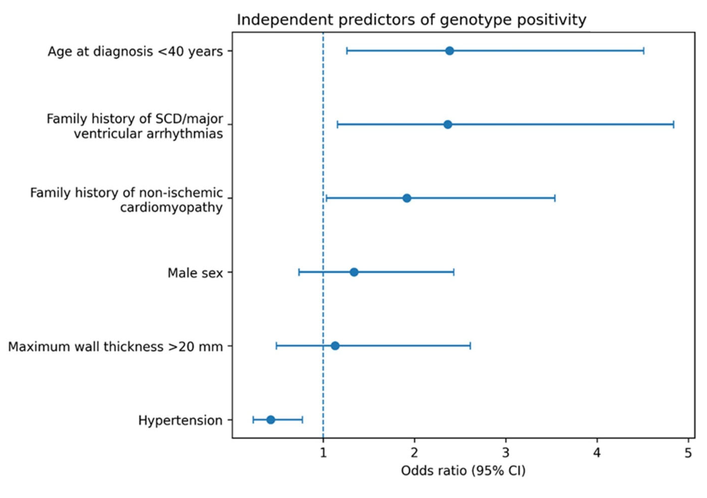
| OR | 95%CI | p-Value | |
|---|---|---|---|
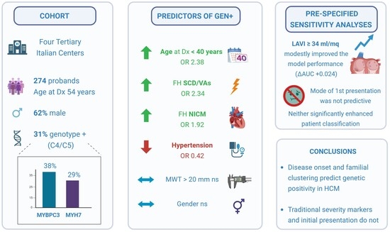
| Age < 40 years at diagnosis | 2.38 | 1.26–4.51 | 0.008 |
| FH of SCD/major VAs | 2.34 | 1.16–4.84 | 0.019 |
| FH of NICM | 1.92 | 1.04–3.54 | 0.038 |
| Hypertension | 0.42 | 0.23–0.77 | 0.005 |
| LV MWT > 20 mm | 1.13 | 0.49–2.61 | 0.776 |
| Male gender | 1.34 | 0.74–2.43 | 0.340 |
Disclaimer/Publisher’s Note: The statements, opinions and data contained in all publications are solely those of the individual author(s) and contributor(s) and not of MDPI and/or the editor(s). MDPI and/or the editor(s) disclaim responsibility for any injury to people or property resulting from any ideas, methods, instructions or products referred to in the content. |
© 2026 by the authors. Licensee MDPI, Basel, Switzerland. This article is an open access article distributed under the terms and conditions of the Creative Commons Attribution (CC BY) license.
Share and Cite
Angelini, F.; Dusi, V.; Feneziani, A.M.; Manai, R.; Bianco, M.; Lonni, E.; Brach Del Prever, G.M.; Bocchino, P.P.; Giannino, G.; Melis, D.; et al. Beyond Wall Thickness: Clinical Predictors of Genotype Positivity in Hypertrophic Cardiomyopathy. Cardiogenetics 2026, 16, 10. https://doi.org/10.3390/cardiogenetics16020010
Angelini F, Dusi V, Feneziani AM, Manai R, Bianco M, Lonni E, Brach Del Prever GM, Bocchino PP, Giannino G, Melis D, et al. Beyond Wall Thickness: Clinical Predictors of Genotype Positivity in Hypertrophic Cardiomyopathy. Cardiogenetics. 2026; 16(2):10. https://doi.org/10.3390/cardiogenetics16020010
Chicago/Turabian StyleAngelini, Filippo, Veronica Dusi, Amedeo Maria Feneziani, Rossella Manai, Matteo Bianco, Enrica Lonni, Giulia Margherita Brach Del Prever, Pier Paolo Bocchino, Giuseppe Giannino, Daniele Melis, and et al. 2026. "Beyond Wall Thickness: Clinical Predictors of Genotype Positivity in Hypertrophic Cardiomyopathy" Cardiogenetics 16, no. 2: 10. https://doi.org/10.3390/cardiogenetics16020010
APA StyleAngelini, F., Dusi, V., Feneziani, A. M., Manai, R., Bianco, M., Lonni, E., Brach Del Prever, G. M., Bocchino, P. P., Giannino, G., Melis, D., Gobello, G., Ravera, F., Laiso, L. E., Juvenal, F., Gallone, G., Pidello, S., Mabritto, B., Giachino, D., Musumeci, G., ... Raineri, C. (2026). Beyond Wall Thickness: Clinical Predictors of Genotype Positivity in Hypertrophic Cardiomyopathy. Cardiogenetics, 16(2), 10. https://doi.org/10.3390/cardiogenetics16020010