


AI-Enabled Smart Monitoring of Bovine Embryo Development Using Time-Lapse Imaging and Transfer Learning


Abstract
1. Introduction
Objectives
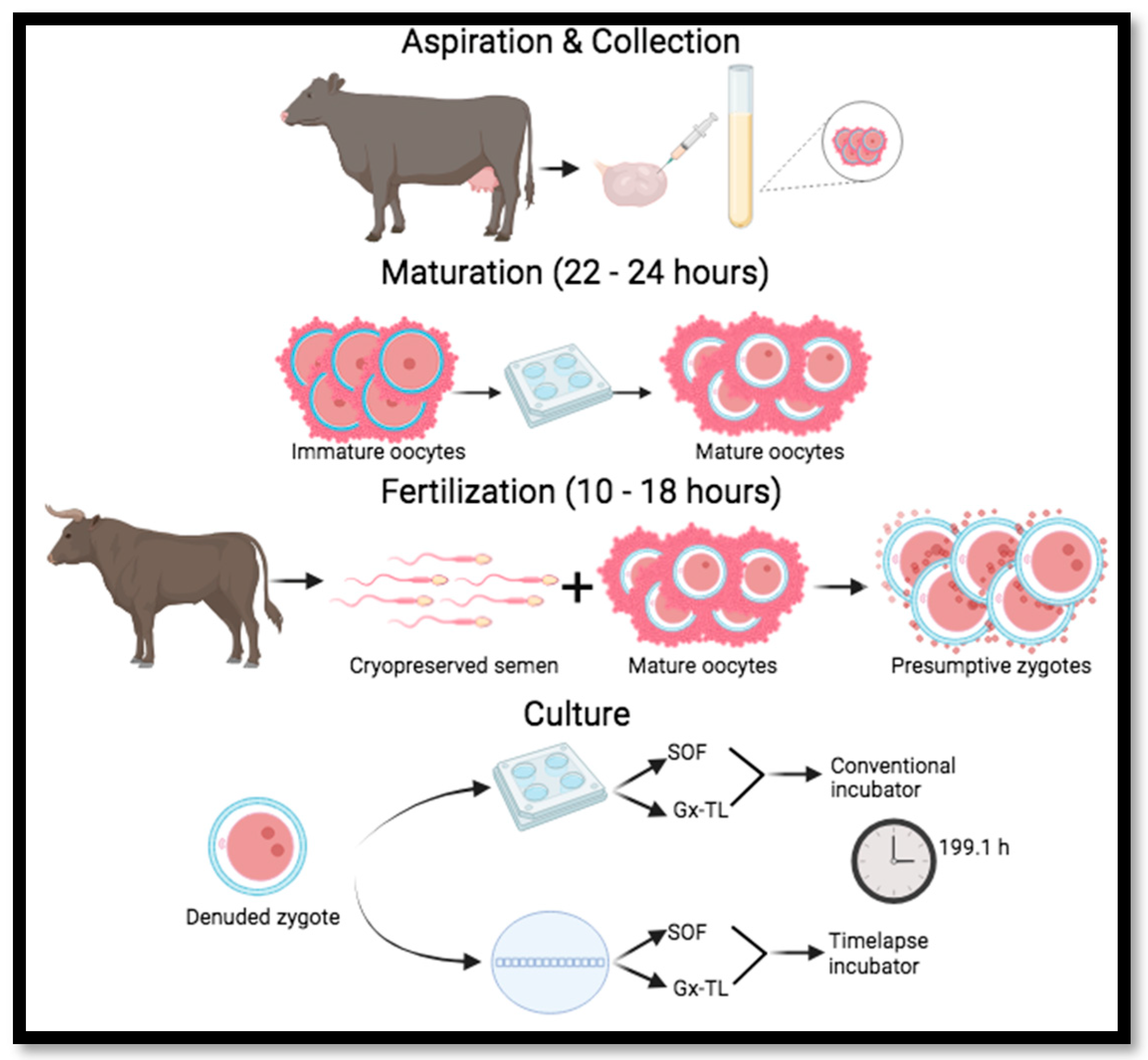
- To develop and evaluate an AI-enabled smart monitoring system for assessing the developmental kinetics and stage-wise progression of bovine embryos cultured in SOF and Vitrolife Gx-TL™ media using MIRI time-lapse imaging, based on the time required and the proportion of embryos reaching each of the nine defined developmental stages;
- To evaluate the performance of transfer learning-based deep convolutional neural networks as an intelligent image analysis component of the smart monitoring system for the automated identification of bovine embryonic developmental stages from image frames extracted from MIRI time-lapse videos.
2. Materials and Methods
2.1. Ovary Collection
2.2. Oocyte Collection and In Vitro Maturation
2.2.1. Media Preparation
2.2.2. Aspiration Set Up
2.2.3. Oocyte Collection
2.2.4. Coin Culture Degassing
2.3. Fertilization
2.3.1. Media Preparation
2.3.2. Swim-Up
2.3.3. SOF and Gx-TL™ Dish Preparation
2.3.4. Coin Culture Plate Preparation
2.3.5. In Vitro Culture
2.4. Development of Deep Learning Classification Models
2.4.1. Developmental Stage Timing and Frame Extraction from Time-Lapse Video
2.4.2. Image Augmentation
2.4.3. Transfer Learning-Based CNN Models and Classification Approaches
- Two-Class Model—Classifies embryos as either developing (positive) or non-developing (negative) at each stage;
- Nine-Class Model—Identifies developing embryos by their specific developmental stage, namely, T2, T3, T4, T5, T6, T7, T8, morula, or blastocyst;
- Ten-Class Model—Expands the nine-class model by including a non-developing embryo (NDE) class.
- Batch size, 16;
- Learning rate, 0.0001;
- Number of epochs, 10;
- Data split ratio (train:validation:test), 70:15:15.
3. Results
3.1. Effect of Culture Medium on Embryo Development
3.2. CNN-Based Classification
3.2.1. Two-Class Model
3.2.2. Nine-Class Model
3.2.3. Ten-Class Model
4. Discussion
4.1. Effect of Culture Medium on Embryo Development
4.2. CNN-Based Classification
4.2.1. Two-Class Model
4.2.2. Nine-Class Model
4.2.3. Ten-Class Model
4.3. Practical and Research Implications
5. Current Limitations and Future Work
6. Conclusions
Author Contributions
Funding
Informed Consent Statement
Data Availability Statement
Conflicts of Interest
References
- Hasler, J.F. Forty years of embryo transfer in cattle: A review focusing on the journal Theriogenology, the growth of the industry in North America, and personal reminisces. Theriogenology 2014, 81, 152–169. [Google Scholar] [CrossRef] [Scilit]
- Hansen, P.J. Review: Some challenges and unrealized opportunities toward widespread use of the in vitro-produced embryo in cattle production. Animal 2023, 17, 100745. [Google Scholar] [CrossRef] [Scilit]
- Kaniyamattam, K.; Block, J.; Hansen, P.J.; De Vries, A. Economic and genetic performance of various combinations of in vitro-produced embryo transfers and artificial insemination in a dairy herd. J. Dairy Sci. 2018, 101, 1540–1553. [Google Scholar] [CrossRef] [Scilit]
- Mandawala, A.A.; Harvey, S.C.; Roy, T.K.; Fowler, K.E. Time-lapse embryo imaging and morphokinetic profiling: Towards a general characterisation of embryogenesis. Anim. Reprod. Sci. 2016, 174, 2–10. [Google Scholar] [CrossRef] [Scilit]
- Magata, F. Time-lapse monitoring technologies for the selection of bovine in vitro fertilized embryos with high implantation potential. J. Reprod. Dev. 2023, 69, 57–64. [Google Scholar] [CrossRef] [Scilit]
- Kim, H.M.; Kang, H.; Lee, C.; Park, J.H.; Chung, M.K.; Kim, M.; Kim, N.Y.; Lee, H.J. Evaluation of the clinical efficacy and trust in AI-Assisted embryo ranking: Survey-based Prospective Study. J. Med. Internet Res. 2024, 26, e52637. [Google Scholar] [CrossRef] [Scilit]
- Cimadomo, D.; Sosa Fernandez, L.; Soscia, D.; Fabozzi, G.; Benini, F.; Cesana, A.; Dal Canto, M.B.; Maggiulli, R.; Muzzì, S.; Scarica, C.; et al. Inter-centre reliability in embryo grading across several IVF clinics is limited: Implications for embryo selection. Reprod. Biomed. Online 2022, 44, 39–48. [Google Scholar] [CrossRef] [Scilit]
- Farin, P.W.; Britt, J.H.; Shaw, D.W.; Slenning, B.D. Agreement among evaluators of bovine embryos produced in vivo or in vitro. Theriogenology 1995, 44, 339–349. [Google Scholar] [CrossRef] [Scilit]
- Shivaani, M.; Madan, P. Application of imaging and spectroscopy techniques for grading of bovine embryos-a review. Front. Vet. Sci. 2024, 11, 1364570. [Google Scholar] [CrossRef] [Scilit]
- Li, X.X.; Cao, P.H.; Han, W.X.; Xu, Y.K.; Wu, H.; Yu, X.L.; Chen, J.Y.; Zhang, F.; Li, Y.H. Non-invasive metabolomic profiling of culture media of ICSI-and IVF-derived early developmental cattle embryos via Raman spectroscopy. Anim. Reprod. Sci. 2018, 196, 99–110. [Google Scholar] [CrossRef] [Scilit]
- Jimenez, L.E.; Juárez, A.C.; Roldán-Olarte, M.; Álvarez, R.M.S. Biochemical changes in the cytoplasm of bovine oocytes during the in vitro maturation process: A Raman microscopy study. Vet. Res. Commun. 2022, 46, 549–562. [Google Scholar] [CrossRef] [Scilit]
- Wiecheć, A.; Opiela, J.; Lipiec, E.; Kwiatek, W.M. Utility of FT-IR imaging spectroscopy in estimating differences between the quality of bovine blastocysts. J. Mol. Struct. 2013, 1049, 227–232. [Google Scholar] [CrossRef] [Scilit]
- De Souza Ciniciato, D.; Takahashi, M.B.; Nogueira, M.F.G.; Rocha, J.C. Potential Use of Smartphone as a Tool to Capture Embryo Digital Images from Stereomicroscope and to Evaluate Them by an Artificial Neural Network. In Proceedings of the International Conference on Computer-Human Interaction Research and Applications (CHIRA 2017); Science and Technology Publications: Setúbal, Portugal, 2017; pp. 185–189. [Google Scholar]
- Rocha, J.C.; Passalia, F.J.; Matos, F.D.; Takahashi, M.B.; Ciniciato, D.D.S.; Maserati, M.P.; Alves, M.F.; De Almeida, T.G.; Cardoso, B.L.; Basso, A.C.; et al. A method based on artificial intelligence to fully automatize the evaluation of bovine blastocyst images. Sci. Rep. 2017, 7, 7659. [Google Scholar] [CrossRef] [Scilit]
- Matos, F.D.; Nogueira, M.F.G.; Rocha, J.C. Artificial intelligence meets the same challenges as humans in morphological classification of bovine blastocysts. Abstract of proceedings of the 28th annual meeting of the embryo technology society (SBTE). A209 supporting biotechnologies: Cryopreservation and cryobiology, image analysis and diagnosis, molecular biology and “omics”. Anim. Reprod. 2014, 11, 489. [Google Scholar]
- Thompson, J. Quantitative Image Assessment of Embryos to Predict Pregnancy in Embryo Transfer Programs. Final Report Submitted to Meat & Livestock Australia for the Project “P.PSH0969”. 2018. Available online: https://www.mla.com.au/research-and-development/reports/2019/quantitative-image-assessment-of-embryos-to-predict-pregnancy-in-embryo-transfer-programs/ (accessed on 15 March 2026).
- Monteiro, C.A.S.; Chow, D.J.; Leal, G.R.; Tan, T.C.; Ferreira, A.M.R.; Thompson, J.G.; Dunning, K.R. Optical imaging of cleavage stage bovine embryos using hyperspectral and confocal approaches reveals metabolic differences between on-time and fast-developing embryos. Theriogenology 2021, 159, 60–68. [Google Scholar] [CrossRef] [Scilit]
- Bhide, P.; Chan, D.Y.; Lanz, D.; Alqawasmeh, O.; Barry, E.; Baxter, D.; Gonzalez Carreras, F.; Choudhury, Y.; Cheong, Y.; Chung, J.P.; et al. Clinical effectiveness and safety of time-lapse imaging systems for embryo incubation and selection in in-vitro fertilisation treatment (TILT): A multicentre, three-parallel-group, double-blind, randomised controlled trial. Lancet 2024, 404, 256–265. [Google Scholar] [CrossRef] [Scilit]
- Zhang, X.-D.; Zhang, Q.; Han, W.; Liu, W.-W.; Shen, X.-L.; Yao, G.-D.; Shi, S.-L.; Hu, L.-L.; Wang, S.-S.; Wang, J.-X.; et al. Comparison of embryo implantation potential between time-lapse incubators and standard incubators: A randomized controlled study. Reprod. Biomed. Online 2022, 45, 858–866. [Google Scholar] [CrossRef] [Scilit]
- Donnay, I.; Van Langendonckt, A.; Auquier, P.; Grisart, B.; Vansteenbrugge, A.; Massip, A.; Dessy, F. Effects of co-culture and embryo number on the in vitro development of bovine embryos. Theriogenology 1997, 47, 1549–1561. [Google Scholar] [CrossRef] [Scilit]
- Keefer, C.L.; Stice, S.L.; Paprocki, A.M.; Golueke, P. In vitro culture of bovine IVM-IVF embryos: Cooperative interaction among embryos and the role of growth factors. Theriogenology 1994, 41, 1323–1331. [Google Scholar] [CrossRef] [Scilit]
- Ferry, L.; Mermillod, P.; Massip, A.; Dessy, F. Bovine embryos cultured in serum-poor oviduct-conditioned medium need cooperation to reach the blastocyst stage. Theriogenology 1994, 42, 445–453. [Google Scholar] [CrossRef] [Scilit]
- Khurana, N.K.; Niemann, H. Effects of oocyte quality, oxygen tension, embryo density, cumulus cells and energy substrates on cleavage and morula/blastocyst formation of bovine embryos. Theriogenology 2000, 54, 741–756. [Google Scholar] [CrossRef] [Scilit]
- John, E.S. In Vitro Production of Bovine Embryos; Lab Manual; Department of Biomedical Science, Ontario Veterinary College, University of Guelph: Guelph, ON, Canada, 2024. [Google Scholar]
- Somfai, T.; Inba, Y.; Aikawa, Y.; Ohtake, M.; Kobayashi, S.; Konishi, K.; Imai, K. Relationship between the length of cell cycles, cleavage pattern and developmental competence in bovine embryos generated by in vitro fertilization or parthenogenesis. J. Reprod. Dev. 2010, 56, 200–207. [Google Scholar] [CrossRef] [Scilit]
- Sugimura, S.; Akai, T.; Imai, K. Selection of viable in vitro-fertilized bovine embryos using time-lapse monitoring in Microwell culture dishes. J. Reprod. Dev. 2017, 63, 353–357. [Google Scholar] [CrossRef] [Scilit]
- Hillyear, L.M.; Zak, L.J.; Beckitt, T.; Griffin, D.K.; Harvey, S.C.; Harvey, K.E. Morphokinetic Profiling Suggests That Rapid First Cleavage Division Accurately Predicts the Chances of Blastulation in Pig In Vitro Produced Embryos. Animals 2024, 14, 783. [Google Scholar] [CrossRef] [Scilit]
- Liu, H.-J.; Oh, S.J.W.Y.; Tay, N.L.; Lim, C.Y.; Hsu, C.-D.; Chua, D.H.H.; Teo, W.K.L.; Loh, Y.-H.; Ng, S.C. Morphokinetic Analyses of Fishing Cat–Domestic Cat Interspecies Somatic Cell Nuclear Transfer Embryos Through A Time-Lapse System. Animals 2025, 15, 148. [Google Scholar] [CrossRef] [Scilit]
- Liu, X. Comparison of four convolutional neural network-based algorithms for Sports Image Classification. In 2023 International Conference on Data Science, Advanced Algorithm and Intelligent Computing; Advances in Intelligent Systems Research; Atlantis Press: Paris, France, 2024; pp. 178–186. [Google Scholar] [CrossRef] [Scilit]
- Sacks, G.C.; Mozes, H.; Ronn, R.; Elder-Geva, T.; Schonberger, O.; Ben-Ami, I.; Srebnik, N. Time-Lapse Incubation for Embryo Culture—Morphokinetics and Environmental Stability May Not Be Enough: Results from a Pilot Randomized Controlled Trial. J. Clin. Med. 2024, 13, 1701. [Google Scholar] [CrossRef] [Scilit]
- Hachani, Y.; Bouthemy, P.; Fromont, E.; Ruffini, S.; Laffont, L.; de Paula Reis, A. Supervised Contrastive Learning for Cell Stage Classification of Animal Embryos. Sci. Rep. 2026, 16, 20290. [Google Scholar] [CrossRef] [Scilit]
- Wells, C.; Hayden, C.; Rea, M.; Cook, B.; Killingsworth, R. Advancing Bovine Embryo Evaluation: Machine Learning to Assess Embryos in Routine Embryo Transfer Practice. J. IVF-Worldw. 2025, 3, 45–57. [Google Scholar] [CrossRef] [Scilit]
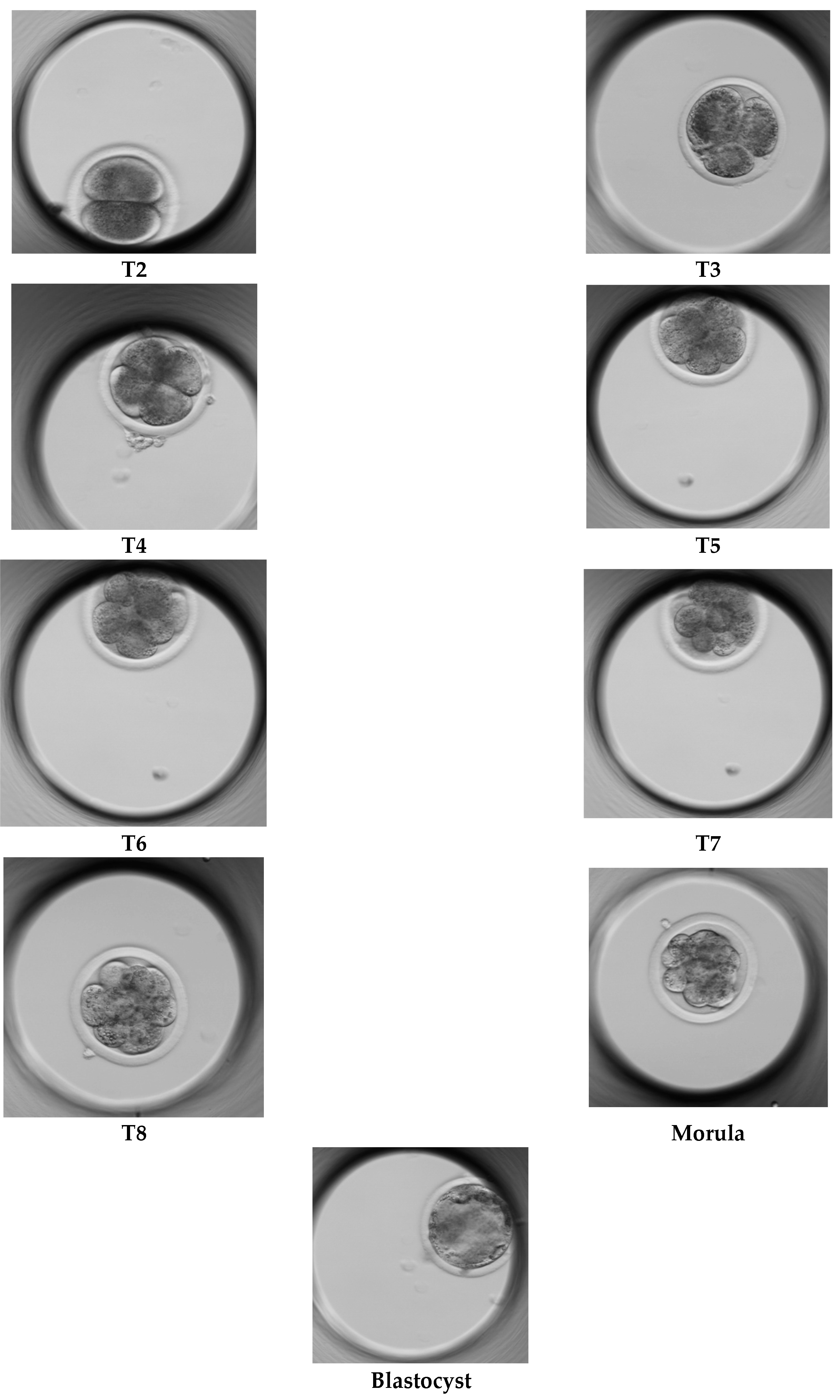
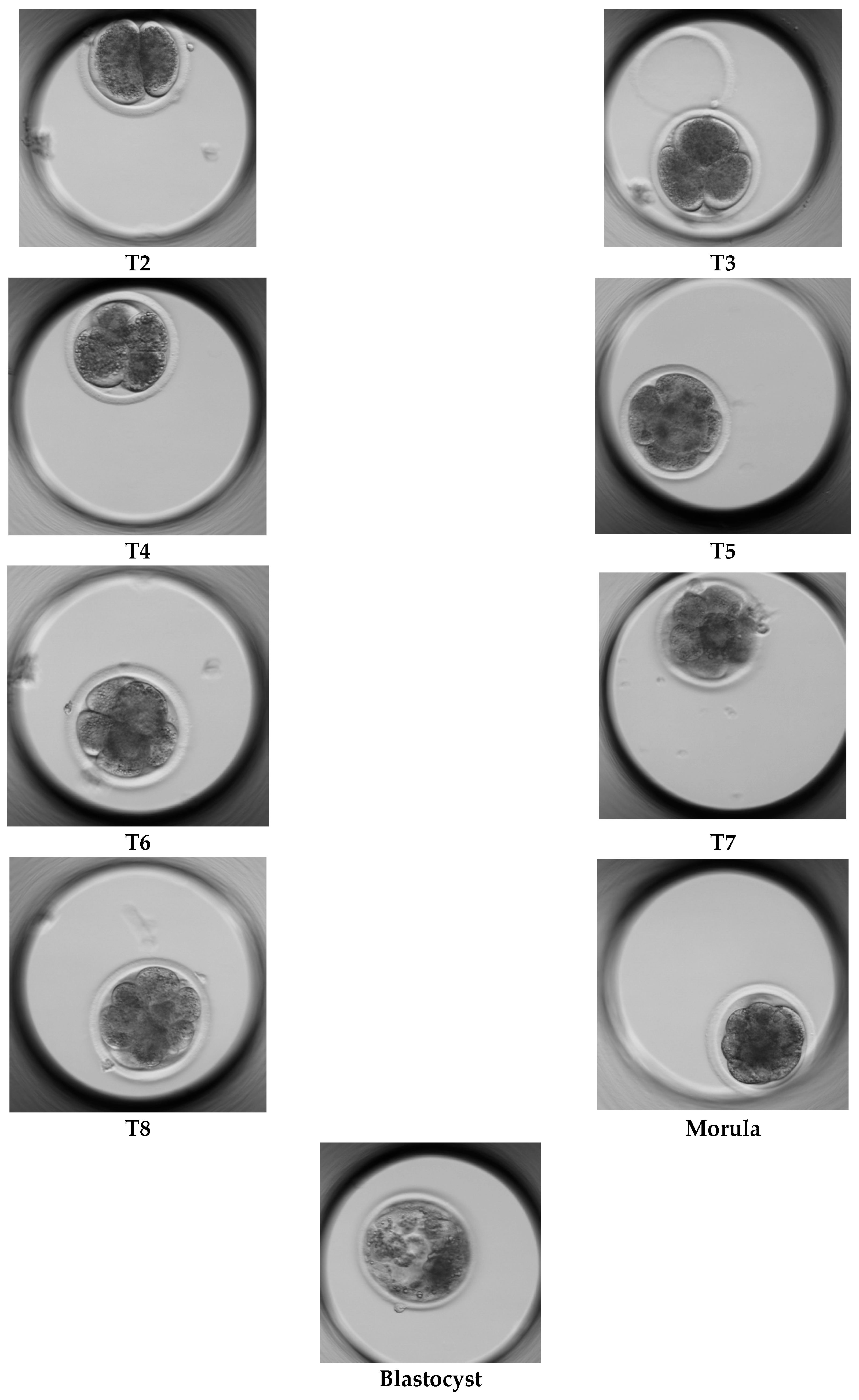

| Culture Medium | No. of Single Cultured Zygotes | Developmental Stages Reached and Clearly Identified in Images | ||||||||
|---|---|---|---|---|---|---|---|---|---|---|
| T2 | T3 | T4 | T5 | T6 | T7 | T8 | Morula | Blastocyst | ||
| SOF | 152 | 81 | 43 | 42 | 29 | 27 | 11 | 14 | 10 | 6 |
| Gx-TL™ | 159 | 77 | 43 | 40 | 28 | 33 | 9 | 8 | 18 | 10 |
| Culture Medium | Mean developmental Time (h) | ||||||||
|---|---|---|---|---|---|---|---|---|---|
| T2 | T3 | T4 | T5 | T6 | T7 | T8 | Morula | Blastocyst | |
| SOF | 44.50± 19.66 | 54.92± 21.23 | 56.02± 27.62 | 59.52± 23.43 | 57.25± 14.25 | 69.53± 25.00 | 86.77± 26.91 | 121.82± 11.01 | 190.71± 4.06 |
| Gx-TL™ | 43.00± 12.40 | 52.05± 10.21 | 58.81± 21.17 | 62.72± 19.74 | 70.06± 24.70 | 84.15± 21.59 | 97.64± 25.70 | 135.69± 12.53 | 179.20± 15.71 |
| Culture Medium | Mean Developmental Time (h) | ||||||||
|---|---|---|---|---|---|---|---|---|---|
| T2 | T3 | T4 | T5 | T6 | T7 | T8 | Morula | Blastocyst | |
| SOF | 33.76± 6.45 | 45.43± 3.74 | 43.94± 8.01 | 56.63± 2.85 | 56.10± 6.08 | 61.72± 3.65 | 99.98± 7.22 | 120.39± 8.41 | 190.71± 4.06 |
| Gx-TL™ | 36.48± 5.63 | 46.29± 7.98 | 50.59± 7.00 | 57.09± 10.45 | 64.85± 18.30 | 83.93± 21.34 | 104.88± 15.02 | 132.37± 12.57 | 179.20± 15.71 |
| Developmental Stage | Average Inference Time/Image (Sec) | Total Training + Evaluation Time (Sec) | Classification Accuracy (%) | ||||||
|---|---|---|---|---|---|---|---|---|---|
| Res. * | Dense. | Eff. | Res. | Dense. | Eff. | Res. | Dense. | Eff. | |
| T2 | 1.133 | 1.460 | 1.141 | 170.0 | 219.1 | 171.2 | 100 | 100 | 100 |
| T3 | 1.170 | 1.378 | 1.150 | 175.6 | 206.8 | 172.6 | 100 | 100 | 100 |
| T4 | 1.132 | 1.375 | 1.153 | 169.9 | 206.3 | 173.0 | 100 | 100 | 100 |
| T5 | 1.121 | 1.354 | 1.123 | 168.1 | 203.2 | 168.4 | 100 | 100 | 100 |
| T6 | 1.147 | 1.373 | 1.129 | 172.1 | 205.9 | 169.4 | 100 | 100 | 100 |
| T7 | 1.115 | 1.352 | 1.181 | 167.3 | 202.8 | 177.2 | 100 | 100 | 100 |
| T8 | 1.169 | 1.361 | 1.167 | 175.4 | 204.1 | 175.1 | 100 | 100 | 100 |
| Morula | 1.183 | 1.335 | 1.122 | 177.5 | 200.3 | 168.3 | 100 | 100 | 100 |
| Blastocyst | 1.248 | 1.369 | 1.163 | 187.2 | 205.4 | 174.5 | 100 | 100 | 100 |
| Developmental Stage | Average Inference Time/Image (S) | Total Training + Evaluation Time (S) | Classification Accuracy (%) | ||||||
|---|---|---|---|---|---|---|---|---|---|
| Res. * | Dense. | Eff. | Res. | Dense. | Eff. | Res. | Dense. | Eff. | |
| T2 | 1.155 | 1.394 | 1.140 | 173.2 | 209.1 | 171.1 | 100 | 100 | 100 |
| T3 | 1.228 | 1.387 | 1.141 | 184.2 | 208.1 | 171.2 | 100 | 100 | 100 |
| T4 | 1.091 | 1.362 | 1.220 | 163.7 | 204.3 | 183.0 | 100 | 100 | 100 |
| T5 | 1.197 | 1.375 | 1.137 | 179.6 | 206.4 | 170.6 | 100 | 100 | 100 |
| T6 | 1.148 | 1.353 | 1.182 | 172.2 | 203.0 | 177.4 | 100 | 100 | 100 |
| T7 | 1.171 | 1.359 | 1.119 | 175.7 | 203.9 | 167.8 | 100 | 100 | 100 |
| T8 | 1.238 | 1.377 | 1.153 | 185.5 | 206.5 | 172.9 | 100 | 100 | 100 |
| Morula | 1.161 | 1.376 | 1.117 | 174.1 | 206.4 | 167.6 | 100 | 100 | 100 |
| Blastocyst | 1.150 | 1.377 | 1.193 | 172.5 | 206.6 | 179.0 | 100 | 100 | 100 |
| Developmental Stage | Average Inference Time/Image (S) | Total Training + Evaluation Time (S) | Classification Accuracy (%) | ||||||
|---|---|---|---|---|---|---|---|---|---|
| Res. * | Dense. | Eff. | Res. | Dense. | Eff. | Res. | Dense. | Eff. | |
| T2 | 1.013 | 1.303 | 1.166 | 303.9 | 391.1 | 349.9 | 100 | 100 | 100 |
| T3 | 0.997 | 1.359 | 1.117 | 299.1 | 407.9 | 335.3 | 100 | 100 | 100 |
| T4 | 0.990 | 1.390 | 1.124 | 297.1 | 417.1 | 337.2 | 100 | 100 | 100 |
| T5 | 1.162 | 1.402 | 1.127 | 348.8 | 420.8 | 338.1 | 100 | 100 | 100 |
| T6 | 1.315 | 1.425 | 1.155 | 394.7 | 427.5 | 346.6 | 100 | 100 | 100 |
| T7 | 0.985 | 1.438 | 1.103 | 295.7 | 431.4 | 330.9 | 100 | 100 | 100 |
| T8 | 0.990 | 1.369 | 1.120 | 297.0 | 410.9 | 336.2 | 100 | 100 | 100 |
| Morula | 0.990 | 1.484 | 1.107 | 297.2 | 445.3 | 332.1 | 100 | 100 | 100 |
| Blastocyst | 0.987 | 1.476 | 1.102 | 296.1 | 443.0 | 330.7 | 100 | 100 | 100 |
| Developmental Stage | Average Inference Time/Image (S) | Total Evaluation Time (S) | Total Training Time (S) | Classification Accuracy (%) | ||||||||
|---|---|---|---|---|---|---|---|---|---|---|---|---|
| Res. * | Dense. | Eff. | Res. | Dense. | Eff. | Res. | Dense. | Eff. | Res. | Dense. | Eff. | |
| T2 | 0.021 | 0.019 | 0.020 | 14.19 | 13.41 | 13.73 | 766.0 | 1022 | 768.0 | 100 | 100 | 100 |
| T3 | 100 | 100 | 100 | |||||||||
| T4 | 100 | 100 | 100 | |||||||||
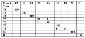
| T5 | 98 | 98 | 100 | |||||||||
| T6 | 98 | 98 | 96 | |||||||||
| T7 | 100 | 97 | 98 | |||||||||
| T8 | 94 | 98 | 94 | |||||||||
| Morula | 100 | 100 | 100 | |||||||||
| Blastocyst | 100 | 100 | 100 | |||||||||
| Overall accuracy | 99.1 | 99.2 | 98.8 | |||||||||
 | 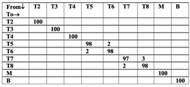 | 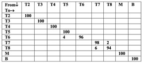 |
| ResNet18 | DenseNet121 | EfficientNet B0 |
| Developmental Stage | Average Inference Time/Image (S) | Total Evaluation Time (S) | Total Training Time (S) | Classification Accuracy (%) | ||||||||
|---|---|---|---|---|---|---|---|---|---|---|---|---|
| Res. * | Dense. | Eff. | Res. | Dense. | Eff. | Res. | Dense. | Eff. | Res. | Dense. | Eff. | |
| T2 | 0.019 | 0.022 | 0.020 | 13.10 | 15.07 | 13.54 | 727.8 | 1687 | 747.3 | 100 | 100 | 96 |
| T3 | 100 | 100 | 98 | |||||||||
| T4 | 100 | 100 | 98 | |||||||||
| T5 | 100 | 100 | 93 | |||||||||
| T6 | 100 | 100 | 98 | |||||||||
| T7 | 100 | 100 | 98 | |||||||||
| T8 | 100 | 100 | 97 | |||||||||
| Morula | 100 | 100 | 98 | |||||||||
| Blastocyst | 100 | 100 | 100 | |||||||||
| Overall accuracy | 100 | 100 | 97.7 | |||||||||
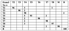 |
| EfficientNet B0 |
| Developmental Stage | Average Inference Time/Image (S) | Total Evaluation Time (S) | Total Training Time (S) | Classification Accuracy (%) | ||||||||
|---|---|---|---|---|---|---|---|---|---|---|---|---|
| Res. * | Dense. | Eff. | Res. | Dense. | Eff. | Res. | Dense. | Eff. | Res. | Dense. | Eff. | |
| T2 | 0.017 | 0.022 | 0.019 | 24.14 | 30.18 | 26.46 | 1487 | 2012 | 1559 | 100 | 100 | 98 |
| T3 | 100 | 98 | 99 | |||||||||
| T4 | 100 | 100 | 98 | |||||||||
| T5 | 100 | 80 | 95 | |||||||||
| T6 | 99 | 99 | 97 | |||||||||
| T7 | 99 | 98 | 98 | |||||||||
| T8 | 98 | 85 | 96 | |||||||||
| Morula | 100 | 100 | 99 | |||||||||
| Blastocyst | 100 | 100 | 100 | |||||||||
| Overall accuracy | 99.5 | 95.5 | 97.7 | |||||||||
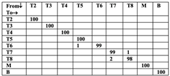 | 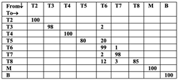 | 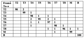 |
| ResNet18 | DenseNet121 | EfficientNet B0 |
| Developmental Stage | Average Inference Time/Image (S) | Total Evaluation Time (S) | Total Training Time (S) | Classification Accuracy (%) | ||||||||
|---|---|---|---|---|---|---|---|---|---|---|---|---|
| Res. * | Dense. | Eff. | Res. | Dense. | Eff. | Res. | Dense. | Eff. | Res. | Dense. | Eff. | |
| T2 | 0.020 | 0.019 | 0.021 | 15.62 | 14.39 | 15.87 | 802.4 | 1028 | 861.7 | 100 | 100 | 100 |
| T3 | 100 | 100 | 100 | |||||||||
| T4 | 100 | 100 | 100 | |||||||||
| T5 | 100 | 100 | 100 | |||||||||
| T6 | 96 | 96 | 96 | |||||||||
| T7 | 98 | 98 | 96 | |||||||||
| T8 | 100 | 100 | 100 | |||||||||
| Morula | 93 | 93 | 93 | |||||||||
| Blastocyst | 100 | 100 | 100 | |||||||||
| NDE ** | 100 | 100 | 98 | |||||||||
| Overall accuracy | 98.8 | 98.8 | 98.4 | |||||||||
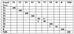 |  | 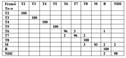 |
| ResNet18 | DenseNet121 | EfficientNet B0 |
| Developmental Stage | Average Inference Time/Image (S) | Total Evaluation Time (S) | Total Training Time (S) | Classification Accuracy (%) | ||||||||
|---|---|---|---|---|---|---|---|---|---|---|---|---|
| Res. * | Dense. | Eff. | Res. | Dense. | Eff. | Res. | Dense. | Eff. | Res. | Dense. | Eff. | |
| T2 | 0.020 | 0.020 | 0.020 | 15.32 | 15.40 | 15.66 | 784.5 | 1026 | 815.5 | 100 | 100 | 100 |
| T3 | 100 | 100 | 100 | |||||||||
| T4 | 100 | 100 | 100 | |||||||||
| T5 | 100 | 100 | 100 | |||||||||
| T6 | 100 | 100 | 100 | |||||||||
| T7 | 100 | 100 | 100 | |||||||||
| T8 | 100 | 100 | 100 | |||||||||
| Morula | 100 | 100 | 100 | |||||||||
| Blastocyst | 100 | 100 | 100 | |||||||||
| NDE ** | 100 | 100 | 100 | |||||||||
| Overall accuracy | 100 | 100 | 100 | |||||||||
| Developmental Stage | Average Inference Time/Image (S) | Total Evaluation Time (S) | Total Training Time (S) | Classification Accuracy (%) | ||||||||
|---|---|---|---|---|---|---|---|---|---|---|---|---|
| Res. * | Dense. | Eff. | Res. | Dense. | Eff. | Res. | Dense. | Eff. | Res. | Dense. | Eff. | |
| T2 | 0.018 | 0.020 | 0.020 | 27.47 | 31.20 | 30.05 | 2019 | 2207 | 1721 | 100 | 100 | 100 |
| T3 | 100 | 100 | 100 | |||||||||
| T4 | 100 | 100 | 100 | |||||||||
| T5 | 100 | 100 | 100 | |||||||||
| T6 | 98 | 99 | 100 | |||||||||
| T7 | 100 | 100 | 100 | |||||||||
| T8 | 98 | 100 | 100 | |||||||||
| Morula | 100 | 99 | 99 | |||||||||
| Blastocyst | 100 | 100 | 100 | |||||||||
| NDE ** | 100 | 100 | 100 | |||||||||
| Overall accuracy | 99.7 | 99.8 | 99.9 | |||||||||
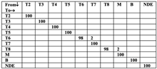 | 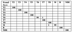 | 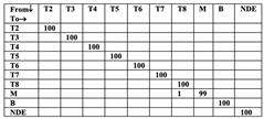 |
| ResNet18 | DenseNet121 | EfficientNet B0 |
Disclaimer/Publisher’s Note: The statements, opinions and data contained in all publications are solely those of the individual author(s) and contributor(s) and not of MDPI and/or the editor(s). MDPI and/or the editor(s) disclaim responsibility for any injury to people or property resulting from any ideas, methods, instructions or products referred to in the content. |
© 2026 by the authors. Licensee MDPI, Basel, Switzerland. This article is an open access article distributed under the terms and conditions of the Creative Commons Attribution (CC BY) license.
Share and Cite
Shivaani, M.; P.L, M.; Madan, P. AI-Enabled Smart Monitoring of Bovine Embryo Development Using Time-Lapse Imaging and Transfer Learning. Future Internet 2026, 18, 476. https://doi.org/10.3390/fi18090476
Shivaani M, P.L M, Madan P. AI-Enabled Smart Monitoring of Bovine Embryo Development Using Time-Lapse Imaging and Transfer Learning. Future Internet. 2026; 18(9):476. https://doi.org/10.3390/fi18090476
Chicago/Turabian StyleShivaani, Manickavasagan, Meenakshi P.L, and Pavneesh Madan. 2026. "AI-Enabled Smart Monitoring of Bovine Embryo Development Using Time-Lapse Imaging and Transfer Learning" Future Internet 18, no. 9: 476. https://doi.org/10.3390/fi18090476
APA StyleShivaani, M., P.L, M., & Madan, P. (2026). AI-Enabled Smart Monitoring of Bovine Embryo Development Using Time-Lapse Imaging and Transfer Learning. Future Internet, 18(9), 476. https://doi.org/10.3390/fi18090476

