


Xenon Encapsulation in Liposomes, Nanobubbles, and Microbubbles: Delivery Strategies, Preclinical Evidence, and Translational Barriers
 and
and
Abstract
1. Introduction
2. Targeted Xenon Delivery
3. Delivery Forms of Active Substances
4. Key Effects of Targeted Xenon Delivery Based on Preclinical Studies
4.1. Cerebral Ischemia–Reperfusion: A Basic Model for Xe-ELIP and Liposomal Development
4.2. The Use of Xenon in the Model of TBI
5. Targeted Xenon Delivery in Models of Renal and Myocardial Injury
6. Safety and Translational Barriers of Xenon-Loaded Carriers
7. Clinical Limitations and Translational Barriers of Xenon-Loaded Carrier Systems
8. Conclusions
Supplementary Materials
Author Contributions
Funding
Institutional Review Board Statement
Informed Consent Statement
Data Availability Statement
Conflicts of Interest
References
- Arola, O.J.; Laitio, R.M.; Roine, R.O.; Grönlund, J.; Saraste, A.; Pietilä, M.; Airaksinen, J.; Perttilä, J.; Scheinin, H.; Olkkola, K.T.; et al. Feasibility and Cardiac Safety of Inhaled Xenon in Combination with Therapeutic Hypothermia Following Out-of-Hospital Cardiac Arrest. Crit. Care Med. 2013, 41, 2116–2124. [Google Scholar] [CrossRef] [Scilit] [PubMed]
- Laitio, R.; Hynninen, M.; Arola, O.; Virtanen, S.; Parkkola, R.; Saunavaara, J.; Roine, R.O.; Grönlund, J.; Ylikoski, E.; Wennervirta, J.; et al. Effect of Inhaled Xenon on Cerebral White Matter Damage in Comatose Survivors of Out-of-Hospital Cardiac Arrest: A Randomized Clinical Trial. JAMA 2016, 315, 1120–1128. [Google Scholar] [CrossRef] [Scilit] [PubMed]
- Arola, O.; Saraste, A.; Laitio, R.; Airaksinen, J.; Hynninen, M.; Bäcklund, M.; Ylikoski, E.; Wennervirta, J.; Pietilä, M.; Roine, R.O.; et al. Inhaled Xenon Attenuates Myocardial Damage in Comatose Survivors of Out-of-Hospital Cardiac Arrest. J. Am. Coll. Cardiol. 2017, 70, 2652–2660. [Google Scholar] [CrossRef] [Scilit] [PubMed]
- Campos-Pires, R.; Onggradito, H.; Ujvari, E.; Karimi, S.; Valeo, F.; Aldhoun, J.; Edge, C.J.; Franks, N.P.; Dickinson, R. Xenon Treatment after Severe Traumatic Brain Injury Improves Locomotor Outcome, Reduces Acute Neuronal Loss and Enhances Early Beneficial Neuroinflammation: A Randomized, Blinded, Controlled Animal Study. Crit. Care 2020, 24, 667. [Google Scholar] [CrossRef] [Scilit] [PubMed]
- Homi, H.M.; Yokoo, N.; Ma, D.; Warner, D.S.; Franks, N.P.; Maze, M.; Grocott, H.P. The Neuroprotective Effect of Xenon Administration during Transient Middle Cerebral Artery Occlusion in Mice. Anesthesiology 2003, 99, 876–881. [Google Scholar] [CrossRef] [Scilit] [PubMed]
- Chen, Q.; Zhu, S.; Wu, M.; Sun, J.; Kimia, M.; Mudiyanselage, D.E.; Lee, H.; Ma, D. Noble Gases Xenon and Argon: From Cellular Signalling Mechanisms to Organoprotection and Clinical Applications. J. Transl. Med. 2026, 24, 513. [Google Scholar] [CrossRef] [Scilit] [PubMed]
- Campos-Pires, R.; Armstrong, S.P.; Sebastiani, A.; Luh, C.; Gruss, M.; Radyushkin, K.; Hirnet, T.; Werner, C.; Engelhard, K.; Franks, N.P.; et al. Xenon Improves Neurologic Outcome and Reduces Secondary Injury Following Trauma in an In Vivo Model of Traumatic Brain Injury. Crit. Care Med. 2015, 43, 149–158. [Google Scholar] [CrossRef] [Scilit] [PubMed]
- Veldeman, M.; Coburn, M.; Rossaint, R.; Clusmann, H.; Nolte, K.; Kremer, B.; Höllig, A. Xenon Reduces Neuronal Hippocampal Damage and Alters the Pattern of Microglial Activation after Experimental Subarachnoid Hemorrhage: A Randomized Controlled Animal Trial. Front. Neurol. 2017, 8, 511. [Google Scholar] [CrossRef] [Scilit] [PubMed]
- Krukov, I.A.; Ershov, A.V.; Lobanov, A.; Grebenchikov, O.A. The effect of xenon exposure duration on the volume of ischemic brain injury in experiment. Patol. Fiziol. Eksp. Ter. (Pathol. Physiol. Exp. Ther.) 2022, 64, 73–78. [Google Scholar] [CrossRef] [Scilit]
- Krukov, I.A.; Ershov, A.V.; Cherpakov, R.A.; Grebenchikov, O.A. Alleviation of neurological and cognitive impairments in rat model of ischemic stroke by 0.5 MAC xenon exposure. Bull. RSMU 2022, 3, 91–97. [Google Scholar] [CrossRef] [Scilit]
- Ershov, A.V.; Krukov, I.A.; Antonova, V.V.; Baeva, A.A. The Effect of Xenon on the Activity of Glycogen Synthase Kinase-Зβ in the Perifocal Zone of Ischemic Cerebral Infarction (Experimental Study). Obŝ. Reanimatol. 2023, 19, 60–67. [Google Scholar] [CrossRef] [Scilit]
- Grebenchikov, O.A.; Evseev, A.K.; Kulabuchov, V.V.; Kuzovlev, A.N.; Petrikov, S.S.; Ramazanov, G.R.; Khusainov, S.G.; Cherpakov, R.A.; Shabanov, A.K.; Spichko, A.I. Neuroprotective Effects of Inhaled Xenon for Sedation Compared with Propofol Intravenous Sedation in Severe Ischemic Stroke. Neotložnaâ Med. Pomoŝ. 2023, 11, 561–572. [Google Scholar] [CrossRef] [Scilit]
- Shpichko, A.I.; Kuzovlev, A.N.; Cherpakov, R.A.; Shpichko, N.P.; Grebenchikov, O.A.; Yevseyev, A.K.; Shabanov, A.K.; Petrikov, S.S. A New Strategy for the Treatment of Patients with Prolonged Impairment of Consciousness Using Xenon. Prospective Pilot Study. Russ. Sklifosovsky J. Emerg. Med. Care 2023, 11, 592–599. [Google Scholar] [CrossRef] [Scilit]
- Hofland, J.; Ouattara, A.; Fellahi, J.-L.; Gruenewald, M.; Hazebroucq, J.; Ecoffey, C.; Joseph, P.; Heringlake, M.; Steib, A.; Coburn, M.; et al. Effect of Xenon Anesthesia Compared to Sevoflurane and Total Intravenous Anesthesia for Coronary Artery Bypass Graft Surgery on Postoperative Cardiac Troponin Release: An International, Multicenter, Phase 3, Single-Blinded, Randomized Noninferiority Trial. Anesthesiology 2017, 127, 918–933. [Google Scholar] [CrossRef] [Scilit] [PubMed]
- Stoppe, C.; Rimek, A.; Rossaint, R.; Rex, S.; Stevanovic, A.; Schälte, G.; Fahlenkamp, A.; Czaplik, M.; Bruells, C.S.; Daviet, C.; et al. Xenon Consumption during General Surgery: A Retrospective Observational Study. Med. Gas Res. 2013, 3, 12. [Google Scholar] [CrossRef] [Scilit] [PubMed]
- Khaliullin, D.M.; Lazarev, V.V.; Shugailov, I.A.; Gracheva, E.S. Pharmacoeconomic Evaluation of Xenon Use in Pediatric Outpatient Dental Practice. Russ. J. Dent. 2025, 29, 224–230. [Google Scholar] [CrossRef] [Scilit]
- Klegerman, M.E.; Moody, M.R.; Hurling, J.R.; Peng, T.; Huang, S.; McPherson, D.D. Gas Chromatography/Mass Spectrometry Measurement of Xenon in Gas-loaded Liposomes for Neuroprotective Applications. Rapid Commun. Mass Spectrom. 2017, 31, 1–8. [Google Scholar] [CrossRef] [Scilit] [PubMed]
- Miao, Y.-F.; Peng, T.; Moody, M.R.; Klegerman, M.E.; Aronowski, J.; Grotta, J.; McPherson, D.D.; Kim, H.; Huang, S.-L. Delivery of Xenon-Containing Echogenic Liposomes Inhibits Early Brain Injury Following Subarachnoid Hemorrhage. Sci. Rep. 2018, 8, 450. [Google Scholar] [CrossRef] [Scilit] [PubMed]
- Britton, G.L.; Kim, H.; Kee, P.H.; Aronowski, J.; Holland, C.K.; McPherson, D.D.; Huang, S.-L. In Vivo Therapeutic Gas Delivery for Neuroprotection With Echogenic Liposomes. Circulation 2010, 122, 1578–1587. [Google Scholar] [CrossRef] [Scilit] [PubMed]
- Dandekar, M.P.; Peng, T.; McPherson, D.D.; Quevedo, J.; Soares, J.C.; Huang, S.-L. Intravenous Infusion of Xenon-Containing Liposomes Generates Rapid Antidepressant-like Effects. Prog. Neuro-Psychopharmacol. Biol. Psychiatry 2018, 86, 140–149. [Google Scholar] [CrossRef] [Scilit] [PubMed]
- Jin, J.; Li, M.; Li, J.; Li, B.; Duan, L.; Yang, F.; Gu, N. Xenon Nanobubbles for the Image-Guided Preemptive Treatment of Acute Ischemic Stroke via Neuroprotection and Microcirculatory Restoration. ACS Appl. Mater. Interfaces 2021, 13, 43880–43891. [Google Scholar] [CrossRef] [Scilit] [PubMed]
- Shekhar, H.; Palaniappan, A.; Peng, T.; Lafond, M.; Moody, M.R.; Haworth, K.J.; Huang, S.; McPherson, D.D.; Holland, C.K. Characterization and Imaging of Lipid-Shelled Microbubbles for Ultrasound-Triggered Release of Xenon. Neurotherapeutics 2019, 16, 878–890. [Google Scholar] [CrossRef] [Scilit] [PubMed]
- Yang, J.; Chen, C.; Miao, X.; Wang, T.; Guan, Y.; Zhang, L.; Chen, S.; Zhang, Z.; Xia, Z.; Kang, J.; et al. Injury Site Specific Xenon Delivered by Platelet Membrane-Mimicking Hybrid Microbubbles to Protect Against Acute Kidney Injury via Inhibition of Cellular Senescence. Adv. Healthc. Mater. 2023, 12, 2203359. [Google Scholar] [CrossRef] [Scilit] [PubMed]
- Peng, T.; Britton, G.L.; Kim, H.; Cattano, D.; Aronowski, J.; Grotta, J.; McPherson, D.D.; Huang, S. Therapeutic Time Window and Dose Dependence of Xenon Delivered via Echogenic Liposomes for Neuroprotection in Stroke. CNS Neurosci. Ther. 2013, 19, 773–784. [Google Scholar] [CrossRef] [Scilit] [PubMed]
- Lee, Y.; Jeong, M.; Park, J.; Jung, H.; Lee, H. Immunogenicity of Lipid Nanoparticles and Its Impact on the Efficacy of mRNA Vaccines and Therapeutics. Exp. Mol. Med. 2023, 55, 2085–2096. [Google Scholar] [CrossRef] [Scilit] [PubMed]
- Szebeni, J. Complement Activation-Related Pseudoallergy Caused by Liposomes, Micellar Carriers of Intravenous Drugs, and Radiocontrast Agents. Crit. Rev. Ther. Drug Carr. Syst. 2001, 18, 567–606. [Google Scholar] [CrossRef] [Scilit]
- Szebeni, J.; Alving, C.R.; Rosivall, L.; Bünger, R.; Baranyi, L.; Bedöcs, P.; Tóth, M.; Barenholz, Y. Animal Models of Complement-Mediated Hypersensitivity Reactions to Liposomes and Other Lipid-Based Nanoparticles. J. Liposome Res. 2007, 17, 107–117. [Google Scholar] [CrossRef] [Scilit] [PubMed]
- Szebeni, J.; Muggia, F.; Gabizon, A.; Barenholz, Y. Activation of Complement by Therapeutic Liposomes and Other Lipid Excipient-Based Therapeutic Products: Prediction and Prevention. Adv. Drug Deliv. Rev. 2011, 63, 1020–1030. [Google Scholar] [CrossRef] [Scilit] [PubMed]
- Romberg, B.; Oussoren, C.; Snel, C.J.; Hennink, W.E.; Storm, G. Effect of Liposome Characteristics and Dose on the Pharmacokinetics of Liposomes Coated with Poly(Amino Acid)s. Pharm. Res. 2007, 24, 2394–2401. [Google Scholar] [CrossRef] [Scilit] [PubMed]
- Raymond, J.L.; Luan, Y.; Peng, T.; Huang, S.-L.; McPherson, D.D.; Versluis, M.; de Jong, N.; Holland, C.K. Loss of gas from echogenic liposomes exposed to pulsed ultrasound. Phys. Med. Biol. 2016, 61, 8321–8339. [Google Scholar] [CrossRef] [Scilit] [PubMed]
- Nsairat, H.; Khater, D.; Sayed, U.; Odeh, F.; Al Bawab, A.; Alshaer, W. Liposomes: Structure, Composition, Types, and Clinical Applications. Heliyon 2022, 8, e09394. [Google Scholar] [CrossRef] [Scilit] [PubMed]
- Tomnikova, A.; Orgonikova, A.; Krizek, T. Liposomes: Preparation and Characterization with a Special Focus on the Application of Capillary Electrophoresis. Monatshefte Chem. 2022, 153, 687–695. [Google Scholar] [CrossRef] [Scilit] [PubMed]
- Zhang, X.; Liu, W.; Chu, H.; Zong, W. Liposomes: A Breakthrough in Advanced Drug Delivery Systems. Adv. Colloid Interface Sci. 2026, 353, 103887. [Google Scholar] [CrossRef] [Scilit] [PubMed]
- Bulbake, U.; Doppalapudi, S.; Kommineni, N.; Khan, W. Liposomal Formulations in Clinical Use: An Updated Review. Pharmaceutics 2017, 9, 12. [Google Scholar] [CrossRef] [Scilit] [PubMed]
- Santos, T.T.; Lages, E.B.; Ricotta, T.N.Q.; De Oliveira, L.G.; Ramos, G.S.; Burlot, J.; Abreu, S.; Chaminade, P.; Legrand, F.-X.; Tran, P.; et al. Long-Circulating Liposomes Codelivering Amphotericin B and Retinoic Acid for Cutaneous Leishmaniasis Treatment. ACS Omega 2025, 10, 48514–48530. [Google Scholar] [CrossRef] [Scilit] [PubMed]
- Gatto, M.S.; Johnson, M.P.; Najahi-Missaoui, W. Targeted Liposomal Drug Delivery: Overview of the Current Applications and Challenges. Life 2024, 14, 672. [Google Scholar] [CrossRef] [Scilit] [PubMed]
- Dalinina, V.D.; Shashkovskaya, V.S.; Khaskhanova, I.M.; Travnikova, D.Y.; Chmelyuk, N.S.; Korzhenevskiy, D.A.; Belousov, V.V.; Abakumova, T.O. Effect of Acoustic Pressure on Temozolomide-Loaded Oleic Acid-Based Liposomes and Its Safety to Brain Tissue. Pharmaceuticals 2025, 18, 910. [Google Scholar] [CrossRef] [Scilit] [PubMed]
- Huang, S.-L.; McPherson, D.D.; MacDonald, R.C. A Method to Co-Encapsulate Gas and Drugs in Liposomes for Ultrasound-Controlled Drug Delivery. Ultrasound Med. Biol. 2008, 34, 1272–1280. [Google Scholar] [CrossRef] [Scilit] [PubMed]
- Venkatesh, A.K.; Zhao, L.; Balamore, D.; Jolesz, F.A.; Albert, M.S. Hyperpolarized 129Xe MRI Using Gas-Filled Liposomes. Acad. Radiol. 2002, 9, S270–S274. [Google Scholar] [CrossRef] [Scilit] [PubMed]
- Peng, T.; Booher, K.; Moody, M.R.; Yin, X.; Aronowski, J.; McPherson, D.D.; Savitz, S.I.; Kim, H.; Huang, S.-L. Enhanced Cerebroprotection of Xenon-Loaded Liposomes in Combination with rtPA Thrombolysis for Embolic Ischemic Stroke. Biomolecules 2023, 13, 1256. [Google Scholar] [CrossRef] [Scilit] [PubMed]
- Hwang, M.; Chattaraj, R.; Sridharan, A.; Shin, S.S.; Viaene, A.N.; Haddad, S.; Khrichenko, D.; Sehgal, C.; Lee, D.; Kilbaugh, T.J. Can Ultrasound-Guided Xenon Delivery Provide Neuroprotection in Traumatic Brain Injury? Neurotrauma Rep. 2022, 3, 97–104. [Google Scholar] [CrossRef] [Scilit] [PubMed]
- Shin, S.S.; Chattaraj, R.; Viaene, A.N.; Karmacharya, M.B.; Haddad, S.; Degani, R.; Sridharan, A.; Sehgal, C.; Lee, D.; Kilbaugh, T.J.; et al. Brain Targeted Xenon Protects Cerebral Vasculature After Traumatic Brain Injury. J. Neurotrauma 2023, 40, 1470–1480. [Google Scholar] [CrossRef] [Scilit] [PubMed]
- Dandekar, M.P.; Yin, X.; Peng, T.; Devaraj, S.; Morales, R.; McPherson, D.D.; Huang, S. Repetitive Xenon Treatment Improves Post-Stroke Sensorimotor and Neuropsychiatric Dysfunction. J. Affect. Disord. 2022, 301, 315–330. [Google Scholar] [CrossRef] [Scilit] [PubMed]
- Trueman, R.C.; Diaz, C.; Farr, T.D.; Harrison, D.J.; Fuller, A.; Tokarczuk, P.F.; Stewart, A.J.; Paisey, S.J.; Dunnett, S.B. Systematic and Detailed Analysis of Behavioural Tests in the Rat Middle Cerebral Artery Occlusion Model of Stroke: Tests for Long-Term Assessment. J. Cereb. Blood Flow Metab. 2017, 37, 1349–1361. [Google Scholar] [CrossRef] [Scilit] [PubMed]
- Freret, T.; Chazalviel, L.; Roussel, S.; Bernaudin, M.; Schumann-Bard, P.; Boulouard, M. Long-Term Functional Outcome Following Transient Middle Cerebral Artery Occlusion in the Rat: Correlation between Brain Damage and Behavioral Impairment. Behav. Neurosci. 2006, 120, 1285–1298. [Google Scholar] [CrossRef] [Scilit] [PubMed]
- David, H.N.; Haelewyn, B.; Risso, J.-J.; Colloc’h, N.; Abraini, J.H. Xenon Is an Inhibitor of Tissue-Plasminogen Activator: Adverse and Beneficial Effects in a Rat Model of Thromboembolic Stroke. J. Cereb. Blood Flow Metab. 2010, 30, 718–728. [Google Scholar] [CrossRef] [Scilit] [PubMed]
- Coburn, M.; Maze, M.; Franks, N.P. The Neuroprotective Effects of Xenon and Helium in an in Vitro Model of Traumatic Brain Injury. Crit. Care Med. 2008, 36, 588–595. [Google Scholar] [CrossRef] [Scilit] [PubMed]
- Campos-Pires, R.; Koziakova, M.; Yonis, A.; Pau, A.; Macdonald, W.; Harris, K.; Edge, C.J.; Franks, N.P.; Mahoney, P.F.; Dickinson, R. Xenon Protects against Blast-Induced Traumatic Brain Injury in an In Vitro Model. J. Neurotrauma 2018, 35, 1037–1044. [Google Scholar] [CrossRef] [Scilit] [PubMed]
- Xu, L.; Chen, Y.; Jin, Q.; Zhang, L.; Fu, W.; Lin, S.; Lin, L.; Wang, R.; Chen, D.; Han, Z.; et al. Targeted Delivery of Therapeutic Gas by Microbubbles. Adv. Ultrasound Diagn. Ther. 2021, 5, 173. [Google Scholar] [CrossRef] [Scilit]
- Yin, X.; Ren, Y.; Klegerman, M.E.; Moody, M.R.; Peng, T.; Kim, H.; McPherson, D.D.; Huang, S.-L. Abstract 259: Xenon-Loaded Echogenic Liposomes Provide Cardioprotection in Acute Myocardial Infarction. Circ. Res. 2015, 117, A259. [Google Scholar] [CrossRef] [Scilit]
- Mdlovu, N.B.; Delila, L.; Wu, S.-H.; Burnouf, T. Platelet Membrane-Coated Nanoparticles: Bioengineering Principles, Quality Control, and Translational Opportunities. APL Bioeng. 2026, 10, 021505. [Google Scholar] [CrossRef] [Scilit] [PubMed]
- Tripisciano, C.; Weiss, R.; Eichhorn, T.; Spittler, A.; Heuser, T.; Fischer, M.B.; Weber, V. Different Potential of Extracellular Vesicles to Support Thrombin Generation: Contributions of Phosphatidylserine, Tissue Factor, and Cellular Origin. Sci. Rep. 2017, 7, 6522. [Google Scholar] [CrossRef] [Scilit] [PubMed]
- Chen, J.; Chen, X.; Cai, L. Engineering Design of Platelet-Mimicking Therapeutic Systems: Multilevel Biomimicry, Gating Strategies, and Translational Boundaries. Front. Bioeng. Biotechnol. 2026, 14, 1787749. [Google Scholar] [CrossRef] [Scilit] [PubMed]
- Chen, C.C.; Borden, M.A. The Role of Poly(Ethylene Glycol) Brush Architecture in Complement Activation on Targeted Microbubble Surfaces. Biomaterials 2011, 32, 6579–6587. [Google Scholar] [CrossRef] [Scilit] [PubMed]
- Etulain, J.; Martinod, K.; Wong, S.L.; Cifuni, S.M.; Schattner, M.; Wagner, D.D. P-Selectin Promotes Neutrophil Extracellular Trap Formation in Mice. Blood 2015, 126, 242–246. [Google Scholar] [CrossRef] [Scilit] [PubMed]
- Finsterbusch, M.; Schrottmaier, W.C.; Kral-Pointner, J.B.; Salzmann, M.; Assinger, A. Measuring and Interpreting Platelet-Leukocyte Aggregates. Platelets 2018, 29, 677–685. [Google Scholar] [CrossRef] [Scilit] [PubMed]
- Qin, P.; Han, T.; Yu, A.C.H.; Xu, L. Mechanistic Understanding the Bioeffects of Ultrasound-Driven Microbubbles to Enhance Macromolecule Delivery. J. Control. Release 2018, 272, 169–181. [Google Scholar] [CrossRef] [Scilit] [PubMed]
- Padilla, F.; Brenner, J.; Prada, F.; Klibanov, A.L. Theranostics in the Vasculature: Bioeffects of Ultrasound and Microbubbles to Induce Vascular Shutdown. Theranostics 2023, 13, 4079–4101. [Google Scholar] [CrossRef] [Scilit] [PubMed]
- Omata, D.; Munakata, L.; Maruyama, K.; Suzuki, R. Enhanced Vascular Permeability by Microbubbles and Ultrasound in Drug Delivery. Biol. Pharm. Bull. 2021, 44, 1391–1398. [Google Scholar] [CrossRef] [Scilit] [PubMed]
- Xie, Y.; Hu, J.; Lei, W.; Qian, S. Prediction of Vascular Injury by Cavitation Microbubbles in a Focused Ultrasound Field. Ultrason. Sonochemistry 2022, 88, 106103. [Google Scholar] [CrossRef] [Scilit] [PubMed]
- Meng, Y.; Pople, C.B.; Lea-Banks, H.; Abrahao, A.; Davidson, B.; Suppiah, S.; Vecchio, L.M.; Samuel, N.; Mahmud, F.; Hynynen, K.; et al. Safety and Efficacy of Focused Ultrasound Induced Blood-Brain Barrier Opening, an Integrative Review of Animal and Human Studies. J. Control. Release 2019, 309, 25–36. [Google Scholar] [CrossRef] [Scilit] [PubMed]
- Escopy, S.; Chaikof, E.L. Targeting the P-Selectin/PSGL-1 Pathway: Discovery of Disease-Modifying Therapeutics for Disorders of Thromboinflammation. Blood Vessel. Thromb. Hemost. 2024, 1, 100015. [Google Scholar] [CrossRef] [Scilit] [PubMed]
- Denorme, F.; Vanhoorelbeke, K.; De Meyer, S.F. Von Willebrand Factor and Platelet Glycoprotein Ib: A Thromboinflammatory Axis in Stroke. Front. Immunol. 2019, 10, 2884. [Google Scholar] [CrossRef] [Scilit] [PubMed]
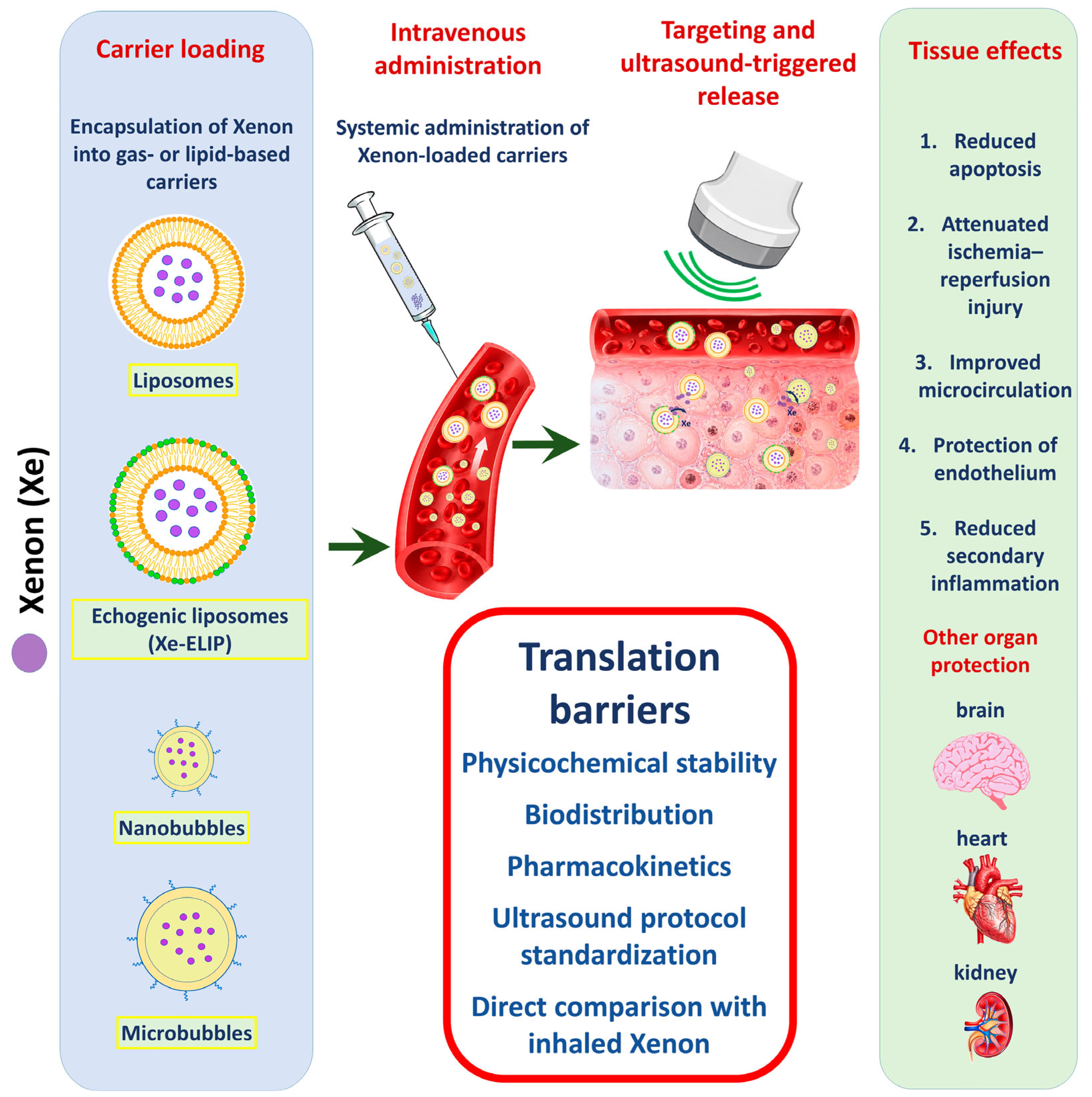

| Platform | Carrier/ Formulation | Xe Loading/ Dose Unit | Trigger/ Localization | Evidence Base | Directly Observed Effects | Main Gaps |
|---|---|---|---|---|---|---|
| Xe-ELIP | Echogenic liposomes; lipid composition varies across studies (e.g., DPPC/DOPC/cholesterol or DPPC/Egg-PC/PEG2000-PE/DPPG/ cholesterol) | Typically ~15–23.38 μL Xe/mg lipid; Miao reports ~20 μL Xe/mg lipid. Doses include 3.5–20 mg/kg or 100–600 μL suspension depending on model | Ultrasound over carotid/internal carotid region; commonly 1 MHz, 0.18 MPa in early stroke studies; 1 MHz, 0.5 W/cm2, 5 min in eMCAO/rtPA study | Rat transient MCAO/reperfusion; prolonged 6 h MCAO with repeated dosing; SAH; embolic MCAO + rtPA | Reduced infarct/hemorrhage volume, TUNEL-positive cells, MMP9 signal with rtPA, and improved behavioral outcomes | Best biological evidence base. Main gaps: no direct tissue Xe PK, no head-to-head inhalation comparison, repeat-dose safety and immunogenicity incomplete |
| Xe-NB | Lipid nanobubbles; DSPE-PEG2K/DSPC; mean diameter about 225 ± 11 nm; particle concentration about 1.95 × 10⁹/mL in the cited study | 73 ± 2 μL Xe/mL suspension; 200 μL intravenously in mouse MCAO/reperfusion model | Used for image-guided accumulation/ultrasound visualization; Image-guided accumulation and ultrasound visualization; available parameters are reported according to the cited study. | Mouse 1 h MCAO followed by reperfusion | Accumulation in ischemic region; CBF recovery to ~88 ± 2% of normal vs. ~75–77% in controls; reduced infarct/apoptosis and improved neurological status | Promising microcirculatory/imaging platform. Conversion to μL/mg lipid not possible without lipid concentration; evidence limited to a narrow model set |
| Xe-MB | Lipid-shell microbubbles; in TBI studies DBPC + DSPE-PEG5000; control gas often perfluorobutane | TBI suspension reported as 4.5 μmol/mL; engineering studies report ~113 μL Xe/mg lipid | Carotid-level ultrasound triggering in piglet TBI studies; a portable ultrasound probe was used for ultrasound-mediated Xe-MB disruption, with available acoustic parameters summarized in Table S1. | Pilot and expanded piglet controlled cortical impact TBI models; engineering characterization studies | Reduced edema/lesion expansion, reactive vascular changes, perivascular inflammation trend/signals, fibrinogen extravasation; in vitro ZO-1 preservation | Large-animal translational bridge. Main gaps: small n, limited functional outcomes, cavitation safety, persistence and repeated exposure |
| Xe-OFP-MB | Lipid-shell microbubbles with Xenon + octafluoropropane (OFP), usually 90:10 gas mixture; DSPC/PEG-lipid shell in Shekhar platform | Reported around 145–151 μL Xe/mg lipid in the cited engineering study | Engineering release under 6 MHz imaging and 220 kHz pulsed ultrasound; OFP improves stability/contrast under undersaturated conditions | Engineering characterization; no definitive therapeutic in vivo neuroprotection model in the current manuscript corpus | Higher loading/stability/contrast than Xe-MB; ultrasound-triggered release demonstrated | Useful platform evidence, not direct therapeutic efficacy evidence. Avoid over-weighting it in biological conclusions |
| Xe-Pla-MB | Hybrid platelet membrane-mimicking microbubbles; platelet membrane promotes adhesion to injured endothelium; gas mixture includes Xe + C3F8/OFP-like stabilizing component | High specific loading reported (~136.9 μL Xe/mg phospholipid in the report), but exact Xe amount per 400 μL therapeutic dose cannot be calculated without lipid mass in working suspension | Ultrasound destruction over injured kidney for 10 min in renal IR model | Rat renal ischemia–reperfusion injury | Improved creatinine/urea/pathology/fibrosis; decreased senescence markers p53/p16/β-gal; targeting to damaged endothelium | Strong targeting concept, but safety questions are central: thrombogenicity, off-target adhesion, immune clearance, inflamed microvasculature |
| Carrier/Model | Directly Measured in Cited Studies | Mechanism Supported | Not Directly Demonstrated | Safe Wording |
|---|---|---|---|---|
| Xe-ELIP; transient MCAO/reperfusion | Infarct volume, behavioral tests, TUNEL; reported BDNF/Akt/MAPK changes in the source literature | Ischemia–reperfusion neuroprotection; anti-apoptotic/survival-signaling support in this model | Direct tissue Xenon exposure; universal microcirculatory mechanism; permanent MCAO protection | “Xe-ELIP reduced infarct volume and apoptosis-related readouts in transient MCAO/reperfusion models” |
| Xe-ELIP; embolic MCAO + rtPA | Clot lysis compatibility, CBF change, infarct size, TUNEL, MMP9, hemorrhage frequency | Compatibility with thrombolysis; reduced neuronal death and MMP9/barrier-associated hemorrhagic risk | Not an enhancer of thrombolysis; not equivalent to transient MCAO; no direct inhaled comparison | “In embolic MCAO, Xe-liposomes did not impair rtPA activity and reduced infarct/TUNEL/MMP9-associated injury” |
| Xe-ELIP; SAH | Hemorrhage volume, TUNEL-positive cells, early brain injury markers | Early brain injury/apoptosis-related protection after SAH | Not an MCAO model; not proof of ischemic–reperfusion mechanism | “SAH evidence supports early brain injury reduction after Xe-ELIP, but should be discussed separately from MCAO” |
| Xe-NB; mouse MCAO/reperfusion | Carrier accumulation/imaging, CBF restoration, infarct volume, apoptosis markers, neurological score | Microcirculatory restoration plus neuroprotection in a mouse reperfusion setting | Not liposome-equivalent; no conversion to μL/mg lipid without lipid mass | “Xe-NB data suggest image-guided delivery and microcirculatory recovery in mouse MCAO/reperfusion” |
| Xe-MB; piglet TBI | MRI FA/RD/MD/AD, perivascular inflammation, reactive vascular changes, GFAP, fibrinogen extravasation; Iba1 not significant | Vascular-barrier protection: edema, BBB leakage/fibrinogen, endothelial/perivascular changes; astrocytic signal is region-dependent | Universal anti-inflammatory effect; microglial suppression; axonal diffusion benefit | “The piglet TBI data mainly support vascular-barrier effects, not broad anti-inflammatory effects” |
| Xe-Pla-MB; renal IR | Creatinine, urea, pathology score, NGAL/KIM-1, fibrosis, p53/p16/β-gal senescence markers, endothelial-targeting imaging | Renal endothelial-targeted protection and senescence-marker reduction after kidney IR | Neurovascular mechanism; brain targeting; generalizable anti-senescence across organs | “Renal Xe-Pla-MB data should be kept as organ-specific evidence and not generalized to CNS mechanisms” |
| Xe-ELIP; myocardial infarction abstract | Reported LV end-systolic volume, cardiac output, infarct volume, but abstract-only and incomplete numerical dataset | Hypothesis-generating cardiac signal only | Established extracranial efficacy; full peer-reviewed cardioprotection evidence | “Cardiac Xe-ELIP evidence is preliminary and should not be weighted equally with full experimental studies” |
| Experimental Block | Treatment Group | Xe-ELIP Dose/Injected Volume | Treatment Timing | Ultrasound Exposure | Normalized Infarct Volume, % | Statistical Comparison/Functional Outcome |
|---|---|---|---|---|---|---|
| Therapeutic time window | Untreated MCAO control | — | — | Not applied | 16 ± 1.8 | Reference group; severe neurological deficit |
| Therapeutic time window | Xe-ELIP | 7 mg/kg; 200 µL | 2 h after stroke onset | 1 MHz, 0.18 MPa | 2.9 ± 0.7 | p < 0.001 vs. control; improvement in limb placement, beam walking, and grid walking tests from day 1 |
| Therapeutic time window | Xe-ELIP | 7 mg/kg; 200 µL | 3 h after stroke onset | 1 MHz, 0.18 MPa | 5.6 ± 1.2 | p < 0.001 vs. control; improvement in behavioral tests, particularly by day 3 |
| Therapeutic time window | Xe-ELIP | 7 mg/kg; 200 µL | 5 h after stroke onset | 1 MHz, 0.18 MPa | 8.5 ± 1.3 | p = 0.004 vs. control; moderate behavioral improvement, weaker than with earlier administration |
| Dose–response | Xe-ELIP | 3.5 mg/kg; 100 µL | 3 h after stroke onset | 1 MHz, 0.18 MPa | 12 ± 3.0 | p > 0.05 vs. corresponding control; no significant behavioral improvement |
| Dose–response | Xe-ELIP | 7 mg/kg; 200 µL | 3 h after stroke onset | 1 MHz, 0.18 MPa | 5.6 ± 1.2 | p < 0.001 vs. corresponding control; significant behavioral improvement |
| Dose–response | Xe-ELIP | 14 mg/kg; 400 µL | 3 h after stroke onset | 1 MHz, 0.18 MPa | 3.3 ± 0.7 | p < 0.001 vs. corresponding control; strongest reduction in infarct volume |
| Ultrasound-triggered release | Untreated MCAO control | — | — | Not applied | 21 ± 2.5 | Reference group for ultrasound comparison |
| Ultrasound-triggered release | Xe-ELIP without ultrasound | 7 mg/kg; 200 µL | 3 h after stroke onset | Not applied | 6.1 ± 0.5 | p < 0.001 vs. control; Xenon-loaded carrier retained neuroprotective activity without triggered release |
| Ultrasound-triggered release | Xe-ELIP + ultrasound | 7 mg/kg; 200 µL | 3 h after stroke onset | 1 MHz, 0.18 MPa | 2.9 ± 0.7 | p < 0.001 vs. control; ultrasound-triggered release further enhanced neuroprotection |
| Test and Indicator | Control | Sham Surgery | MCAO Group | Xe-ELIP | Significance |
|---|---|---|---|---|---|
| Elevated Plus Maze: Time in the Open Arms, % | ≈3.3 | ≈5.0 | ≈0.8–1.0 | ≈9.0 | The overall effect: F(4.37) = 7.738; p = 0.00011 |
| Elevated Plus Maze: Open Arm Entrances, % | ≈10 | ≈13 | ≈2 | ≈14% | The overall effect: F(4.37) = 3.295; p = 0.02092 |
| Elevated Plus Maze: Entrances to Closed Arms, % | ≈90 | ≈86–87 | ≈97–98 | ≈85–86 | p > 0.053 |
| Forced Swim Test: Active Swim Time, sec | ≈78 | ≈99–100 | ≈51–52 | ≈105 | p < 0.00124 |
| 120 min | 240 min | ||
|---|---|---|---|
| Time | Action | Time | Action |
| 0 min | Embolic occlusion of the MCA | 0 min | Embolic occlusion of the MCA |
| 110 min | Xe-ELIP injection in the relevant group | 110 min | Xe-ELIP injection in the relevant group |
| 120 min | Start of thrombolysis | 4 h | Start of thrombolysis |
| 120–180 min | Alteplase infusion within 60 min | 4–5 h | Alteplase infusion within 60 min |
| 1–3 days | Behavioral tests | 1–3 days | Behavioral tests |
| 3rd day | TTC infarction score | 3rd day | TTC, TUNEL, hemorrhagic component, MMP9 |
| Study/Carrier | Injury/Reperfusion Phenotype | Experimental Emphasis | Main Positive Signal | Mechanistic Signal Supported by the Study | Interpretation for This Review |
|---|---|---|---|---|---|
| Britton et al., Xe-ELIP | 2 h transient MCAO followed by reperfusion | First in vivo proof-of-concept for ultrasound-triggered Xe-ELIP delivery | Reduced normalized infarct volume; effect enhanced by ultrasound | Local carrier release; reduction in ischemic tissue injury | Demonstrates feasibility of intravenous Xe-ELIPs with ultrasound-triggered release in transient ischemia–reperfusion |
| Peng et al., Xe-ELIP | 2 h transient MCAO followed by reperfusion | Therapeutic window and dose–response | Protection preserved up to 5 h; higher Xe-ELIP dose produced stronger reduction in infarct volume | Reduced infarct volume and behavioral improvement; molecular survival/apoptosis signals reported | Defines dose/window relationships within transient MCAO, not permanent MCAO |
| Dandekar et al., repeated Xe-ELIP | Prolonged 6 h transient MCAO followed by reperfusion | Repeated dosing and long-term behavioral outcome | Improved sensorimotor recovery and anxiety/depressive-like behavioral endpoints | Long-term functional recovery; cytokine/microbiota signals exploratory | Extends Xe-ELIP evidence to a more severe prolonged ischemia–reperfusion paradigm |
| Peng et al., Xe-liposomes + rtPA | Embolic MCAO with pharmacological reperfusion | Compatibility with thrombolysis and late rtPA-associated injury | Combination with rtPA reduced infarct volume; late treatment reduced hemorrhagic complications, TUNEL-positive cells, and MMP9 signal | Apoptosis and vascular-barrier protection in the context of thrombolysis | Supports adjunctive use with reperfusion therapy; mechanistically distinct from filament transient MCAO |
| Miao et al., Xe-ELIP | Subarachnoid hemorrhage by endovascular perforation | Early brain injury after hemorrhagic cerebrovascular damage | Reduced hemorrhage volume and TUNEL-positive cells; empty carriers and Xe-saturated solution were ineffective | Reduced early cell death after SAH | Should be discussed as hemorrhagic brain injury, not as MCAO/stroke ischemia model |
| Jin et al., Xe-NB | 1 h transient MCAO followed by reperfusion in mice | Nanobubble accumulation, ultrasound imaging, microcirculatory recovery | Nanobubbles accumulated in ischemic tissue, improved microcirculatory recovery, reduced infarct/apoptosis, improved neurological status | Microcirculatory restoration and neuroprotection | Represents a distinct nanobubble ischemia–reperfusion platform, not directly comparable with Xe-ELIP dose–response studies |
| Stage | Purpose | Scheme | Evaluation Period | Primary Endpoint |
|---|---|---|---|---|
| Xenon before and after injury | To check the basic protection with the most optimal circuit | 75% Xenon/25% O2 2 h before injury and 2 h after TBI | 24 h | Neurological outcome, contusion volume |
| Xenon only after injury | To test a clinically more realistic option for early initiation of therapy | 75% Xenon/25% O2, onset 15 min after TBI, duration 3 h | 24 h | Neurological outcome, contusion volume |
| Therapeutic window | To determine the effectiveness of delayed onset | 75% Xenon/25% O2, onset 15 min, 1 h, 3 h or 6 h after TBI, duration 3 h | 24 h | Neurological outcome, contusion volume |
| Application of different concentrations | Assessment of dose–response effect | 30%, 50%, or 75% Xenon with 25% O2, onset at 15 min, duration 3 h | 24 h | Contusion volume, neurological outcome |
| Physiological assessment | To eliminate impact on system performance | 75% Xenon/25% O2 vs. control gas | During exposure | Blood pressure, heart rate, temperature |
| Dynamics up to 5 days | To check the preservation of the early functional effect | 75% Xenon/25% O2, onset 15 min, duration 3 h | Days 1–5 | Neurological outcome |
| Long-term motor effects | Assessment of the durability of effects | 75% Xenon/25% O2, onset 15 min, duration 3 h | 1 month | Rotarod, automated gait analysis |
| Indicator | Control Microbubbles with Perfluorobutane | Xe-MBa | p |
|---|---|---|---|
| Change in edema volume according to MRI from 1 to 5 days | An increase of approximately 160% | Around 0%/slight decrease | ≤0.03 |
| Changes in the nucleus and hemorrhagic component according to MRI | Increase of about 75% | A reduction of approximately 40% | The difference is statistically insignificant |
| Change in the total volume of damage according to MRI | An increase of approximately 145% | Around −10% | ≤0.01 |
| Reactive vascular changes and endothelial proliferation | ≈0.85 points | ≈0.15 points | 0.002 |
| Perivascular inflammation | ≈1.2 points | ≈0.15 points | Lower in the Xenon group but without statistical significance |
| Evaluation Domain | Direct Endpoint/Method | Endpoint Timing | n Used for This Endpoint | Direct Factual Result/Statistical Comparison |
|---|---|---|---|---|
| White matter diffusion | Fractional anisotropy, diffusion tensor MRI | Day 1 | CtMB n = 5; Xe-MB n = 8 | Fractional anisotropy was higher in the Xe-MB group than in the CtMB group in both corona radiata, both cerebral peduncles, and in the genu and splenium of the corpus callosum; p < 0.05 for the indicated regions |
| White matter diffusion | Fractional anisotropy, diffusion tensor MRI | Day 5 | CtMB n = 5; Xe-MB n = 8 | Most day 1 differences were no longer present; higher fractional anisotropy persisted only in a small region of the right cerebral peduncle; p < 0.05 |
| Vasogenic edema-related diffusion | Radial diffusivity, diffusion tensor MRI | Day 1 | CtMB n = 5; Xe-MB n = 8 | Radial diffusivity was higher in the CtMB group than in the Xe-MB group in both corona radiata, the genu and splenium of the corpus callosum, and the contralateral cerebral peduncle; p < 0.05 for the indicated regions |
| General tissue diffusion | Mean diffusivity, diffusion tensor MRI | Day 1 | CtMB n = 5; Xe-MB n = 8 | Mean diffusivity was lower in the Xe-MB group only in the contralateral corona radiata; p < 0.05 |
| General tissue diffusion | Mean diffusivity, diffusion tensor MRI | Day 5 | CtMB n = 5; Xe-MB n = 8 | No significant differences between groups were detected; p > 0.05 |
| Axial diffusion | Axial diffusivity, diffusion tensor MRI | Days 1 and 5 | CtMB n = 5; Xe-MB n = 8 | No significant differences between groups were detected at either time point; p > 0.05 |
| Perivascular inflammation | H&E staining; semi-quantitative perivascular inflammation score | Day 5 | CtMB n = 4; Xe-MB n = 8 | On the injured side, perivascular inflammation was lower in the Xe-MB group than in the CtMB group; p < 0.05. On the contralateral side, the difference did not reach statistical significance; p = 0.052 |
| Reactive vascular changes | H&E staining; semi-quantitative endothelial reactivity/vascular proliferation score | Day 5 | CtMB n = 4; Xe-MB n = 8 | On the injured side, reactive vascular changes were lower in the Xe-MB group than in the CtMB group; p < 0.05 |
| Astrocytic reactivity | GFAP immunostaining; percentage of stained area | Day 5 | CtMB n = 4; Xe-MB n = 8 | GFAP-positive stained area was lower in the Xe-MB group in the contralateral cortex and ipsilateral white matter; p < 0.05. Differences in the ipsilateral cortex and contralateral white matter did not reach statistical significance; p = 0.08 and p = 0.06, respectively |
| Microglial reactivity | Iba1 immunostaining; percentage of stained area | Day 5 | CtMB n = 4; Xe-MB n = 8 | No significant differences between groups were detected; p > 0.05 |
| Blood–brain barrier disruption | Fibrinogen extravasation immunostaining | Day 5 | CtMB n = 4; Xe-MB n = 8 | Fibrinogen extravasation was lower in the Xe-MB group in ipsilateral white matter; p = 0.01. In the perifocal cortex, the difference did not reach statistical significance; p = 0.17. Contralateral staining was minimal, without significant group differences |
Disclaimer/Publisher’s Note: The statements, opinions and data contained in all publications are solely those of the individual author(s) and contributor(s) and not of MDPI and/or the editor(s). MDPI and/or the editor(s) disclaim responsibility for any injury to people or property resulting from any ideas, methods, instructions or products referred to in the content. |
© 2026 by the authors. Licensee MDPI, Basel, Switzerland. This article is an open access article distributed under the terms and conditions of the Creative Commons Attribution (CC BY) license.
Share and Cite
Cherpakov, R.A.; Shashkovskaya, V.S.; Grebenchikov, O.A.; Sergunova, V. Xenon Encapsulation in Liposomes, Nanobubbles, and Microbubbles: Delivery Strategies, Preclinical Evidence, and Translational Barriers. Pharmaceutics 2026, 18, 855. https://doi.org/10.3390/pharmaceutics18070855
Cherpakov RA, Shashkovskaya VS, Grebenchikov OA, Sergunova V. Xenon Encapsulation in Liposomes, Nanobubbles, and Microbubbles: Delivery Strategies, Preclinical Evidence, and Translational Barriers. Pharmaceutics. 2026; 18(7):855. https://doi.org/10.3390/pharmaceutics18070855
Chicago/Turabian StyleCherpakov, Rostislav A., Vera S. Shashkovskaya, Oleg A. Grebenchikov, and Viktoria Sergunova. 2026. "Xenon Encapsulation in Liposomes, Nanobubbles, and Microbubbles: Delivery Strategies, Preclinical Evidence, and Translational Barriers" Pharmaceutics 18, no. 7: 855. https://doi.org/10.3390/pharmaceutics18070855
APA StyleCherpakov, R. A., Shashkovskaya, V. S., Grebenchikov, O. A., & Sergunova, V. (2026). Xenon Encapsulation in Liposomes, Nanobubbles, and Microbubbles: Delivery Strategies, Preclinical Evidence, and Translational Barriers. Pharmaceutics, 18(7), 855. https://doi.org/10.3390/pharmaceutics18070855

