
Article

Sexual Dimorphism in the Sensory Organs of Monochamus saltuarius Gebler (Coleoptera: Cerambycidae): A Morphometric Analysis of Compound Eyes and Stridulatory Files


Key Laboratory of Beijing for the Control of Forest Pests, Beijing Forestry University, Beijing 100083, China
*
Author to whom correspondence should be addressed.
These authors contributed equally to this work.
Forests 2026, 17(1), 145; https://doi.org/10.3390/f17010145
Submission received: 17 December 2025 / Revised: 17 January 2026 / Accepted: 21 January 2026 / Published: 22 January 2026
(This article belongs to the Special Issue Pest Infestation in Trees: Mechanisms, Effects and Control Methods)

Abstract

The Sakhalin pine sawyer, Monochamus saltuarius Gebler, 1830 (Coleoptera: Cerambycidae), is a newly discovered insect vector of the pine wood nematode (Bursaphelenchus xylophilus) in China. Despite its ecological importance, the detailed morphology of its sensory systems remains largely unexplored. This study presents comprehensive ultrastructural analysis of the compound eyes and stridulatory organs in adult M. saltuarius, with a focus on sexual differences. Our morphometric results revealed pronounced sexual dimorphism: males possessed significantly larger compound eyes in terms of total area and perimeter. Furthermore, all three types of ommatidial facets (hexagonal, pentagonal, and quadrilateral) exhibited significantly greater area, perimeter, and diameter in males. Interestingly, while the total number of ommatidia and the counts within the dorsal and ventral eye regions showed no significant sexual difference, they were strongly positively correlated with body length in both sexes. Regarding the stridulatory organs, key morphological features including the transverse diameter, longitudinal diameter, and width of the lateral sulcus of the stridulatory file were also significantly greater in males. In contrast, no sexual dimorphism was detected in the width or density of the stridulatory teeth. These findings collectively indicate a significant investment in sexually dimorphic sensory organs in M. saltuarius, which we hypothesize is closely linked to sex-specific behavioral roles, particularly in active mate searching, courtship displays, and intrasexual competition. This research provides a crucial morphological foundation for understanding the species’ visual and acoustic communication, supporting future work on its sensory ecology and the development of behavior-based control strategies for pine wilt disease.

1. Introduction

Insects inhabit a world dominated by sensory information, relying on sophisticated sensory organs to detect and interpret a myriad of external chemical, auditory, and visual cues. These inputs facilitate a suite of essential survival behaviors, including predator avoidance, host and resource localization, intraspecific communication, and adaptation to fluctuating environmental conditions [1,2]. Among these sensory modalities, vision, mediated primarily by the compound eye, stands as a critical channel for navigation, foraging, predator evasion, and reproductive activities across diverse insect taxa [3].
Structurally, the insect compound eye is a remarkable visual system composed of numerous repeating optical units called ommatidia. Each ommatidium typically consists of a corneal lens, a crystalline cone, and a cluster of photoreceptor cells, forming a self-contained light-guiding and sensing structure [4,5,6,7]. The external surface of the eye presents a distinctive facet array, most commonly hexagonal, which is a direct reflection of the dense packing of these ommatidia [8]. Key morphological parameters—such as the total number of ommatidia, overall eye size, and surface area of individual facets—are fundamental in governing the optical performance of the compound eye, including its sensitivity to light and its spatial resolution [9,10]. These traits are not static but have evolved under long-term ecological and behavioral selection pressures to meet specific functional demands.
The morphology of the compound eye directly determines its optical capacity. For instance, larger facets enhance light capture under low-intensity conditions, improving sensitivity, while a greater number of ommatidia expands the visual field and can improve spatial resolution [11,12]. This intimate structure-function relationship makes the compound eye a classic model for studying ecological adaptation. For instance, diurnal visual predators typically possess larger eyes with higher ommatidial density than nocturnal species [13]. Furthermore, compound eyes frequently exhibit marked sexual dimorphism and developmental plasticity, reflecting divergent life-history strategies between the sexes and ontogenetic sensory optimization [14]. The behavioral significance of vision is particularly evident during courtship and mating in many cerambycid beetles. Studies on species such as Glenea cantor (Fabricius, 1787), Anoplophora chinensis (Forster, 1771), Anoplophora malasiaca (Thomson, 1865), and Monochamus alternatus (Hope, 1843) have highlighted the role of visual cues in mate location and recognition [15,16,17,18,19]. Experimental evidence, involving eye occlusion, has been shown to significantly reduce mating success in several cerambycids, including Anoplophora glabripennis (Motschulsky, 1854) and M. alternatus [17,20], underscoring the critical role of vision. However, some taxa, such as Batocera lineolata Chevrolat, 1852, and Massicus raddei (Blessig, 1872), appear to compensate for visual impairment through other sensory modalities, suggesting a degree of ecological flexibility [21,22].
Beyond vision, acoustic signaling represents another essential sensory channel in insects, supporting key behaviors such as courtship, mating, rivalry, and alarm communication. This is exemplified across diverse groups including crickets [23], katydids [24], grasshoppers [25], cicadas [26,27,28], moths [29], lacewings [30], bark beetles [31], and numerous cerambycid species [32,33,34]. Longhorn beetles produce sound via two distinct methods. Beetles in the subfamilies Prioninae and Parandrinae do so by rubbing their ridged hind femora against the margins of their elytra [35,36], whereas others employ a stridulatory mechanism involving the forward and backward rubbing of the prothorax against the mesothorax [32,37,38]. Ultrastructural studies using scanning electron microscopy have revealed intricate details and significant interspecific and sexual differences in the morphology, size, and tooth count of stridulatory organs in beetles like Leptura arcuata Panzer, 1793, A. glabripennis, and M. raddei [39]. These studies also documented ancillary structures such as neatly arranged bristles along the pronotal margin and the presence of a lateral sulcus on the stridulatory file of A. glabripennis. Subsequent research on Monochamus sartor urussovii (Fischer von Waldheim, 1805) and M. sutor (Linnaeus, 1758) confirmed the presence of a lateral sulcus, with notable interspecific variation in file morphology and size, supporting its potential use as a taxonomic character [40]. In B. lineolata, Luo and Huang [33] successfully linked stridulatory morphology to temporal and amplitude differences between forward and backward chirps. Similarly, a comparative analysis of stridulatory structures and sound traits in G. cantor, Moechotypa diphysis (Pascoe, 1871), and Psacothea hilaris (Pascoe, 1857) revealed clear distinctions, highlighting a direct structure–function relationship [34]. Such morphological diversity corresponds to functional variation; acoustic signals often facilitate courtship and mating, with males emitting species-specific sounds to stimulate partners and enable intersex communication. Nevertheless, the full behavioral significance and functional context of stridulation—particularly in intrasexual interactions and mating—remain inadequately understood for many species, including those within the genus Monochamus.
In forest ecosystems affected by the pine wood nematode (PWN), Bursaphelenchus xylophilus (Steiner and Buhrer, 1934) Nickle, 1970—which causes the lethal pine wilt disease across vast regions of Asia and Europe [41]—beetles of the genus Monochamus (Coleoptera: Cerambycidae) serve as the primary vectors for nematode transmission among host trees [42,43]. In Asia, the Japanese pine sawyer, M. alternatus, has long been recognized as the major vector. However, the Sakhalin pine sawyer, M. saltuarius Gebler, 1830, has also been identified as an important and competent vector in Japan [44], the Republic of Korea [43], and, more recently, in Liaoning Province, China [45,46,47]. Notably, M. saltuarius has been confirmed to transmit PWN to Korean white pine (Pinus koraiensis Siebold & Zucc.), an economically significant forest species, in both Korea [48] and China [45,46], raising serious concerns for pine forest management and health in newly affected regions.
Although previous studies have addressed certain aspects of M. saltuarius biology—such as its life history, host selection preferences, and chemical ecology [49,50]—the structural and functional bases of its sensory systems remain poorly characterized. As a cerambycid beetle likely dependent on multimodal signaling (integrating chemical, visual, and potentially acoustic cues) for critical behaviors such as mate location, courtship, and host recognition, a thorough understanding of the morphology of its compound eyes and stridulatory organs is paramount. However, a significant gap exists in the scientific literature: no research to date has systematically described the fine ultrastructure of either the compound eyes or the stridulatory organs in M. saltuarius. This lack of basic morphological knowledge hinders a comprehensive understanding of its sensory ecology, communication mechanisms, and the potential for developing sensory-based pest control strategies.
To address this critical knowledge gap, the present study employs scanning electron microscopy to investigate the ultrastructure of the compound eyes and stridulatory organs in M. saltuarius. Specifically, this study is guided by two primary research questions: (1) What are the detailed ultrastructural characteristics of these sensory organs? (2) Do they exhibit significant sexual dimorphism? Based on the common ecological roles of male cerambycids in active mate searching and competition, we hypothesized the following: (1) Males possess larger compound eyes with larger ommatidia to enhance visual sensitivity. (2) Males have larger stridulatory files, potentially associated with sound production for communication. By integrating detailed description with morphometric comparisons, this work seeks to establish a foundational morphological dataset and test for adaptive sexual dimorphism linked to sex-specific behaviors. The findings are expected to provide crucial morphological insights into the species’ sensory adaptations, thereby supporting future behavioral and ecological studies and aiding the development of integrated, behavior-based management strategies for combating the spread of pine wilt disease.

2. Materials and Methods

2.1. Insect Collection, Rearing and Experimental Rationale

2.1.1. Collection and Rearing Protocol

Log sections of Korean white pine, P. koraiensis (length range: 57.6–74.3 cm), containing larvae of M. saltuarius, were carefully collected from the Dahuofang Forest Farm located in Fushun City, Liaoning Province, China (41°56′23.028″ N, 124°13′3.925″ E). All specimens examined in this study belong to the nominotypical subspecies, M. saltuarius saltuarius [51]. Immediately after collection, both exposed ends of each log were meticulously sealed with a layer of molten paraffin wax to minimize desiccation and preserve the moisture content essential for larval survival and development. The sealed logs were then placed within cylindrical 16-mesh wire netting cages (100 cm in height × 60 cm in diameter).
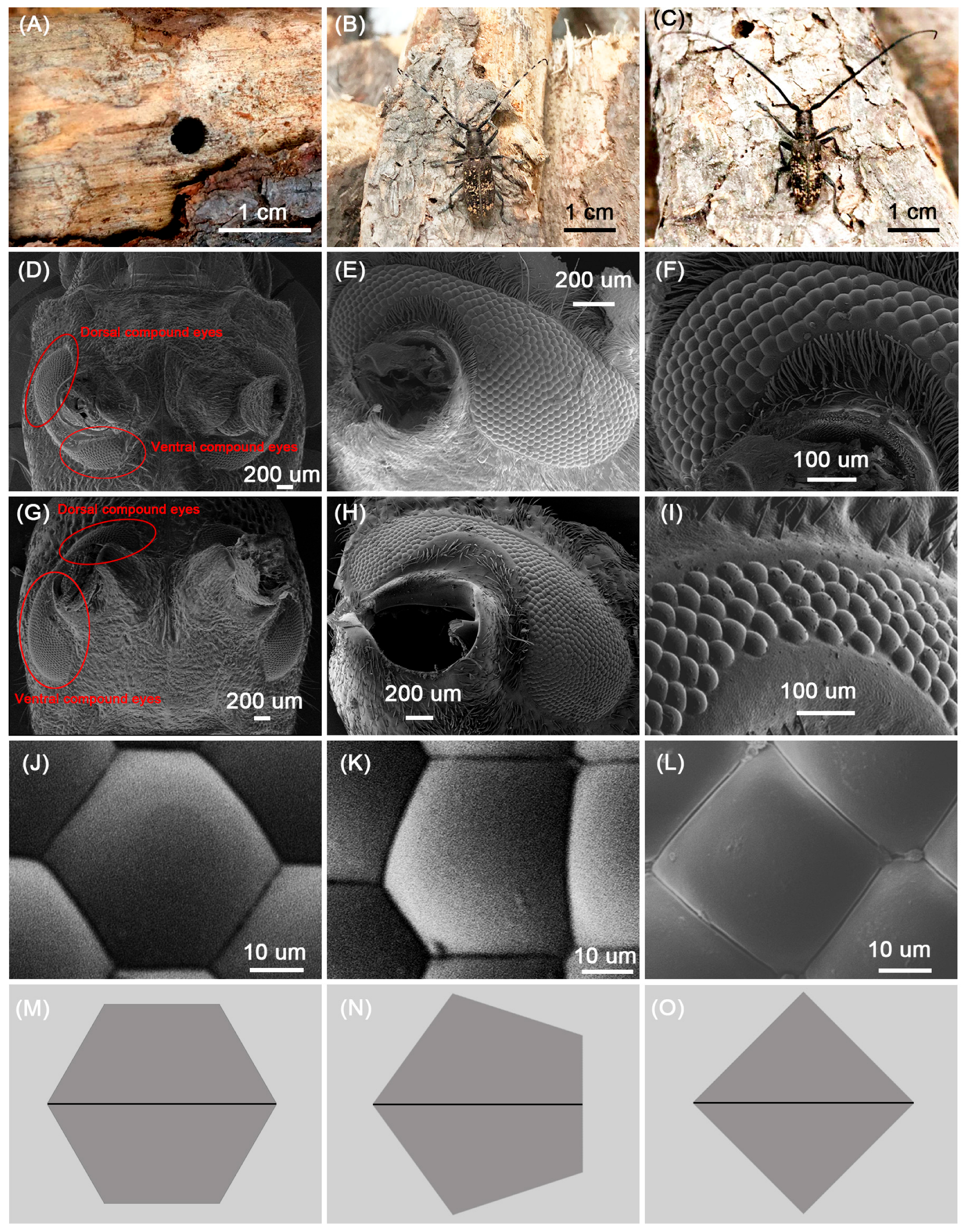
Rearing was conducted in a controlled climate chamber maintained at a constant temperature of 22.3 ± 2.3 °C and a relative humidity of 33.0% ± 7.0%. Newly emerged adult beetles (Figure 1A–C) were collected from the cages each morning to ensure accurate age determination. Upon collection, individuals were immediately isolated in separate 800 mL transparent plastic containers to prevent mating and inter-individual interactions, thus ensuring their virgin status for subsequent experiments. All beetles were provided with an ad libitum supply of fresh, tender shoots of P. koraiensis as a food and moisture source. These shoots were replaced every other day to maintain freshness and nutritional quality.

2.1.2. Rationale for Using Laboratory-Reared, Age-Standardized, Virgin Adults

While field-collected adults are commonly used in morphological studies, we specifically reared adults from larval-infested logs to obtain individuals of known, standardized age and virgin status. This controlled approach is essential for isolating inherent sexual dimorphism from confounding variables.
First, the ultrastructure and functional sensitivity of insect sensory organs, such as compound eyes, are known to undergo changes with age, as documented in other insect groups [14,52,53,54]. Precise age determination is therefore critical in this study. Using adults of a standardized, sexually mature age ensures that the sensory structures are fully developed and functionally representative, thereby isolating sex as the variable of interest and eliminating age-related variation as a confounding factor.
Second, maintaining virgin status is essential because mating induces significant physiological and behavioral shifts. In insects, sensory structures can be subject to wear and tear through use in mating-related behaviors (e.g., stridulation) [55]. By using virgin individuals, we examine the morphology optimized for pre-mating activities before any potential modification by mating experience or differential behavioral wear. This controlled design allows for a precise comparison of the inherent, sex-linked investment in sensory morphology, providing a clean baseline for understanding sexual dimorphism in M. saltuarius.

2.1.3. Determination of Age and Sexual Maturity

The age of sexual maturity is a critical factor for behavioral and morphological studies. Based on established literature, females of M. saltuarius typically reach sexual maturity approximately 7 days post-emergence [56], whereas males are capable of mating almost immediately upon emergence [57]. To standardize our study population and ensure physiological relevance, we utilized 7-day-old virgin females and 5-day-old virgin males for all morphological examinations and measurements. This age selection ensured that the sensory structures were fully developed and representative of reproductively active individuals.

2.2. Scanning Electron Microscopy (SEM)

2.2.1. Sample Preparation for Compound Eyes

The heads of M. saltuarius adults were carefully excised using fine-point dissection scissors and precision forceps under a stereomicroscope. The samples were fixed with a mixture of 2% paraformaldehyde and 2.5% glutaraldehyde (prepared from 10% paraformaldehyde, 25% glutaraldehyde, 0.2 mol/L phosphate buffer, and distilled water in a ratio of 2:1:5:2, v/v; pH 7.2) at 4 °C for 24 h. After fixation, the specimens were rinsed three times with 0.1 mol/L phosphate buffer (pH 7.2), for 10 min each, followed by sequential dehydration in 30%, 50%, 70%, 80%, and 90% ethanol for 10 min per step, and finally three times in 100% ethanol for 30 min each. The dehydrated samples were treated with isoamyl acetate twice (30 min each), subjected to critical point drying with CO2, mounted on stubs using double-sided conductive tape, and sputter-coated with gold using an E-1010 ion sputter coater (Hitachi Ltd., Tokyo, Japan) for 5 min.

2.2.2. Sample Preparation for Stridulatory Organs

The pronotum and mesonotum of M. saltuarius were carefully removed using forceps. The dissected sections were immersed in a 20% sodium hydroxide (NaOH) solution for 24 h, which was intended to ensure gentle yet effective removal of muscle tissues while minimizing potential damage to the delicate cuticular teeth of the stridulatory file. This protocol has been successfully employed in previous ultrastructural studies of cerambycid stridulatory organs [39,58] and was found to yield clean, intact specimens suitable for precise SEM measurement. After immersion, surrounding muscle tissues were meticulously removed. The specimens were then washed with distilled water and cleaned ultrasonically three times (3 min each). Subsequent dehydration was performed sequentially in 60%, 80%, 95%, and 100% ethanol solutions, each for 2 min, followed by air-drying at ambient temperature. The dried samples were mounted on stubs and sputter-coated with gold using an E-1010 ion sputter coater for 5 min.

2.2.3. SEM Imaging and Observation

All prepared samples were examined and imaged using a Hitachi S-3400 N scanning electron microscope (Hitachi, Tokyo, Japan). The microscope was operated at an accelerating voltage ranging from 5.0 to 15.0 kV. Multiple micrographs were captured at various magnifications to comprehensively document the morphology of the compound eyes and stridulatory files.

2.3. Terminology

The terminology used to describe the structures of the compound eyes of M. saltuarius was primarily adapted from recent detailed works on cerambycid eyes, specifically Wen et al. [18,19]. The terminology for describing the stridulatory organs was based on the foundational work of Cheng [32] and subsequent studies by Li et al. [58], Luo and Huang [33], and Wei et al. [34].

2.4. Morphometric Measurements and Data Analysis

The body length of each M. saltuarius individual was measured to the nearest 0.01 mm using a digital caliper (Shengtai Electronic Technology Co., Ltd., Shenzhen, China). A total of 10 females and 10 males were utilized for body length measurement and ommatidia counting. For the compound eyes, several parameters were measured or calculated from the SEM micrographs using ImageJ software (version 1.51k, National Institutes of Health, Bethesda, MD, USA). The total ommatidial count was manually determined from SEM images encompassing the entire eye surface. The eye was digitally divided along the constriction near the antennal socket to separately count ommatidia in the dorsal and ventral regions. To ensure consistency in the partitioned counts, the “constriction near the antennal socket” was defined as the narrowest region connecting the dorsal and ventral eye areas, comprising a width of only 3–4 ommatidia (see Figure 1F,I), and served as the digital dividing line. The perimeter and surface area of each compound eye were obtained by tracing its outline, with the software performing the subsequent calculations. Furthermore, for a detailed analysis of ommatidial size, 10 ommatidia of each shape type (hexagonal, pentagonal, and quadrilateral) were randomly selected from the central region of the ventral eye area for each specimen. Their maximum diameter (feret diameter), perimeter, and cross-sectional area were measured; these detailed measurements were conducted on a subset of five females and five males.
Key morphological characteristics of the stridulatory organs were measured directly from SEM images. The transverse and longitudinal diameters of the stridulatory file were measured at their maximum dimensions, along with the width of the lateral sulcus. Additionally, the width of individual stridulatory teeth was measured for 10 randomly selected teeth per individual. Tooth density was determined by counting the number of tooth rows within a 100 μm span in the central region of the file and converting this value to rows per millimeter. These stridulatory organ measurements were also performed on five females and five males.
All statistical analyses were conducted using IBM SPSS Statistics software (Version 22.0) (IBM Corp., Armonk, NY, USA). An independent samples t-test was employed to compare all morphological features between males and females. Prior to conducting t-tests, the assumptions of normality of data distribution and homogeneity of variances were verified using the Shapiro–Wilk test and Levene’s test, respectively. Data are presented as mean ± standard deviation (SD). Pearson’s correlation coefficient was used to assess the relationships between body length and eye parameters (ommatidia number and compound eye area). For all tests, statistical significance was set at α = 0.05.

3. Results

3.1. Gross Morphology and Sexual Dimorphism of the Compound Eye

The compound eyes of adult M. saltuarius are prominently positioned on the lateral aspects of the head capsule, exhibiting the typical reniform (kidney-shaped) configuration that encircles the base of the antennal socket (Figure 1D,G). Each compound eye is distinctly divided into two primary regions by a constriction located adjacent to the antennal insertion point: a dorsal portion and a substantially larger ventral portion (Figure 1D,E,G,H). At its narrowest point, the isthmus connecting the dorsal and ventral regions comprises only 3–4 ommatidia (Figure 1F,I). The corneal surface of the eye is smooth and glossy, lacking any superficial specialized structures such as interommatidial sensory setae or corneal nipple arrays, which are present in some other insect groups. The vast majority of the corneal facets are hexagonal in shape, forming the characteristic honeycomb pattern (Figure 1J,M). However, intermingled within this hexagonal array are smaller proportions of pentagonal (Figure 1K,N) and quadrilateral facets (Figure 1L,O).
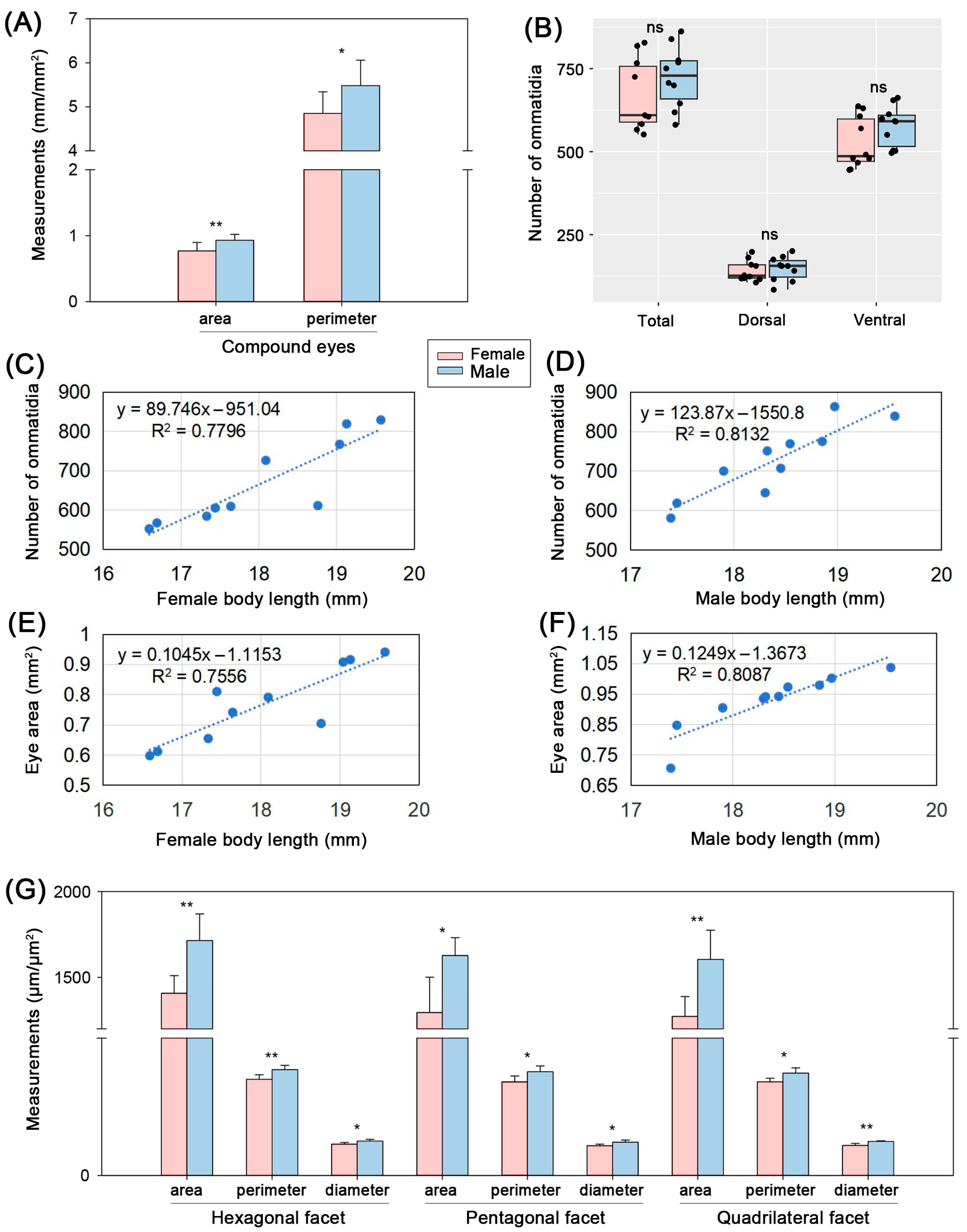
Quantitative analyses revealed significant sexual dimorphism in the overall size of the compound eyes. The compound eye area of male M. saltuarius was significantly larger than that of females (females: 0.77 ± 0.13 mm2; males: 0.93 ± 0.09 mm2; t18 = −3.2, p = 0.005; Figure 2A). Consistently, the eye perimeter was also significantly greater in males (females: 4.85 ± 0.49 mm; males: 5.48 ± 0.58 mm; t18 = −2.654, p = 0.016; Figure 2A and Table S1).
In contrast to the size differences, the total number of ommatidia per compound eye did not differ significantly between the sexes (females: 666.9 ± 107.1; males: 724.9 ± 92.2; t18 = −1.298, p = 0.211). Similarly, no significant sexual difference was found when ommatidia were counted separately in the dorsal region (females: 141.0 ± 31.1; males: 148.3 ± 36.1; t18 = −0.485, p = 0.634) or the ventral region (females: 525.9 ± 76.8; males: 576.6 ± 61.1; t18 = −1.634, p = 0.12). The numerical dominance of the ventral region was consistent, containing approximately four times the number of ommatidia found in the dorsal region in both sexes (Figure 2B and Table S1).
A strong positive relationship was observed between visual investment and body size. The total number of ommatidia per compound eye increased significantly with increasing body length in both females (r = 0.883, p < 0.001, n = 10; Figure 2C) and males (r = 0.902, p < 0.001, n = 10; Figure 2D). Similarly, the compound eye area was positively correlated with body length in both females (r = 0.869, p = 0.001, n = 10; Figure 2E) and males (r = 0.899, p < 0.001, n = 10; Figure 2F).

3.2. Ommatidial Size and Facet-Specific Dimorphism

Detailed morphometry of individual ommatidia revealed a consistent pattern of males possessing larger facets across all three shape types. For hexagonal ommatidia, the area (females: 1406.74 ± 102.99 μm2; males: 1714.29 ± 155.56 μm2; t8 = −3.686, p = 0.006), perimeter (females: 140.18 ± 6.68 μm; males: 154.22 ± 6.37 μm; t8 = −3.402, p = 0.009), and diameter (females: 45.75 ± 2.42 μm; males: 50.14 ± 2.75 μm; t8 = −2.675, p = 0.028) were all significantly larger in males (Figure 2G and Table S1).
This trend was equally evident in pentagonal ommatidia, with males exhibiting significantly greater area (females: 1294.61 ± 206.20 μm2; males: 1626.81 ± 104.77 μm2; t8 = −3.212, p = 0.012); perimeter (females: 136.57 ± 8.61 μm; males: 151.29 ± 8.36 μm; t8 = −2.744, p = 0.025); and diameter (females: 43.45 ± 2.32 μm; males: 48.61 ± 3.15 μm; t8 = −2.942, p = 0.019) (Figure 2G and Table S1).
Quadrilateral ommatidia also followed the same pattern, showing significant male-biased size in area (females: 1272.23 ± 115.92 μm2; males: 1604.85 ± 169.85 μm2; t8 = −3.617, p = 0.007); perimeter (females: 136.43 ± 5.38 μm; males: 149.18 ± 7.63 μm; t8 = −3.053, p = 0.016); and diameter (females: 43.86 ± 2.97 μm; males: 49.57 ± 1.41 μm; t8 = −3.884, p = 0.009) (Figure 2G and Table S1).

3.3. Morphology and Dimorphism of the Stridulatory File

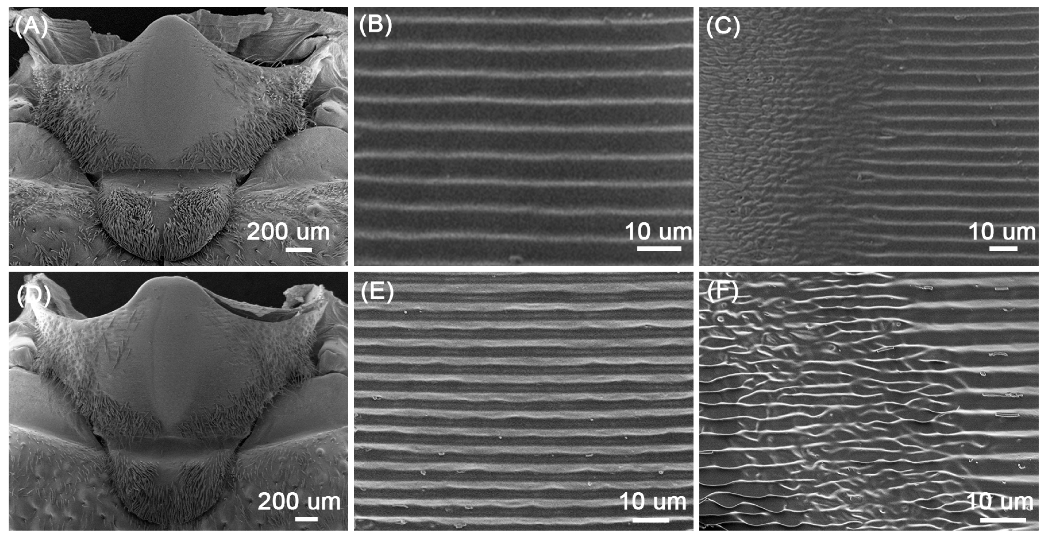
Both female and male M. saltuarius possess well-developed stridulatory organs. Stridulatory file is located centrally on the mesonotum. Under natural conditions, it is concealed beneath the posterior margin of the pronotum. When the pronotum is retracted, the file is exposed as an elongated, roughly oval-shaped structure covered in parallel, transverse ridges (Figure 3A,D). The file is composed of numerous finely cuticular sound teeth (Figure 3B,E). A prominent and consistent morphological feature is a curved longitudinal stripe, identified as the lateral sulcus, located on the left side of the file (Figure 3A,D). This sulcus is formed by the successive bifurcation and fusion of the sound teeth, creating a visible groove (Figure 3C,F). The presence of this lateral sulcus divides the file into two asymmetrical parts: a narrower left portion and a broader, more extensive right portion (Figure 3A,D).
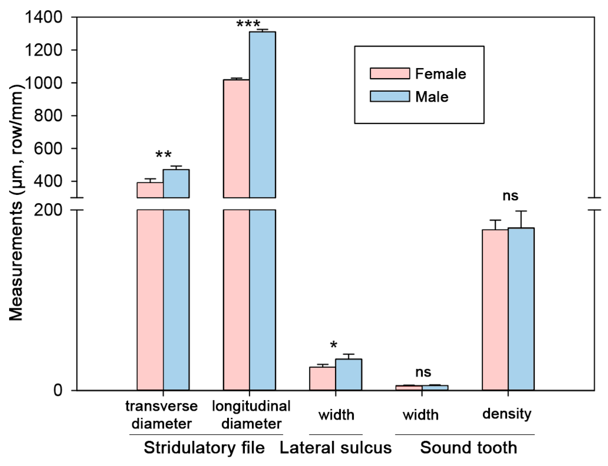
Measurements of the stridulatory file revealed significant sexual dimorphism. The transverse diameter of the file was significantly larger in males (females: 391.74 ± 23.53 μm; males: 471.27 ± 21.41 μm; t8 = −5.589, p = 0.001). The longitudinal diameter exhibited an even more pronounced difference, being substantially greater in males (females: 1018.43 ± 10.67 μm; males: 1310.36 ± 15.47 μm; t8 = −34.731, p < 0.001) (Figure 4). Furthermore, the width of the lateral sulcus was also significantly greater in males (females: 25.66 ± 3.25 μm; males: 34.67 ± 5.63 μm; t8 = −3.1, p = 0.015) (Figure 4 and Table S1).
In contrast, the microstructure of the sound teeth themselves showed no sexual dimorphism. The width of sound teeth did not differ significantly between females and males (females: 5.09 ± 0.70 μm; males: 5.40 ± 0.73 μm; t8 = −0.680, p = 0.516). Similarly, the density of the sound teeth, measured as the number of tooth rows per millimeter, was virtually identical between the sexes (females: 177.97 ± 10.73 rows/mm; males: 180.10 ± 18.77 rows/mm; t8 = −0.220, p = 0.831) (Figure 4 and Table S1).

4. Discussion

4.1. Structural Simplicity and Diurnal Adaptation of the Compound Eye

The compound eye surface of adult M. saltuarius presents a morphology of functional simplicity, lacking specialized structures such as interommatidial sensory hairs and corneal nipple arrays. This morphological theme is shared with other diurnally active cerambycids, such as A. chinensis and A. glabripennis [19,59], suggesting a convergent adaptation to a lifestyle dominated by daylight activity. There is an absence of corneal nipples, which are known to reduce surface reflection and enhance photon capture in nocturnal insects by creating an anti-reflective coating [60]. Its absence in M. saltuarius aligns well with field observations reporting that its primary mating and oviposition activities occur during the daytime [61]. The pronounced reniform shape, with a large ventral region containing roughly four times more ommatidia than the smaller dorsal region, mirrors the configuration found in A. glabripennis [59]. This anatomical division likely underpins a partitioned visual field: the extensive ventral region presumably facilitates acute forward and lateral vision critical for flight navigation through complex forest canopies, precise landing on host branches, and close-range mate assessment, while the smaller dorsal region may be specialized for monitoring the upper hemisphere for predators or competitors.

4.2. Sexual Dimorphism in Eye Morphology: Implications for Behavior and Visual Performance

Our study reveals pronounced sexual dimorphism in the compound eyes of M. saltuarius. Males not only possess significantly larger overall eyes (in area and perimeter) but also have consistently larger individual ommatidia (in area, perimeter, and diameter) across all facet types (hexagonal, pentagonal, and quadrilateral), despite the lack of a significant difference in the total number of ommatidia. This pattern suggests that the male visual advantage is achieved primarily through the enlargement of individual optical units rather than an increase in their number. This strategy is consistent with findings in various other insects where males typically play more active roles in patrolling, mate searching, and intrasexual competition [11,13]. Larger facets possess a greater aperture, which enhances their light capture capacity and sensitivity [4]. This could be particularly advantageous for males under variable light conditions, such as during crepuscular activity or in shaded forest understories, even if the species is primarily diurnal. A congeneric species, M. alternatus, possesses an apposition eye capable of dynamic adaptation to low light, suggesting some flexibility in the genus [18]. The superior light-gathering capability afforded by larger ommatidia in male M. saltuarius likely supports crucial visually mediated behaviors, including long-range detection of potential mates, tracking of female movement, and assessment of rivals during male–male competitions, as documented in other cerambycids like G. cantor [15].

4.3. Allometric Scaling and Potential Trade-Offs in Eye Design

We found that both compound eye area and the total number of ommatidia increased significantly with body length in M. saltuarius. Given that body length is a reliable proxy for overall body size in this species [62], this reflects a widespread pattern in insects where sensory organs scale with body size to maintain functional capacity and optical performance as the animal grows [63,64,65]. However, a fundamental constraint exists: on a finite corneal surface, larger ommatidia (as seen in males) could potentially limit the total number that can be packed, thereby reducing the spatial sampling density (ommatidial density) and potentially limiting resolution. Insects can mitigate this trade-off by increasing the overall eye size to accommodate both larger facets and a sufficient number of them, or by increasing the density of packing [66]. The strong positive correlation between body size and ommatidial number in M. saltuarius indicates that larger individuals effectively expand their corneal surface area to incorporate more ommatidia, thereby likely maintaining or even improving spatial resolution despite the general enlargement of facets, particularly in males. This represents an adaptive solution to prevent inadequate image sampling as individuals grow larger, a phenomenon also observed in other Diptera like Chrysomya megacephala (Fabricius, 1794) [66,67].

4.4. Stridulatory File Morphology: Taxonomic and Functional Implications

The stridulatory file of M. saltuarius possesses a distinct lateral sulcus on its left side, formed by the successive bifurcation of the striae. The presence, position, and morphology of such a sulcus are considered key taxonomic characters for distinguishing among cerambycid subfamilies and genera [68,69]. Its occurrence in M. saltuarius aligns perfectly with its consistent presence within the Lamiinae subfamily, as previously reported in congeners like M. urussovi and M. sutor [40] and a range of other lamiine species [58]. More importantly, our study uncovered significant sexual dimorphism in the file structure: males had significantly larger files in both transverse and longitudinal diameters and a wider lateral sulcus. As the morphology of the file directly governs the physical interaction with the plectrum and consequently influences the temporal structure, frequency spectrum, and amplitude of the produced sound [33,34], these morphological differences strongly suggest the production of sexually dimorphic acoustic signals in M. saltuarius. While stridulation in longhorn beetles can serve diverse functions, including alarm signaling (e.g., in A. glabripennis [70]; in Xylotrechus rusticus (Linnaeus, 1758) [71]), interference during rivalry (e.g., in M. alternatus [72] and Tetraopes tetrophthalmus (Forster, 1771) [73]), and predator deterrence (e.g., in Anoplophora horsfieldi (Hope, 1843) [74]) the pronounced sexual dimorphism in M. saltuarius points towards a role in sexual communication. The larger file in males could potentially produce louder, lower-frequency, or more complex sounds used in courtship displays to attract females or in acoustic contests with rival males. However, the functional significance and behavioral context of these dimorphic sounds require definitive validation through integrated bioacoustic recordings and detailed behavioral experiments.

4.5. Implications for Pest Management and Future Directions

The pronounced sexual dimorphism in sensory organs, particularly the male-biased investment in vision and sound production, provides a direct morphological rationale for developing innovative, behavior-based control strategies. We propose that an effective trap for M. saltuarius could synergistically combine stimuli targeting multiple sensory channels. Specifically, a multimodal trapping system could integrate (1) visual decoys designed to exploit male visual sensitivity (e.g., black silhouettes mimicking female body shape or panels with specific UV-reflective patterns); (2) acoustic lures emitting synthetic or recorded sounds based on the predicted output of the dimorphic stridulatory files (e.g., male rivalry or courtship chirps); and (3) existing female sex pheromones for attraction and orientation. This integrated approach is predicted to significantly enhance trap efficacy and selectivity compared to conventional single-modality lures by extending attraction range through complementary signals and potentially increasing male-specific capture.
However, these interpretations, particularly those concerning acoustic communication, remain inferential due to a lack of direct bioacoustic evidence. Our findings are also limited to a single laboratory population from Northeast China. Considering the species’ broad Eurasian distribution [75,76] and the documented intraspecific morphological variation in cerambycids [51], future research should investigate potential geographic variation in traits such as sexual dimorphism or sensory organ size, which may reflect local ecological adaptation or genetic divergence. Validating our predictions will require bioacoustic recordings and playback experiments. Furthermore, examining the effects of age, mating experience, and structural wear will be essential to fully understand the species’ sensory ecology and to optimize management strategies tailored to regional conditions.

5. Conclusions

This study establishes significant sexual dimorphism in the primary sensory organs of M. saltuarius, with clear functional implications. Males possess larger compound eyes featuring enlarged ommatidia for greater light sensitivity, and more substantial stridulatory files capable of producing more prominent acoustic signals. This suite of traits suggests that males may rely on a multimodal combination of visual and acoustic cues for active mate searching, courtship, and intrasexual competition. Conversely, the relatively smaller sensory structures in females may indicate a greater dependence on close-range chemical and tactile cues for final mate assessment and precise host selection. These findings fill a critical gap in the sensory biology of this important vector insect. More importantly, the documented sexual dimorphism provides a concrete morphological foundation for advancing targeted, multimodal pest management strategies. We hypothesize that a trapping system combining male-optimized visual cues (based on their larger eyes), species-specific acoustic signals (inferred from their larger stridulatory files), and female sex pheromones would create a synergistic “sensory trap” with greater range and specificity than current tools.

Supplementary Materials

The following supporting information can be downloaded at: https://www.mdpi.com/article/10.3390/f17010145/s1.

Author Contributions

Conceptualization, Z.H.; Methodology, J.Y. and Y.X.; Software, J.Y. and Y.X.; Validation, Z.H.; Formal Analysis, J.Y. and Y.X.; Investigation, J.Y. and Y.X.; Resources, Z.H.; Data Curation, J.Y. and Y.X.; Writing—Original Draft Preparation, J.Y. and Y.X.; Writing—Review & Editing, Z.H.; Visualization, J.Y. and Y.X.; Supervision, Z.H.; Project Administration, Z.H.; Funding Acquisition, Z.H. All authors have read and agreed to the published version of the manuscript.

Funding

This study was funded by the National Natural Science Foundation of China (Grant No. 32301293) (for Z.H.).

Data Availability Statement

The data that support the findings of this study are available from the corresponding author upon reasonable request.

Acknowledgments

We would like to express our deep gratitude to three anonymous workers at Dahuofang Forest Farm located in Fushun City, Liaoning Province for their help with specimen collection.

Conflicts of Interest

The authors declare no conflicts of interest.

References

  1. Desiraju, G.R.; Steiner, T. The Insects: Structure and Function, 5th ed.; Cambridge University Press: Cambridge, UK, 2013. [Google Scholar]
  2. Tan, Z.; Zhang, Y.; Jiang, X.; Zhang, G.; Huo, W.; Hou, Z. Sensilla on Organs of Adults of the Pistachio-Seed Wasp Eurytoma plotnikovi (Hymenoptera: Eurytomidae). Microsc. Res. Tech. 2025, 88, 2210–2221. [Google Scholar] [CrossRef] [Scilit]
  3. Feller, K.D.; Sharkey, C.R.; McDuffee-Altekruse, A.; Bracken-Grissom, H.D.; Lord, N.P.; Porter, M.L.; Schweikert, L.E. Surf and Turf Vision: Patterns and Predictors of Visual Acuity in Compound Eye Evolution. Arthropod Struct. Dev. 2021, 60, 101002. [Google Scholar] [CrossRef] [Scilit]
  4. Warrant, E.J. Visual Tracking: Hot Pursuit with Tiny Eyes. Curr. Biol. 2017, 27, R234–R237. [Google Scholar] [CrossRef] [Scilit]
  5. Lan, Y.; Wei, C. Morphology, Histology and Ultrastructure of the Compound Eyes of the Last Instar Nymphs and Adults of Meimuna mongolica (Hemiptera: Cicadidae). Acta Entomol. Sin. 2020, 63, 1441–1451. [Google Scholar]
  6. Nilsson, D.E. The Diversity of Eyes and Vision. Annu. Rev. Vis. Sci. 2021, 7, 19–41. [Google Scholar] [CrossRef] [Scilit]
  7. Yang, X.; Ran, H.; Jiang, Y.; Lu, Z.; Wei, G.; Li, J. Fine Structure of the Compound Eyes of the Crepuscular Moth Grapholita molesta (Busck 1916) (Lepidoptera: Tortricidae). Front. Physiol. 2024, 15, 1343702. [Google Scholar] [CrossRef] [Scilit]
  8. Makarova, A.A.; Diakov, A.A.; Chaika, S.; Polilov, A.A. Scaling of the Sense Organs of Insects. 1. Introduction. Compound Eyes. Entomol. Rev. 2022, 102, 161–181. [Google Scholar] [CrossRef] [Scilit]
  9. Singh, S.P.; Mohan, L. Variations in the Ommatidia and Compound Eyes of Three Species of Mosquito Vectors. J. Entomol. Zool. Stud. 2013, 1, 16–21. [Google Scholar]
  10. Pichaud, F.; Casares, F. Shaping an Optical Dome: The Size and Shape of the Insect Compound Eye. Semin. Cell Dev. Biol. 2022, 130, 37–44. [Google Scholar] [CrossRef] [Scilit] [PubMed]
  11. Rutowski, R.L. Variation of Eye Size in Butterflies: Inter- and Intraspecific Patterns. J. Zool. 2000, 252, 187–195. [Google Scholar] [CrossRef]
  12. Zagorski, E.R.; Marry, J.W. How Do Eye Size and Facet Lens Size Vary by Age and Sex in Acheta domesticus? Bios 2014, 85, 151–159. [Google Scholar] [CrossRef] [Scilit]
  13. Bauer, T.; Kredler, M. Morphology of the Compound Eyes as an Indicator of Lifestyle in Carabid Beetles. Can. J. Zool. 1993, 71, 799–810. [Google Scholar] [CrossRef] [Scilit]
  14. Meyer-Rochow, V.B. Compound Eyes of Insects and Crustaceans: Some Examples That Show There Is Still a Lot of Work Left to Be Done. Insect Sci. 2015, 22, 461–481. [Google Scholar] [CrossRef] [Scilit]
  15. Lu, W.; Wang, Q.; Tian, M.Y.; He, X.Z.; Zeng, X.L.; Zhong, Y.X. Mate Location and Recognition in Glenea cantor (Fabr.) (Coleoptera: Cerambycidae: Lamiinae): Roles of Host Plant Health, Female Sex Pheromone, and Vision. Environ. Entomol. 2007, 36, 864–870. [Google Scholar] [CrossRef] [Scilit] [PubMed]
  16. Yasui, H. Chemical Communication in Mate Location and Recognition in the White-Spotted Longicorn Beetle, Anoplophora malasiaca (Coleoptera: Cerambycidae). Appl. Entomol. Zool. 2009, 44, 183–194. [Google Scholar] [CrossRef] [Scilit]
  17. Liu, B.; Xu, H.C.; Meng, J.G.; Sun, J.H.; Fan, J.T. Visual Cues for the Host-Finding and Mating Locations of Monochamus alternatus (Coleoptera: Cerambycidae). J. Zhejiang A&F Univ. 2012, 29, 617–620. [Google Scholar]
  18. Wen, C.; Ma, T.; Deng, Y.; Liu, C.; Liang, S.; Wen, J.; Wang, C.; Wen, X. Morphological and Optical Features of the Apposition Compound Eye of Monochamus alternatus Hope (Coleoptera: Cerambycidae). Micron 2020, 128, 102769. [Google Scholar] [CrossRef] [Scilit]
  19. Wen, C.; He, J.Y.; Zhong, H.N.; Qiu, H.L.; Wen, X.J.; Wang, S.; Wen, J.B.; Xiao, H.J. External Morphology and Internal Ultrastructure of Compound Eyes in Adult Anoplophora chinensis Adult. J. Environ. Entomol. 2024, 46, 773–780. [Google Scholar]
  20. He, P.; Huang, J.F. Adult Behavior of Anoplophora glabripennis. Acta Entomol. Sin. 1993, 36, 51–55. [Google Scholar]
  21. Pan, X.Y.; Luo, S.L.; Chen, L.Z.; Wang, M.Q. The Role of Vision in the Mate Location Process of Monochamus alternatus. Insect Res. Cent. China 2012, 8, 82–85. [Google Scholar]
  22. Wei, J.R.; Gao, C.; Gao, J.C.; Dong, L.J. Roles of Vision and Antennae in the Short-Distance Mate Searching Behavior of Male Adults of Massicus raddei (Coleoptera: Cerambycidae). Acta Entomol. Sin. 2013, 56, 824–830. [Google Scholar]
  23. Hagg, L.; Zuk, M. Lack of Perceived Sperm Competition Risk Increases Post-Copulatory Song in Pacific Field Crickets. Behav. Ecol. 2025, 36, araf081. [Google Scholar] [CrossRef] [Scilit]
  24. Korsunovskaya, O.S.; Zhantiev, R.D. Acoustic and Vibrational Signaling in True Katydid Nesoecia nigrispina: Three Means of Sound Production in One Species. PeerJ 2022, 10, e13749. [Google Scholar] [CrossRef] [Scilit]
  25. Sevastianov, N.; Neretina, T.; Vedenina, V. Evolution of Calling Songs in the Grasshopper Subfamily Gomphocerinae (Orthoptera, Acrididae). Zool. Scr. 2023, 52, 154–175. [Google Scholar] [CrossRef] [Scilit]
  26. Hou, Z.; Luo, C.; Roberts, J.D.; Wei, C. Sexual Pair-Formation in a Cicada Mediated by Acoustic Behaviour of Females and Positive Phonotaxis of Males. Sci. Rep. 2017, 7, 6453. [Google Scholar] [CrossRef] [Scilit]
  27. Hou, Z.; Liu, Y.; Wei, S.; Wei, C. Females Prefer Males Producing a High-Rate Song with Shorter Timbal–Stridulatory Sound Intervals in a Cicada Species. Curr. Zool. 2021, 68, 103–112. [Google Scholar] [CrossRef] [Scilit]
  28. Hou, Z.; Wei, S.; Wei, C. The Best of Both Worlds: Cicada Males Change Costly Signals to Achieve Mates While Females Choose a Mate Based on Both Calling and Courtship Songs. Curr. Zool. 2022, 68, 716–725. [Google Scholar] [CrossRef] [Scilit]
  29. Nakano, R.; Takanashi, T.; Surlykke, A.; Skals, N.; Ishikawa, Y. Evolution of Deceptive and True Courtship Songs in Moths. Sci. Rep. 2013, 3, 2003. [Google Scholar] [CrossRef] [Scilit]
  30. Henry, C.S.; Wells, M.L.M. Courtship Songs of Green Lacewings Filmed in Slow Motion: How a Simple Vibrating Structure can Generate Complex Signals (Neuroptera: Chrysopidae: Chrysoperla). J. Insect Behav. 2015, 28, 89–106. [Google Scholar] [CrossRef] [Scilit]
  31. Lindeman, A.A.; Yack, J.E. Bark Beetles Use a Spring-Loaded Mechanism to Produce Variable Song Patterns. J. Exp. Biol. 2019, 222, jeb190660. [Google Scholar] [CrossRef] [Scilit] [PubMed]
  32. Cheng, J.Q. Sound Production in Longhorned Beetles: Stridulation and Associated Behavior of the Adult (Coleoptera: Cerambycidae). Sci. Silvae Sin. 1991, 27, 234–237. [Google Scholar]
  33. Luo, C.Q.; Huang, S.H. Stridulatory Sound Production and Acoustic Signals of the Longhorn Beetle Batocera lineolata (Coleoptera: Cerambycidae). Bioacoustics 2021, 31, 148–159. [Google Scholar] [CrossRef] [Scilit]
  34. Wei, J.Q.; Wang, X.Y.; Zheng, X.L.; Tong, X. Stridulatory Organs and Sound Recognition of Three Species of Longhorn Beetles (Coleoptera: Cerambycidae). Insects 2024, 15, 849. [Google Scholar] [CrossRef] [Scilit]
  35. Linsley, E.G. Ecology of Cerambycidae. Annu. Rev. Entomol. 1959, 4, 99–138. [Google Scholar] [CrossRef] [Scilit]
  36. Hepp, F.; Bezerra, A.M.; Botero, J.P. First Description of Sound Emission of Rhaphiptera affinis Thomson, 1868 (Cerambycidae: Lamiinae: Pteropliini). Zootaxa 2019, 4567, 580–582. [Google Scholar] [CrossRef] [Scilit]
  37. Finn, W.E.; Mastro, V.C.; Payne, T.S. Stridulatory Apparatus and Analysis of the Acoustics of Four Species of the Subfamily Lamiinae (Coleoptera: Cerambycidae). Ann. Entomol. Soc. Am. 1972, 65, 644–647. [Google Scholar] [CrossRef] [Scilit]
  38. Breidbach, O. Zur Stridulation der Bockkäfer (Cerambycidae, Coleoptera). Dtsch. Entomol. Z. 1988, 35, 417–425. [Google Scholar] [CrossRef] [Scilit]
  39. Li, L.; Chi, D.F.; Li, Y.; Yin, Y.B.; Zhang, Z. Stridulatory Organs Ultrastructure of Three Longhorned Beetles and Their Stridulation Mechanism. J. Nanjing For. Univ. (Nat. Sci. Ed.) 2013, 37, 71–77. [Google Scholar]
  40. Ma, Y.K.; Sun, F.; Li, L. Comparative Study on the Ultra-Structure of Stridulatory Apparatus in Two Species of Monochamus. J. Anhui Agric. Sci. 2014, 42, 5895–5896. [Google Scholar]
  41. Zhao, B.G.; Futai, K.; Sutherland, J.R.; Takeuchi, Y. Pine Wilt Disease; Springer: Tokyo, Japan, 2008. [Google Scholar]
  42. Kishi, Y. Pine Wood Nematode and the Japanese Pine Sawyer; Thomas Company Limited: Tokyo, Japan, 1995. [Google Scholar]
  43. Kwon, T.S.; Lim, J.H.; Sim, S.J.; Kwon, Y.D.; Son, S.K.; Lee, K.Y.; Kim, Y.T.; Park, J.W.; Shin, C.H.; Ryu, S.B.; et al. Distribution Patterns of Monochamus alternatus and M. saltuarius (Coleoptera: Cerambycidae) in Korea. J. Korean For. Soc. 2006, 95, 543–550. [Google Scholar]
  44. Heisuke, S.; Takeshi, S.; Mitsunori, K. Transmission of Bursaphelenchus xylophilus (Steiner et Buhrer) Nickle (Nematoda: Aphelenchoididae) by Monochamus saltuarius (Gebler) (Coleoptera, Cerambycidae). J. Jpn. For. Soc. 1987, 69, 492–496. [Google Scholar]
  45. Yu, H.; Wu, H. New Host Plants and New Vector Insects Found in Pine Wood Nematode in Liaoning. For. Pest Dis. 2018, 37, 61. [Google Scholar]
  46. Li, M.; Li, H.; Sheng, R.C.; Sun, H.; Sun, S.H.; Chen, F.M. The First Record of Monochamus saltuarius (Coleoptera; Cerambycidae) as Vector of Bursaphelenchus xylophilus and Its New Potential Hosts in China. Insects 2020, 11, 636. [Google Scholar] [CrossRef] [Scilit] [PubMed]
  47. Pan, L.; Li, Y.; Cui, R.; Liu, Z.; Zhang, X. Monochamus saltuarius Endangers Pinus tabuliformis Carr. and Carries Bursaphelenchus xylophilus (Steiner and Buhrer) in China. Forests 2020, 11, 1051. [Google Scholar] [CrossRef] [Scilit]
  48. Kim, M.K.; Kim, J.S.; Han, J.H.; Kim, Y.J.; Yoon, C.; Kim, G.H. Mating Behaviour of Pine Sawyer, Monochamus saltuarius Gebler (Coleoptera: Cerambycidae). Asia-Pac. Entomol. 2006, 9, 275–280. [Google Scholar] [CrossRef] [Scilit]
  49. Han, J.H.; Kim, H.K.; Kang, W.J.; Kim, G.-H. Feeding and Oviposition Preference of the Sakhalin Pine Sawyer Monochamus saltuarius (Coleoptera: Cerambycidae) for Various Tree Species. Entomol. Res. 2016, 46, 331–336. [Google Scholar] [CrossRef] [Scilit]
  50. Hou, Z.; Shi, F.; Ge, S.; Tao, J.; Ren, L.; Wu, H.; Zong, S. Comparative Transcriptome Analysis of the Newly Discovered Insect Vector of the Pine Wood Nematode in China, Revealing Putative Genes Related to Host Plant Adaptation. BMC Genom. 2021, 22, 189. [Google Scholar] [CrossRef] [Scilit]
  51. Sláma, M. A contribution to the recognition of two longicorn species Cerambyx cerdo Linnaeus, 1758 and Monochamus saltuarius (Gebler, 1830) (Coleoptera, Cerambycidae). Humanit. Space-Int. Alm. 2017, 6, 933–938. [Google Scholar]
  52. Butler, L.; Roppel, R.; Zeigler, J. Post emergence maturation of the eye of the adult black carpet beetle, Attagenus megatoma (Fab.). J. Morphol. 1970, 130, 103–128. [Google Scholar] [CrossRef] [Scilit]
  53. Guo, A. Elektrophysiologische Untersuchungen zur Spektral- und Polarisationsempfindlichkeit an den Sehzellen von Calliphora erythrocephala II. Sci. Sin. 1980, 23, 1461–1468. [Google Scholar]
  54. Guo, A. Elektrophysiologische Untersuchungen zur Spektral- und Polarisationsempfindlichkeit an den Sehzellen von Calliphora erythrocephala IV. Sci. Sin. 1981, 24, 542–553. [Google Scholar]
  55. Dambach, M. Wear and tear on the stridulatory file of the field cricket, Gryllus campestris (Orthoptera: Gryllidae). Entomol. Gen. 2005, 27, 277–285. [Google Scholar] [CrossRef] [Scilit]
  56. Jung, J.K.; Kwon, H.; Kim, J.; Nam, Y.; Kim, D.; Jung, C. Changes in Catch Rate of Monochamus saltuarius (Coleoptera: Cerambycidae) in Relation to Sexual Maturation. Korean J. Appl. Entomol. 2020, 59, 295–301. [Google Scholar]
  57. Jikumaru, S.; Togashi, K.; Taketsune, A.; Takahashi, F. Oviposition Biology of Monochamus saltuarius (Coleoptera: Cerambycidae) at a Constant Temperature. Appl. Entomol. Zool. 1994, 29, 555–561. [Google Scholar] [CrossRef] [Scilit]
  58. Li, L.; Sun, F.; Ma, Y.K. Observations of Stridulatory Field Fine Structure in 6 Species of Lamiinae Beetles. J. For. Environ. 2019, 39, 220–224. [Google Scholar]
  59. Li, H.P. Structural Differences and Transcriptomic Data Analysis Between Dorsal and Ventral of Compound Eye of Anoplophora glabripennis Adults. Master’s Thesis, Hebei Agricultural University, Baoding, China, 2024. [Google Scholar]
  60. Bernhard, C.G.; Gemne, G.; Sällström, J. Comparative Ultrastructure of Corneal Surface Topography in Insects with Aspects on Phylogenesis and Function. Z. Vergl. Physiol. 1970, 67, 1–25. [Google Scholar] [CrossRef] [Scilit]
  61. Wang, J.; Shi, Y.; Fan, L.C.; Zhang, Y.L.; Zheng, Y.N. Reproductive Behavior of Monochamus saltuarius (Coleoptera: Cerambycidae). For. Res. 2023, 36, 22–30. [Google Scholar]
  62. Zhang, C.C.; Wu, H.; Hou, Z.H.; Zong, S.X. Mating Behavior and Sexual Selection in Monochamus saltuarius (Gebler). Forests 2023, 14, 2312. [Google Scholar] [CrossRef] [Scilit]
  63. Baker, G.T.; Ma, P.W.K. Morphology and Number of Ommatidia in the Compound Eyes of Solenopsis invicta, Solenopsis richteri, and Their Hybrid (Hymenoptera: Formicidae). Zool. Anz. 2006, 245, 121–125. [Google Scholar] [CrossRef] [Scilit]
  64. Moser, J.C.; Reeve, J.D.; Bento, S.; Lucia, T.; Cameron, R.S.; Heck, N.M. Eye Size and Behavior of Day- and Night-Flying Leafcutting Ant Alates. J. Zool. 2004, 264, 69–75. [Google Scholar] [CrossRef] [Scilit]
  65. Schwarz, S.; Narendra, A.; Zeil, J. The Properties of the Visual System in the Australian Desert Ant Melophorus bagoti. Arthropod Struct. Dev. 2011, 40, 128–134. [Google Scholar] [CrossRef] [Scilit]
  66. van Hateren, J.H.; Hardie, R.C.; Rudolph, A.; Laughlin, S.B.; Stavenga, D.G. The Bright Zone, a Specialized Dorsal Eye Region in the Male Blowfly Chrysomyia megacephala. J. Comp. Physiol. A 1989, 164, 297–308. [Google Scholar] [CrossRef] [Scilit]
  67. Smith, J.L.; Palermo, N.A.; Theobald, J.C.; Wells, J.D. Body Size, Rather Than Male Eye Allometry, Explains Chrysomya megacephala (Diptera: Calliphoridae) Activity in Low Light. J. Insect Sci. 2015, 15, 150. [Google Scholar] [CrossRef] [Scilit]
  68. Pu, F.J. Coleoptera, Cerambycidae. In Economic Insect Fauna of China; Science Press: Beijing, China, 1980; Volume 19. [Google Scholar]
  69. Li, C.D. Forest Entomology; China Forestry Press: Beijing, China, 2004. [Google Scholar]
  70. Luo, X.C.; Huo, Q.F.; Yang, L.; Wang, Y.C.; Yan, S.C.; Sun, F. The Stress Sound and Intra-Specific Spatial Movement Behavior of Anoplophora glabripennis. J. Northeast For. Univ. 2024, 52, 119–122. [Google Scholar]
  71. Li, L.; Sun, F.; Hu, J.H.; Yin, Y.; Chi, D.F. Ultrastructure of Stridulating Organ of Xylotrechus rusticus L. (Coleoptera, Cerambycidae) and Behavioral Responses to Alarm Sounds. J. For. Res. 2013, 24, 547–552. [Google Scholar] [CrossRef] [Scilit]
  72. Zhang, S.M. Regional Differences in Body Size and Cold Resistance and Acoustic Characteristics of Monochamus alternatus Hope (Coleoptera, Cerambycidae). Master’s Thesis, Jiangxi Agricultural University, Nanchang, China, 2023. [Google Scholar]
  73. Delong, S.A.; Desjonquères, C.; Cirino, L.A. Vibrational Signals Differ between Contests and Copulatory Courtship in the Red Milkweed Beetle Tetraopes tetrophthalmus. Behaviour 2024, 161, 949. [Google Scholar] [CrossRef] [Scilit]
  74. Cheng, J.Q. A Study on the Acoustical Properties of Thoracic Stridulation and Elytral Vibration Sounding in Beetle Anoplophora horsfieldi (Hope) (Coleoptera: Cerambycidae). Acta Entomol. Sin. 1993, 36, 150–157. [Google Scholar]
  75. Sláma, M.E.F. Tesaříkovití—Cerambycidae České Republiky a Slovenské Republiky; Milan Sláma Private Printing: Krhanice, Czech Republic, 1998; pp. 276–278. [Google Scholar]
  76. Ledebour, C. Allgemeine Bemerkungen über die im Kolywan-Woskresensktschen Hüttcnbezirke vorkommenden Insekten. In Teil 2; Bunge, A., Ed.; De Gruyter: Berlin, Germany; Boston, MA, USA, 1830; pp. 525–547. [Google Scholar]
Figure 1. The external morphology and ultrastructure of the compound eye of Monochamus saltuarius. (A) Adult emergence hole. (B) Female adult. (C) Male adult. (D) Female head, frontal view, showing the bipartite compound eye. (E) Lateral view of the female compound eye. (F) Detail of the female eye’s narrowest interocular span. (G) Male head, frontal view, showing the bipartite compound eye. (H) Lateral view of the male compound eye. (I) Detail of the male eye’s narrowest interocular span. (J) Ommatidial facets: hexagonal. (K) Ommatidial facets: pentagonal. (L) Ommatidial facets: quadrilateral. (M) Measurement of a hexagonal facet diameter. (N) Measurement of a pentagonal facet diameter. (O) Measurement of a quadrilateral facet diameter.
Forests 17 00145 g001
Figure 2. Morphological parameters of the compound eyes in adult Monochamus saltuarius. (A) Compound eye area and perimeter. (B) Total ommatidial number. (C) Correlation between female body length and ommatidial number. (D) Correlation between male body length and ommatidial number. (E) Correlation between female body length and compound eye area. (F) Correlation between male body length and compound eye area. (G) Ommatidial area, perimeter, and diameter. Data are presented as mean ± SD. Significance levels: * p < 0.05, ** p < 0.01, ns: not significant.
Forests 17 00145 g002
Figure 3. Structure of the stridulatory plate in adult Monochamus saltuarius. (A) Female stridulatory plate; (B) Female sound teeth; (C) Female lateral sulcus; (D) Male stridulatory plate; (E) Male sound teeth; (F) Male lateral sulcus.
Forests 17 00145 g003
Figure 4. Morphological parameters of the stridulatory plate in adult M. saltuarius. Data are presented as mean ± SD. Significance levels: * p < 0.05, ** p < 0.01, *** p < 0.001; ns: not significant.
Forests 17 00145 g004
Disclaimer/Publisher’s Note: The statements, opinions and data contained in all publications are solely those of the individual author(s) and contributor(s) and not of MDPI and/or the editor(s). MDPI and/or the editor(s) disclaim responsibility for any injury to people or property resulting from any ideas, methods, instructions or products referred to in the content.